Embed Size (px)
Citation preview

Berger L, Speare R, Hyatt AD. Chytrid fungi and amphibian declines:overview, implications and future directions. In: Campbell A. (ed)Declines and disappearances of Australian frogs. Environment Australia:Canberra. 1999: 23-33.
23
Chytrid fungi and amphibiandeclines: Overview, implicationsand future directions
Lee Berger1, 2, Rick Speare2 and Alex Hyatt1
1 CSIRO, Australian Animal Health Laboratory Ryrie St, Geelong, Vic 32202 School of Public Health and Tropical Medicine James Cook University,Townsville, Qld 4218
ABSTRACT
A recently described chytrid fungus, genus
Batrachochytrium, killed free-living and captive
amphibians in Australia, Central America and the
USA. There is epidemiological, pathological, and
experimental evidence that some amphibian
populations suddenly declined due to mass
mortalities caused by chytridiomycosis.
These were notably high altitude, stream dwelling
rainforest anurans in protected areas of Queensland
and Panama. Chytrid fungi caused a widespread
infection of the skin resulting in hyperkeratosis,
sloughing and erosions of the epidermis, and
occasional ulcerations.There was minimal
inflammation in the skin. Infection occurs through
waterborne zoospores that invade the superficial
layers of epidermis, and experimentally infected
frogs became terminally ill 10–47 days after
exposure. Tadpoles appear to be unaffected by the
fungus which infects their keratinised mouthparts.
Batrachochytrium can probably also survive and grow
in the environment. Based on the epidemiology of
the amphibian declines, chytridiomycosis appears to
be an emerging disease causing mortality in many
species of anurans and has caused the disappearance
and presumed extinction of some species. These
species may have been more vulnerable to
extinction due to a combination of characteristics of
their distribution and biology which suited
Batrachochytrium, as well as rendering them less able
to recover from population declines. Here we
present an overview of the published and
unpublished data on the amphibian chytrid fungus,
discuss the implications of these findings, and
suggest future directions that should be taken to
investigate and manage this problem.
24
INTRODUCTION
Amphibian declines in some regions have been attributed tohabitat disturbance including pollution, cattle damage, fishintroduction and habitat destruction such as logging andwetland degradation (Hayes and Jennings 1986,Tyler 1997).However, habitat disturbance does not explain the rapiddisappearance of high-altitude stream-dwelling rainforestamphibians from many protected areas in Australia (Richardset al. 1993, Mahony 1996) and Central America (Lips 1998).There was no correlation between frog population declinesand changes in ground level solar UV-B radiation inQueensland (Moise, unpubl. data). Several factors in thedeclines indicated that a waterborne infectious disease, ofhigh virulence to adults of some species, had entered apopulation previously unexposed to it. These factors were:
1) sudden, severe declines occurred over a few months;
2) declines were asynchronous and spread as a front;
3) adults died while tadpoles survived and metamorphs died when they subsequently emerged;
4) no environmental changes were detected;
5) only stream dwelling frogs disappeared and,
6) in two intensively monitored sites, mass mortalities wereobserved at the time of significant population declines(Laurance et al. 1996, Lips 1999,Trenerry et al. 1994).
In these two montane rainforest locations — Big Tableland,Australia (1993) and Fortuna, Panama (1997) — sick anddying anurans (including Taudactylus acutirostris, Litoria rheocolaand L. nannotis) were collected for pathological examinationand found to be infected with chytrid fungi in the skin(Berger et al. 1998). This fungus has been placed in a newgenus, Batrachochytrium (Longcore et al. 1999).
Along with the epidemiological evidence above, we presentpathological and experimental data that demonstrate thatchytrids are pathogenic to amphibians. The pattern of thepopulation declines is consistent with being caused byBatrachochytrium as it is waterborne, is virulent to adults, doesnot kill tadpoles (Berger et al. 1999), prefers coolertemperatures (Longcore et al. 1999), and is not dependentupon the highly susceptible species for its continuedexistence. Similar waves of mass mortalities, described as thepost-metamorphic death syndrome, have been reported invarious amphibian populations in western North America(Scott 1993). Although the cause(s) was not determined,chytridiomycosis has recently been discovered in populationsof endangered north American frogs, including leopard frogs(Rana yavapiensis and R. chiricahuensis) in Arizona (Nichols etal. 1998, Morell, 1999) and chytrids were also seen asincidental findings in six percent of a group of wild cricketfrogs (Acris crepitans) in Illinois (Pessier et al. 1999).
Although Batrachochytrium has a broad amphibian host range andis currently widespread, not all susceptible species have declined.The selectivity of the declines may be due to a combination ofenvironmental factors and host biology that provide thenecessary conditions for expression of disease, as well asrendering species less able to recover after population crashes.Declining species from high altitude rainforests have restrictedranges and smaller clutch sizes (Williams and Hero 1998).
In this paper we collate the data on the amphibian chytridand expand on previously presented hypotheses, with a focuson Australian circumstances.
BIOLOGY OF BATRACHOCHYTRIUMAND THE CHYTRIDIOMYCOTA
The amphibian chytrid has been placed in a new genus,Batrachochytrium (Phylum Chytridiomycota, ClassChytridiomycetes, Order Chytridiales) and an isolate from acaptive blue poison dart frog (Dendrobates azureus) that diedat the National Zoological Park in Washington has beendescribed as B. dendrobatidis (Longcore et al. 1999). Theultrastructural morphology, amphibian host and 18S rDNAsequence of Batrachochytrium show that it is distinctlydifferent from other chytrid fungi (Berger et al. 1998,Longcore et al. 1999).
Chytridiomycete fungi are a large and diverse group and havebeen found in almost every type of environment, includingrainforests, deserts and arctic tundra (Powell 1993). They arefrequently found in soil and water where they digestsubstrates such as chitin from insect cadavers, cellulose fromvegetable matter, keratin from hair and skin, or pollen. Thesespecies function as important primary biodegraders and arepossibly vital to the ecosystem. Others are parasites ofinsects, fungi, algae, plants and nematodes and a few of thesecause significant disease (Barr 1990, Powell 1993).
Powell (1993) discusses the significance and inherent value ofchytridiomycetes and reviews the ability of parasitic species tocause disease. The onset of chytridiomycete parasitism ofphytoplankton is often correlated with a rapid decline in hostpopulation and so has a major impact on the ecology of thehost. Synchytrium endobioticum causes black wart disease ofpotatoes in Europe and Canada, and was introduced to the USAin the early 1900’s but has since been eradicated. Coelomomyceshas been considered for use in biological control of mosquitoes.Apart from species found among the normal rumen flora ofruminants, chytridiomycetes have not been found in vertebratesother than amphibians (Barr 1990, Berger et al 1998).
Sparrow (1960) describes the evanescent nature of chytridepidemics, with their sudden appearance, brief period of rapidmultiplication and then decline and disappearance.This pattern isrelated to their virulence, ability for rapid reproduction, and theloss of optimal environmental conditions. Factors affecting theepidemiology of chytrid blooms include seasonal temperaturechanges, water pH, light, nutrition and dissolved oxygen (Sparrow1968). These may be relevant considerations when attempting toisolate Batrachochytrium from the environment and wheninvestigating the causes of outbreaks of chytridiomycosis. Forexample, epidemics in populations of Litoria caerulea in southernQueensland and northern NSW occurred in the winters of1996, 1997 and 1998, demonstrating seasonal regularity (Table 1).
Findings from studies of other aquatic zoosporic fungi may bepertinent here. The abundance of Saprolegniaceae inCalifornia was correlated with altitude (Sparrow 1968).As aquatic phycomycetes are probably very sensitive tocontaminants they are considered good biological indicatorsof pollution (Sparrow 1968).
25
Most chytrids (i.e. members of the order Chytridiales) occurin aquatic habitats. They have motile flagellated zoosporeswhich develop within a stationary sporangium. Sporangia ofsome species form one or more discharge tubes throughwhich the zoospores are released. Zoospores often displaychemotaxis towards their particular substrate enabling themto reach hosts or nutrients in the vicinity which are notabundant, although water flow is probably the main methodof dissemination (Sparrow 1968). Zoospores ofBatrachochytrium are waterborne, can live for over 24 hours(Berger, unpubl.) and are infective to frogs and tadpoles.Zoospores of many fungi produce an adhesive as they encyston their host (Bartnicki-Garcia and Sing 1986). Encystedzoospores of Batrachochytrium in culture take 4-5 days togrow into mature sporangia containing numerous zoospores(Longcore et al. 1999). Sporangia of Batrachochytrium grow inthe keratinised epidermis of amphibians, but as they can begrown in culture and grew on boiled snake skin (keratin),they may also be able to exist and proliferate as saprobes inthe environment (Longcore et al. 1999). Rhizoids supply thedeveloping sporangia with nutrients, and are formed whetherthe sporangia are in the epidermis or in culture (Longcore etal. 1999). Batrachochytrium is inoperculate and develops eithermonocentrically or colonially (Longcore et al. 1999).
Some chytrids have a thick walled, resistant resting sporestage which can survive for decades in extreme conditions(Powell 1993) but such a stage has not been observed inBatrachochytrium (Longcore et al. 1999), which may be arelatively fragile species.
Culture media for the amphibian chytrid contained tryptone,gelatin hydrolysate and lactose (Longcore et al. 1999). Inculture B. dendrobatidis developed most rapidly at 23C andgrew at 28C, but did not grow significantly at 29C (Longcoreet al. 1999). Cultures grew well at 15C and survived formore than three months at 4C (Longcore, unpubl. data).Chytridiomycetes do not generally survive freezing well,although some success with storage in liquid nitrogen hasbeen achieved (Hohl and Iselin 1986). Species without restingspores are less able to be preserved in an inactive state(Hohl and Iselin 1986).
No significant ultrastructural morphological differences wereobserved between isolates from Australia, the USA andCentral America (Longcore et al. 1999, Berger et al. 1998)and DNA comparisons are needed to determine how manyamphibian chytrid species exist. It is likely all isolates belong toa single species. The 18S rDNA sequence of chytrids from awild caught Australian L. caerulea and a captive American D. azureus had only five base pairs different out of about 1700bp sequenced, and four of these differences were deletionswhich may be due to error (James, Porter and Longcore,unpubl.). Preliminary sequencing of a more variable region, therDNA internal transcribed spacers (ITS), demonstrates thatsimilar strains (<2% sequence divergence) can infect a rangeof Australian frog species (Morgan, unpubl.). More studies areneeded to define significant variations.
DISTRIBUTION
In Australia, Batrachochytrium has been found in frogs since1989 and has been observed in various regions — includingrainforests of southern, central and northern Queensland and
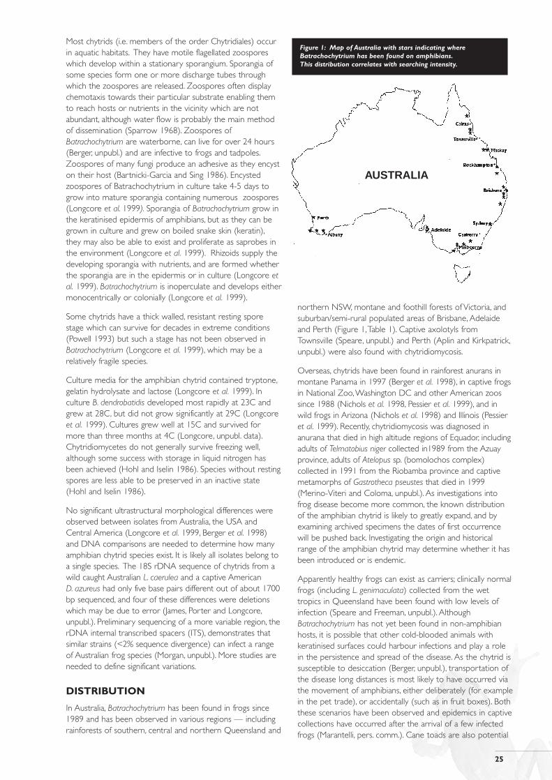
northern NSW, montane and foothill forests of Victoria, andsuburban/semi-rural populated areas of Brisbane, Adelaideand Perth (Figure 1,Table 1). Captive axolotyls fromTownsville (Speare, unpubl.) and Perth (Aplin and Kirkpatrick,unpubl.) were also found with chytridiomycosis.
Overseas, chytrids have been found in rainforest anurans inmontane Panama in 1997 (Berger et al. 1998), in captive frogsin National Zoo,Washington DC and other American zoossince 1988 (Nichols et al. 1998, Pessier et al. 1999), and inwild frogs in Arizona (Nichols et al. 1998) and Illinois (Pessieret al. 1999). Recently, chytridiomycosis was diagnosed inanurana that died in high altitude regions of Equador, includingadults of Telmatobius niger collected in1989 from the Azuayprovince, adults of Atelopus sp. (bomolochos complex)collected in 1991 from the Riobamba province and captivemetamorphs of Gastrotheca pseustes that died in 1999(Merino-Viteri and Coloma, unpubl.). As investigations intofrog disease become more common, the known distributionof the amphibian chytrid is likely to greatly expand, and byexamining archived specimens the dates of first occurrencewill be pushed back. Investigating the origin and historicalrange of the amphibian chytrid may determine whether it hasbeen introduced or is endemic.
Apparently healthy frogs can exist as carriers; clinically normalfrogs (including L. genimaculata) collected from the wettropics in Queensland have been found with low levels ofinfection (Speare and Freeman, unpubl.). AlthoughBatrachochytrium has not yet been found in non-amphibianhosts, it is possible that other cold-blooded animals withkeratinised surfaces could harbour infections and play a rolein the persistence and spread of the disease. As the chytrid issusceptible to desiccation (Berger, unpubl.), transportation ofthe disease long distances is most likely to have occurred viathe movement of amphibians, either deliberately (for examplein the pet trade), or accidentally (such as in fruit boxes). Boththese scenarios have been observed and epidemics in captivecollections have occurred after the arrival of a few infectedfrogs (Marantelli, pers. comm.). Cane toads are also potential
Figure 1: Map of Australia with stars indicating whereBatrachochytrium has been found on amphibians.This distribution correlates with searching intensity.
AUSTRALIA
26
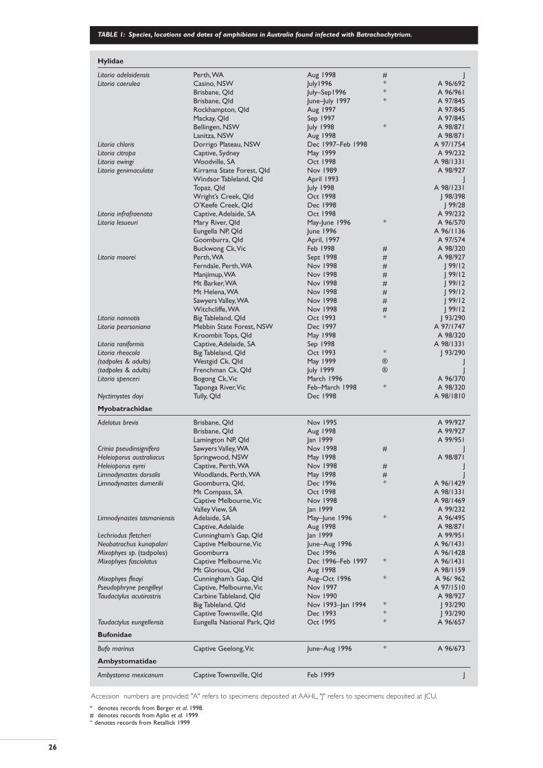
TABLE 1: Species, locations and dates of amphibians in Australia found infected with Batrachochytrium.
* denotes records from Berger et al. 1998.# denotes records from Aplin et al. 1999“ denotes records from Retallick 1999
Hylidae
Litoria adelaidensis Perth,WA Aug 1998 # JLitoria caerulea Casino, NSW July1996 * A 96/692
Brisbane, Qld July–Sep1996 * A 96/961Brisbane, Qld June–July 1997 * A 97/845Rockhampton, Qld Aug 1997 A 97/845Mackay, Qld Sep 1997 A 97/845Bellingen, NSW July 1998 * A 98/871Lanitza, NSW Aug 1998 A 98/871
Litoria chloris Dorrigo Plateau, NSW Dec 1997–Feb 1998 A 97/1754Litoria citropa Captive, Sydney May 1999 A 99/232Litoria ewingi Woodville, SA Oct 1998 A 98/1331Litoria genimaculata Kirrama State Forest, Qld Nov 1989 A 98/927
Windsor Tableland, Qld April 1993 JTopaz, Qld July 1998 A 98/1231Wright’s Creek, Qld Oct 1998 J 98/398O’Keefe Creek, Qld Dec 1998 J 99/28
Litoria infrafraenata Captive,Adelaide, SA Oct 1998 A 99/232Litoria lesueuri Mary River, Qld May-June 1996 * A 96/570
Eungella NP, Qld June 1996 A 96/1136Goomburra, Qld April, 1997 A 97/574Buckwong Ck,Vic Feb 1998 # A 98/320
Litoria moorei Perth,WA Sept 1998 # A 98/927Ferndale, Perth,WA Nov 1998 # J 99/12Manjimup,WA Nov 1998 # J 99/12Mt Barker,WA Nov 1998 # J 99/12Mt Helena,WA Nov 1998 # J 99/12Sawyers Valley,WA Nov 1998 # J 99/12Witchcliffe,WA Nov 1998 # J 99/12
Litoria nannotis Big Tableland, Qld Oct 1993 * J 93/290Litoria pearsoniana Mebbin State Forest, NSW Dec 1997 A 97/1747
Kroombit Tops, Qld May 1998 A 98/320Litoria raniformis Captive,Adelaide, SA Sep 1998 A 98/1331Litoria rheocola Big Tableland, Qld Oct 1993 * J 93/290(tadpoles & adults) Westgid Ck, Qld May 1999 ® J(tadpoles & adults) Frenchman Ck, Qld July 1999 ® JLitoria spenceri Bogong Ck,Vic March 1996 A 96/370
Taponga River,Vic Feb–March 1998 * A 98/320Nyctimystes dayi Tully, Qld Dec 1998 A 98/1810
Myobatrachidae
Adelotus brevis Brisbane, Qld Nov 1995 A 99/927Brisbane, Qld Aug 1998 A 99/927Lamington NP, Qld Jan 1999 A 99/951
Crinia pseudinsignifera Sawyers Valley,WA Nov 1998 # JHeleioporus australiacus Springwood, NSW May 1998 A 98/871Heleioporus eyrei Captive, Perth,WA Nov 1998 # JLimnodynastes dorsalis Woodlands, Perth,WA May 1998 # JLimnodynastes dumerilii Goomburra, Qld, Dec 1996 * A 96/1429
Mt Compass, SA Oct 1998 A 98/1331Captive Melbourne,Vic Nov 1998 A 98/1469Valley View, SA Jan 1999 A 99/232
Limnodynastes tasmaniensis Adelaide, SA May–June 1996 * A 96/495Captive,Adelaide Aug 1998 A 98/871
Lechriodus fletcheri Cunningham’s Gap, Qld Jan 1999 A 99/951Neobatrachus kunapalari Captive Melbourne,Vic June–Aug 1996 A 96/1431Mixophyes sp. (tadpoles) Goomburra Dec 1996 A 96/1428Mixophyes fasciolatus Captive Melbourne,Vic Dec 1996–Feb 1997 * A 96/1431
Mt Glorious, Qld Aug 1998 A 98/1159Mixophyes fleayi Cunningham’s Gap, Qld Aug–Oct 1996 * A 96/ 962Pseudophryne pengilleyi Captive, Melbourne,Vic Nov 1997 A 97/1510Taudactylus acutirostris Carbine Tableland, Qld Nov 1990 A 98/927
Big Tableland, Qld Nov 1993–Jan 1994 * J 93/290Captive Townsville, Qld Dec 1993 * J 93/290
Taudactylus eungellensis Eungella National Park, Qld Oct 1995 * A 96/657
Bufonidae
Bufo marinus Captive Geelong,Vic June–Aug 1996 * A 96/673
Ambystomatidae
Ambystoma mexicanum Captive Townsville, Qld Feb 1999 J
Accession numbers are provided: "A" refers to specimens deposited at AAHL, "J" refers to specimens deposited at JCU.
27
carriers as they continue to expand their distribution inAustralia, as evidence suggests that free-ranging toads areinfected with chytrids1 (Parkes, pers. comm.).
We hypothesise that Batrachochytrium was introduced toAustralia in the 1970’s around Brisbane (where the firstprecipitous declines occurred), although as yet there are nohard data to support this assumption. Batrachochytrium hassince become established in many areas on the east coast,around Adelaide and in south west Western Australia.
CHYTRIDIOMYCOSIS:THE DISEASE
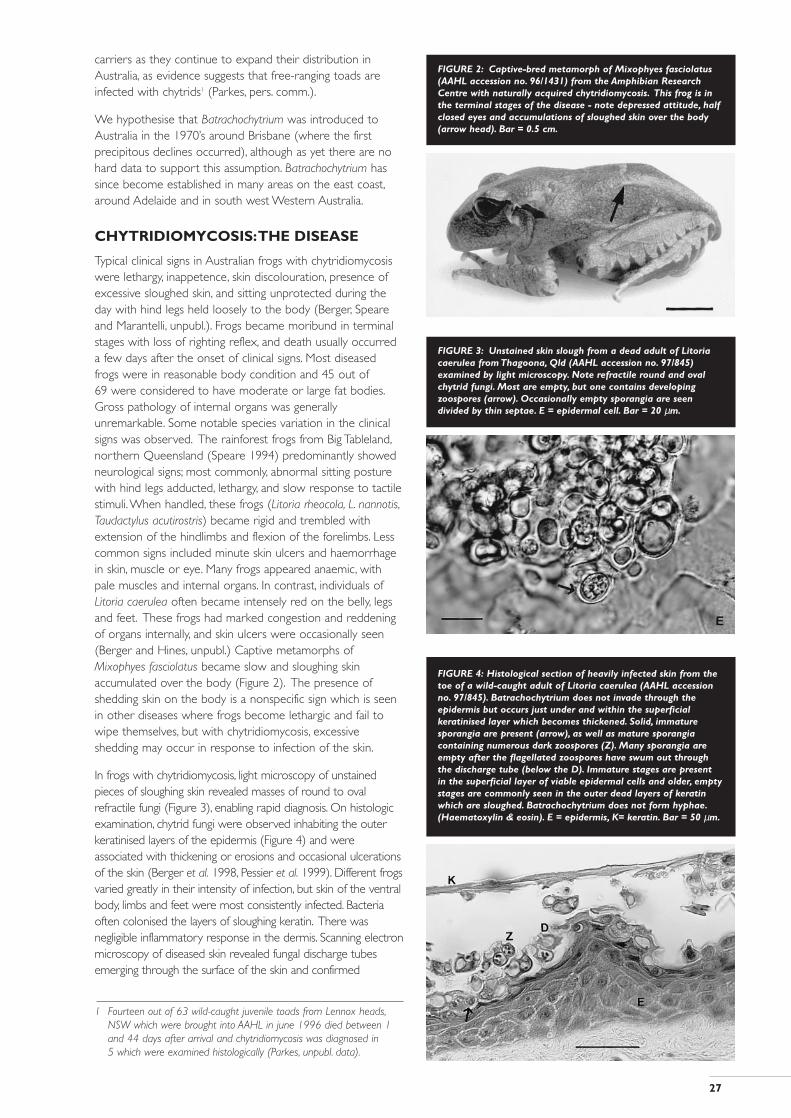
Typical clinical signs in Australian frogs with chytridiomycosiswere lethargy, inappetence, skin discolouration, presence ofexcessive sloughed skin, and sitting unprotected during theday with hind legs held loosely to the body (Berger, Speareand Marantelli, unpubl.). Frogs became moribund in terminalstages with loss of righting reflex, and death usually occurreda few days after the onset of clinical signs. Most diseasedfrogs were in reasonable body condition and 45 out of 69 were considered to have moderate or large fat bodies.Gross pathology of internal organs was generallyunremarkable. Some notable species variation in the clinicalsigns was observed. The rainforest frogs from Big Tableland,northern Queensland (Speare 1994) predominantly showedneurological signs; most commonly, abnormal sitting posturewith hind legs adducted, lethargy, and slow response to tactilestimuli.When handled, these frogs (Litoria rheocola, L. nannotis,Taudactylus acutirostris) became rigid and trembled withextension of the hindlimbs and flexion of the forelimbs. Lesscommon signs included minute skin ulcers and haemorrhagein skin, muscle or eye. Many frogs appeared anaemic, withpale muscles and internal organs. In contrast, individuals ofLitoria caerulea often became intensely red on the belly, legsand feet. These frogs had marked congestion and reddeningof organs internally, and skin ulcers were occasionally seen(Berger and Hines, unpubl.) Captive metamorphs ofMixophyes fasciolatus became slow and sloughing skinaccumulated over the body (Figure 2). The presence ofshedding skin on the body is a nonspecific sign which is seenin other diseases where frogs become lethargic and fail towipe themselves, but with chytridiomycosis, excessiveshedding may occur in response to infection of the skin.
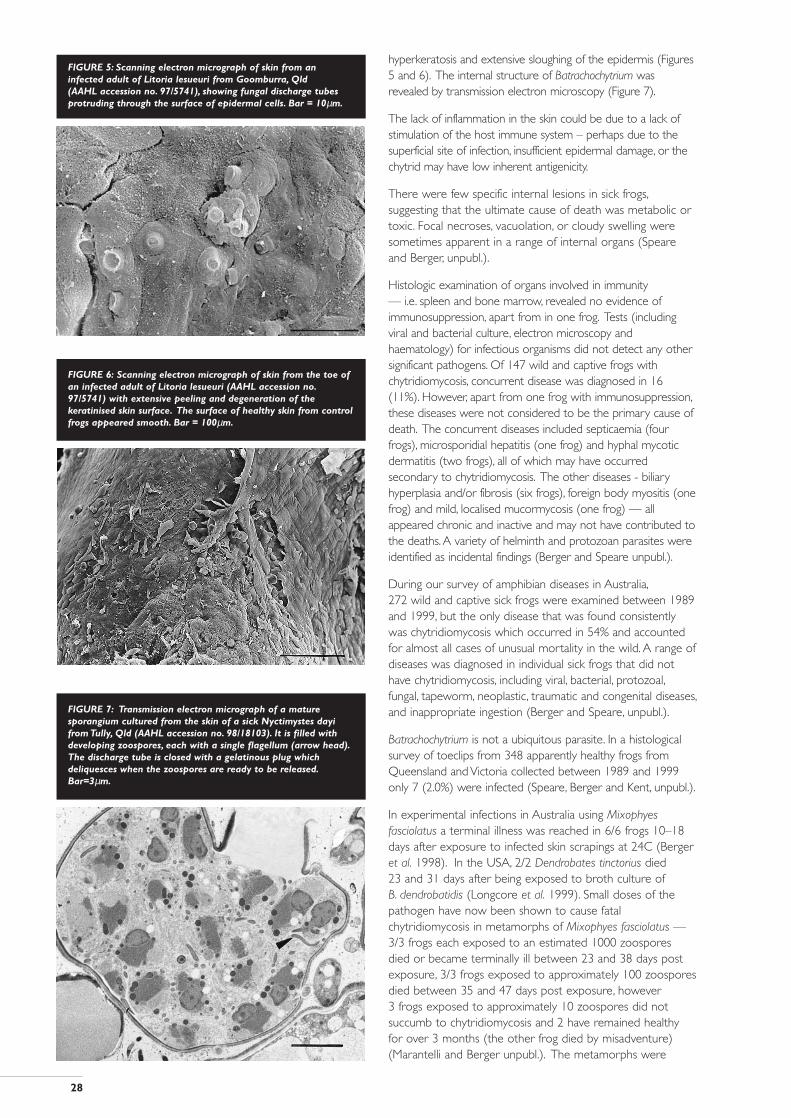
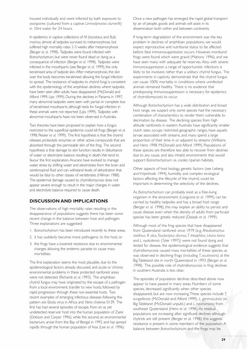
In frogs with chytridiomycosis, light microscopy of unstainedpieces of sloughing skin revealed masses of round to ovalrefractile fungi (Figure 3), enabling rapid diagnosis. On histologicexamination, chytrid fungi were observed inhabiting the outerkeratinised layers of the epidermis (Figure 4) and wereassociated with thickening or erosions and occasional ulcerationsof the skin (Berger et al. 1998, Pessier et al. 1999). Different frogsvaried greatly in their intensity of infection, but skin of the ventralbody, limbs and feet were most consistently infected. Bacteriaoften colonised the layers of sloughing keratin. There wasnegligible inflammatory response in the dermis. Scanning electronmicroscopy of diseased skin revealed fungal discharge tubesemerging through the surface of the skin and confirmed
1 Fourteen out of 63 wild-caught juvenile toads from Lennox heads,NSW which were brought into AAHL in june 1996 died between 1and 44 days after arrival and chytridiomycosis was diagnosed in 5 which were examined histologically (Parkes, unpubl. data).
FIGURE 2: Captive-bred metamorph of Mixophyes fasciolatus(AAHL accession no. 96/1431) from the Amphibian ResearchCentre with naturally acquired chytridiomycosis. This frog is inthe terminal stages of the disease - note depressed attitude, halfclosed eyes and accumulations of sloughed skin over the body(arrow head). Bar = 0.5 cm.
FIGURE 3: Unstained skin slough from a dead adult of Litoriacaerulea from Thagoona, Qld (AAHL accession no. 97/845)examined by light microscopy. Note refractile round and ovalchytrid fungi. Most are empty, but one contains developingzoospores (arrow). Occasionally empty sporangia are seendivided by thin septae. E = epidermal cell. Bar = 20 µm.
FIGURE 4: Histological section of heavily infected skin from thetoe of a wild-caught adult of Litoria caerulea (AAHL accessionno. 97/845). Batrachochytrium does not invade through theepidermis but occurs just under and within the superficialkeratinised layer which becomes thickened. Solid, immaturesporangia are present (arrow), as well as mature sporangiacontaining numerous dark zoospores (Z). Many sporangia areempty after the flagellated zoospores have swum out throughthe discharge tube (below the D). Immature stages are present in the superficial layer of viable epidermal cells and older, emptystages are commonly seen in the outer dead layers of keratinwhich are sloughed. Batrachochytrium does not form hyphae.(Haematoxylin & eosin). E = epidermis, K= keratin. Bar = 50 µm.
28
hyperkeratosis and extensive sloughing of the epidermis (Figures5 and 6). The internal structure of Batrachochytrium wasrevealed by transmission electron microscopy (Figure 7).
The lack of inflammation in the skin could be due to a lack ofstimulation of the host immune system – perhaps due to thesuperficial site of infection, insufficient epidermal damage, or thechytrid may have low inherent antigenicity.
There were few specific internal lesions in sick frogs,suggesting that the ultimate cause of death was metabolic ortoxic. Focal necroses, vacuolation, or cloudy swelling weresometimes apparent in a range of internal organs (Speareand Berger, unpubl.).
Histologic examination of organs involved in immunity — i.e. spleen and bone marrow, revealed no evidence ofimmunosuppression, apart from in one frog. Tests (includingviral and bacterial culture, electron microscopy andhaematology) for infectious organisms did not detect any othersignificant pathogens. Of 147 wild and captive frogs withchytridiomycosis, concurrent disease was diagnosed in 16(11%). However, apart from one frog with immunosuppression,these diseases were not considered to be the primary cause ofdeath. The concurrent diseases included septicaemia (fourfrogs), microsporidial hepatitis (one frog) and hyphal mycoticdermatitis (two frogs), all of which may have occurredsecondary to chytridiomycosis. The other diseases - biliaryhyperplasia and/or fibrosis (six frogs), foreign body myositis (onefrog) and mild, localised mucormycosis (one frog) — allappeared chronic and inactive and may not have contributed tothe deaths.A variety of helminth and protozoan parasites wereidentified as incidental findings (Berger and Speare unpubl.).
During our survey of amphibian diseases in Australia,272 wild and captive sick frogs were examined between 1989and 1999, but the only disease that was found consistentlywas chytridiomycosis which occurred in 54% and accountedfor almost all cases of unusual mortality in the wild. A range ofdiseases was diagnosed in individual sick frogs that did nothave chytridiomycosis, including viral, bacterial, protozoal,fungal, tapeworm, neoplastic, traumatic and congenital diseases,and inappropriate ingestion (Berger and Speare, unpubl.).
Batrachochytrium is not a ubiquitous parasite. In a histologicalsurvey of toeclips from 348 apparently healthy frogs fromQueensland and Victoria collected between 1989 and 1999only 7 (2.0%) were infected (Speare, Berger and Kent, unpubl.).
In experimental infections in Australia using Mixophyesfasciolatus a terminal illness was reached in 6/6 frogs 10–18days after exposure to infected skin scrapings at 24C (Bergeret al. 1998). In the USA, 2/2 Dendrobates tinctorius died 23 and 31 days after being exposed to broth culture of B. dendrobatidis (Longcore et al. 1999). Small doses of thepathogen have now been shown to cause fatalchytridiomycosis in metamorphs of Mixophyes fasciolatus —3/3 frogs each exposed to an estimated 1000 zoospores died or became terminally ill between 23 and 38 days postexposure, 3/3 frogs exposed to approximately 100 zoosporesdied between 35 and 47 days post exposure, however 3 frogs exposed to approximately 10 zoospores did notsuccumb to chytridiomycosis and 2 have remained healthy for over 3 months (the other frog died by misadventure)(Marantelli and Berger unpubl.). The metamorphs were
FIGURE 5: Scanning electron micrograph of skin from aninfected adult of Litoria lesueuri from Goomburra, Qld (AAHL accession no. 97/5741), showing fungal discharge tubesprotruding through the surface of epidermal cells. Bar = 10µm.
FIGURE 6: Scanning electron micrograph of skin from the toe ofan infected adult of Litoria lesueuri (AAHL accession no.97/5741) with extensive peeling and degeneration of thekeratinised skin surface. The surface of healthy skin from controlfrogs appeared smooth. Bar = 100µm.
FIGURE 7: Transmission electron micrograph of a maturesporangium cultured from the skin of a sick Nyctimystes dayifrom Tully, Qld (AAHL accession no. 98/18103). It is filled withdeveloping zoospores, each with a single flagellum (arrow head).The discharge tube is closed with a gelatinous plug whichdeliquesces when the zoospores are ready to be released.Bar=3µm.
29
housed individually and were infected by bath exposure tozoospores (cultured from a captive Limnodynastes dumerilli) in 10ml water for 24 hours.
In epidemics in captive collections of M. fasciolatus and Bufomarinus, almost all tadpoles survived to metamorphose, butsuffered high mortality rates 2-3 weeks after metamorphosis(Berger et al. 1998). Tadpoles were found infected withBatrachochytrium, but were never found dead or dying as aconsequence of infection (Berger et al. 1998). Tadpoles wereinfected in the mouthparts (see Berger et al. 1999), the onlykeratinised area of tadpole skin.After metamorphosis, the skinover the body becomes keratinised allowing the fungal infectionto spread. The resistance of tadpoles to chytrid fungi is consistentwith the epidemiology of the amphibian declines where tadpoleshave been seen after adults have disappeared (McDonald andAlford 1999, Lips 1999). During the declines in Panama in 1997,many abnormal tadpoles were seen with partial or complete lossof keratinised mouthparts, although tests for fungal infection inthese animals were not reported (Lips 1999). Tadpoles withabnormal mouthparts have not been observed in Australia.
Two theories have been proposed to explain how a fungusrestricted to the superficial epidermis could kill frogs (Berger et al.1998, Pessier et al. 1999). The first hypothesis is that the chytridreleases proteolytic enzymes or other active compounds that areabsorbed through the permeable skin of the frog. The secondhypothesis is that damage to skin function results in disturbanceof water or electrolyte balance resulting in death.We tend tofavour the first explanation.Anurans have evolved to managewater stress by shifting water and electrolytes from the brain andcerebrospinal fluid and can withstand levels of dehydration thatwould be fatal to other classes of vertebrates (Hillman 1988).The epidermal damage caused by chytridiomycosis does notappear severe enough to result in the major changes in waterand electrolyte balance required to cause death.
DISCUSSION AND IMPLICATIONS
The observations of high mortality rates resulting in thedisappearance of populations suggests there has been somerecent change in the balance between host and pathogen.Three explanations are suggested:
1. Batrachochytrium has been introduced recently to these areas,
2. it has suddenly become more pathogenic to the host, or
3. the frogs have a lowered resistance due to environmentalchanges allowing the endemic parasite to cause massmortalities.
The first explanation seems the most plausible, due to theepidemiological factors already discussed, and acute or chronicenvironmental problems in these protected rainforest areaswere not detected (Richards et al. 1993). The amphibianchytrid fungus may have originated by the escape of a pathogenfrom a local environment, transfer to new hosts, followed byrapid progression through these non-essential hosts. Tworecent examples of emerging infectious diseases following thispattern are Ebola virus in Africa and Vibrio cholerae 0139. Thefirst has had several episodes of escape, from an as yetundetected reservoir host into the human population of Zaire(Dobson and Carper 1996), while the second, an environmentalbacterium, arose from the Bay of Bengal in 1992 and has spreadrapidly through the human population of Asia (Lee et al. 1996).
Once a new pathogen has emerged, the rapid global transportby air of people, goods and animals will assist in itsdissemination both within and between continents.
If long-term degradation of the environment was the keyproblem in declines of amphibian populations, we wouldexpect reproductive and nutritional status to be affectedbefore fatal immunosuppression occurs. However, moribundfrogs were found which were gravid (Mahony 1996), and wehave seen many with adequate fat reserves. Also, with severeimmunosuppression a range of opportunistic infections islikely to be involved, rather than a solitary chytrid fungus. Theexperiments in captivity demonstrate that the chytrid funguscan cause 100% mortality in conditions where uninfectedanimals remained healthy. There is no evidence thatpredisposing immunosuppression is necessary for epidemicsof chytridiomycosis to occur.
Although Batrachochytrium has a wide distribution and broadhost range, we suspect only some species had the necessarycombination of characteristics to render them vulnerable todecimation by disease. The declining species from highaltitude rainforests in eastern Australia have significantly smallerclutch sizes, occupy restricted geographic ranges, have aquaticlarvae associated with streams, and many spend a largeproportion of their time in or adjacent to streams (Williamsand Hero 1998, McDonald and Alford 1999). Populations ofthese species are therefore less able to recover from declinesdue to any cause, and also inhabit environments that wouldsupport Batrachochytrium i.e. cooler, riparian habitats.
Other aspects of host biology, genetic factors (see Lebergand Vrijenhoek 1994), humidity and complex ecologicalfactors affecting the lifecycle of the chytrid, could beimportant in determining the selectivity of the declines.
As Batrachochytrium can probably exist as a free-livingorganism in the environment (Longcore et al. 1999), can becarried by healthy tadpoles and has a broad host range(Berger et al. 1998), this may explain an ability to persist andcause disease even when the density of adults from particularspecies has been greatly reduced (Daszak et al. 1999).
Although most of the frog species that have disappearedfrom Queensland rainforest since 1979 (e.g. Rheobatrachusvitellinus, R. silus,Taudactylus diurnus,T. rheophilus, Litoria loricaand L. nyakalensis (Tyler 1997)) were not found dying andtested for disease, the epidemiological evidence suggests thatchytridiomycosis caused mass mortalities of these species, aswas observed in declining frogs (including T. acutirostris) at theBig Tableland site in north Queensland in 1993 (Berger et al.1998). The possible role of chytridiomycosis in frog declinesin southern Australia is less clear.
The episodes of population declines described above nowappear to have passed in many areas. Numbers of somespecies, decreased significantly when other speciesdisappeared, but are now increasing.These species include T.eungellensis (McDonald and Alford 1999), L. genimaculata onBig Tableland (McDonald unpubl.) and L. pearsoniana fromsoutheast Queensland (Hero et al. 1998). As residualpopulations are increasing after significant declines althoughchytrids are still present (Berger et al. 1998), this suggestsresistance is present in some members of the population. Abalance between Batrachochytrium and the frogs may be
30
developing in areas where we suspect it has been present forat least five years, and although disease still occurs, it does notdevastate the populations. Batrachochytrium may now behaveas an endemic pathogen with outbreaks of disease occurringwhen conditions are optimal. The long term prognosis maybe good for species which have survived and are recovering,as long as remaining habitats are protected to allow damagedpopulations to reestablish. However, since we currently knowlittle about the interaction between the amphibian chytridand hosts, and many frog species are currently consideredthreatened or their status is insufficiently known (Tyler 1997),we should not be complacent. Also, if uninfected areas stillexist in Australia then locally endemic species in those areasmay be at high risk.
Infectious disease is important in the population biology of wildanimals, as it is in humans and domestic animals (May 1988).Areview of infectious disease and animal populations concludedthat disease is an important factor affecting survival,reproduction, dispersal, community structure and geneticdiversity, and should therefore be considered by ecologistsexamining host population-dynamics (Scott 1988). Diseasebecomes a threatening force when environmental degradationputs pressure on populations, and international trade andsmuggling continually threaten to introduce new pathogens towhich the native fauna has no resistance (Scott 1988, Daszak etal. 1999). Exotic diseases can have effects similar to those of feralpredators, with susceptible native species facing extinction whilethe ecosystem readjusts, and are another example of howincreased global homogeneity leads to reduced biodiversity.Although the initial >99% mortality rate of myxomatosis wasnot sufficient to exterminate rabbits from Australia (Fenner andRatcliffe 1965), it is plausible that a similar mortality rate couldwipe out frog species with limited distributions and relativelyinfrequent breeding. The introduction of avian malaria issuspected to have caused the extinction of birds in Hawaii(Warner 1968).A protozoan parasite of cats (Toxoplasma gondii)was probably introduced to Australia during the Europeaninvasion, and marsupials are among the most susceptible animals(Reddacliff et al. 1993). Phytophthora cinnamomi is an example ofa pathogenic, introduced zoosporic fungus which threatens manynative Australian plant species, and quarantine measures arerecommended to prevent the invasion of the pathogen intonew areas. Some plant species are highly susceptible, whereasothers only become diseased after periods of stress such as adrought (Dawson and Weste 1985;Wills 1993). Infrastructureexists to prevent and manage exotic disease outbreaks indomestic animals, but little concern is shown for wildlife wheremany diseases are yet to be discovered and understood, andmonitoring the disease status of populations currently appears tobe the responsibility of no one.
FUTURE DIRECTIONS
Work on amphibian chytridiomycosis must continue — toconfirm or reject the hypothesis that it is the primary cause ofthe declines, to determine how the epidemic began, to find waysto manage the problem in areas where the fungus is established,and to prevent it occurring in new regions. Knowledge gainedfrom these investigations will be useful in preventing similarpopulation crashes occurring in frogs again, and in developingmanagement strategies for other wildlife species.
We will continue to map the temporal and geographic
distribution of chytridiomycosis by examination of frogspreserved in museum collections to determine whetherchytrids were introduced to Australia or to these habitats andto determine from where and how chytrids have spread.Information on the current distribution may also help informulating management plans to prevent new outbreaks —for example, in deciding where exposed or uninfected frogsshould be released after captive breeding. This survey relieson a collaborative approach (see Appendix 1). As manyhealthy tadpoles may carry chytrids for extended periods,sampling of tadpoles may provide a more sensitive means of assessing a location. Examination of sick frogs is the most sensitive way to detect chytrids, but these are not always available.
Mycological studies on the chytrid are needed to learn moreabout its lifecycle, requirements and survival in the wild.Knowledge about the ecology and hosts of the fungus isessential to understanding the spread, and therefore to themanagement of the disease. Information about the particularconditions that encourage growth may help in understandingthe factors which precipitate disease epidemics.
Further transmission experiments are required to confirm thepathogenicity in a range of species, and also to determinewhat environmental conditions e.g. temperature, are requiredfor expression of the disease in frogs. Treatments for adultsand tadpoles are also being tested which will aid captivebreeding of endangered species. By producing large numbersof frogs in captivity, it may be possible to help species tosurvive and evolve immunity.
Studies of the pathogenesis of chytridiomycosis are importantto understanding this disease, and immunologists in the USAwill investigate the innate and acquired immune response offrogs to Batrachochytrium.
Further work will be done using DNA analysis to comparechytrids from various species from localities across Australia,Central America and the USA. The number of species ofBatrachochytrium can be determined by using this informationcombined with morphological taxonomy. Molecular biology asa tool for molecular epidemiology can also be used toprovide clues about the origins and spread of the fungus.
Data are being collected to evaluate diagnostic tests.Histology and examination of skin scrapings are highly specifictests, but may not be very sensitive when used to detectchytrids on healthy specimens. Production of antibodies hascommenced to enable more sensitive testing, and perhaps foruse in detecting chytrids in the environment.
Regulations regarding quarantine, testing, treatment andmovement of amphibians need to be introduced to preventfurther spread of Batrachochytrium within Australia andinternationally. Although our understanding of the role ofchytridiomycosis in amphibian declines is far from complete, webelieve it is crucial to take immediate preventative measuresrather than risk waiting for more scientific data to be accrued.
ACKNOWLEDGEMENTS
Amphibian disease investigations would not have been possiblewithout the tremendous support of herpetologists throughoutAustralia.A huge collaborative effort was involved in collecting

31
sick frogs from the field and urgently forwarding them to thelab.We are indebted to many people who have been involved,including those who provided sick or healthy specimens,allowed us access to their collections or gave advice, especiallyGerry Marantelli, Harry Hines, Keith McDonald, Mike Tyler, KenAplin,Alastair Freeman, Craig Williams, Rebecca Short, GraemeGillespie, Nick Sheppard, David Page, David Byrnes, SteveWilliams, Craig Taylor, D.A. Stewart, Michael Mahony, LanceTarvey, Michael Smith, Richard Retallick, Marc Hero, John Clarke,Michael Healey, Brent Dadds, Lothar Voigt,Adrian Wayne,Jeremy Morante, Danny Wotherspoon and members of thepublic.We are very grateful to Lee Skerratt, Deborah Middletonand Murray Littlejohn for comments on the manuscript. Thanksto Frank Fillipi for photography, Julia Hammond for bacteriology,and Megan Braun, Gail Russell,Terry Wise and Andrew Kent forthe many tissue sections prepared. Peter Hooper and MarkWilliamson are thanked for their support and pathologyexpertise.We also thank people who allowed us to cite theirunpublished data. Lee Berger has been supported by a grantfrom Environment Australia.
REFERENCES
Aplin, K., Speare, R., Berger, L. and Cowan, M. A., (1999)Outbreak of fungal disease in Western Australian Frogs.J Royal Soc WA (submitted).
Barr, D. J. S., (1990) Phylum Chytridiomycota. Pp. 454-466 inHandbook of Protoctista. ed by L. Margulis, J. O. Corliss,M. Melkonian and D. J. Chapman. Lubrecht and Kramer,Monticello, New York.
Bartnicki-Garcia, S. and Sing,V. O. (1986) Adhesion ofzoospores of Phytophthora to solid surfaces. Pp. 279-283 in Zoosporic Fungi in Teaching and Research. ed by M. S. Fuller and A. Jaworski. Dept of Botany, University ofGeorgia, Athens GA.
Berger, L. Speare, R. Daszak, P., Green, D. E, Cunningham,A.A.,Goggin, C. L., Slocombe, R. Ragan, M.A. Hyatt,A. D., McDonald,K. R. Hines, H. B., Lips, K. R., Marantelli, G. and Parkes, H., (1998)Chytridiomycosis causes amphibian mortality associated withpopulation declines in the rainforests of Australia and CentralAmerica. Proc Natl Acad Sci, 95: 9031-9036.
Berger, L., Speare, R. and Kent, A., (1999) Diagnosis ofchytridiomycosis in amphibians by histologic examination.Proc Frog Symposium: Frogs in the Community, QueenslandMuseum, Brisbane, February 1999 (submitted).
Daszak, P.1, Berger, L., Cunningham, A. A., Hyatt, A. D., Green, D.E. and Speare, R., (1999) Role of Emerging InfectiousDiseases in Amphibian Population Declines and GlobalImplications. Emerg Inf Dis, (in press).
Dawson, P. and Weste, G., (1985) Changes in the distributionof Phytophthora cinnamomi in the Brisbane Ranges NationalPark between 1970 and 1980-81. Aust J Bot, 33: 309-315.
Dobson, A. P and Carper, E. R., (1996) Infectious disease andhuman population history. Bioscience, 46: 115-126.
Fenner, F. and Ratcliffe, F. N., (1965) Myxomatosis, CambridgeUniversity Press, New York.
Groff, J. M., Mughannam, A., McDowell,T. S.,Wong, A. Dykstra,M. J., Frye, F. L. and Hedrick, R. P., (1991) An epidemic ofcutaneous zygomycosis in cultured dwarf African clawedfrogs (Hymenochirus curtipes) due to Basidiobolus ranarum. JMed Vet Mycology, 29: 215-223.
Hayes, M. P. and Jennings, M. R., (1986) Decline of ranid frogsspecies in Western North America: Are bullfrogs (Ranacatesbiana) responsible? J Herpetol, 20: 490-509.
Hero, J-M., Hines, H., Meyer, E., Morrison, C., Streatfeild, C. andRoberts, L., (1998) New records of “declining” frogs inQueensland, Australia (-February 1998). Froglog 29.
Hillman, S. S., (1988) Dehydrational effects on brain andcerebrospinal fluid electrolytes in two amphibians. PhysiolZool, 61: 254-259.
Hohl, H. R. and Iselin, K., (1986) Liquid nitrogen preservationof zoosporic fungi. Pp 143-145 in Zoosporic Fungi inTeaching and Research. ed by M. S. Fuller and A. Jaworski.Dept of Botany, University of Georgia, Athens GA.
Laurance,W. F., McDonald, K. R. and Speare, R., (1996)Epidemic disease and the catastrophic decline of Australianrainforest frogs. Conserv Biol, 77: 203-212.
Leberg, P. L. and Vrijenhoek, R. C., (1994) Variation amongdesert topminnows in their susceptibility to attack byexotic parasites. Conserv Biol, 8: 419-424.
Lee, S. H., Lai, S. T., Lai, J.Y. and Leung, N. K., (1996) Resurgenceof cholera in Hong Kong. Epidemiol Infect, 117: 43-49.
Lips, K. R., (1998) Decline of a tropical montane fauna.Conserv Biol, 12: 106-117.
Lips, K. R., (1999) Mass mortality and population declines ofanurans at an upland site in western Panama. Conserv Biol,13: 117-125.
Longcore, J. E., Pessier, A. P. and Nichols, D. K., (1999)Batrachochytrium dendrobatidis gen. et sp. nov., a chytridpathogenic to amphibians. Mycologia, 91: 219-227.
Mahony, M., (1996) The decline of the green and golden bellfrog Litoria aurea viewed in the context of declines anddisappearances of other Australian frogs. Aust Zool, 30:237-247.
Marantelli, G., (1999) Husbandry: science or art ? — Arecaptive technologies ready to contribute to recoveryprocesses for Australian frogs? Pp 168-176 in Declines andDisappearances of Australian Frogs ed by A.Campbell.Environment Australia, Canberra.
May, R. M., (1988) Conservation and Disease. Conserv Biol,2: 28-30.
McDonald, K.R. and Alford, R.A., (1999) A review of decliningfrogs in northern Queensland. Pp 14-22 in Declines andDisappearances of Australian Frogs ed by A.Campbell.Environment Australia, Canberra.
Morell,V. (1999) Are pathogens felling frogs? Science, 284:728-731.
Nichols, D.K., Pessier, A.P., and Longcore, J.E., (1998)Cutaneous chytridiomycosis: an emerging disease? Proc AmAssoc Zoo Vet, 1998:269-271.
Pessier, A. P. Nichols, D. K., Longcore, J. E. and Fuller, M. S.,(1999) Cutaneous chytridiomycosis in poison dart frogs(Dendrobates spp.) and White’s tree frogs (Litoria caerulea).J Vet Diag Invest, 11: 194-199.
Powell, M. J., (1993) Looking at mycology with a Janus face:A glimpse of chytridiomycetes active in the environment.Mycologia, 85: 1-20.
Reddacliff, G.L., Hartley,W.J., Dubey, J.P. and Cooper, D.W.,(1993) Pathology of experimentally-induced, acutetoxoplasmosis in macropods. Aust Vet J.70: 4-6.
32
Retallick, R., (1999) Translocations and experimental ecologyof declining frogs. Update for the North Queensland FrogRecovery Team, August 1999. Unpublished report.
Richards, S. J., McDonald, K R. and Alford, R. A., (1993)Declines in populations of Australia’s endemic tropicalrainforest frogs. Pacific Conserv Biol, 1: 66-77.
Scott, M. E., (1988) The impact of infection and disease onanimal populations: Implications for conservation biology.Conserv Biol, 2: 40-56.
Scott, N. J., (1993) Post metamorphic death syndrome.Froglog, 7: 1-2.
Sparrow, F. K., (1960) Aquatic Phycomycetes. 2nd re. ed.University of Michigan Press, Ann Arbor, Michigan.
Sparrow, F. K., (1968) Ecology of Freshwater Fungi. pp 41-93in The Fungi. ed by G. C. Gainsworth and A. S. Sussman,Academic Press, New York.
Speare, R., (1994) Preliminary study on diseases in AustralianWet Tropics amphibians. Deaths of rainforest frogs at O’Keefe Creek, Big Tableland. Final report to QueenslandDepartment of Environment and Heritage. Unpublishedreport, Queensland Department of Environment andHeritage.
Taylor, S. K.,Williams, E. S.,Thorne, E. T., Mills, K.W.,Withers,D. I. and Pier, A. C., (1999) Causes of mortality of theWyoming toad. J Wildl Dis, 35: 49-57.
Trenerry, M. P., Laurance,W. F. and McDonald, K. R., (1994)Further evidence for the precipitous decline of endemicrainforest frogs in tropical Australia. Pacific Conserv Biol,1: 150-153.
Tyler, M. J., (1997) The Action Plan for Australian Frogs. WildlifeAustralia, Canberra.
Viggers, K. L., Lindenmayer, D. B. and Spratt, D. M., (1993) The importance of disease in reintroduction programmes.Wildl Res, 20: 687-98.
Warner, R. E., (1968) The role of introduced diseases in theextinction of the endemic Hawaiian avifauna. The Condor,70: 101-120.
Williams S. E. and Hero J-M., (1998) Rainforest frogs of theAustralian Wet Tropics: guild classification and theecological similarity of declining species. Proc Roy Soc Lond,B 265: 597-602.
Wills, R. T., (1993) The ecological impact of Phytophthoracinnamomi in the Stirling Range National Park,WesternAustralia. Aust J Ecol, 18: 145-159.
APPENDIX 1
What herpetologists can do to assist
SURVEY OF SICK AND HEALTHY FROGS
We wish to examine any diseased frogs or cane toads thatare found in order to determine the cause of death and toscreen for the presence of chytrids. Frog tissues deterioratevery rapidly after death, so if a sick frog is found that is likelyto survive another 24 hours, it should be sent by courier tothe Australian Animal Health Laboratory or James CookUniversity after contacting us. Frogs or tadpoles found deadshould be fixed or frozen immediately to preserve thetissues. They should be fixed in 10% buffered neutralformalin, but 70% ethanol can also be used. It is important toslit open the belly, and to ensure the frog is well covered infixative so that tissues are preserved rapidly. Details of whatto do with sick or dead frogs have been posted on theWorld Wide Web at http://www.jcu.edu.au/dept/school/phtm/PHTM/frogs/pmfrog.htm.
Please send collection data with any frog submitted, and wewill keep you informed about the results of the post mortem.Pathology is required for diagnosis, as the clinical signs ofchytridiomycosis are not highly specific.
We have prepared a frog mortality questionnaire(http://www.jcu.edu.au/school/phtm/PHTM/frogs/pmques.htm)which details the type of data that are important to observeand record if you encounter a mass mortality event.
For our examination of archived frogs for chytrids, we needskin samples from amphibians from a wide range of localitiesand dates.We want skin from the pelvic areas and toes fromany formalin-fixed or ethanol-fixed frog that has collectiondata.We especially require frogs from inland Australia,Northern Territory and northern WA, as none have beenexamined from these regions. A protocol is available athttp://www.jcu.edu.au/school/phtm/frogs/pmskin.htm.
People doing skeletochronology on histological sections oftoes could simultaneously check the skin for chytrids (seeFigure 4). In healthy frogs, the level of infection may be verylow, with only occasional sporangia present along the skinsurface. For detailed diagnostic histological features, seeBerger et al. (1999).
We are attempting to maintain a comprehensive list of confirmed cases of chytridiomycosis(http://www.jcu.edu.au/school/phtm/PHTM/frogs/chyspec.htm)and hope that data will be submitted for inclusion. This listwill enable management decisions to be made based oncurrent knowledge.
33
MANAGEMENT OF CHYTRIDIOMYCOSIS IN CAPTIVITY
If any epidemics of chytridiomycosis occur in captivecollections, various antifungal drugs could be administered,and the results communicated.
Benzalkonium chloride is a disinfectant that has been used at2 mg/l to successfully treat a similar superficial mycoticdermatitis in dwarf African clawed frogs (Hymenochiruscurtipes) reported to be caused by Basidiobolus ranarum(Groff et al. 1991). The regime used experimentally was 30 minutes of bath treatment, on three alternate days.This was repeated in 8 days (i.e. 6 treatments in total). Oralitraconazole has also been used to treat B. ranarum infections(Taylor et al. 1999). One micro bead from 100mgitraconazole capsules was administered daily for 9 days toWyoming toads (Bufo baxteri) at the first signs of disease.Benzalkonium chloride (1mg/L), amphotericin and fluconazoleare effective against Batrachochytrium in vitro (Berger, unpubl.).
In captivity, routine quarantine procedures (Marantelli 1999)have been adequate in restricting outbreaks to certain tanks,and no airborne transmission has been observed (Marantelli,unpubl.). Each group of frogs should be kept completelyseparate to ensure no water borne transmission of diseasecan occur. By changing and discarding gloves between everytank, avoiding splashing water between tanks, and disinfectionof tanks and implements before reuse using 2% hypochlorite,many frogs have been housed in close proximity withouttransmission of disease.
PREVENTING SPREAD OF DISEASE IN THE WILD
To prevent the spread of chytrids or other diseases whenperforming field work, disinfection of equipment should beperformed.We need more information on the resistance ofthis fungus to heat, desiccation and disinfectants; so at presentare recommending measures for disinfection that have beenproven against highly resistant organisms. (see protocol —http://www.jcu.edu.au/school/phtm/PHTM/frogs/prevent.htm).
Disease status should always be considered whentranslocating animals (Marantelli 1999,Viggers et al. 1993) andattempts should be made to reduce the chance ofintroducing disease to a naïve population.We recommendthe screening of healthy frogs by histologic examination oftoe clips, or by sacrificing a few in the group for moreextensive skin examination (Berger et al. 1999). Testing forchytrids on sick frogs is more sensitive, so any deaths in avaluable group of animals should be submitted to a pathologylaboratory for testing. To screen a group of healthy tadpoles,some need to be sacrificed so that their mouths can beexamined histologically (see Berger et al. 1999).We have littleinformation on the sensitivity of these tests, so it is impossibleto recommend a statistically significant number of animals.Also, until we have more data on the distribution of chytridsin amphibian populations around Australia, it will be difficultto make decisions about the release of infected animals.
New regulations are being proposed to control themovement and trade of adult amphibians and tadpoles, butbefore these are introduced, quarantine measures shouldbecome routine.