Embed Size (px)
Citation preview
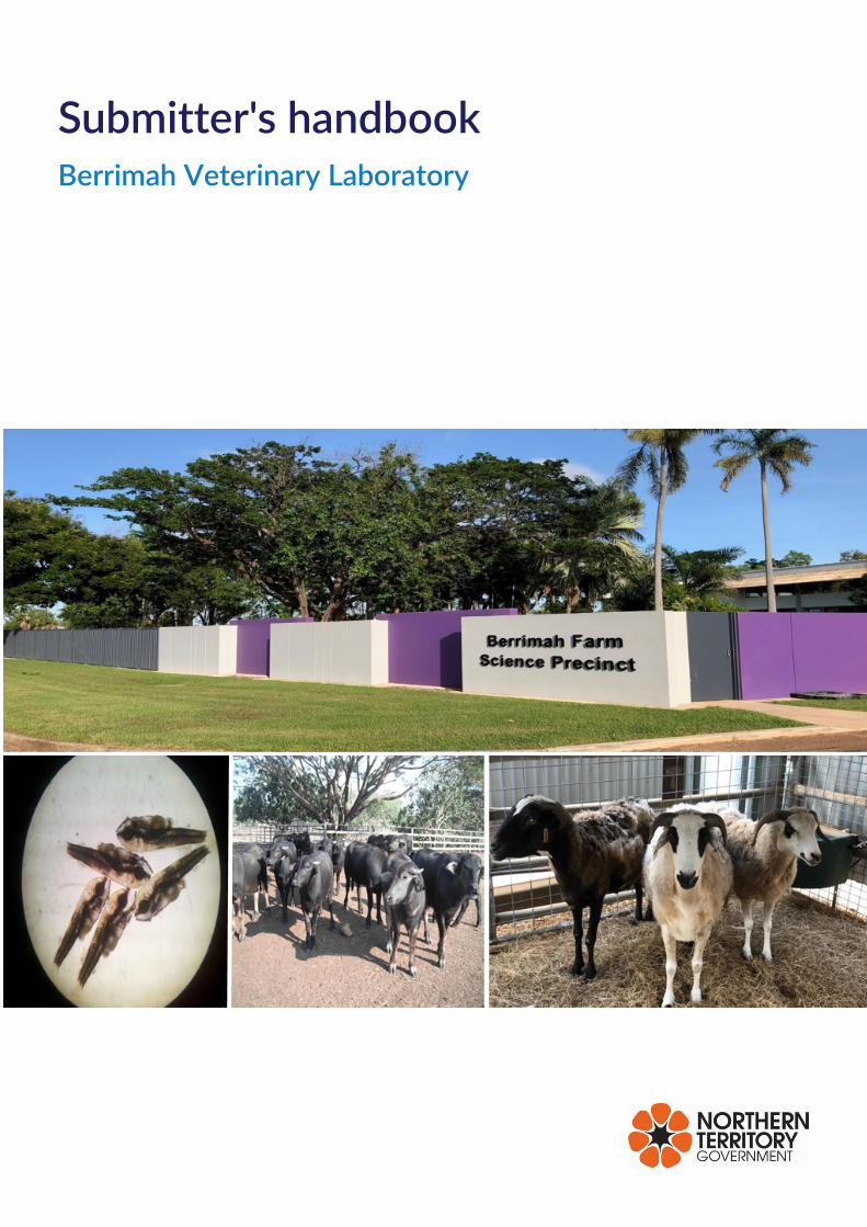
Submitter's handbook Berrimah Veterinary Laboratory
Contents 1. Introduction to Berrimah Veterinary Laboratory .................................................................................................... 4
2. Address and contact numbers for BVL ...................................................................................................................... 4
3. General submission policies ......................................................................................................................................... 4
4. Turnaround times ........................................................................................................................................................... 5
5. Export testing and movement certification .............................................................................................................. 5
6. Fee for service ................................................................................................................................................................. 5
7. List of tests and charges ............................................................................................................................................... 6
8. Reporting of results ....................................................................................................................................................... 6
9. Completing a specimen advice note (SAN) ............................................................................................................... 6
10. General advice on collection of specimens ............................................................................................................ 7 10.1. Three basic principles for specimen collection ............................................................................................... 7 10.2. Appropriate specimens ........................................................................................................................................ 7 10.3. Labelling of specimens ......................................................................................................................................... 7 10.4. Specimen containers ............................................................................................................................................ 8 10.5. Rejection of specimens ........................................................................................................................................ 8
11. General advice on storage and packaging of specimens ..................................................................................... 9 11.1. Storage of specimens ........................................................................................................................................... 9 11.2. Packaging of specimens....................................................................................................................................... 9
12. General advice on transporting specimens ......................................................................................................... 10 12.1. Notification of consignment ............................................................................................................................ 10 12.2. Freight dockets ................................................................................................................................................... 11
13. Specific advice for each section ............................................................................................................................. 11 13.1. Bacteriology ........................................................................................................................................................ 11
13.1.1. Specimen collection for general culture ................................................................................................ 11 13.1.2. Specimen collection for fungal culture .................................................................................................. 13 13.1.3. Bovine campylobacter (vibriosis) and trichomonas infections .......................................................... 14 13.1.4. Leptospirosis ............................................................................................................................................... 14
13.2. Clinical chemistry ............................................................................................................................................... 14 13.3. Cytology ............................................................................................................................................................... 14
13.3.1. Fine needle aspiration (FNA) .................................................................................................................... 14 13.3.2. Imprints, scrapings and swabs ................................................................................................................. 15 13.3.3. Body fluids ................................................................................................................................................... 15
13.4. Haematology....................................................................................................................................................... 16 13.5. Urinalysis ............................................................................................................................................................. 16 13.6. Histology.............................................................................................................................................................. 16 13.7. Necropsy ............................................................................................................................................................. 16 13.8. Parasitology......................................................................................................................................................... 17
Submitter's handbook
13.9. Serology ............................................................................................................................................................... 18 13.9.1. Leptospirosis serology ............................................................................................................................... 19
13.10. Entomology ....................................................................................................................................................... 19 13.11. Virology ............................................................................................................................................................. 19 13.12. Molecular .......................................................................................................................................................... 20
14. Appendices ................................................................................................................................................................. 23 14.1. Summary of tests available at BVL and specimens required. Tests marked with * are accredited (accreditation # 13626). .............................................................................................................................................. 23 14.2. List of preferred referring laboratories .......................................................................................................... 26 14.3. Sample sheet ....................................................................................................................................................... 31 14.4. What blood tube to use .................................................................................................................................... 32 14.5. Making a good blood smear............................................................................................................................. 33 14.6. Collecting samples for African swine fever. ................................................................................................. 34 14.7. Submission of samples for the National Transmissible Spongiform Encephalopathy Surveillance Program (NTSESP) ....................................................................................................................................................... 35 14.8. Collection of samples for bovine Johne's disease testing ......................................................................... 36 14.9. Submission of bats for Bat Lyssavirus testing .............................................................................................. 38 14.10. Submission of specimens to test for Chlamydia ....................................................................................... 39
14.10.1. From birds ................................................................................................................................................. 39 14.10.2. From crocodiles ........................................................................................................................................ 40
14.11. Bovine infertility testing ................................................................................................................................. 40 14.12. Collection of specimens from cattle with suspected tick fever ............................................................. 42 14.13. Antibiotic sensitivity testing at BVL ............................................................................................................ 43
Submitter's handbook
Department of PRIMARY INDUSTRY AND RESOURCES 28 February 2020 | Issue 9 Page 4 of 47
1. Introduction to Berrimah Veterinary Laboratory Berrimah Veterinary Laboratory (BVL) is part of the Northern Territory Department of Primary Industry and Resources (DPIR). The core function of BVL is testing for diseases of production animals and aquaculture species for diagnostic, surveillance, regulatory, research, export and exotic disease exclusion purposes. The laboratory also provides a testing service for companion animals, performance animals, aviary birds and native fauna on a fee for service basis.
Management and staff of BVL are fully committed to providing a quality testing service to our clients. BVL is accredited with the National Association of Testing Authorities, Australia (NATA) in the field of Animal Health (ISO/IEC 17025). Detailed information on the scope of our accredited services can be found on NATA’s website.
2. Address and contact numbers for BVL Delivery address:
Berrimah Veterinary Laboratory, DPIR Berrimah Farm Science Precinct 29 Makagon Road Berrimah NT 0828
Postal address:
Berrimah Veterinary Laboratory, DPIR, GPO Box 3000 Darwin NT 0801 Do not post biological samples
Contacts:
Phone: 08 8999 2249 Fax: 08 8999 2024 Email: [email protected]
All queries regarding specimen collection and submission, or results of testing, should be directed to BVL Specimen Reception.
3. General submission policies All submissions to BVL must be made through a veterinarian or animal health expert. A completed Specimen Advice Note (SAN) must accompany all submissions. Specifying requested tests on the SAN is the responsibility of the submitter. If no tests are specified, and testing does not incur a fee, testing conducted will be at the discretion of the Pathologist. Where fee for service applies, failure to specify required tests may result in delays in processing (refer to Section 9 for further instructions on completing a SAN). Additional tests may be required in order to reach a diagnosis. Where tests incur a fee, this will be discussed with the submitter before proceeding.
Submitted samples must be adequate, submitted in appropriate containers and clearly marked/labelled. Inappropriate or incomplete submissions may be rejected or processing of the submission delayed.
Staff at the Berrimah Veterinary Laboratories work under NT Public Service conditions of employment, which include a working day from 8.00 am to 4.20pm and no weekend or public holiday work, under normal circumstances.
BVL will not subcontract test services for which we are accredited except where extenuating circumstances require referral to another accredited or suitably competent facility, and agreed to by the client. For tests not conducted at BVL, or where services are temporarily unavailable, please refer to Appendix 12.2 for our list of preferred referral services.
BVL does not provide a courier service. It is the responsibility of the submitter to transport all samples at their cost. This includes samples that do not attract a test fee. There is no postal service to Berrimah Farm, please use a courier for sample submission if samples are perishable. For detailed information on samples packaging, freight and consignment notification please refer to Sections 10 and 11.
Submitter's handbook
Department of PRIMARY INDUSTRY AND RESOURCES 28 February 2020 | Issue 9 Page 5 of 47
4. Turnaround times Priority testing is given to disease outbreaks in both production and aquaculture submissions. Under normal circumstance, the following turnaround times apply to samples received before 3pm.
• Cytology, and faecal parasitology results are normally reported the following day. For parasitology submissions, interim results should be available on the following working day after submission, but identifications of parasites may need to be referred to another laboratory.
• Necropsies are usually done as soon as the body is received and an interim report should be available the same or following day. Please submit animals for necropsy before 3:00pm, or contact the laboratory before submission if after this time. Note that a disposal fee on a per kg basis applies in addition to the necropsy fee. When a necropsy is done, specimens for possible follow up testing will be collected as appropriate, but will be held until the duty pathologist has discussed further testing and charges with the submitting veterinarian.
• Histopathology is normally reported within two to four days. Samples require at least 24 hours for fixation before processing.
• Bacteriology turnaround times depend on a number of factors but for routine aerobic cultures interim results are usually reported within 48 hours. Fungal cultures may take several weeks. Specimens received on a Thursday or Friday will be held over until Monday before culturing.
• Serology and PCR tests are usually batched, thus results may not be available for up to several days. To test for a rising antibody titre, two serum samples should be taken about two weeks apart. Both samples should then be tested together.
• Please contact the laboratory if urgent testing is required. If testing is urgent, please indicate this on the SAN
5. Export testing and movement certification Please contact the laboratory if export testing is planned, what testing will be required, how many animals are involved, the likely date of bleeding and the expected date of departure.
Specimens for serological testing should be submitted 10 working days before the results are required.
Note that faecal culture for Johne's disease requires at least 10 weeks and is referred to another laboratory. Molecular testing on faeces is performed at BVL.
6. Fee for service Testing for diagnostic purposes in production animals (agricultural livestock and aquaculture species) from NT properties is provided free of charge. Testing of specimens from production animals e.g. for health monitoring or research, is charged for, unless arrangements are made with the laboratory in advance for funding under specific projects.
Other testing that attracts a charge includes: submissions by private veterinary practitioners from companion animals, horses, caged and aviary birds, backyard chickens, and wildlife, testing of specimens from cattle or other livestock for export; serological testing of groups of cattle for herd disease surveillance, including serological investigations of possible reproductive problems, and research or ad hoc disease survey by DPIR or other government departments. In general, submissions of chickens from backyard flocks not experiencing significant mortality or morbidity will be charged; assessed on a case-by-case basis at the discretion of the Duty Pathologist.
All fees and charges will be invoiced to the submitter. If you are unsure of the availability of a test and/or the charging arrangements, please contact Specimen Reception at BVL.
Submitter's handbook
Department of PRIMARY INDUSTRY AND RESOURCES 28 February 2020 | Issue 9 Page 6 of 47
7. List of tests and charges All private veterinary practices that submit to BVL are provided with a copy of BVL's List of Tests and Charges for companion animals, cage and aviary birds and wildlife. Please contact the laboratory to discuss tests that are not included on this list or to obtain a quote.
BVL provides testing in a range of fields. These include: clinical chemistry and haematology (on production animals only); bacteriology; cytology; parasitology; gross pathology (necropsy); histopathology; molecular testing; serology; virology; and viral entomology. BVL can also arrange testing by other DPIR laboratories, e.g. the Entomology or Chemistry laboratories, or refer specimens to other veterinary laboratories if the testing is not available through DPIR. Testing for suspected exotic diseases is done by the Australian Animal Health Laboratory (AAHL) in Geelong, and specimens are referred to them by BVL.
Please refer to Appendix 12.1 for a summary of tests available and required samples. Please refer to section specific information on samples collection and storage for required tests. Failure to submit an appropriate sample or incorrect storage may result in your submission being rejected. Full details on the scope of our accredited services can be obtained from NATA’s website.
8. Reporting of results Results are confidential, and only reported to the submitting veterinarian. However, BVL reserves the right to disclose test results and other relevant information to the appropriate authorities (including the Northern Territory Chief Veterinary Officer) where results indicate the presence of a disease which is notifiable or must be disclosed to the relevant authority under any applicable legislation or in the public interest.
Submission of samples to the BVL does not relieve any person of any legal obligations. Samples and relevant information submitted for testing become the property of BVL and may be used for training, education, or any purpose deemed necessary to protect our primary industries and public of the Northern Territory. This includes sharing of de-identified data among animal health agencies within Australia.
Results are routinely reported by email. If required, results may be phoned through. Submitters outside the DPIR will always receive a copy, either by email, or mail. Please indicate on the Specimen Advice Note the preferred reporting method and include the details i.e. email address, postal address.
9. Completing a specimen advice note (SAN) Please note all submissions must be made via a veterinarian or animal health expert.
A completed Specimen Advice Note (SAN) must accompany all submissions to BVL. Submitters without a SAN book can print off an electronic copy of the SAN, available at the end of in this handbook; or complete a SAN at specimen reception when they drop off the specimens. Ensure the following information is included as a minimum:
• Name, address, phone and fax numbers of submitter • Property/Locality • Date collected • Date submitted • Animal identification • Previous submission • Detailed history/clinical findings/post-mortem findings
The SAN should be clearly and legibly filled out in pen.
Submitter's handbook
Department of PRIMARY INDUSTRY AND RESOURCES 28 February 2020 | Issue 9 Page 7 of 47
• All SANs have a SAN number, or "B number" (BXXXXX), at the top right hand corner. This is the submitter's reference number. Use this number when contacting the laboratory for results or information about a submission. When using a printed electronic SAN please contact the lab to be given an appropriate SAN number.
• Specimens from a group of animals from the same species, with the same owner, can be submitted on the same SAN.
• Specimens from different owners require separate SANs. • Specimens from different species require separate SANs. • Sign and date the SAN before submitting. Print your name for reporting purposes.
10. General advice on collection of specimens
10.1. Three basic principles for specimen collection 1. The quality and value of the laboratories' results depends on the quality and appropriateness of the
specimens submitted. 2. It is always better to send extra specimens in case of need, rather than find that the specimens
required for a diagnosis are missing. 3. Specimens must be collected, stored and transported appropriately so they arrive at the lab in a
suitable condition for testing.
10.2. Appropriate specimens
• The specimens collected must be suitable for the tests required. • Specific details on the specimens required for particular tests are given in section 11 and are
summarised in appendix 1. • See appendix 5 for information on the type of blood collection tube to use. • See appendices 7 and 12 for the range of specimens required for some specific diagnostic tests • If you are still unsure of the specimens to take, and how to collect or preserve them, phone the
laboratory on 08 8999 2249 for advice, before collection.
10.3. Labelling of specimens All specimens submitted should be in individually labelled containers, using an indelible marker. Labelling should be legible and consistent with the information on the SAN.
Specifically, specimen containers should be labelled:
• With the animal identification. • With the SAN number (i.e. the ‘B’ number), especially if more than one submission is dispatched to
BVL at the same time. If using a printed SAN contact the lab for a SAN number. • If there are large numbers of specimens of the same type (e.g. cattle blood samples for export
testing) there is no need to label each tube with the SAN number, but the tubes should be packed together, separate from other specimens, with the exterior of the package clearly labelled with the SAN number.
• With the type of tissue, when submitting fresh and formalin fixed tissues. Ensure fresh tissues are in separate containers.
• With the date, especially if the same sample is collected from the same animal on different occasions.
Submitter's handbook
Department of PRIMARY INDUSTRY AND RESOURCES 28 February 2020 | Issue 9 Page 8 of 47
10.4. Specimen containers
Containers supplied
BVL supplies the regional veterinary officers and stock inspectors in the NT with a range of specimen containers, as well as packaging materials to send specimens to the laboratory.
Private veterinarians who submit specimens for “fee for service” are entitled to the following consumables for specimen collection:
• sterile specimen jars • sterile swabs in transport media • Biohazard bags • formalin • pots and buckets for larger formalin fixed specimens • blood tubes - EDTA, lithium heparin, plain tubes: 5-10mL size • glass slides • slide holders • Viral transport media • Swabs
Submit clean containers
When submitted, the outside of specimen containers should be clean. We realise that specimens are often collected in difficult and dirty conditions in the field, but please remove gross contamination from the outside of containers before submission, or put the dirty container inside a clean one or into a clean plastic bag. For those bleeding cattle, we suggest a bucket of fresh water be used to rinse the blood tubes of blood and faecal material immediately after sampling.
10.5. Rejection of specimens It is the submitter's responsibility to submit appropriate specimens in suitable condition for testing. However, if specimens are unsuitable, the duty pathologist will try to contact the submitter to clarify details, discuss other possible testing, and suggest appropriate samples.
Criteria used in deciding that a submission is unsuitable include:
• Lack of information on the SAN form and/or incomplete or no labelling of specimen containers so that it is impossible to determine the nature of the specimen or which animal is being tested.
• Illegible SAN form due to leakage of blood or other tissue fluids. • Leaking or broken specimen containers. • Samples in inappropriate containers (e.g. samples submitted in gloves or syringes with needles still
attached).Insufficient sample. • Wrong specimen for tests requested. • Pooled samples for bacteriological or viral isolation. • Test not specified on SAN. • Discrepancies in numbers of specimens. • Samples in inappropriate containers (e.g. samples submitted in gloves or syringes with needles still
attached.
Submitter's handbook
Department of PRIMARY INDUSTRY AND RESOURCES 28 February 2020 | Issue 9 Page 9 of 47
11. General advice on storage and packaging of specimens
11.1. Storage of specimens General principles are given here. Specific details for particular specimens may be found in section 11. Contact the laboratory for advice if still unsure.
• Blood in a tube with anti-coagulant (e.g. EDTA, lithium heparin) should be refrigerated immediately after collection. If collecting in the field, place the tubes in an esky with a cold ice brick straight away. It is often convenient to have a small esky at the crush or yards, and transfer the specimens in batches to a car fridge or larger esky as required.
• Blood in a serum tube should be allowed to clot at room temperature (usually less than 1 hour) before refrigerating. Keep out of direct sunlight. In the field, place in the shade, and transfer to an esky or car fridge as soon as it has clotted. If it is very hot, then put the tubes in an esky straight away.
• Blood smears should be air-dried then kept clean and dry. In the field, place the smears in a covered container immediately, to keep away from dust and flies and out of sunlight. Keep dry (not in an esky or fridge with ice). Keep away from formalin.
• Fresh tissues for bacterial or viral culture should be chilled to 4°C as soon as possible after collection. Collect different tissues into individual containers. When doing post-mortem examinations in the field, have an esky with a cold ice brick available to put tissue specimens into straight away.
• Swabs in transport medium for bacterial culture should be held at room temperature if they can be transported to the lab quickly. From experience samples sent from local clinics to the laboratory by couriers usually arrive ‘hot’. If collecting in the field, place swabs in an esky with a cold ice brick. If delivery to the lab will be delayed (e.g. several hours or overnight), they should be refrigerated.
• Formalin-fixed tissues should be kept at room temperature. Do not refrigerate or freeze.
11.2. Packaging of specimens Specimens delivered direct to BVL
When specimens are delivered by hand to the laboratory, the specimen containers should be placed inside clean plastic bags, eskies or boxes, with as much detail as possible written on the SAN and the outside of all containers labelled with the SAN number.
Specimens sent by courier or mail
The specimens must be packed to conform with IATA (International Air Transport Association) regulations, as well as the guidelines of the transport company or Australia Post. It is the submitter's responsibility to ensure they comply with all the relevant requirements. For diagnostic specimens, IATA Packing Instruction 650 applies. The Con Note must include the words ‘Biological Substance Category B’ and ‘UN3373’.
Helpful hints on routine packaging of specimens
Blood tubes
Lie blood tubes on newspaper or cotton wool. If there are a number of tubes, group them in packs of 10. Roll the samples up to make a firm bunch, making sure there is a layer between the tubes. Place in a plastic bag and tape up. (This ensures that if one tube breaks the cotton wool or newspaper will soak up the blood and the plastic bag will stop the leaks.) Alternatively they can be stood in foam racks and covered in absorbent material sealed in a plastic bag. When taping up do not tape directly over tubes as when the tape is removed it will remove any labelling including the animal ID.
Submitter's handbook
Department of PRIMARY INDUSTRY AND RESOURCES 28 February 2020 | Issue 9 Page 10 of 47
Specimen jars - containing fluid
Pack as for blood samples. Plastic jars should not need as much cushioning, but still use a bit of cotton wool or paper in the bottom of the plastic bag to soak up any leaks. Place in a plastic bag and seal. Ensure lids are firmly screwed on.
Specimen jars - containing faeces or fresh tissues
These can be grouped in plastic bags. Normally about 4-5 jars can fit in a plastic bag with some cotton wool. Seal the bag. Under no circumstances are faeces to be delivered in collection gloves. You will be asked to transfer the sample into a container before submitting.
Blood smears
The laboratory can supply you with blood smear containers. These are plastic and hold up to 5 slides. They are not airtight and should not be sent in the same esky as the formalin fixed tissues as the fumes affect the smears. They should be kept dry and out of extreme temperatures. Smears should be labelled with the animal identification on the glass slide.
Ice bricks
Packages may sit for prolonged periods out in the sun before loading, so ensure that there are adequate ice bricks.
Packing the esky or container
Place a layer of paper or cotton wool on the bottom of the esky to soak up any possible leaks. Add the packed specimens making sure you leave room for the ice brick. It is always better to send two eskies rather than try and fit too much into one. If there are spaces, fill them with paper. There should be no movement in the container once the lid is on. Pack everything with the assumption that the container will be tipped upside down, thrown around and have something heavy placed on top during transport.
Place the esky or eskies inside a cardboard box, with some extra cushioning if needed, and tape up. The cardboard box protects the esky. Couriers sometimes off-load eskies which are not packed in boxes.
12. General advice on transporting specimens For EAD (Emergency Animal Disease) exclusion, contact your local Animal Biosecurity Branch and they can assist with the transport of samples. For all other submissions to BVL it is the responsibility of the submitter to arrange transport of samples.
• Please ensure that the specimens will be delivered to the laboratory, not held at the airport or depot.
• Please notify the laboratory in advance by emailing [email protected], or phone 0889992249. If we don’t get notification, and your samples go astray, we will not know they are missing.
• Do not send specimens for overnight delivery on a Friday, unless they are urgent and you have discussed testing with the laboratory. When specimens are sent on Fridays they are not delivered until the following Monday, and may not be stored appropriately over the weekend.
12.1. Notification of consignment
• On the day the parcel is sent, phone or email ([email protected]) the details of the consignment note to the laboratory. This will alert us to expect delivery and we can chase up the parcel if it doesn't arrive.
• Notification in advance, particularly of submissions with large numbers of specimens, also assists laboratory staff to plan their workload efficiently.
Submitter's handbook
Department of PRIMARY INDUSTRY AND RESOURCES 28 February 2020 | Issue 9 Page 11 of 47
12.2. Freight dockets When completing the freight docket ensure that the following address appears:
Attention: Specimen reception Phone 08 89992249
Berrimah Veterinary Laboratory Department of Primary Industry and Resources BerrimahFarm Science Precinct 29 Makagon Road Berrimah NT 0828
For routine diagnostic specimens write "Biological Substance Category B and UN3373” in the contents box.
If your samples require refrigeration clearly write on the external package in large letters “Refrigerate Only”. This will ensure that if it is delayed it will be placed in a cool room or refrigerator before delivery. Also ensure you have ice bricks enclosed.
13. Specific advice for each section
13.1. Bacteriology
13.1.1. Specimen collection for general culture Tissue samples
• Collect specimens in as clean a manner as possible (preferably using sterile technique). Instruments used to open the gastrointestinal tract should not be used to open other organs, and samples from ‘clean’ organs e.g. liver, kidney, lung, should be collected prior to examination of the gastrointestinal tract.
• Use sterile specimen containers, and pack tissues separately to avoid cross contamination. Sealable bags may be used, but are not preferred
• Do not send the whole organ. A piece of tissue about 4cm3 in size is preferred. • If intestine is submitted, ligate the ends with string before cutting and submit the tied off section. • Ensure each tissue is identified clearly on the container. • Do not freeze tissues but refrigerate immediately and keep chilled during transport to the lab.
Contaminants will outgrow pathogens at room temperature.
Culture swabs
• Always use swabs with transport medium. Bacteria survive much better on the moist type of transport swabs. Dry swabs usually give no bacterial growth. Transport medium also helps to prevent overgrowth of contaminants.
• Be sure each swab is identified as to animal and site. • Take swab from deep in the tissue or lesion and not from the surface, where contamination is
common.
Blood culture
• Not usually used for recovery of animal pathogens because bacteraemia is intermittent. • A syringe full of blood or a swab soaked in blood cannot be used • If you suspect septicaemia (bacteraemia), phone the laboratory for information on blood culture.
The laboratory can supply blood culture bottles, with culture medium that must be inoculated as soon as the blood is collected. Add 1 part blood to 10 parts culture medium. Use strict aseptic
Submitter's handbook
Department of PRIMARY INDUSTRY AND RESOURCES 28 February 2020 | Issue 9 Page 12 of 47
technique to collect and inoculate the sample. Submit to lab immediately after collection, at room temperature.
Pus, exudate and drainage
• Using a sterile needle and syringe, aspirate material from undrained abscesses. Place the material in a sterile container. Do not submit syringes with the needle still attached. Do not submit syringes with the needle still attached.
Body fluids (pleural, synovial and peritoneal)
• Specimens are collected aseptically and placed in sterile containers.
Respiratory
• Specimens from the mouth can be collected onto a swab and placed into aerobic transport medium. • Specimens from the nose may include biopsy and nasal flush and should be transported in a sterile
tube. Nasal swabs are usually not sufficient for diagnosis. Sterile saline without a preservative may be added to the biopsy to prevent drying. Specimens should be cultured promptly but if there is a delay, store overnight at 4°C.
• Transport bronchial, transtracheal, or tracheal washes or aspirates in sterile tubes. These may be stored overnight at 4°C.
Vaginal and uterine
• Collect vaginal specimens on a swab with an aerobic transport medium, and uterine specimens either on a swab or in a sterile tube. Either may be stored overnight at 4°C if lab submission delay expected.
Urine
• For culture, urine should be collected by catheter, cystocentesis or mid-stream catch into a sterile, leak-proof container.
• Refrigerate but do not freeze. Send to the lab as quickly as possible. • If a delay of more than six hours is unavoidable before the sample reaches the laboratory, it is
suggested the sample be split into two portions: 1. Keep a well mixed sample, refrigerated, for bacterial culture. Alternatively, take a swab of the
fresh urine sample and place into transport medium. 2. To 10mL of well-mixed sample, add two drops of 10% formalin used for preserving histology
samples. The formalin will prevent the growth of organisms and will preserve structures such as cells and casts. This sample can be used for microscopy.
Milk samples
• Clean the teats and strip several streams of milk before starting collection. • Collect milk samples into clearly labelled, sterile containers. • Collect milk samples before treatment. • Milk should be refrigerated at 4°C immediately following collection and delivered to the laboratory
as soon as possible, adequately packed with cold bricks. If culture cannot be performed within 24 hours, samples may be frozen (once only) for up to two weeks without altering recoverability of pathogens.
Faecal samples
• Submit faecal samples in a sealed specimen container, no more than three-quarters full (50 grams of faeces is more than enough sample).
• Faecal swabs are satisfactory if they are placed in a tube containing transport medium, and are not dried out.
Submitter's handbook
Department of PRIMARY INDUSTRY AND RESOURCES 28 February 2020 | Issue 9 Page 13 of 47
Abortion
• Submit foetal tissues (in particular lung, spleen and kidney - in separate containers), foetal stomach content, and a small portion of placenta (containing cotyledons in the case of ruminants).
Samples for anaerobic culture
• For meaningful results from anaerobic culture, good quality specimens are essential. Aseptic technique in collection is important, and prompt delivery to the laboratory is required.
• Swabs in transport medium can be used for anaerobic culture. • Fluid specimens can be sent in the syringe in which the specimen was collected. If a syringe cap is
available, remove the needle, expel any air from the syringe and seal with the cap. Alternately, fill a small sterile container with the fluid and close the lid tightly. Do not submit syringes to the lab with the needle still attached.
• Tissue specimens can be collected and sent as for general culture.
Useful specimens for anaerobic culture include:
• foul smelling discharge, material from infected deep wounds, joint fluid • thoracic and abdominal fluids • transtracheal aspirate from pneumonic animals • necrotic tissue • aspirate from chronic otitis media and interna • blood from live animals, when anaerobic bacteraemia is suspected (use a proper blood culture vial,
contact the lab for more information.)
Antimicrobial susceptibility testing
The antibiotics included routinely in microbial sensitivity testing at BVL depend on the animal species involved and the site of collection of the specimens submitted. A summary of the antimicrobial sensitivity tests, with the routine antibiotic discs used, are outlined in appendix 13. It is the responsibility of the prescribing veterinarian to use appropriate antibiotics in food producing and non-food producing animals. BVL takes no responsibility for any inappropriate use of antibiotics in animals.
13.1.2. Specimen collection for fungal culture In general, specimens can be collected, stored and transported as for bacterial culture. For dermatophytes and superficial mycotic infections, the following guidelines apply:
Hair
• No cleaning of the site is needed. • With forceps, pluck at least 10 hairs. Choose hairs at the periphery of the lesion, particularly hairs
that are broken, thickened or irregular. For hairs broken off at skin level, use a scalpel to scrape out. Include any hairs that fluoresce under a Woods lamp.
• Place hairs between two clean glass slides, or into a clean envelope or an appropriately labelled sterile container.
Skin
• Scrape the surface of the lesion with a sterile scalpel. • Place scrapings between two clean glass slides or in a clean envelope or appropriately labelled
sterile container.
Tissue
• Collect tissue specimens aseptically from the centre and edge of the lesion.
Submitter's handbook
Department of PRIMARY INDUSTRY AND RESOURCES 28 February 2020 | Issue 9 Page 14 of 47
• Place the specimens between two pieces of sterile gauze moistened with sterile saline, or in a sterile container with a small amount of sterile saline. Refrigerate until processed.
• Storage at 4°C for up to 8-10 hours is acceptable except if a zygomycete or Pythium is suspected. These organisms do not survive well when stored at 4°C. Contact the lab for more information.
13.1.3. Bovine campylobacter (vibriosis) and trichomonas infections These tests are referred to an interstate laboratory. Discuss with the laboratory before collecting samples for bovine Campylobacter and Trichomonas cultures. Special medium is required for immediate inoculation of the sample after collection which needs to be ordered from interstate. The method is included in appendix 11.
13.1.4. Leptospirosis Leptospires are fastidious organisms and are very difficult to grow. Contact the laboratory if you are considering attempted isolation of leptospires. Kidney is the organ of choice for isolation. Samples must be aseptically collected and transported to BVL as soon as possible. From live animals, urine is considered suitable sample, but is not recommended. If delayed, refrigerate specimens.
13.2. Clinical chemistry
• Serum is the preferred sample for clinical chemistry. Blood in lithium heparin anticoagulant may be submitted.
• At least 0.5mL serum or plasma (ie at least 1mL of whole blood) is preferable. This allows for a range of tests to be done, with repeat testing if necessary.
• Check with the laboratory if a specific test is required and you are unsure of the correct specimen to submit.
• To collect serum, use a plain tube (no additive) or a tube with clot activator and allow the blood to clot at room temperature for 30 min. before refrigerating.
• If the serum cannot be submitted to the laboratory the same day, separate the serum from the cells. Once the blood has clotted and red cells settled on the bottom of the tube, aspirate off the serum with a pipette, or needle and syringe, and place into a sterile tube with no additive. Alternatively, specimens can be centrifuged and the serum removed. If using the tubes with a gel plug, the serum can remain in the tube after centrifuging, because the gel separates the serum from the cells.
13.3. Cytology
13.3.1. Fine needle aspiration (FNA)
• To obtain a FNA, insert a 22 gauge needle attached to a 10mL syringe into a superficial lump or tumor, lymph node or internal lesion, and exert suction to acquire some cells for microscopic examination.
• For optimal results, the smears made should be thin (single cell layer), and rapidly air-dried. Use only new, clean, dry slides. All glass slides must be labelled with the animal number.
• Thin smears can be made: o in a manner similar to making a blood smear (ie spreading a drop of fluid along a slide o by spraying material onto the slide, then spreading by placing another slide on top and
sliding gently apart o by squashing thick lumps of material or tissue fragments between two glass slides, then
sliding apart • When possible, several smears should be prepared, so different stains can be used if required.
Submitter's handbook
Department of PRIMARY INDUSTRY AND RESOURCES 28 February 2020 | Issue 9 Page 15 of 47
• The smears should be rapidly air-dried using a hair dryer or fan, or by waving in the air. All glass slides must be labelled with the animal number.
Note that thick slides and slides that dry slowly often have unacceptable levels of artefact.
13.3.2. Imprints, scrapings and swabs Imprints
Imprints for cytological evaluation may be obtained from external lesions on the living animal, or from tissues removed during surgery or necropsy. For example, a “touch preparation” from the cut surface of a lymph node, which has been removed for histological processing, may give a more rapid diagnosis by cytological examination.
Ensure excess blood or fluid is blotted off the surface of the tissue before touching to the slide.
Scrapings
Hold a scalpel blade perpendicular to the lesion’s cleaned, blotted surface, preferably at the edge of normal and abnormal tissue, and pull the blade across the lesion several times.
The material collected on the blade is then transferred to the microscope slide(s) and spread.
Swabs
• The most common use of swabs is for exfoliative cytological diagnosis, particularly for stage of oestrus in dogs.
• Rub the swab across the surface being sampled, then roll the swab onto several slides and rapidly air-dry
• Note that for staging oestrus, repeat smears should be examined over several days. • Swabs submitted in bacterial transport medium are not suitable for making cytology smears. If both
bacterial culture and cytology are required, submit a swab in transport medium for culture and make smears for cytology from a second swab.
13.3.3. Body fluids
• Collect fluid from a body cavity or joint aseptically. • Place a portion of the fluid into an EDTA tube to prevent clotting, particularly for cell counts. • Also place a portion of the fluid into a sterile container. This can be used for bacteriological culture
if required, as well as for cytological examination. • Body fluids should be kept cold after collection and submitted without delay, as cells degenerate
rapidly. In particular, CSF samples should be received at the laboratory within an hour of collection. Please phone the laboratory before collection of a CSF sample, to arrange for rapid examination (see below).
Cerebrospinal fluid (CSF)
Cerebrospinal fluid should be collected into a sterile tube with EDTA anticoagulant for cytological evaluation, and a separate small sample placed in a sterile plain (red-topped serum) tube in the event culture is required. Cells in CSF degrade quickly with time, therefore samples must be examined shortly after collection (i.e. can't be stored overnight). BVL uses a sedimentation technique to prepare smears for cerebrospinal fluid. This, and the manual cell counting procedures required for CSF evaluation, renders cytological evaluation of CSF at least a 1 hour-long procedure. Therefore, BVL should be notified prior to sampling CSF to advise BVL that a sample is pending, and samples must be submitted before 3 pm.
Submitter's handbook
Department of PRIMARY INDUSTRY AND RESOURCES 28 February 2020 | Issue 9 Page 16 of 47
13.4. Haematology Note that BVL only routinely accepts haematology samples from production animals.
• Whole blood in EDTA is required for haematology. • EDTA causes degeneration of white blood cells. If blood cannot be delivered to BVL the same day
it is collected, then it is essential that blood smears are made at or soon after collection, so that a differential white cell count and evaluation of white cell morphology can be done. See appendix 6 for advice on making blood smears.
• If blood is taken with a syringe and needle and then transferred to a Vacutainer, release the vacuum first by taking the cap off the Vacutainer tube, then gently squirt the blood into the tube from the syringe. This will reduce cell damage.
• Preferably fill the tube to the level indicated, but it is better to under fill than overfill. Cap the tube and invert gently several times to mix the blood with the anticoagulant.
• Tubes that are overfilled or that have exceeded the expiry date may provide inadequate anticoagulation of the blood and resultant inaccurate haematology results.
• Label the blood tubes with the date of collection and the animal identification. • Refrigerate the blood immediately after collection, and transport with an ice brick. • Keep blood smears dry, at room temperature. Keep the slides away from formalin fumes. Label
slides in pencil on the frosted end with the date and animal identification • All glass slides must be labelled with the animal number.
13.5. Urinalysis
• Routine urinalysis testing is no longer offered at BVL. (Urine can still be submitted for culture (see Bacteriology section).
13.6. Histology
• Collect samples into 10% neutral buffered formalin, which can be supplied by the laboratory. Formalin deteriorates with time (particularly if kept in a hot car), so try to use fresh formalin.
• Pieces of tissue should be no more than about 1cm thick. Slice large lesions, aiming to include the margin between normal and abnormal tissue.
• The volume of formalin should be ten times the volume of the pieces of tissue. Use suitable size, clean containers, with well-sealing lids.
• For general purposes, a range of tissues from the same animal can be placed together in one pot of formalin, but for more specific purposes when identification of particular tissues is important, put single samples into separate containers.
• Label the container with the relevant animal identification and data. Where necessary, identify the tissue sample(s).
13.7. Necropsy Bodies for post-mortem examination should be submitted as soon as possible after death. At the usual NT outdoor ambient temperatures, carcasses undergo rapid autolysis, becoming markedly autolysed after a few hours in the sun, and are largely unsuitable for examination if left outside overnight.
• Bodies should be refrigerated immediately after death. BVL has a large, walk-in cold room so submit large bodies to BVL straight away, even if the post-mortem examination has to be delayed until the following day. Carcasses submitted for necropsy after 3pm will likely be stored under refrigeration until the next day, or the following Monday if submitted late on a Friday afternoon.
• Avoid freezing bodies that are destined for post-mortem examination (unless submission is delayed for more than 48 hours).
Submitter's handbook
Department of PRIMARY INDUSTRY AND RESOURCES 28 February 2020 | Issue 9 Page 17 of 47
• Large animal post-mortems (e.g. adult horse or cattle) are best done by a veterinary pathologist or veterinarian at the property, where the carcass can be buried by the owner, and samples brought in to BVL. A government field vet should be the first point of contact to arrange a large animal post-mortem at a property. Although BVL has facilities for unloading and handling large carcasses (i.e. an overhead gantry and winch), large carcass disposal becomes problematic since it must be cut into small pieces and put into bags for burial. Most carcasses that undergo post-mortem at BVL are collected by a contractor and are buried at the dump. There are fees for carcass disposal of non-production animals by burial at BVL (see fee schedule).
• Due to biosafety concerns, return of animal bodies to owners for burial following post-mortem at BVL is extremely discouraged. Therefore, if the owners require the carcass following post-mortem, ideally the post-mortem should be done elsewhere by the submitter and samples from the necropsy submitted to BVL.
• Fish, crustaceans or molluscs submitted for post mortem examination are best submitted live. A selection of typically affected and moribund animals should be submitted. In such cases, typically affected animals should be selected, placed in plastic bags and buried in plentiful of wet ice in a well-sealed esky for transport to BVL. In addition, samples of tissues from affected animals may be taken on the farm, preserved in 10% formalin (see section 11 and submitted together with the whole fish on ice. In the event that live or chilled animals cannot be submitted, a full range of tissues preserved in 10% formalin may be submitted.
13.8. Parasitology The Northern Territory Government no longer employs a parasitologist at BVL, therefore parasitology laboratory expertise and services are limited. Routine parasitology services offered are examination of faeces for egg counts and identification of common parasites.
Faecal samples
• Faeces should be refrigerated after collection and submitted fresh as soon as possible. If there will be a delay in submission the faeces can be preserved in 5% formalin.
• Faeces should be collected directly from the rectum. Ground samples may contain the eggs of free living nematodes.
Note: DO NOT SUBMIT FAECAL SAMPLES IN GLOVES. Transfer to a Specimen Jar.
Cattle, sheep, pigs, and horses
• Use a 70mL yellow top pot and fill to exclude air. Ensure it is tightly sealed. Note: a separate faecal sample must be submitted if bacterial culture is desired, see the bacteriology section.
• As a minimum, 10g of ruminant faeces should be submitted for a faecal egg count and oocyst count. If larval culture is required a further 20g of faeces needs to be submitted.
• Cestode egg and larval culture on horse faeces requires at least 20g. • Faecal cultures for larval differentiation (in ruminants) will be performed depending on the
outcome of the egg count results. Results are available 11 days after receiving the faecal sample.
Note: A faecal egg count is the number of strongyle type eggs per gram of faeces.
Companion animals
As a minimum, 5g of faeces should be submitted for examination.
Birds
• For chickens approximately 5g is sufficient to be sampled. Birds can be confined above a plastic sheet and the droppings then collected into a container with little air. Chickens and all birds with a caecum produce two types of faeces:
Submitter's handbook
Department of PRIMARY INDUSTRY AND RESOURCES 28 February 2020 | Issue 9 Page 18 of 47
o from the caecum - fine particles, pasty, green-brown colour o from the intestine – coarse grained, loose, various colour.
• Ensure that both types are collected, as caecal worm eggs will only be present in caecal faeces. • From smaller birds, smaller samples are accepted, but negative results may not exclude infection.
Storage and submission of specimens
• Samples should be cooled immediately to prevent death of larvae, or egg hatching. Faeces should be submitted fresh and cooled. An insulated container (esky) with a cooler brick is ideal. If there will be a delay in submission (>24 hours) or no cooling available, the faeces can be preserved in 5% formalin (ie mix faeces with an equal volume of 10% formalin).
• Larval cultures cannot be performed on preserved faeces and are not as successful if there is a delay after collection, or if the faeces have been refrigerated. Split the sample after collection and refrigerate half for egg and oocyst counts and keep the other half at room temperature for larval culture.
• If protozoans are suspected, eg trichomonads, fresh samples must be submitted as quickly as possible and maintained at room temperature.
Maggots (for screw-worm rule out)
• Collect a number of larvae from the site. Drop into boiling water and then into 70% ethanol. • Screw worm kits can be obtained from reception at BVL or your regional DPIR office in Katherine,
Tennant and Alice Springs regions. If you would like a kit posted to you email [email protected] with your details.
13.9. Serology For serological testing:
Collect blood into a plain tube (no additive) or a tube with clot activator. Use of the tubes with a gel plug in the base is optional.
• Ideally, the blood should be collected with minimal trauma (to prevent haemolysis), in an aseptic manner.
• Allow the blood to clot at room temperature. • If delivery to the laboratory cannot be made the same day, separate the serum from the cells. Once
the blood has clotted, suck off the serum with a pipette, or needle and syringe, and place into a sterile tube with no additive. Alternatively, specimens can be centrifuged and the serum removed. If using the tubes with a gel plug, the serum can remain in the tube after centrifuging, because the gel separates the serum from the cells.
• Refrigerate the specimens and submit as soon as possible, packed with ice bricks. • A minimum of 1mL of serum (ie 2mL of blood) should be collected. However, if possible, collect
10mL of blood.
Interpretation of serological tests
Serological tests identify antibodies in serum. To be diagnostically useful (ie to indicate recent infection with an agent) it is essential to show at least a four-fold increase in the antibody titre between acute and convalescent serum samples. In other words, we need to test two serum samples - one taken when the animal was showing signs of disease, and the second taken 10-14 days later, when it has had time to mount an immune response. This can be very difficult to organise in an extensive field situation, but if it is possible to yard sick or suspect animals (and possibly some cohorts) and hold them for a repeat bleed in 10-14 days it can greatly increase the value of the serological tests.
Submitter's handbook
Department of PRIMARY INDUSTRY AND RESOURCES 28 February 2020 | Issue 9 Page 19 of 47
13.9.1. Leptospirosis serology BVL currently refers all Leptospirosis Serology testing. It is good practice to submit convalescent serum 10 to 14 days after the first bleed. The test may be negative in the early stages, but the second specimen may be positive or show a rise in titre compared with the first. It is always difficult to interpret the test result from a single sample. In cattle however, results from a single sample can be meaningful to determine herd prevalence due to previous herd exposure/vaccination and to conduct epidemiological studies. In dogs, acute serum samples are held pending submission of convalescent sera. The test will be performed both on the initial sample and convalescent serum at the same time to demonstrate significant changes in titre, if there is any.
13.10. Entomology Insect collections for the National Arbovirus Monitoring Program (NAMP) should be submitted on their own SAN and not included with specimens from other species (eg sentinel herd bloods).
Specimens should be submitted promptly after collection, ie within two or three days of collection. To ensure adequate preservation the collected insects should not be more that 50% of the bottle’s volume. The remaining space should be filled with 70% Ethanol.
For transport, the lid of the bottle should be firmly screwed on and secured by adhesive tape wrapped two or three times around the interface of the lid and bottle.
If material is to be posted, the alcohol should be carefully decanted leaving as little as possible in the container.
13.11. Virology Note that it can take at least three - five weeks to grow a virus isolate, and identification of the isolate may then take anything from two weeks to months, depending on the virus.
Blood samples
• If you suspect a possible viral disease, take blood in a lithium heparin tube and blood in an EDTA tube for virus isolation.
• Also collect blood in a plain (serum) tube for clinical chemistry and serology if required. The EDTA blood can also be used for haematology (preferably take two tubes). Make blood smears at the time of collection if the samples will not arrive at the laboratory the same day.
• If possible, take blood from the sick animal(s), plus some apparently normal animals from the same group.
• Store EDTA and lithium heparin blood samples at 4°C, and send to the lab as soon as possible. Serum should be allowed to clot before refrigeration. If delivery to the laboratory will be delayed, separate the serum from the clot (see section 11.8 Serology).
• If possible take a further set of samples from the same animals 2-3 weeks after the initial sampling.
Swabs and tissue specimens
• Swabs from live or dead animals should be collected using dry, sterile swabs, then immediately placed into sterile heart-brain broth. Heart-brain broth can be ordered from the laboratory and stored frozen until required.
• If a post-mortem examination is done, a range of fresh tissues should be collected, particularly liver, kidney, spleen, heart and lung, and any other tissue that appears relevant to the case.
• Take tissues as cleanly as possible (preferably using sterile technique) and place each tissue in a separate, clearly labelled, sterile container
Submitter's handbook
Department of PRIMARY INDUSTRY AND RESOURCES 28 February 2020 | Issue 9 Page 20 of 47
• Take reasonably large pieces of tissue, and include the capsule, so the surface can be decontaminated at the laboratory and the sample can be taken from the interior.
• Chill the tissues to 4°C as quickly as possible after collection, and keep chilled during transport. • Try to get the samples to BVL within 48 hours after collection. • If there will be a long delay in transport to the laboratory, freeze the tissues, and ensure they
remain frozen during transport.
13.12. Molecular Sample requirements for Molecular Testing.
Successful infectious agent detection is dependent upon a number of critical factors including:
• targeting of appropriate animals for investigation, i.e. animals actively excreting virus – generally during the acute stages of disease, and before an effective immune response has been mounted;
• collection of appropriate samples containing infectious agents; and • maintenance of an adequate level of intact infectious agent during transit to the diagnostic
laboratory.
Accuracy of results from nucleic acid detection methods is only as good as the initial samples provided for testing. Poor or denatured samples (particularly in the case of RNA viruses) will provide results from testing that may be difficult to interpret accurately. If the number of target sequences in the sample is very small, a false negative may be obtained if degradation has occurred. If samples of marginal quality, quantity or integrity are received by the laboratory the submitter will be notified and a repeat collection requested.
Sample collection
Wherever possible when sampling, endeavor to collect sufficient samples to allow nucleic acid detection tests to be performed on dedicated samples. Separation of samples at all stages of the sampling process must be ensured as minor degrees of cross-contamination that would not be significant for other types of test, may result in erroneous results by nucleic acid amplification. Contamination must be minimized to avoid false positive detection.
All samples should be collected aseptically using single use, disposable equipment and placed in sterile nuclease free containers. All sample containers must be adequately labelled, with the animal identification, using a permanent marker.
Upper respiratory tract and cloacal swabs must be stored in viral transport medium (supplied on request from the laboratory) during transit. Cloacal swabs should be taken avoiding excess solid faecal material or visible blood. Descriptions of the preferred sample type for the molecular tests currently available at BVL are provided below.
Transport
Time in transit should be minimised to maintain the integrity of the sample for analysis. Unless samples are placed in an appropriate preservative, fixative or stabilizer, they should be kept cold (or frozen) throughout transport to the laboratory. If delivering samples to the laboratory on the same day they are collected, hold at 4°C during transport. If the delivery time is expected to be in excess of 24 hours post sampling then freeze (below -20°C) and transport on dry ice, or packed with ice bricks.
Important: Swabs used for Molecular testing cannot be transported in bacterial culture transport media. Ideally swabs for molecular testing are transported in virus transport media, or in a small plain sterile container.
Submitter's handbook
Department of PRIMARY INDUSTRY AND RESOURCES 28 February 2020 | Issue 9 Page 21 of 47
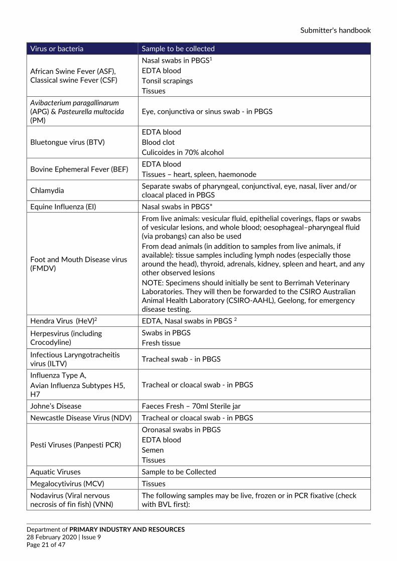
Virus or bacteria Sample to be collected
African Swine Fever (ASF), Classical swine Fever (CSF)
Nasal swabs in PBGS1 EDTA blood Tonsil scrapings Tissues
Avibacterium paragallinarum (APG) & Pasteurella multocida (PM)
Eye, conjunctiva or sinus swab - in PBGS
Bluetongue virus (BTV) EDTA blood Blood clot Culicoides in 70% alcohol
Bovine Ephemeral Fever (BEF) EDTA blood Tissues – heart, spleen, haemonode
Chlamydia Separate swabs of pharyngeal, conjunctival, eye, nasal, liver and/or cloacal placed in PBGS
Equine Influenza (EI) Nasal swabs in PBGS*
Foot and Mouth Disease virus (FMDV)
From live animals: vesicular fluid, epithelial coverings, flaps or swabs of vesicular lesions, and whole blood; oesophageal–pharyngeal fluid (via probangs) can also be used From dead animals (in addition to samples from live animals, if available): tissue samples including lymph nodes (especially those around the head), thyroid, adrenals, kidney, spleen and heart, and any other observed lesions NOTE: Specimens should initially be sent to Berrimah Veterinary Laboratories. They will then be forwarded to the CSIRO Australian Animal Health Laboratory (CSIRO-AAHL), Geelong, for emergency disease testing.
Hendra Virus (HeV)2 EDTA, Nasal swabs in PBGS 2
Herpesvirus (including Crocodyline)
Swabs in PBGS Fresh tissue
Infectious Laryngotracheitis virus (ILTV) Tracheal swab - in PBGS
Influenza Type A, Avian Influenza Subtypes H5, H7
Tracheal or cloacal swab - in PBGS
Johne’s Disease Faeces Fresh – 70ml Sterile jar Newcastle Disease Virus (NDV) Tracheal or cloacal swab - in PBGS
Pesti Viruses (Panpesti PCR)
Oronasal swabs in PBGS EDTA blood Semen Tissues
Aquatic Viruses Sample to be Collected Megalocytivirus (MCV) Tissues Nodavirus (Viral nervous necrosis of fin fish) (VNN)
The following samples may be live, frozen or in PCR fixative (check with BVL first):
Submitter's handbook
Department of PRIMARY INDUSTRY AND RESOURCES 28 February 2020 | Issue 9 Page 22 of 47
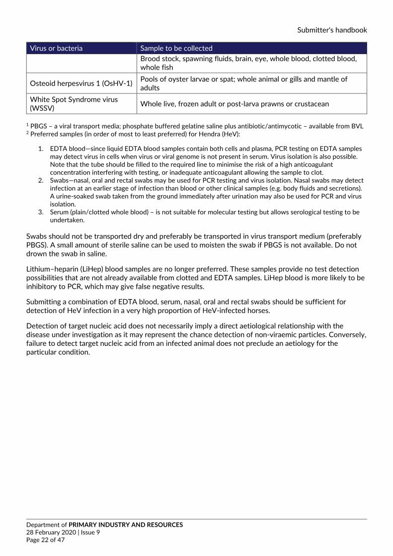
Virus or bacteria Sample to be collected Brood stock, spawning fluids, brain, eye, whole blood, clotted blood, whole fish
Osteoid herpesvirus 1 (OsHV-1) Pools of oyster larvae or spat; whole animal or gills and mantle of adults
White Spot Syndrome virus (WSSV) Whole live, frozen adult or post-larva prawns or crustacean
1 PBGS – a viral transport media; phosphate buffered gelatine saline plus antibiotic/antimycotic – available from BVL 2 Preferred samples (in order of most to least preferred) for Hendra (HeV):
1. EDTA blood—since liquid EDTA blood samples contain both cells and plasma, PCR testing on EDTA samples may detect virus in cells when virus or viral genome is not present in serum. Virus isolation is also possible. Note that the tube should be filled to the required line to minimise the risk of a high anticoagulant concentration interfering with testing, or inadequate anticoagulant allowing the sample to clot.
2. Swabs—nasal, oral and rectal swabs may be used for PCR testing and virus isolation. Nasal swabs may detect infection at an earlier stage of infection than blood or other clinical samples (e.g. body fluids and secretions). A urine-soaked swab taken from the ground immediately after urination may also be used for PCR and virus isolation.
3. Serum (plain/clotted whole blood) – is not suitable for molecular testing but allows serological testing to be undertaken.
Swabs should not be transported dry and preferably be transported in virus transport medium (preferably PBGS). A small amount of sterile saline can be used to moisten the swab if PBGS is not available. Do not drown the swab in saline.
Lithium–heparin (LiHep) blood samples are no longer preferred. These samples provide no test detection possibilities that are not already available from clotted and EDTA samples. LiHep blood is more likely to be inhibitory to PCR, which may give false negative results.
Submitting a combination of EDTA blood, serum, nasal, oral and rectal swabs should be sufficient for detection of HeV infection in a very high proportion of HeV-infected horses.
Detection of target nucleic acid does not necessarily imply a direct aetiological relationship with the disease under investigation as it may represent the chance detection of non-viraemic particles. Conversely, failure to detect target nucleic acid from an infected animal does not preclude an aetiology for the particular condition.
Submitter's handbook
Department of PRIMARY INDUSTRY AND RESOURCES 28 February 2020 | Issue 9 Page 23 of 47
14. Appendices
14.1. Summary of tests available at BVL and specimens required. Tests marked with * are accredited (accreditation # 13626).
SECTION TEST SPECIMEN REQUIRED
Bacteriology
aerobic culture (& sensitivity)* fresh tissue, fluid, swab in transport medium, etc anaerobic culture* fresh tissue, fluid, swab in transport medium, etc fungal culture* fresh tissue, fluid, swab in transport medium, hair/skin scrape, etc
blood culture* aseptically collected blood placed immediately into culture broth (broth can be obtained from the laboratory)
Leptospira culture
fresh tissues (especially kidney, liver) with minimal delay after collection
Clinical chemistry
multiple biochemical analysis (Production animals only) Serum is the preferred sample, lithium heparin (not all tests organ/system profiles serum, lithium heparin (not all tests) single tests (as requested) serum, lithium heparin (not all tests)
Cytology fluid analysis freshly collected fluid (plain tube and EDTA) smear examination unstained, air-dried, thin smears
Haematology full blood count (Production animals only) EDTA (and smears if not same day delivery)
Parasitology
faecal egg count fresh faeces (minimum 30 grams) faecal oocyst count fresh faeces (minimum 30 grams) faecal flotation fresh faeces (minimum 30 grams) faecal smear fresh faeces liver fluke sedimentation test (equine) fresh faeces (minimum 40 grams) microfilaria detection EDTA blood bovine tick fevers EDTA blood, air-dried capillary blood smears, brain and organ smears
Pathology necropsy* body
Submitter's handbook
Department of PRIMARY INDUSTRY AND RESOURCES 28 February 2020 | Issue 9 Page 24 of 47
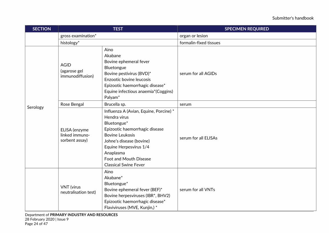
SECTION TEST SPECIMEN REQUIRED gross examination* organ or lesion histology* formalin-fixed tissues
Serology
AGID (agarose gel immunodiffusion)
Aino Akabane Bovine ephemeral fever Bluetongue Bovine pestivirus (BVD)* Enzootic bovine leucosis Epizootic haemorrhagic disease* Equine infectious anaemia*(Coggins) Palyam*
serum for all AGIDs
Rose Bengal Brucella sp. serum
ELISA (enzyme linked immuno-sorbent assay)
Influenza A (Avian, Equine, Porcine) * Hendra virus Bluetongue* Epizootic haemorrhagic disease Bovine Leukosis Johne’s disease (bovine) Equine Herpesvirus 1/4 Anaplasma Foot and Mouth Disease Classical Swine Fever
serum for all ELISAs
VNT (virus neutralisation test)
Aino Akabane* Bluetongue* Bovine ephemeral fever (BEF)* Bovine herpesviruses (IBR*, BHV2) Epizootic haemorrhagic disease* Flaviviruses (MVE, Kunjin,) *
serum for all VNTs
Submitter's handbook
Department of PRIMARY INDUSTRY AND RESOURCES 28 February 2020 | Issue 9 Page 25 of 47
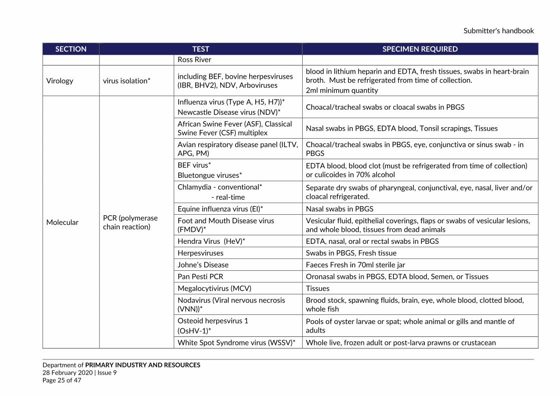
SECTION TEST SPECIMEN REQUIRED Ross River
Virology virus isolation* including BEF, bovine herpesviruses (IBR, BHV2), NDV, Arboviruses
blood in lithium heparin and EDTA, fresh tissues, swabs in heart-brain broth. Must be refrigerated from time of collection. 2ml minimum quantity
Molecular PCR (polymerase chain reaction)
Influenza virus (Type A, H5, H7))* Newcastle Disease virus (NDV)*
Choacal/tracheal swabs or cloacal swabs in PBGS
African Swine Fever (ASF), Classical Swine Fever (CSF) multiplex Nasal swabs in PBGS, EDTA blood, Tonsil scrapings, Tissues
Avian respiratory disease panel (ILTV, APG, PM)
Choacal/tracheal swabs in PBGS, eye, conjunctiva or sinus swab - in PBGS
BEF virus* Bluetongue viruses*
EDTA blood, blood clot (must be refrigerated from time of collection) or culicoides in 70% alcohol
Chlamydia - conventional* - real-time
Separate dry swabs of pharyngeal, conjunctival, eye, nasal, liver and/or cloacal refrigerated.
Equine influenza virus (EI)* Nasal swabs in PBGS Foot and Mouth Disease virus (FMDV)*
Vesicular fluid, epithelial coverings, flaps or swabs of vesicular lesions, and whole blood, tissues from dead animals
Hendra Virus (HeV)* EDTA, nasal, oral or rectal swabs in PBGS Herpesviruses Swabs in PBGS, Fresh tissue Johne’s Disease Faeces Fresh in 70ml sterile jar Pan Pesti PCR Oronasal swabs in PBGS, EDTA blood, Semen, or Tissues Megalocytivirus (MCV) Tissues Nodavirus (Viral nervous necrosis (VNN))*
Brood stock, spawning fluids, brain, eye, whole blood, clotted blood, whole fish
Osteoid herpesvirus 1 (OsHV-1)*
Pools of oyster larvae or spat; whole animal or gills and mantle of adults
White Spot Syndrome virus (WSSV)* Whole live, frozen adult or post-larva prawns or crustacean
Submitter's handbook
Department of PRIMARY INDUSTRY AND RESOURCES 28 February 2020 | Issue 9 Page 26 of 47
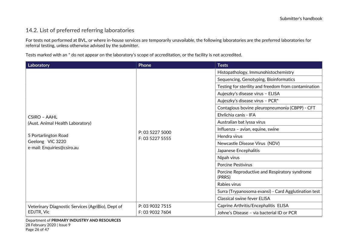
14.2. List of preferred referring laboratories For tests not performed at BVL, or where in-house services are temporarily unavailable, the following laboratories are the preferred laboratories for referral testing, unless otherwise advised by the submitter.
Tests marked with an * do not appear on the laboratory’s scope of accreditation, or the facility is not accredited.
Laboratory Phone Tests
CSIRO – AAHL (Aust. Animal Health Laboratory) 5 Portarlington Road Geelong VIC 3220 e-mail: [email protected]
P: 03 5227 5000 F: 03 5227 5555
Histopathology, Immunohistochemistry Sequencing, Genotyping, Bioinformatics Testing for sterility and freedom from contamination Aujeszky’s disease virus – ELISA Aujeszky’s disease virus – PCR* Contagious bovine pleuropneumonia (CBPP) - CFT Ehrlichia canis - IFA Australian bat lyssa virus Influenza – avian, equine, swine Hendra virus Newcastle Disease Virus (NDV) Japanese Encephalitis Nipah virus Porcine Pestivirus Porcine Reproductive and Respiratory syndrome (PRRS) Rabies virus Surra (Trypanosoma evansi) - Card Agglutination test Classical swine fever ELISA
Veterinary Diagnostic Services (AgriBio), Dept of EDJTR, Vic
P: 03 9032 7515 F: 03 9032 7604
Caprine Arthritis/Encephalitis ELISA Johne’s Disease – via bacterial ID or PCR
Submitter's handbook
Department of PRIMARY INDUSTRY AND RESOURCES 28 February 2020 | Issue 9 Page 27 of 47
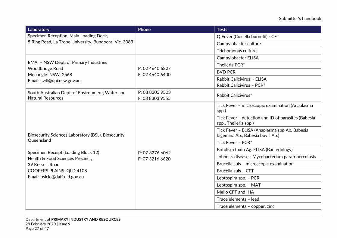
Laboratory Phone Tests Specimen Reception, Main Loading Dock, 5 Ring Road, La Trobe University, Bundoora Vic. 3083
Q Fever (Coxiella burnetii) - CFT Campylobacter culture Trichomonas culture
EMAI – NSW Dept. of Primary Industries Woodbridge Road Menangle NSW 2568 Email: [email protected]
P: 02 4640 6327 F: 02 4640 6400
Campylobacter ELISA Theileria PCR* BVD PCR Rabbit Calicivirus - ELISA Rabbit Calicivirus – PCR*
South Australian Dept. of Environment, Water and Natural Resources
P: 08 8303 9503 F: 08 8303 9555
Rabbit Calicivirus*
Biosecurity Sciences Laboratory (BSL), Biosecurity Queensland Specimen Receipt (Loading Block 12) Health & Food Sciences Precinct, 39 Kessels Road COOPERS PLAINS QLD 4108 Email: [email protected]
P: 07 3276 6062 F: 07 3216 6620
Tick Fever – microscopic examination (Anaplasma spp.) Tick Fever – detection and ID of parasites (Babesia spp., Theileria spp.) Tick Fever – ELISA (Anaplasma spp Ab, Babesia bigemina Ab., Babesia bovis Ab.) Tick Fever – PCR* Botulism toxin Ag. ELISA (Bacteriology) Johnes’s disease - Mycobacterium paratuberculosis Brucella suis – microscopic examination Brucella suis – CFT Leptospira spp. – PCR Leptospira spp. – MAT Melio CFT and IHA Trace elements – lead Trace elements – copper, zinc
Submitter's handbook
Department of PRIMARY INDUSTRY AND RESOURCES 28 February 2020 | Issue 9 Page 28 of 47
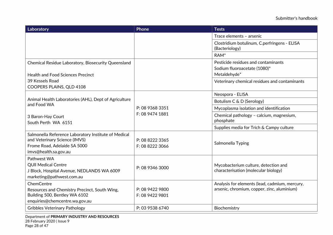
Laboratory Phone Tests Trace elements – arsenic Clostridium botulinum, C.perfringens - ELISA (Bacteriology) RAM*
Chemical Residue Laboratory, Biosecurity Queensland Health and Food Sciences Precinct 39 Kessels Road COOPERS PLAINS, QLD 4108
Pesticide residues and contaminants Sodium fluoroacetate (1080)* Metaldehyde* Veterinary chemical residues and contaminants
Animal Health Laboratories (AHL), Dept of Agriculture and Food WA 3 Baron-Hay Court South Perth WA 6151
P: 08 9368 3351 F: 08 9474 1881
Neospora - ELISA Botulism C & D (Serology) Mycoplasma isolation and identification Chemical pathology – calcium, magnesium, phosphate Supplies media for Trich & Campy culture
Salmonella Reference Laboratory Institute of Medical and Veterinary Science (IMVS) Frome Road, Adelaide SA 5000 [email protected]
P: 08 8222 3365 F: 08 8222 3066
Salmonella Typing
Pathwest WA QUll Medical Centre J Block, Hospital Avenue, NEDLANDS WA 6009 [email protected]
P: 08 9346 3000 Mycobacterium culture, detection and characterisation (molecular biology)
ChemCentre Resources and Chemistry Precinct, South Wing, Building 500, Bentley WA 6102 [email protected]
P: 08 9422 9800 F: 08 9422 9801
Analysis for elements (lead, cadmium, mercury, arsenic, chromium, copper, zinc, aluminium)
Gribbles Veterinary Pathology P: 03 9538 6740 Biochemistry
Submitter's handbook
Department of PRIMARY INDUSTRY AND RESOURCES 28 February 2020 | Issue 9 Page 29 of 47
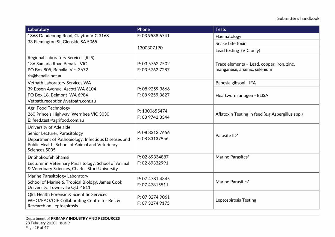
Laboratory Phone Tests 1868 Dandenong Road, Clayton VIC 3168 33 Flemington St, Glenside SA 5065
F: 03 9538 6741 1300307190
Haematology Snake bite toxin Lead testing (VIC only)
Regional Laboratory Services (RLS) 136 Samaria Road,Benalla VIC PO Box 805, Benalla Vic 3672 [email protected]
P: 03 5762 7502 F: 03 5762 7287
Trace elements – Lead, copper, iron, zinc, manganese, arsenic, selenium
Vetpath Laboratory Services WA 39 Epson Avenue, Ascott WA 6104 PO Box 18, Belmont WA 6984 [email protected]
P: 08 9259 3666 F: 08 9259 3627
Babesia gibsoni - IFA
Heartworm antigen - ELISA
Agri Food Technology 260 Prince’s Highway, Werribee VIC 3030 E: [email protected]
P: 1300655474 F: 03 9742 3344
Aflatoxin Testing in feed (e.g Aspergillus spp.)
University of Adelaide Senior Lecturer, Parasitology Department of Pathobiology, Infectious Diseases and Public Health, School of Animal and Veterinary Sciences 5005
P: 08 8313 7656 F: 08 83137956
Parasite ID*
Dr Shokoofeh Shamsi Lecturer in Veterinary Parasitology, School of Animal & Veterinary Sciences, Charles Sturt University
P: 02 69334887 F: 02 69332991
Marine Parasites*
Marine Parasitology Laboratory School of Marine & Tropical Biology, James Cook University, Townsville Qld 4811
P: 07 4781 4345 F: 07 47815511
Marine Parasites*
Qld. Health Forensic & Scientific Services WHO/FAO/OIE Collaborating Centre for Ref. & Research on Leptospirosis
P: 07 3274 9061 F: 07 3274 9175
Leptospirosis Testing
Submitter's handbook
Department of PRIMARY INDUSTRY AND RESOURCES 28 February 2020 | Issue 9 Page 30 of 47
Laboratory Phone Tests 39 Kessels Rd, Coopers Plains Qld 4108
Melbourne University Department of Microbiology & Immunology, Building 184, Ground Floor, Royal Parade PARKVILLE, VIC 3010
(03) 8344 5701 (03) 8344 5713
E.coli serotyping
Murdoch University, School of Veterinary and Life Sciences, South Street, Murdoch University, Murdoch, WA 6150
P: 08 9360 6000
Sunshine virus*
Trypanosome PCR*
Australian Water Quality Centre 250 Victoria Square, Tarntanyangga, Adelaide, SA, 5000 PO BOX 1751, Adelaide, SA, 5001 [email protected]
P: 1300653366 F: 1300883171
Algal toxins
IDEXX 3 Overend Street East, Brisbane QLD 4169
P: 0734566000 Canine Diarrhea, parvovirus, parvo 2, Clostridium perfrignes, Giardia spp., Salmonella spp. Campylobacter jejuni - PCR
Submitter's handbook
Department of PRIMARY INDUSTRY AND RESOURCES 28 February 2020 | Issue 9 Page 31 of 47
14.3. Sample sheet
Sample sheet SAN number: Lab number: Species: Owner: Sample number
Animal number or identification
Sample number
Animal number or identification
Sample number
Animal number or identification
1 34 67 2 35 68 3 36 69 4 37 70 5 38 71 6 39 72 7 40 73 8 41 74 9 42 75 10 43 76 11 44 77 12 45 78 13 46 79 14 47 80 15 48 81 16 49 82 17 50 83 18 51 84 19 52 85 20 53 86 21 54 87 22 55 88 23 56 89 24 57 90 25 58 91 26 59 92 27 60 93 28 61 94 29 62 95 30 63 96 31 64 97 32 65 98 33 66 99 100
Submitter's handbook
Department of PRIMARY INDUSTRY AND RESOURCES 28 February 2020 | Issue 9 Page 32 of 47
14.4. What blood tube to use The vast array of blood collecting tubes available, with different coloured lids, from different manufacturers, can lead to confusion, resulting in blood being submitted to the laboratory in inappropriate tubes for the tests required.
• Check the tube label to confirm what anticoagulant is present - don't just go by the colour of the lid.
• If in doubt, collect one EDTA, one lithium heparin and one clotted blood (no additive) • Make blood smears as soon as convenient from well-mixed EDTA blood (or directly after collection,
from the syringe).
EDTA
• EDTA is used for haematology, because it preserves the white blood cell and red blood cell morphology well, for up to six hours. EDTA chelates the calcium to prevent clotting.
• Blood mixed with EDTA is unsuitable for the majority of biochemical tests, coagulation studies or serological tests, however it can be used for some tests if serum is unavailable.
• EDTA is the anti-coagulant of choice for buffy coat preparations for Erhlichia detection and for PCR for bovine ephemeral fever (BEF).
Lithium heparin
• Lithium heparin is used as the anticoagulant for blood that requires viral culture • The plasma is not the preferred specimen for serological tests, but can be used for some tests if
serum is unavailable. • Lithium heparin causes clumping of the white cells and platelets, and produces a strange stain
reaction, so is unsuitable for haematology. • The plasma can be used for most biochemical tests
Clotted blood
Serum is the preferred specimen for all biochemistry and serological tests
The serum should be separated from the cells soon after clotting
TEST EDTA Lithium heparin Serum (clotted blood) Full blood count YES no no Clinical chemistry (MBA) no YES (some tests) YES Virus isolation YES YES possible from clot Agar gel diffusion tests (AGID) no no YES
ELISA no no YES Complement fixation test (CFT) no no YES
Pestivirus antigen YES from cells YES from cells YES (and clot)
BEF and BTV PCR YES no Yes from clot
Leptospirosis (MAT) No No YES
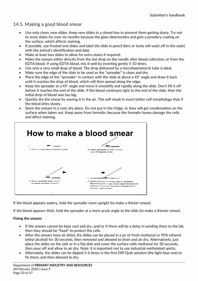
Submitter's handbook
Department of PRIMARY INDUSTRY AND RESOURCES 28 February 2020 | Issue 9 Page 33 of 47
14.5. Making a good blood smear
• Use only clean, new slides. Keep new slides in a closed box to prevent them getting dusty. Try not to store slides for over six months because the glass deteriorates and gets a powdery coating on the surface, which affects staining.
• If possible, use frosted end slides and label the slide in pencil (biro or texta will wash off in the stain) with the animal's identification and date.
• Make at least two slides to allow for extra stains if required. • Make the smears either directly from the last drop on the needle after blood collection, or from the
EDTA blood. If using EDTA blood, mix it well by inverting gently 5-10 times. • Use only a very small drop of blood. The drop delivered by a microhaematocrit tube is ideal. • Make sure the edge of the slide to be used as the "spreader" is clean and dry. • Place the edge of the "spreader" in contact with the slide at about a 45° angle and draw it back
until it reaches the drop of blood, which will then spread along the edge. • Keep the spreader at a 45° angle and move it smoothly and rapidly along the slide. Don't lift it off
before it reaches the end of the slide. If the blood continues right to the end of the slide, then the initial drop of blood was too big.
• Quickly dry the smear by waving it in the air. This will result in much better cell morphology than if the blood dries slowly.
• Store the smears in a cool, dry place. Do not put in the fridge, or they will get condensation on the surface when taken out. Keep away from formalin, because the formalin fumes damage the cells and affect staining.
If the blood appears watery, hold the spreader more upright (to make a thicker smear).
If the blood appears thick, hold the spreader at a more acute angle to the slide (to make a thinner smear).
Fixing the smears
• If the smears cannot be kept cool and dry, and/or if there will be a delay in sending them to the lab, then they should be "fixed" to protect the cells.
• After the smears have air dried, the slides can be placed in a jar of fresh methanol or 95% ethanol (ethyl alcohol) for 30 seconds, then removed and allowed to drain and air dry. Alternatively, just place the slides on the sink or in a flat dish and cover the surface with methanol for 30 seconds, then pour off and allow to air dry. Note: it is important not to use industrial methylated spirits.
• Alternately, the slides can be dipped 5-6 times in the first Diff Quik solution (the light blue one) to fix them, and then allowed to dry.
How to make a blood smear
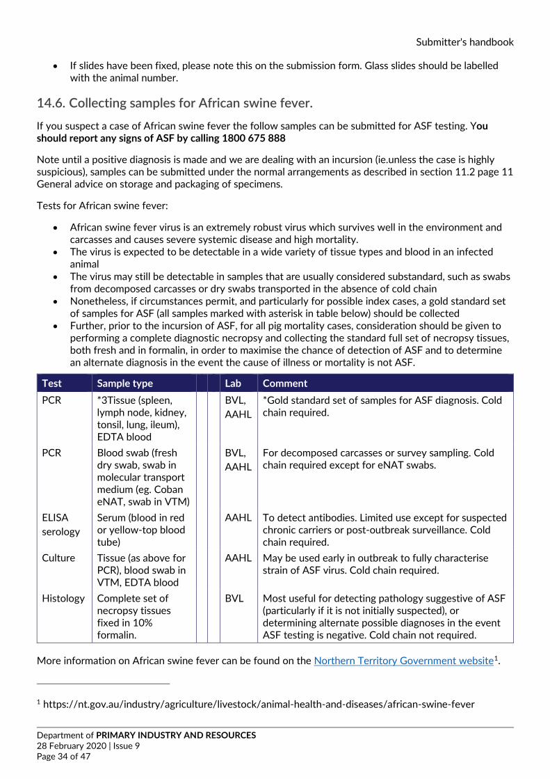
Submitter's handbook
Department of PRIMARY INDUSTRY AND RESOURCES 28 February 2020 | Issue 9 Page 34 of 47
• If slides have been fixed, please note this on the submission form. Glass slides should be labelled with the animal number.
14.6. Collecting samples for African swine fever. If you suspect a case of African swine fever the follow samples can be submitted for ASF testing. You should report any signs of ASF by calling 1800 675 888
Note until a positive diagnosis is made and we are dealing with an incursion (ie.unless the case is highly suspicious), samples can be submitted under the normal arrangements as described in section 11.2 page 11 General advice on storage and packaging of specimens.
Tests for African swine fever:
• African swine fever virus is an extremely robust virus which survives well in the environment and carcasses and causes severe systemic disease and high mortality.
• The virus is expected to be detectable in a wide variety of tissue types and blood in an infected animal
• The virus may still be detectable in samples that are usually considered substandard, such as swabs from decomposed carcasses or dry swabs transported in the absence of cold chain
• Nonetheless, if circumstances permit, and particularly for possible index cases, a gold standard set of samples for ASF (all samples marked with asterisk in table below) should be collected
• Further, prior to the incursion of ASF, for all pig mortality cases, consideration should be given to performing a complete diagnostic necropsy and collecting the standard full set of necropsy tissues, both fresh and in formalin, in order to maximise the chance of detection of ASF and to determine an alternate diagnosis in the event the cause of illness or mortality is not ASF.
Test Sample type Lab Comment PCR *3Tissue (spleen,
lymph node, kidney, tonsil, lung, ileum), EDTA blood
BVL, AAHL
*Gold standard set of samples for ASF diagnosis. Cold chain required.
PCR Blood swab (fresh dry swab, swab in molecular transport medium (eg. Coban eNAT, swab in VTM)
BVL, AAHL
For decomposed carcasses or survey sampling. Cold chain required except for eNAT swabs.
ELISA serology
Serum (blood in red or yellow-top blood tube)
AAHL To detect antibodies. Limited use except for suspected chronic carriers or post-outbreak surveillance. Cold chain required.
Culture Tissue (as above for PCR), blood swab in VTM, EDTA blood
AAHL May be used early in outbreak to fully characterise strain of ASF virus. Cold chain required.
Histology Complete set of necropsy tissues fixed in 10% formalin.
BVL Most useful for detecting pathology suggestive of ASF (particularly if it is not initially suspected), or determining alternate possible diagnoses in the event ASF testing is negative. Cold chain not required.
More information on African swine fever can be found on the Northern Territory Government website1.
1 https://nt.gov.au/industry/agriculture/livestock/animal-health-and-diseases/african-swine-fever
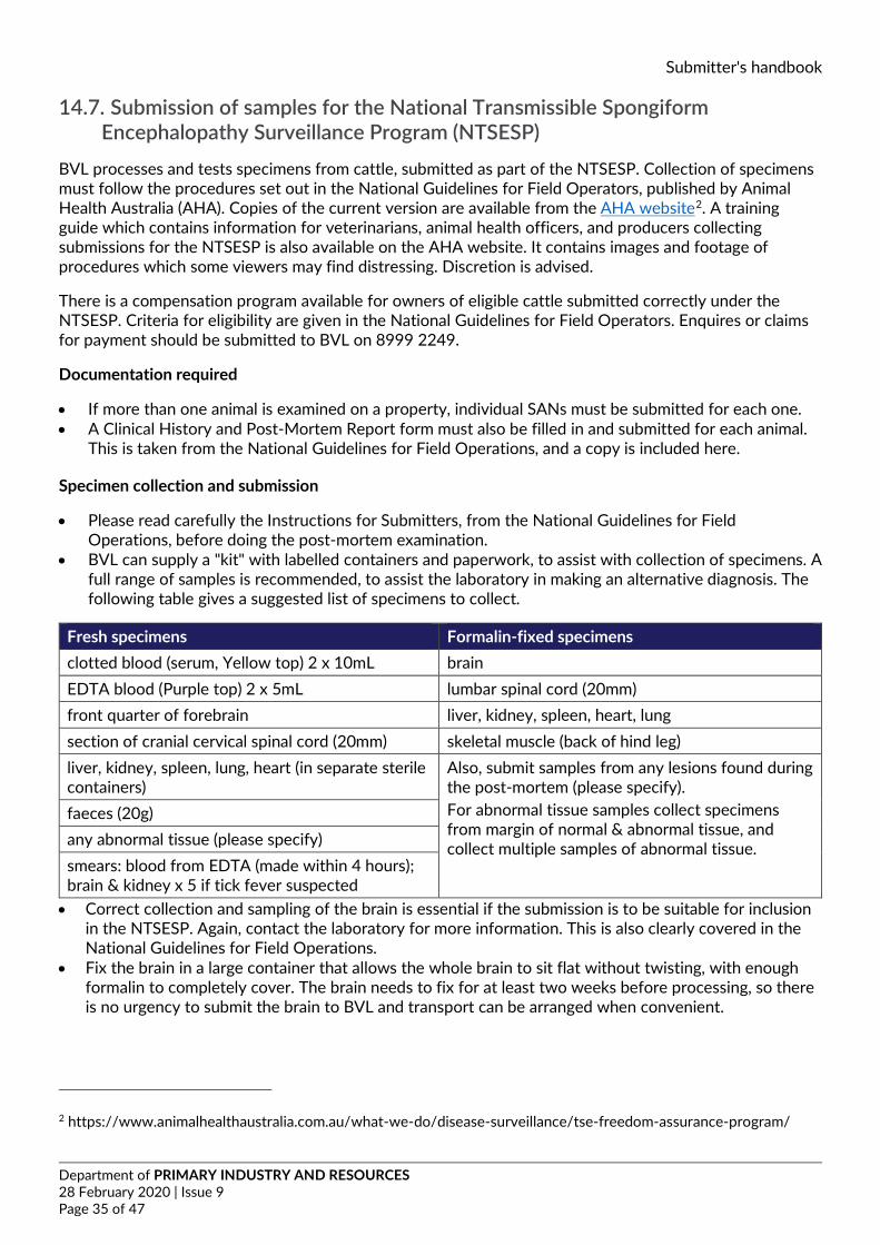
Submitter's handbook
Department of PRIMARY INDUSTRY AND RESOURCES 28 February 2020 | Issue 9 Page 35 of 47
14.7. Submission of samples for the National Transmissible Spongiform Encephalopathy Surveillance Program (NTSESP)
BVL processes and tests specimens from cattle, submitted as part of the NTSESP. Collection of specimens must follow the procedures set out in the National Guidelines for Field Operators, published by Animal Health Australia (AHA). Copies of the current version are available from the AHA website2. A training guide which contains information for veterinarians, animal health officers, and producers collecting submissions for the NTSESP is also available on the AHA website. It contains images and footage of procedures which some viewers may find distressing. Discretion is advised.
There is a compensation program available for owners of eligible cattle submitted correctly under the NTSESP. Criteria for eligibility are given in the National Guidelines for Field Operators. Enquires or claims for payment should be submitted to BVL on 8999 2249.
Documentation required
• If more than one animal is examined on a property, individual SANs must be submitted for each one. • A Clinical History and Post-Mortem Report form must also be filled in and submitted for each animal.
This is taken from the National Guidelines for Field Operations, and a copy is included here.
Specimen collection and submission
• Please read carefully the Instructions for Submitters, from the National Guidelines for Field Operations, before doing the post-mortem examination.
• BVL can supply a "kit" with labelled containers and paperwork, to assist with collection of specimens. A full range of samples is recommended, to assist the laboratory in making an alternative diagnosis. The following table gives a suggested list of specimens to collect.
Fresh specimens Formalin-fixed specimens clotted blood (serum, Yellow top) 2 x 10mL brain EDTA blood (Purple top) 2 x 5mL lumbar spinal cord (20mm) front quarter of forebrain liver, kidney, spleen, heart, lung section of cranial cervical spinal cord (20mm) skeletal muscle (back of hind leg) liver, kidney, spleen, lung, heart (in separate sterile containers)
Also, submit samples from any lesions found during the post-mortem (please specify). For abnormal tissue samples collect specimens from margin of normal & abnormal tissue, and collect multiple samples of abnormal tissue.
faeces (20g) any abnormal tissue (please specify) smears: blood from EDTA (made within 4 hours); brain & kidney x 5 if tick fever suspected • Correct collection and sampling of the brain is essential if the submission is to be suitable for inclusion
in the NTSESP. Again, contact the laboratory for more information. This is also clearly covered in the National Guidelines for Field Operations.
• Fix the brain in a large container that allows the whole brain to sit flat without twisting, with enough formalin to completely cover. The brain needs to fix for at least two weeks before processing, so there is no urgency to submit the brain to BVL and transport can be arranged when convenient.
2 https://www.animalhealthaustralia.com.au/what-we-do/disease-surveillance/tse-freedom-assurance-program/
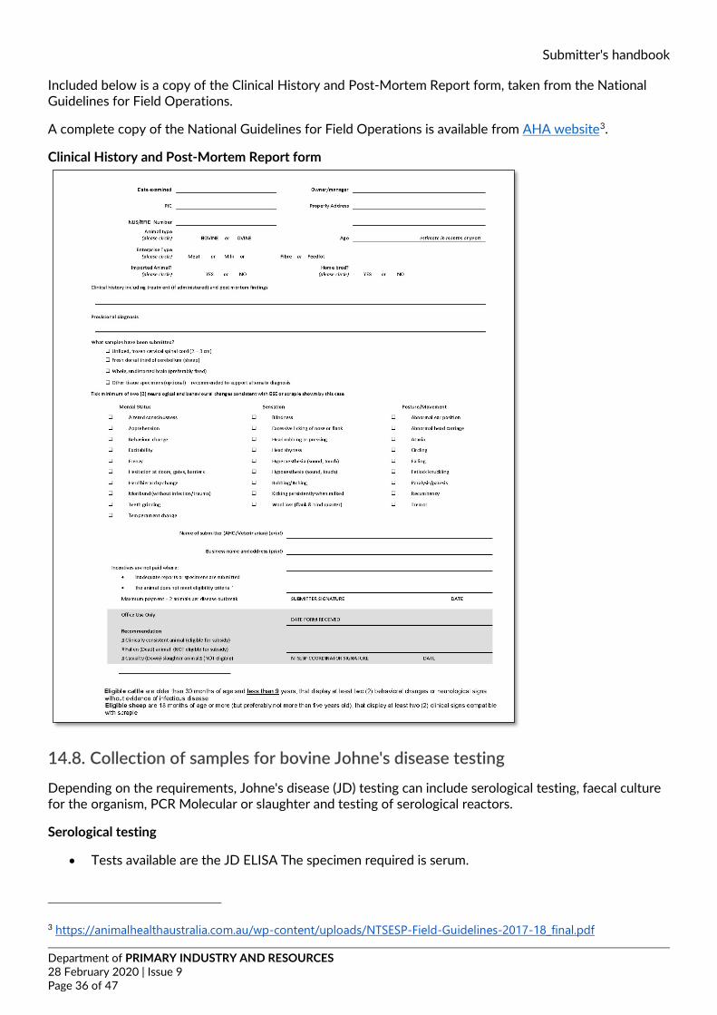
Submitter's handbook
Department of PRIMARY INDUSTRY AND RESOURCES 28 February 2020 | Issue 9 Page 36 of 47
Included below is a copy of the Clinical History and Post-Mortem Report form, taken from the National Guidelines for Field Operations.
A complete copy of the National Guidelines for Field Operations is available from AHA website3.
Clinical History and Post-Mortem Report form
14.8. Collection of samples for bovine Johne's disease testing Depending on the requirements, Johne's disease (JD) testing can include serological testing, faecal culture for the organism, PCR Molecular or slaughter and testing of serological reactors.
Serological testing
• Tests available are the JD ELISA The specimen required is serum.
3 https://animalhealthaustralia.com.au/wp-content/uploads/NTSESP-Field-Guidelines-2017-18_final.pdf
Submitter's handbook
Department of PRIMARY INDUSTRY AND RESOURCES 28 February 2020 | Issue 9 Page 37 of 47
Molecular
• PCR test:pooled HT-J faecal PCR
To minimise the cost of laboratory testing for Johne’s disease, faecal samples from up to five individual cattle may be pooled into a single HT-J PCR test. Faeces should always be collected and submitted as individual animal samples, with pooling of samples conducted at the laboratory. This provides for samples to be precisely measured as equal contributors to a pool, and for individual re-testing of contributors in the event of a positive result for a pooled sample.
Faecal culture
• The specimen required for faecal culture is fresh faeces, collected from the rectum. Fill a 70mL yellow top specimen jar with the faeces.
• The specimen should be chilled immediately (placed in an esky with a cold brick, or in a car fridge) and sent to BVL as soon as possible after collection. It is very important to keep the faecal samples chilled throughout transport.
• Faecal culture can take up to 10 weeks. Contamination of cultures is common, and in that case, collection of another sample is required to repeat the test. Keeping samples chilled from the time they are collected minimises the risk of contamination.
• If there is to be a delay in transporting the faeces to BVL, the specimen can be frozen, but it is important that it is then kept frozen during transport.
BVL will forward the specimens to the Johne's disease Reference Laboratory, Primary Industries AgriBio, where the faecal cultures are done, or regional centres can arrange to send the samples directly to AgriBio. JD PCR testing is performed by BVL
Post mortem examination
• If slaughter and testing is required of serologically positive animals, then the approved protocol must be followed. This protocol is taken from the current manual of John’s Disease in Cattle Definitions and Guidelines for the Australian Beef and Dairy Industry4. Please inform BVL before doing the post-mortem, to discuss the requirements.
• Faecal samples and fresh tissues for JD culture must be chilled immediately after collection and submitted to BVL as soon as possible. It is important that the specimens are kept chilled throughout transport. Specimens for culture are then forwarded to the Johne's Disease Reference Laboratory, for testing. Again, regional centres may choose to send samples directly to Veterinary diagnostic services Bundoora Vic.
4 https://animalhealthaustralia.com.au/wp-content/uploads/JD-in-cattle-definitions-and-guidelines.pdf
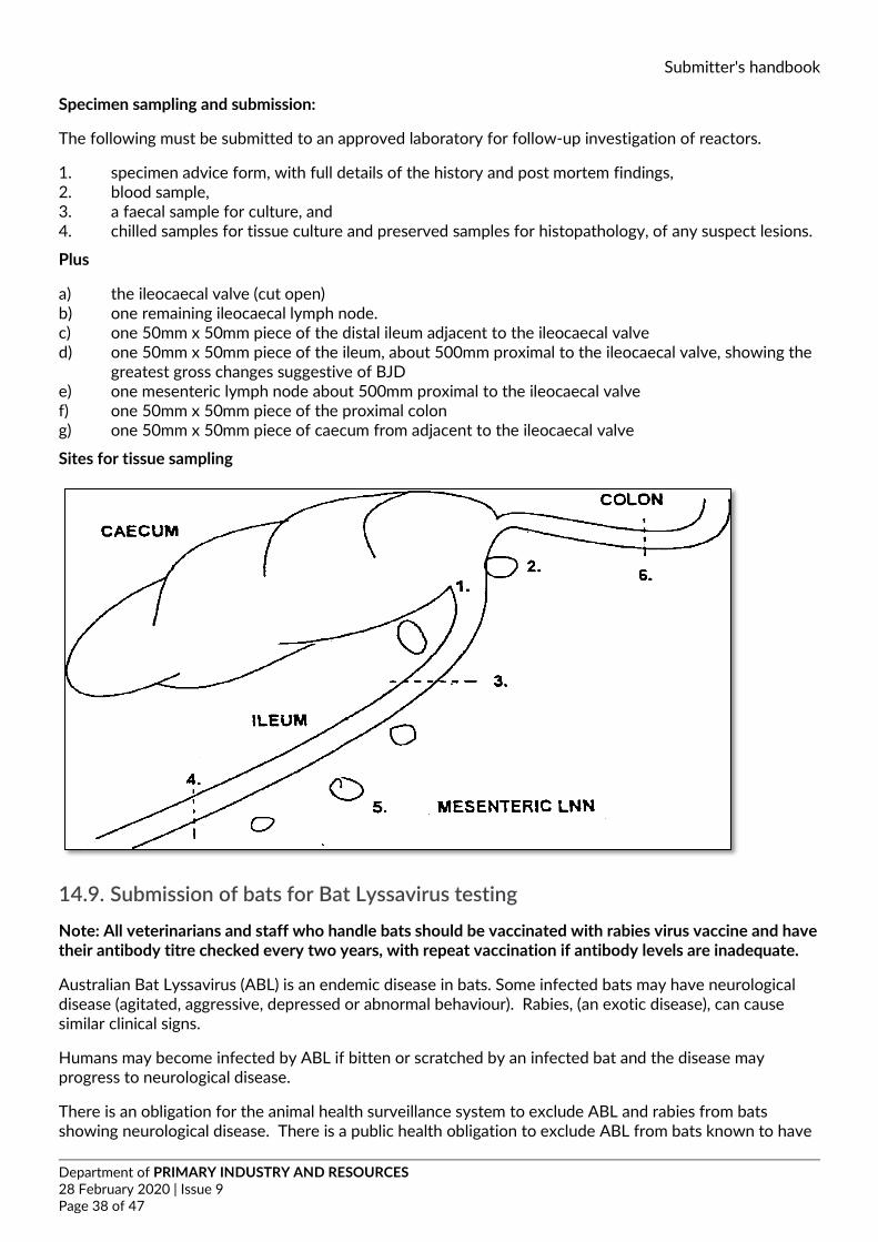
Submitter's handbook
Department of PRIMARY INDUSTRY AND RESOURCES 28 February 2020 | Issue 9 Page 38 of 47
Specimen sampling and submission:
The following must be submitted to an approved laboratory for follow-up investigation of reactors.
1. specimen advice form, with full details of the history and post mortem findings, 2. blood sample, 3. a faecal sample for culture, and 4. chilled samples for tissue culture and preserved samples for histopathology, of any suspect lesions.
Plus
a) the ileocaecal valve (cut open) b) one remaining ileocaecal lymph node. c) one 50mm x 50mm piece of the distal ileum adjacent to the ileocaecal valve d) one 50mm x 50mm piece of the ileum, about 500mm proximal to the ileocaecal valve, showing the
greatest gross changes suggestive of BJD e) one mesenteric lymph node about 500mm proximal to the ileocaecal valve f) one 50mm x 50mm piece of the proximal colon g) one 50mm x 50mm piece of caecum from adjacent to the ileocaecal valve
Sites for tissue sampling
14.9. Submission of bats for Bat Lyssavirus testing Note: All veterinarians and staff who handle bats should be vaccinated with rabies virus vaccine and have their antibody titre checked every two years, with repeat vaccination if antibody levels are inadequate.
Australian Bat Lyssavirus (ABL) is an endemic disease in bats. Some infected bats may have neurological disease (agitated, aggressive, depressed or abnormal behaviour). Rabies, (an exotic disease), can cause similar clinical signs.
Humans may become infected by ABL if bitten or scratched by an infected bat and the disease may progress to neurological disease.
There is an obligation for the animal health surveillance system to exclude ABL and rabies from bats showing neurological disease. There is a public health obligation to exclude ABL from bats known to have
Submitter's handbook
Department of PRIMARY INDUSTRY AND RESOURCES 28 February 2020 | Issue 9 Page 39 of 47
bitten or scratched humans. Regarding pet (dog or cat) exposure, there has never been a case of ABLV in a dog or cat, and they are not known to be susceptible to the virus. However, if dogs or cats are exposed to bats, given the theoretical possibility of infection, the bat can be tested for ABLV.
Dead bats can be submitted for laboratory examination including exclusion of ABL and rabies to diagnose the cause of death under the following conditions.
1. Bats found alive with abnormal nervous signs
• exclude ABL and rabies • other tests at the discretion of the pathologist • no charges to submitter
2. Bats known to have bitten or scratched a person or that have been exposed to pets
• (dogs or cats) • exclude ABL • No charges to submitter
4. Bats found dead
• tests requested by submitter • other tests at the discretion of the pathologist with the knowledge of the submitter • submitter will be charged for laboratory testing.
For background information on bat lyssavirus, go to the link below to see the information sheet ‘Australian Bat Lyssavirus, Hendra Virus and Menangle Virus information for Veterinary Practitioners’.
• The Department of Health (Australian Government) website5 • Northern Territory Government website6
The Parks and Wildlife Service of the NT no longer provide a rescue service for injured, ill or orphaned native wildlife. This service is now provided by organisations within the community that continue to work closely with the Parks and Wildlife Service.
There are volunteer organisations who; where possible, will collect, care and release injured or orphaned animals. If you find an injured animal please contact on one of these organisations for assistance.
For more information visit the Northern Territory Government website7
14.10. Submission of specimens to test for Chlamydia
14.10.1. From birds Specimens required for this test can be taken from live or dead birds.
Note: Chlamydiosis is a zoonotic disease. All birds are potentially susceptible to infection with Chlamydophila psittaci, but captive psittacines (parrots) and pigeons are most commonly infected. Dead birds with suspected chlamydiosis should be submitted to BVL for post-mortem examination, where the PM can be done in a biological safety cabinet.
5 https://www1.health.gov.au/internet/main/publishing.nsf/Content/cdna-song-abvl-rabies.htm 6 https://nt.gov.au/environment/animals/australian-bat-lyssavirus-and-your-pet 7 https://nt.gov.au/environment/animals/report-injured-wildlife-or-rescue
Submitter's handbook
Department of PRIMARY INDUSTRY AND RESOURCES 28 February 2020 | Issue 9 Page 40 of 47
Specimens from live birds
• Swabs of the conjunctiva, nasal discharge or cloaca can be taken from the live bird. False positives can occur with cloacal swabs
• Swabs should not be transported dry and preferably be transported in virus transport medium (preferably PBGS). A small amount of sterile saline can be used to moisten the swab if PBGS is not available. Do not drown the swab in saline.
• Swabs can be stored refrigerated for up to 5 days before testing.
Specimens from dead birds
• From freshly dead birds, the same specimens can be collected as for live birds (ie swabs of conjunctiva, nasal discharge and cloaca)
• Preferably, submit dead birds with suspected chlamydiosis to BVL for post-mortem examination. If a post-mortem is done on site, submit pieces of fresh liver and spleen, as well as formalin-fixed liver and spleen, plus a full range of tissues in formalin. If delay in submitting specimens is likely (over 24 hours) also take swabs of the cut surface of the liver and spleen. These should be made using a sterile swab and placed in PBGS, A small amount of sterile saline can be used to moisten the swab if PBGS is not available. Do not drown the swab in saline moistened with sterile saline (break swab off into a sterile container and add a few drops of sterile saline). Swabs can be stored refrigerated for up to 5 days before testing.
14.10.2. From crocodiles BVL use a diagnostic PCR to determine the presence of Chlamydia (for either birds or crocodile submissions). BVL prefer to receive swabs in PBGS for all aspects of testing for Chlamydia.
Samples required:
• pharyngeal and/or conjunctival swabs (preferred) • liver or cloacal swabs (can be included)
14.11. Bovine infertility testing Collection of specimens from bulls for Trichomonas culture
Note: This procedure requires specific transport and culture media, which must be inoculated at the time of collection. BVL refers this testing interstate and culture media will need to be obtained from the referral testing laboratory. Please contact the laboratory prior to organising bull testing.
Equipment needed:
• Sterile tube with flat bevelled end, with an external diameter of 1cm and about 60cm in length • Firm rubber bulb with a capacity of about 85mL • Universal container containing 5mL saline • Plastic pipette, graduated to 1mL with a 5mL bulb • Additional sterile saline to dilute viscous samples.
Procedure:
Collecting the specimen:
• Put the bull in crush and fasten nearest hind leg with leg rope • Using the tube with the bulb attached, draw up the sterile saline • Introduce the tube to the full length of the preputial cavity. Squeeze the bulb to release the saline
and hold the preputial orifice firmly with one hand around the tube to prevent the saline from escaping
Submitter's handbook
Department of PRIMARY INDUSTRY AND RESOURCES 28 February 2020 | Issue 9 Page 41 of 47
• Squeeze the bulb and collect material in the ventral fornix and then suck up material along dorsal surfaces of penis and surrounding preputial mucosa for 30 seconds to one minute, controlling placement of the flat bevelled end of pipette through wall of prepuce by hand. The flat bevelled end of the pipette is directed onto these surfaces during collection
• Withdraw the tube, and place the collected material back into the universal container. Allow the material to settle for about 10 minutes. Add additional sterile saline if the collected material is very viscous
• 0.5mL of the sediment that settles to the bottom of the container will be used for the Trichomonas inoculation.
Inoculation of the “InPouch TF" pouch for Trichomonas
• Remove the pouch from the box. Fold pouch back over itself to reduce the crease. Make sure that the liquid in the upper chamber is below the closure tape to prevent fluid from leaking upon opening. Tear open the pouch at the notch just above the closure. Open the pouch, sufficiently to admit the pipette, by pulling the closure tape’s middle tabs apart
• Using the plastic pipette, suck up approximately 0.5mL of sedimented material from the bottom of the sample container. Insert the pipette tip into the liquid of the pouch’s upper chamber. The pouch is inoculated by expelling approximately 0.5mL and no more than 1mL of the sample out of the pipette
• Express the contents of the upper pouch chamber into the lower chamber. Roll down the pouch until the tape is at the bottom of the medium; the label should still be visible. Fold the wire tape’s end tabs to lock the roll.
• Sample transport to the laboratory Forward samples to the laboratory as soon as possible. Samples MUST be received at the laboratory within two days.
During transit, store the samples at ROOM Temperature. DO NOT REFRIGATE samples, or expose them to direct sunlight or temperatures above 35°C.
If sending samples as freight, they must be packaged according to requirements for biological samples.
Submission of specimens for bovine venereal campylobacteriosis testing by ELISA.
Scope: The ELISA detects IgA antibodies in the vaginal mucus. The test is a useful aid for abortion and/or infertility investigations. This test is performed at EMAI, NSW.
Materials needed
• 4.5mL phosphate buffered saline containing 0.05% Tween 20 (PBST) • Sterile swab
Note: These materials can be directly obtained from EMAI
Procedure
• Following cleaning of the perineum the swab is introduced into the vagina as cranially as possible • The swab should be pressed against the vaginal wall and turned a few times to ensure full
saturation • Following sampling, the cotton head of the swab should be cut and placed in PBST, refrigerated
and then despatched with a completed specimen submission form to EMAI as soon as possible.
Please note for regional submitters: It is advised to contact EMAI on 02 4640 6327 to obtain collecting material directly. This will save time, as the kits can be sent directly to you.
Submitter's handbook
Department of PRIMARY INDUSTRY AND RESOURCES 28 February 2020 | Issue 9 Page 42 of 47
14.12. Collection of specimens from cattle with suspected tick fever Tick fevers of cattle can be caused by three blood parasites: Babesia bovis, Babesia bigemina or Anaplasma marginale. The following samples should be collected from animals with suspected tick fever.
Sick animals Take blood in EDTA and serum tubes. Make thin blood smears from the EDTA blood and from the tail tip or ear. Also, bleed a few healthy animals from the same group.
Freshly dead animals Make thin blood smears from the tail tip or ear. Make organ smears (kidney, heart muscle, spleen, liver, brain). Also sample any sick animals and a few healthy ones, as above.
Decomposing animals As above, but the best organs to use are spleen and brain.
Making tail tip and ear tip smears
• These are difficult to make well. The tail or ear tip must be clean and dry. Clip the hair off and rub the skin really well with a damp cloth to get the surface dirt and cells off, then rub again with a dry cloth.
• Make the smear basically as for a blood smear (see appendix 6). • Use a needle or scalpel tip to make a small incision, then squeeze to form a drop of blood. The blood
should ooze, not run from the incision. • Don't drop the blood straight onto the slide. Instead, use the spreader to collect a small drop and
transfer it to the slide to make the smear. • The smears should be thin. Air dry rapidly. • Label the smears in pencil on the frosted end with the animal identification and the date.
Making brain and organ smears
• Brain and organ smears are mainly for detecting Babesia bovis, which causes infected red blood cells to sludge in the capillaries.
• Organ smears should be thin. Kidney, spleen and heart muscle are the most useful. They can be made either as organ blood smears or impression smears.
• For organ blood smears squeeze out a drop of blood from the cut surface of the organ and transfer it to a slide to make a thin smear, as for a blood smear.
• For impression smears take a freshly cut surface of a small piece of the organ, wipe it with a clean knife or scalpel blade to remove excess surface blood, then lightly press onto the slide. Air dry rapidly. If the tissue is too bloody, the slide will be too thick. Brain smears also need to be thin. Don't use too much brain - a match head size piece or less is all that is required. Place this onto one slide. Place a second slide on top. Gently squash the brain between the slides, then drag the slides apart without lifting them, to smear the brain between the two slides. Air dry rapidly. If the slides don't dry after waving in the air for a few seconds they are too thick.
• Ensure all smears are labelled in pencil on the frosted end with the animal identification, organ and date.
Points to note
• Take ordinary blood samples as well as the tail or ear tip smears from live animals, ie an EDTA tube and a plain tube for serum. Make smears from the EDTA blood within a couple of hours after collection if possible. Anaplasma can be found in jugular blood, but tends to fall off the cells during storage, so if smears are not made until the blood reaches the lab then the organism will have gone.
• All blood and organ smears should be air-dried rapidly (ie wave them around for a few seconds until they are dry) and stored dry and tightly sealed. They must not get wet and they must not be exposed to formalin fumes. If transporting the smears with samples that need an ice brick, make sure the smears will not get condensation on them. Pack separately to any formalin samples.
Submitter's handbook
Department of PRIMARY INDUSTRY AND RESOURCES 28 February 2020 | Issue 9 Page 43 of 47
• The smears can be methanol fixed (see appendix 6) to protect them. However, some of the staining techniques for blood parasites require unfixed smears, so if smears are made in duplicate only fix one smear from each pair. Record which smears are fixed.
Anaplasma antibody detection ELISA
BVL also has an antibody ELISA for Anaplasma. This test is not NATA accredited and is mainly used for research and surveillance purposes. Serum is the sample of choice.
14.13. Antibiotic sensitivity testing at BVL The Bacteriology section follows the guidelines from the Performance Standards for Antimicrobial Disk and Dilution Susceptibility Tests for Bacteria isolated from Animals, by the CLSI (Clinical and Laboratory Standards Institute). The CLSI standard provides three interpretative categories:
1. Sensitive – an infection due to the organism can be inhibited by achievable serum or tissue levels of the antibiotic when used appropriately
2. Intermediate – borderline results, where attainable serum or tissue levels MAY be sufficient to inhibit the organism, but response rates may be lower than for organisms in the “Sensitive” category.
3. Resistant – the organism is not inhibited by the usually achievable systemic concentrations of the antibiotic.
Table 1 shows the antibiotics that are tested on a routine basis for different species when the submitter requests general culture and sensitivity. There are some situations where the antibiotic reported (eg cephalothin) is not the antibiotic routinely used in practice. This is because the standard stipulates the antibiotic to be tested, and the result can then be extrapolated to the antibiotics regularly used (see footnotes to the table, and Table 2).
Table 1. Antibiotics tested routinely for different species for general culture and sensitivity
Antibiotic disc used dogs♠ horses cattle pigs poultry crocs♣ Ampicillin1 yes Amoxicillin/clavulanic acid yes Cephalothin2 yes Gentamicin yes Neomycin3 yes yes Penicillin yes yes yes Sulphafurazole4 yes yes yes Tetracycline5 yes yes yes yes yes yes Trimethoprim/sulfamethoxazole6 yes yes yes yes yes yes
1 used to represent ampicillin and amoxicillin 2 used to represent the first generation cephalosporins, eg cephalexin, cefadroxil 3 CLSI interpretative criteria are not given for use of neomycin in animals. Results are interpreted according to criteria used for humans. 4 used to represent all of the commercially available sulfonamides 5 used to represent all tetracyclines, and the results can be applied to chlortetracycline, oxytetracycline and doxycycline. However, certain organisms may be more susceptible to doxycycline than to tetracycline 6 used to represent all the potentiated sulfonamides.
• CLSI interpretative criteria are not specified for cats. Results are reported by the laboratory according to the CLSI criteria for dogs.
• CLSI interpretative criteria are not available for crocodiles.
Submitter's handbook
Department of PRIMARY INDUSTRY AND RESOURCES 28 February 2020 | Issue 9 Page 44 of 47
Non-routine antibiotics for sensitivity testing
If the submitting veterinarian requires other antibiotics to be tested in a particular situation, then these antibiotics need to be requested on the submission form. The antibiotics available for testing on request, with comments on the CLSI criteria and reporting, are listed in Table 3.
Table 2: Groups of antibiotics considered to have the same antibacterial activity
Antibiotic tested Other antibiotics in the same group ampicillin amoxycillin cephalothin first generation cephalosporins clindamycin lincomycin oxacillin cloxacillin sulphafurazole commercially available sulfonamides tetracycline includes oxytetracycline, chlortetracycline and doxycycline, but doxycycline
may have additional activity trimethoprim/sulpha-methoxazole
potentiated sulfonamides
Table 3: Antibiotics available for testing on request
Antibiotic CLSI criteria available for: Comments Ceftiofur pigs, cattle horses dogs Third generation cephalosporin available as
large animal, injectable preparation Chloramphenicol dogs Will not be reported for any food-producing
animal Clindamycin dogs Clindamycin is tested as the class
representative for both clindamycin and lincomycin
Enrofloxacin dogs, cats, poultry Use of enrofloxacin on food producing animals is not recommended.
Erythromycin pigs, cattle, poultry Lincomycin See clindamycin See clindamycin Oxacillin bovine mastitis Used to detect sensitivity of Staphylococci to
cloxacillin Streptomycin not included Will not be reported for any food-producing
animal Ticarcillin horses (extra label)
It is up to the submitter to choose and request relevant antibiotics for the clinical condition. The laboratory does not routinely report different lists of antibiotics for different situations, eg mastitis, urinary tract infections, wounds, bone infections. The only specific list of antibiotics reported is for ear infections in small animals. These are topical antibiotics, and include the following: enrofloxacin, framycetin, gentamicin, neomycin, polymixin B.
Submitter's handbook
Department of PRIMARY INDUSTRY AND RESOURCES 28 February 2020 | Issue 9 Page 45 of 47
Antibiotic sensitivity testing for anaerobic organisms.
This is not routinely done. The choice of antibiotics for anaerobic bacteria is usually empirical. The following table can be used to assist with drug choice.
Table 4: Antibiotics useful for anaerobic bacteria
Bacteria Drugs of choice Alternatives Anaerobes - Gram positive (eg Clostridium spp.)
Penicillin Clindamycin, Tetracycline
Anaerobes - Gram negative (except Bacteroides fragilis)
Penicillin Clindamycin, Metronidazole, Chloramphenicol (not food producing animals)
Anaerobes - Bacteroides fragilis
Clindamycin, Metronidazole,
Chloramphenicol (not food producing animals)
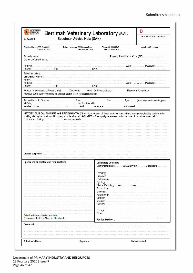
Submitter's handbook
Department of PRIMARY INDUSTRY AND RESOURCES 28 February 2020 | Issue 9 Page 46 of 47
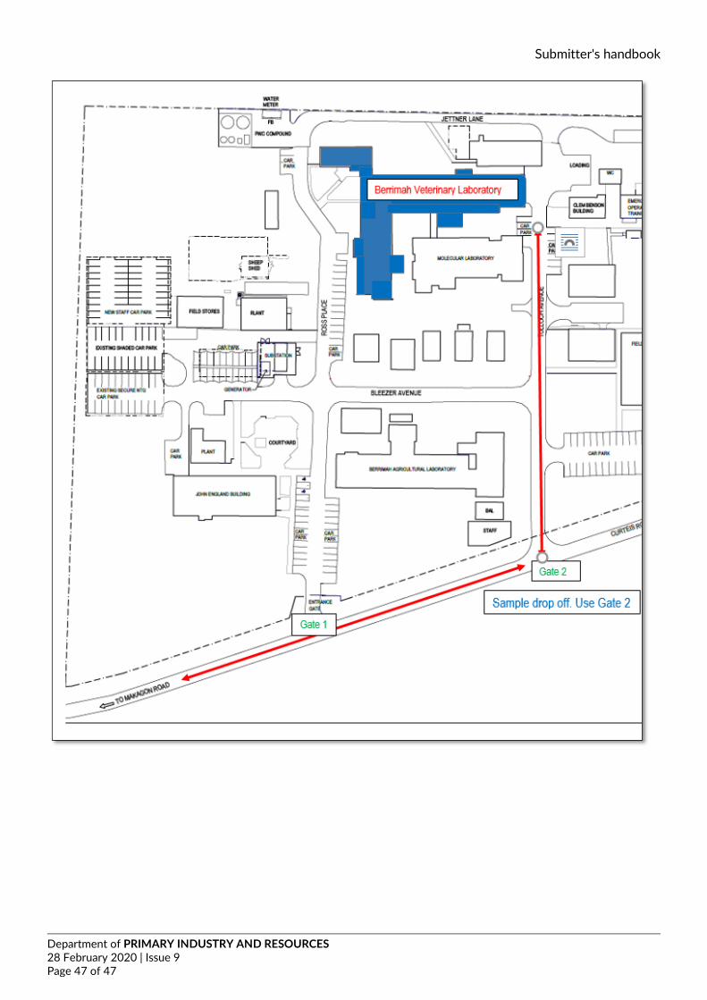
Submitter's handbook
Department of PRIMARY INDUSTRY AND RESOURCES 28 February 2020 | Issue 9 Page 47 of 47