Embed Size (px)
Citation preview
Available online on www.thepharmaresearch.info
43 | P a g e
THE PHARMA RES EARCH
A J o u r n a l o f P h a r m a c y R e s e a r c h
O r i g i n a l A r t i c l e I S S N - 0 9 7 5 - 8 2 1 6
I m p a c t - 0 . 5 2 9 Refer website
BIOADHESIVE BRAIN TARGETED NASAL DELIVERY OF AN ANT ISCHEMIC
DRUG
N. M. Morsi, D.M. Ghorab, H.A.Badie*
Affiliation:
Department of Pharmaceutics, Faculty of Pharmacy Cairo University, Kasr El Ainy street 11562, Cairo, Egypt
ABSTRACT
Transnasal delivery is a non-invasive method of bypassing the BBB to deliver the drug substances and
peptides to the CNS. Hence, in this work Vinpocetine SLNs was prepared by the high shear
homogenization and ultrasonication method. Ten different bioadhesive nasal gels of Vinpocetine
SLNs were prepared. Particle size analysis before and after SLNs dispersion and after 30 days
storage in the refrigerator, Rheological Measurements, ex-vivo bioadhesive strength,
histopathological studies and ex vivo permeation studies were performed to evaluate the prepared
Vinpocetine SLNs bioadhesive nasal gels. Moreover, Vinpocetine SLNs tissue distribution and an in
vivo pharmacokinetic study were carried. Bond strength ranged from 53.86 N/m2 to 2002.104
N/m2.The drug targeting index (DTI%) was 380.46% and the nose to brain direct transport
percentage (DTP%) was 73.71% suggesting a high targeting efficiency of the prepared Vinpocetine
SLNs bioadhesive nasal gel formula G10.
Key Words: Vinpocetine, Tissue distribution, Bioadhesion, Nasal gel, Mucoadhesive.
Available online on www.thepharmaresearch.info
44 | P a g e
INTRODUCTION
The euphoria derived from the sniffing of
cocaine in conscious subjects occurs rapidly
(within 3–5 min). It has been suggested that the
reason for such rapid effects is, apart from a
rapid nasal absorption, the presence of a direct
pathway from the nasal cavity to the CNS and
the capacity of the drug to concentrate
selectively in specific regions in the brain.
Various studies in animal models have
confirmed that, at early time points after nasal
administration, the concentration of cocaine in
the brain was higher after nasal administration
than after intravenous injection, thereby
showing the existence of the pathway from
nose to brain.[1, 2]
It was reported that nasal mucosa is composed
of respiratory mucosa and olfactory mucosa.
Under the former there are plenty of vascular,
through which preparations can be absorbed
into systemic circulation, and from the latter
preparations can be directly delivered to CNS,
bypassing the BBB.[1]
The use of solid lipid nanoparticles (SLNs) may
offer an improvement to nose-to-brain drug
delivery since they are able to protect the
encapsulated drug from biological and/or
chemical degradation, and extracellular
transport by efflux proteins such as P-gp.
Particle size is an important property that is
associated with the mucosal transport, and
particles smaller than 100 nm, in general, have
higher transport. [3, 4] Furthermore, it has been
previously reported that a small diameter
potentially allows nanoparticles to be
transported transcellularly through olfactory
neurons to the brain via various endocytic
pathways of sustentacular or neuronal cells in
the olfactory membrane.[5-7]
An effective formulation strategy for nasal
administration is the use of bioadhesive
delivery systems. The aim is to promote
adhesion of the formulation to the nasal
mucosa, allowing an extended period of contact
for drug absorption to occur. Nasal
administration of bioadhesive polymer gels can
be technically challenging and may require a
specialized device, and there will also be a limit
to the viscosity of gel that can be formulated for
convenient nasal administration.
Vinpocetine is used in ischemic stroke. It lowers
blood viscosity in patients with cerebrovascular
disease, as a result of its vasodilating properties
Vinpocetine, when taken on an empty stomach,
has an absorption rate of 6.7 percent. The
elimination half-life of the oral form is one to
two hours and the majority of vinpocetine is
eliminated from the body within eight hours.
The aim of this work is to formulate Vinpocetine
SLNs bioadhesive nasal gel as a targeted drug
delivery system able to provide a sustained,
effective delivery of Vinpocetine in the brain so
Available online on www.thepharmaresearch.info
45 | P a g e
as to decrease the dose, dosing frequency and
increase patient compliance.
Materials and Methods
Vinpocetine (Batch No.: 099011-000) was
kindly supplied by ACAPI (Badr City, Egypt).
Glyceryl Monostearate (GMS), Tween 80,
Pluronic F68 were purchased from Sigma-
Aldrich (St. Louis, Missouri, USA). Carbopol
934p, (Goodrich Chemical Company, Ohio,
USA). Hydroxypropylmethylcellulose, HPMC
(Aqualon, U.K.). Methyl Cellulose (MC),
Polyethylene Oxide (PEO), Carboxymethyl
Cellulose (CMC) and Triethanolamine, TEM (E.
Merck, Germany). Acetonitrile, Methanol, and
Water were of HPLC reagent grade; Romil,
London, UK. Glacial acetic acid, Perchloric acid
(70%) and Triethylamine were of analytical
reagent grade (J.T. Baker, Phillipsburg, NJ). All
other reagents were of analytical grade.
Preparation of Vinpocetine SLNs
Bioadhesive Nasal Gels.
The preparation of Vinpocetine SLNs was carried
out by the high shear homogenization and the
ultrasonication method [8-11]. 5% glyceryl
monostearate (GMS) was used as the lipid
component while the surfactant mixture was
formed of tween 80: pluroinc F68 (1:1). Five
different bioadhesive gel forming polymers
namely Carbapol, MC, HPMC, CMC and PEO
were used in two different concentrations
as shown in table (I). 10% glycerol was
added as humectant. [12]
Particle Size Analysis
Particle size analysis was performed by Photon
Correlation Spectroscopy (PCS) using Zetasizer
Nano ZS, Malvern Instruments (Malvern, UK).
Particle size analysis and polydispersity index
(PDI) were performed for the prepared SLNs
before and after dispersion in the hydrogels.
Further particle size analysis of formulae for
formulae hadan average particle size diameter (Z-
average) below 150NMm was performed after 30
days of storage in refrigerator[13]
Rheological Measurements of the Prepared
Vinpocetine SLNs bioadhesive nasal gels.
The freshly prepared Vinpocetine SLNs
bioadhesive nasal gels were subjected to tests
for their rheological characteristics at 25 C
using Brookfield Viscometer D.V.-I. Using
spindle 41 (radius = 2.4cm, angle = 0.8 degrees).
Evaluation of the Ex-Vivo Bioadhesive Strength
of the Prepared Vinpocetine SLNs Bioadhesive
Nasal Gels.
The modified balance method [14-18] was
used. The membrane used for mucoadhesive
testing was fresh nasal sheep membrane[19-22]
where it was cut into two pieces (2.25cm2 in
Available online on www.thepharmaresearch.info
46 | P a g e
surface area) and glued to the stainless steel
plate and to the fixed glass plate using
cyanoacrylate adhesive. [23] The force of
adhesion (N) as well as the bond strength
(N/m2) [18] were determined in replicates (n=3)
and the mean values determined using
equations 1 and 2 as follow:
………Eq1
………………...Eq2
Statistical analysis was done using the SPSS
software program (V.17) and post Hoc LSD test
(least square difference) with 95% confidence
level for the force of adhesion (N) and z-
average. Differences were considered
significant when P ≤ 0.05. Statistical tests were
done to show the effect of the entrapped
Vinpocetine SLNs formula, gel forming polymer
type and gel forming polymer concentration on
the force of adhesion (N) or the z-average.
Histopathological studies
Histopathological studies were carried out
using autopsy samples taken from fresh nasal
sheep mucosa in different groups. [24] Five
groups were examined where group I was
treated with phosphate buffer saline pH 6.4
(as negative control), group II was treated
with Vinpocetine dispersion in phosphate
buffer saline pH 6.4, group III was treated
with Vinpocetine SLNs formula F32, group IV
treated with Vinpocetine SLNs bioadhesive
nasal gels formula G10 and group V was
treated isopropyl alcohol (nasal mucociliary
toxicity agent used as a positive control),
respectively[24]. Samples were fixed in 10%
formol saline for twenty four hour then washed,
dehydrated, deparaffinized and stained by
hematoxylin and eosin stain as well as Masson
trichrom for the collagen then examined
through the electric light microscope.[25]
Ex vivo permeation studies:
It was carried out by following the procedure
described by Steffen Lang et al. [26] Sheep nasal
mucosa was excised and used at no more than
30 min after the excision. Samples were taken
and inserted into the assimilated Franz diffusion
chambers, the apical side of the tissue typically
facing the donor compartment. The hydrogel
equivalent to 150 mg was placed on the upper
side of the nasal mucosa. The donor and the
receiver compartment containing phosphate
buffer pH 6.8 as the diffusion medium to
maintain sink conditions and the temperature
was maintained at 32°C. After 3 hours a sample
from the receptor compartment was taken and
examined using scanning electron microscope.
Scanning electron microscope Model Quanta
Available online on www.thepharmaresearch.info
47 | P a g e
250 FEG (Field Emission Gun) attached with EDX
Unit (Energy Dispersive X-ray Analyses), with
accelerating voltage 30 K.V., magnification14x
up to 1000000 and resolution for Gun.1n). It
was used to determine the shape of the
permeated Vinpocetine SLNs and to
demonstrate any possible distortion in the
shape of the penetrated Vinpocetine SLNs
achieved either due to preparation step or after
nasal mucosal permeation.
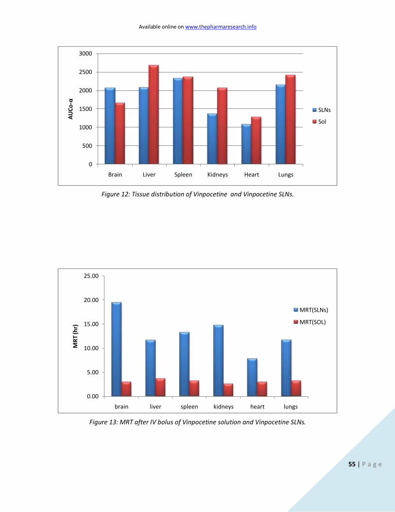
Vinpocetine SLNs Tissue distribution
Forty Two rats were divided randomly into
14groups. Each group was composed of three
rats. Eight groups received an IV injection of
Vinpocetine SLNs at a dose level (0.44mg
Vinpocetine/kg animal body mass)[27] in the
tail vein with a slow IV bolus dose by a no. 7
needle, another six groups for the control
received Vinpocetine Solution (in 10% ether), at
the same dose level. Rats were sacrificed at
different time intervals, the brain, heart, liver,
spleen, lungs and kidneys were taken, squeezed
and washed. A 20% by weight tissue
homogenate of each organ was prepared Total
Vinpocetine concentration in each organ was
determined by HPLC analysis.
Pharmacokinetic analysis and brain targeting
efficiency of Vinpocetine from the prepared
Vinpocetine SLNs bioadhesive nasal gels.
Male Wistar Albino rats ( aged 4–5 months)
weighing between 200 and 250 g were selected
for the study. Vinpocetine SLNs bioadhesive
nasal gel formula G10 was prepared. For each
rat either 100 µl gel was administered in its
right nostril or an equivalent Vinpocetine dose
administrated by IV bolus injection in the tail
vein. Subsequently, Rat brains were dissected,
washed and homogenized. Vinpocetine
concentration in each rat was determined by
HPLC analysis.
Pharmacokinetic parameters [28],Drug
targeting index (DTI) [29, 30] and Nose to brain
direct transport percentage (DTP%) were
calculated where:
………………………………..Eq3
……………………………………………..Eq4
Where Bx = (Bi.v./Pi.v.) × Pi.n., Bx is the brain AUC fraction contributed by systemic circulation through the BBB following intranasal administration,
Bi.v. is the ( (brain) following intravenous administration, Pi.v. is the (blood) following intravenous administration, Bi.n. is the (brain) following intranasal administration, Pi.n. is the (blood) following intranasal administration and AUC is the area under the curve.
Full factorial statistical analysis was done using
SPSS software program (V.17) and post Hoc LSD
test (least square difference) with 95%
confidence level for the area under the curve
(AUC , and mean residence time (MRT) and
Available online on www.thepharmaresearch.info
48 | P a g e
differences were considered significant when P
≤ 0.05.
One way ANOVA statistical analysis was done
using SPSS software program (V.17) and post
Hoc LSD test (least square difference) with
95% confidence level for the area under the
curve (AUC , and mean residence time (MRT)
and differences were considered significant
when P ≤ 0.05.
Chromatographic conditions
The concentrations of Vinpocetine in rat plasma
were determined by HPLC [31]. The HPLC
system was composed of a model HP Agilent
1100 series with G1311A Quaternary Pump,
G1315A Diode Array Detector and G1313A
Autosampler. The analytical column was
Hypersil C18 (100 × 4.6 mm, 5 μm) (Thermo,
UK). The injection volume was 20 µl auto
adjusted by the Autosampler; the mobile phase
was methanol:water (80:20 (v/v)), containing
0.1% w/w triethylamine and adjusted to pH 7
using glacial acetic acid.[32]; the flow rate was
1.5 ml/min; the UV detector wavelength was
273 nm; the column temperature was 35 oC.
Results and discussion
The chosen Vinpocetine SLNs formula was
selected after long study as it was the most
promising Vinpocetine SLNs that can be
effectively targeted to the brain. Where average
particle size diameter (z- average) were 123 nm
± 10.58 , PDI = 0.141, entrapment efficiency
percent of 89.09% ±1.49, zeta potential of -
11.33 mv ±0.97, cumulative released percent
after 96 hours (Q96) of 72.12 %±4.52 with zero
order sustained release kinetics.
In order to exhibit gel-forming properties, the
carboxylic acid groups (in the used Carbopol
934p) have to be neutralized. However, if
sodium hydroxide was used it could reduce
the zeta potential of the particles [33], resulting
in destabilization of SLNs dispersions leading to
the particle growth and subsequent formation
of semisolid gels. This phenomenon is well
known for lipid nanoemulsions. [34] So
Triethanolamine (TEA) was used as the alkali
of choice.
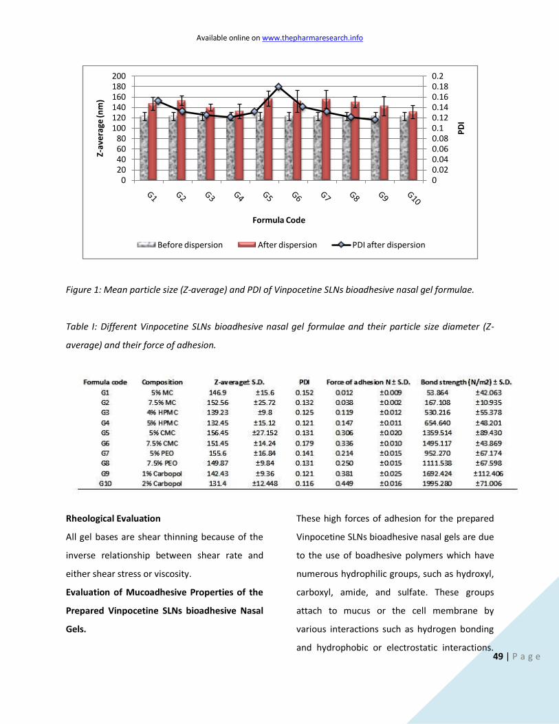
Particle size Analysis
A slight increase in the measured mean particle
size and the polydispersity index as shown in as
shown in table (I) and figure (1), but the
nanoparticulate structure could be maintained.
Similar results were previously obtained. [12]
This can be explained by the maintenance of
integrity of the strong hydrogel network, rather
than the existence of nanoparticle aggregations
[35, 36] which can further be attributed to the
presence of the gelling agent those obstacles a
good dispersion of nanoparticles.
Available online on www.thepharmaresearch.info
49 | P a g e
Figure 1: Mean particle size (Z-average) and PDI of Vinpocetine SLNs bioadhesive nasal gel formulae.
Table I: Different Vinpocetine SLNs bioadhesive nasal gel formulae and their particle size diameter (Z-
average) and their force of adhesion.
Rheological Evaluation
All gel bases are shear thinning because of the
inverse relationship between shear rate and
either shear stress or viscosity.
Evaluation of Mucoadhesive Properties of the
Prepared Vinpocetine SLNs bioadhesive Nasal
Gels.
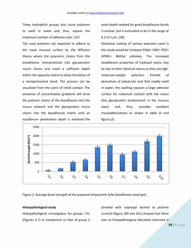
These high forces of adhesion for the prepared
Vinpocetine SLNs bioadhesive nasal gels are due
to the use of boadhesive polymers which have
numerous hydrophilic groups, such as hydroxyl,
carboxyl, amide, and sulfate. These groups
attach to mucus or the cell membrane by
various interactions such as hydrogen bonding
and hydrophobic or electrostatic interactions.
00.020.040.060.080.10.120.140.160.180.2
020406080
100120140160180200
PD
I
Z-av
era
ge (n
m)
Formula Code
Before dispersion After dispersion PDI after dispersion
Available online on www.thepharmaresearch.info
50 | P a g e
These hydrophilic groups also cause polymers
to swell in water and, thus, expose the
maximum number of adhesive sites. [37]
The used polymers are expected to adhere to
the nasal mucosal surface by the diffusion
theory where the polymeric chains from the
bioadhesive interpenetrate into glycoprotein
mucin chains and reach a sufficient depth
within the opposite matrix to allow formation of
a semipermanent bond. The process can be
visualized from the point of initial contact. The
existence of concentration gradients will drive
the polymer chains of the bioadhesive into the
mucus network and the glycoprotein mucin
chains into the bioadhesive matrix until an
equilibrium penetration depth is achieved.The
exact depth needed for good bioadhesive bonds
is unclear, but is estimated to be in the range of
0.2–0.5 μm. [38]
Statistical ranking of various polymers used in
this study would be Carbopol 934p> CMC< PEO<
HPMC< Methyl cellulose. The increased
bioadhesive properties of Carbopol resins may
be due to their chemical nature as they are high-
molecular-weight polymers formed of
derivatives of polyacrylic acid that readily swell
in water; the swelling exposes a large adhesive
surface for maximum contact with the mucin
(the glycoprotein predominant in the mucous
layer) and, thus, provides excellent
mucoadhesiveness as shown in table (I) and
figure (2).
Figure 2: Average bond strength of the prepared Vinpocetine SLNs bioadhesive nasal gels.
Histopathological study
Histopathological investigation for groups I-IV,
(Figures 3-7) in comparison to that of group V
(treated with isopropyl alcohol as positive
control) (figure 160 and 161) showed that there
was no histopathological alteration observed in
0
500
1000
1500
2000
2500
Bo
nd
Str
en
gth
(N/m
2)
Available online on www.thepharmaresearch.info
51 | P a g e
the mucosal layer with the glandular structure
and muscularis indicating that neither the drug
being investigated (group II) nor the prepared
Vinpocetine SLNs (group III) nor the prepared
Vinpocetine SLNs bioadhesive nasal gel formula
G10 (group IV) had any harmful effect on the
nasal mucosa preserving the normal cell
structure of the nasal sheep mucosa similar to
the negative control used which was phosphate
buffer saline pH 6.4 (group I negative
control). On the other hand histopathological
investigation for group V treated with isopropyl
alcohol (nasal mucociliary toxicity agent used
as a positive control) (figures 8 and 9) show
lysis and destruction of the nasal mucosal cells.
Figure 3:Mucosal layer of sheep nasal cavity for
group I showing normal histopathological
structure of the lining mucosal epithelium (mu)
and glandular structure (g).
Figure 4:Mucosal layer of sheep nasal cavity for
group II showing the intact normal
histopathological structure of the lining mucosal
epithelium (mu).
Figure 5:Mucosal layer of sheep nasal cavity for
group III showing the intact normal
histopathological structure of the mucosa (mu)
and glandular structure (g).
Available online on www.thepharmaresearch.info
52 | P a g e
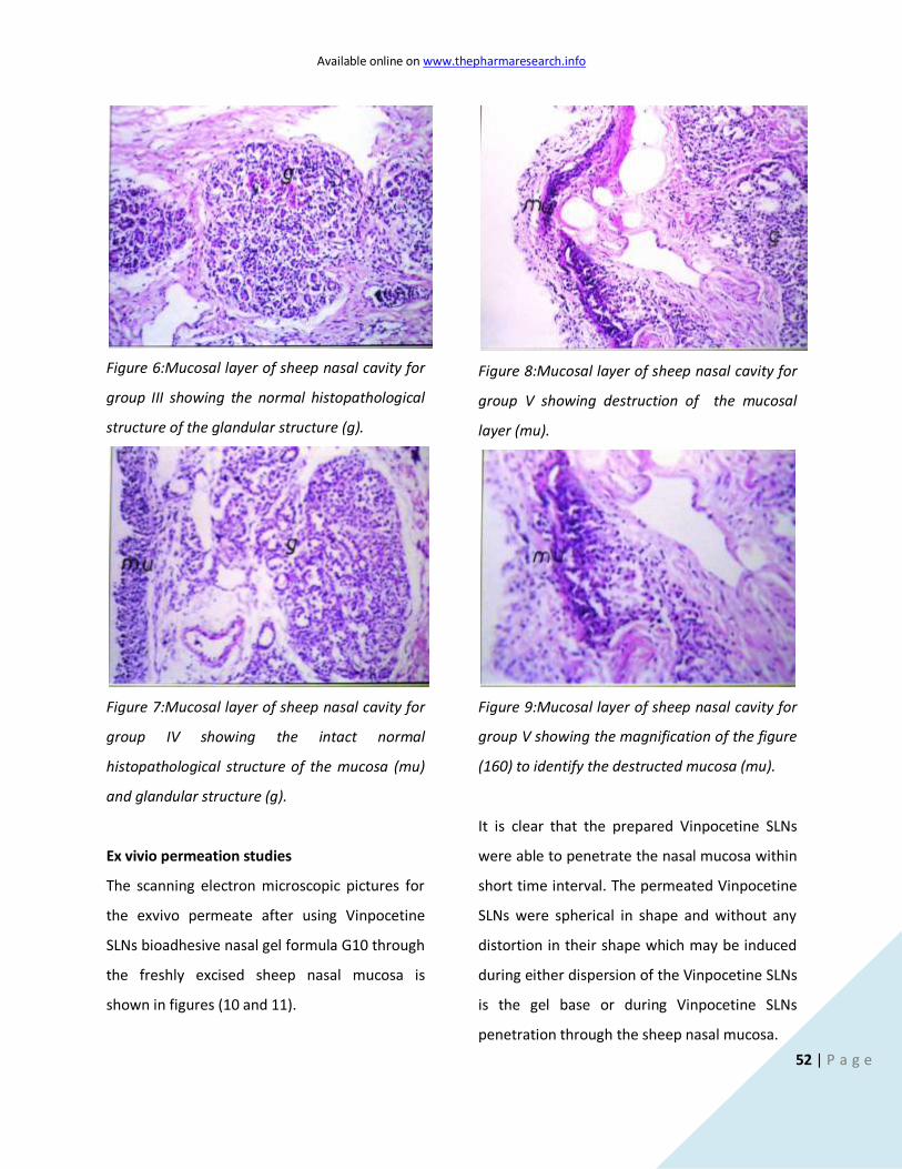
Figure 6:Mucosal layer of sheep nasal cavity for
group III showing the normal histopathological
structure of the glandular structure (g).
Figure 7:Mucosal layer of sheep nasal cavity for
group IV showing the intact normal
histopathological structure of the mucosa (mu)
and glandular structure (g).
Figure 8:Mucosal layer of sheep nasal cavity for
group V showing destruction of the mucosal
layer (mu).
Figure 9:Mucosal layer of sheep nasal cavity for
group V showing the magnification of the figure
(160) to identify the destructed mucosa (mu).
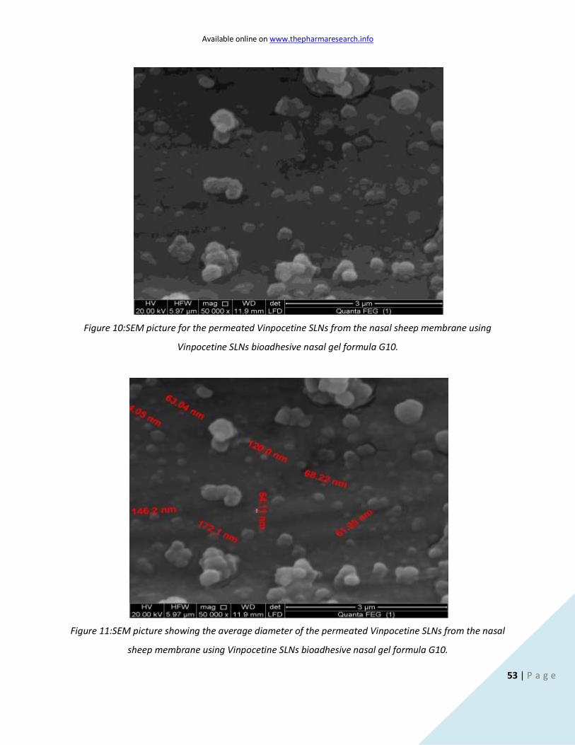
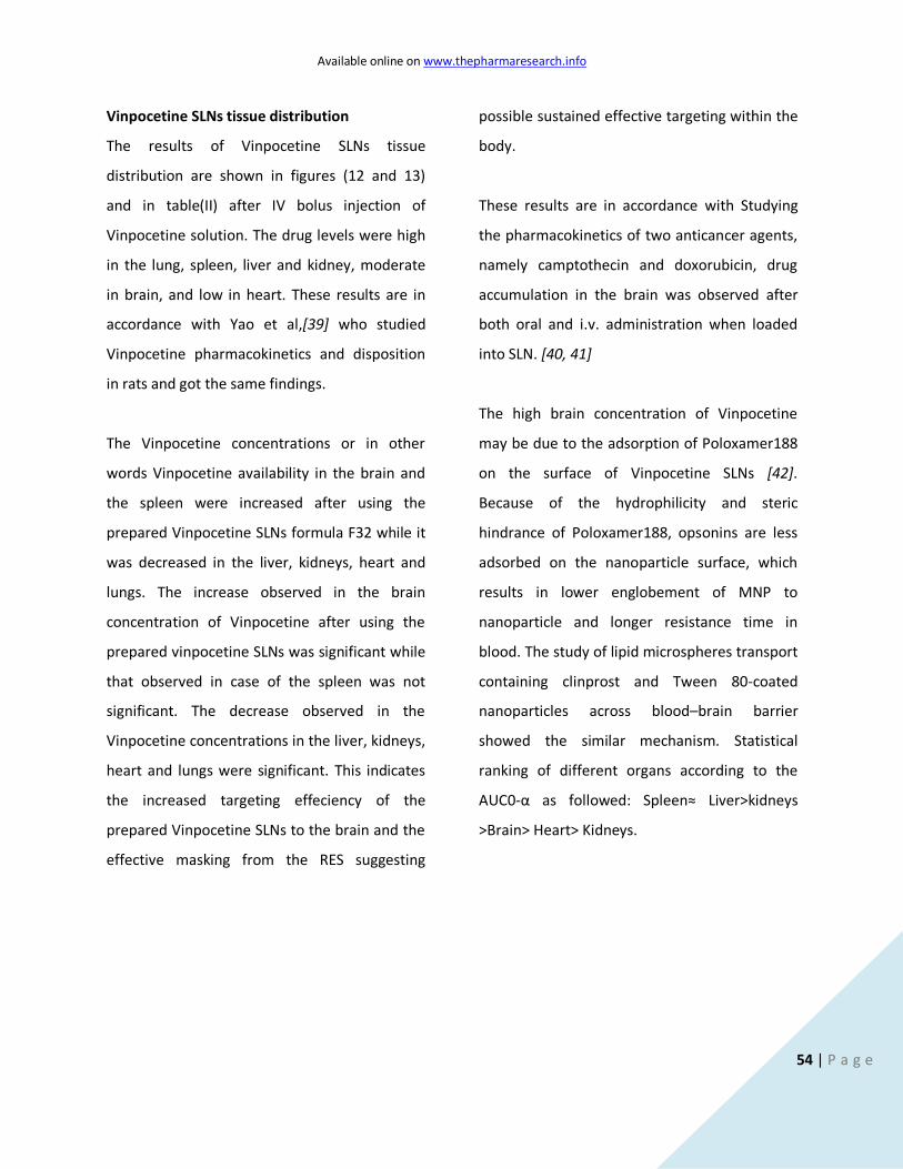
Ex vivio permeation studies
The scanning electron microscopic pictures for
the exvivo permeate after using Vinpocetine
SLNs bioadhesive nasal gel formula G10 through
the freshly excised sheep nasal mucosa is
shown in figures (10 and 11).
It is clear that the prepared Vinpocetine SLNs
were able to penetrate the nasal mucosa within
short time interval. The permeated Vinpocetine
SLNs were spherical in shape and without any
distortion in their shape which may be induced
during either dispersion of the Vinpocetine SLNs
is the gel base or during Vinpocetine SLNs
penetration through the sheep nasal mucosa.
Available online on www.thepharmaresearch.info
53 | P a g e
Figure 10:SEM picture for the permeated Vinpocetine SLNs from the nasal sheep membrane using
Vinpocetine SLNs bioadhesive nasal gel formula G10.
Figure 11:SEM picture showing the average diameter of the permeated Vinpocetine SLNs from the nasal
sheep membrane using Vinpocetine SLNs bioadhesive nasal gel formula G10.
Available online on www.thepharmaresearch.info
54 | P a g e
Vinpocetine SLNs tissue distribution
The results of Vinpocetine SLNs tissue
distribution are shown in figures (12 and 13)
and in table(II) after IV bolus injection of
Vinpocetine solution. The drug levels were high
in the lung, spleen, liver and kidney, moderate
in brain, and low in heart. These results are in
accordance with Yao et al,[39] who studied
Vinpocetine pharmacokinetics and disposition
in rats and got the same findings.
The Vinpocetine concentrations or in other
words Vinpocetine availability in the brain and
the spleen were increased after using the
prepared Vinpocetine SLNs formula F32 while it
was decreased in the liver, kidneys, heart and
lungs. The increase observed in the brain
concentration of Vinpocetine after using the
prepared vinpocetine SLNs was significant while
that observed in case of the spleen was not
significant. The decrease observed in the
Vinpocetine concentrations in the liver, kidneys,
heart and lungs were significant. This indicates
the increased targeting effeciency of the
prepared Vinpocetine SLNs to the brain and the
effective masking from the RES suggesting
possible sustained effective targeting within the
body.
These results are in accordance with Studying
the pharmacokinetics of two anticancer agents,
namely camptothecin and doxorubicin, drug
accumulation in the brain was observed after
both oral and i.v. administration when loaded
into SLN. [40, 41]
The high brain concentration of Vinpocetine
may be due to the adsorption of Poloxamer188
on the surface of Vinpocetine SLNs [42].
Because of the hydrophilicity and steric
hindrance of Poloxamer188, opsonins are less
adsorbed on the nanoparticle surface, which
results in lower englobement of MNP to
nanoparticle and longer resistance time in
blood. The study of lipid microspheres transport
containing clinprost and Tween 80-coated
nanoparticles across blood–brain barrier
showed the similar mechanism. Statistical
ranking of different organs according to the
AUC0-α as followed: Spleen≈ Liver>kidneys
>Brain> Heart> Kidneys.
Available online on www.thepharmaresearch.info
55 | P a g e
Figure 12: Tissue distribution of Vinpocetine and Vinpocetine SLNs.
Figure 13: MRT after IV bolus of Vinpocetine solution and Vinpocetine SLNs.
0
500
1000
1500
2000
2500
3000
Brain Liver Spleen Kidneys Heart Lungs
AU
Co
-α
SLNs
Sol
0.00
5.00
10.00
15.00
20.00
25.00
brain liver spleen kidneys heart lungs
MR
T (h
r)
MRT(SLNs)
MRT(SOL)
Available online on www.thepharmaresearch.info
56 | P a g e
Table II:Pharmacokinetic parameters and tissue distribution of Vinpocetine SLNs and Vinpocetine
Solution in rats.
Time (hr) Brain Liver Spleen Kidneys Heart Lungs
SLNs SOL SLNs SOL SLNs SOL SLNs SOL SLNs SOL SLNs SOL
K (hr-1) 0.05 0.417 0.068 0.34 0.062 0.376 0.064 0.395 0.068 0.4 0.087 0.361
AUC0-24(ng/g.hr) 2070.40 1607.40 2076.15 2472.79 2331.09 2247.53 1370.26 2008.91 1075.66 1229.40 2147.21 2310.41
AUC0-α(ng/g.hr) 2954.77 1663.52 2407.99 2681.55 2833.84 2364.58 1729.50 2060.15 1210.61 1276.90 2511.07 2405.88
AUMC0-24 (ng/g.hr) 18788.53 4228.93 15115.85 7394.25 17306.95 6091.75 11294.00 4776.32 6876.30 3264.92 16474.58 6415.19
AUMC0-α(ng/g.hr) 577700.79 4868.61 27960.10 9887.11 37427.10 7456.41 25528.96 5367.25 9411.48 3812.40 29389.51 7538.98
MRT (hr) 19.53 2.93 11.61 3.69 13.21 3.15 14.76 2.61 7.77 2.99 11.70 3.13
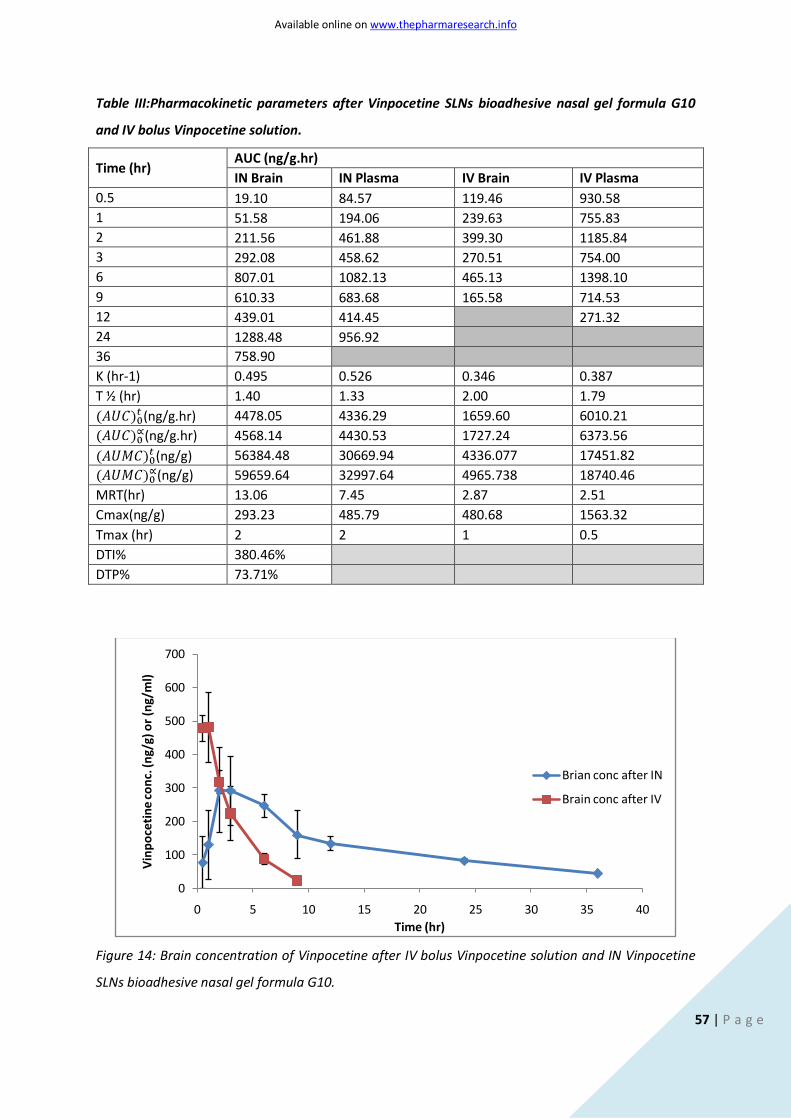
Pharmacokinetic analysis and brain
Targetting efficiency of Vinpocetine form the
prepared Vinpocetine SLNs bioadhesive nasal
gels
Pharmacokinetic parameters of Vinpocetine
were determined using non-compartmental
analysis. Table (III) and figure (4) show
Vinpocentine pharmacokinetic parameters
after intravenous administration of the
calculated animal dose. The calculated values
of AUC0-α of Vinpocetine calculated in the
brain after the IV bolus and the IN
Vinpocetine SLNs bioadhesive nasal gel
formula G20 which are 1727.24 ng/g.hr and
4568.14 ng/g.hr respectively. The high
Vinpocetine brain AUC0-α in comparison to
that after IV bolus injection is indicative of
direct nose to brain transport bypassing the
blood–brain barrier, [43, 44] hence prove the
superiority of nose to brain delivery of
Vinpocetine SLNs.
Reports in the literature [1, 45, 46] reveal
that the drug uptake into the brain from the
nasal mucosa mainly occurs via two different
pathways. One is the systemic pathway by
which some of the drug is absorbed into the
systemic circulation and subsequently reaches
the brain by crossing the BBB. The other is the
olfactory pathway by which the drug partly
travels from the nasal cavity to CSF and/or
brain tissue. It can be concluded, that the
amount of drug in the brain tissue after nasal
administration is attributed to these two
pathways. The DTP% and DTI% represent the
percentage of drug directly transported to the
brain via the olfactory pathway.
Available online on www.thepharmaresearch.info
57 | P a g e
Table III:Pharmacokinetic parameters after Vinpocetine SLNs bioadhesive nasal gel formula G10
and IV bolus Vinpocetine solution.
Time (hr) AUC (ng/g.hr)
IN Brain IN Plasma IV Brain IV Plasma
0.5 19.10 84.57 119.46 930.58
1 51.58 194.06 239.63 755.83
2 211.56 461.88 399.30 1185.84
3 292.08 458.62 270.51 754.00
6 807.01 1082.13 465.13 1398.10
9 610.33 683.68 165.58 714.53
12 439.01 414.45
271.32
24 1288.48 956.92 36 758.90
K (hr-1) 0.495 0.526 0.346 0.387
T ½ (hr) 1.40 1.33 2.00 1.79
(ng/g.hr) 4478.05 4336.29 1659.60 6010.21
(ng/g.hr) 4568.14 4430.53 1727.24 6373.56
(ng/g) 56384.48 30669.94 4336.077 17451.82
(ng/g) 59659.64 32997.64 4965.738 18740.46
MRT(hr) 13.06 7.45 2.87 2.51
Cmax(ng/g) 293.23 485.79 480.68 1563.32
Tmax (hr) 2 2 1 0.5
DTI% 380.46%
DTP% 73.71%
Figure 14: Brain concentration of Vinpocetine after IV bolus Vinpocetine solution and IN Vinpocetine
SLNs bioadhesive nasal gel formula G10.
0
100
200
300
400
500
600
700
0 5 10 15 20 25 30 35 40
Vin
po
ceti
ne
con
c. (n
g/g)
or
(ng/
ml)
Time (hr)
Brian conc after IN
Brain conc after IV
Available online on www.thepharmaresearch.info
58 | P a g e
Conclusion
The use of Vinpocetine SLNs in the form a
bioadhesive nasal gel effectively increased the
nose to brain delivery in comparison of using
Vinpocetine solution intravenously also the
presence of the drug in the form of SLNs
effectively sustained its release pattern and
finally the extremely small size of the
prepared Vinpocetine SLNs allowed for an
effective escape from the RES allowing for
long residence time in vivo.
Acknowledgement
The authors would like to thank all colleagues
and staff members in Pharmaceutics
Department, College of Pharmacy, Cairo
University.
Declaration of interest
The authors report no conflict of interest. The
authors alone are responsible for the content
and writing of this paper.
References
1. Illum, L., Transport of drugs from the
nasal cavity to the central nervous
system. Eur J Pharm Sci, 2000. 11(1): p. 1-
18.
2. Chow, H.S., Z. Chen, and G.T. Matsuura,
Direct transport of cocaine from the nasal
cavity to the brain following intranasal
cocaine administration in rats. J Pharm
Sci, 1999. 88(8): p. 754-8.
3. Patel, M.M., et al., Getting into the brain:
approaches to enhance brain drug
delivery. CNS Drugs, 2009. 23(1): p. 35-
58.
4. Brooking, J., S.S. Davis, and L. Illum,
Transport of nanoparticles across the rat
nasal mucosa. J Drug Target, 2001. 9(4):
p. 267-79.
5. Mistry, A., S. Stolnik, and L. Illum,
Nanoparticles for direct nose-to-brain
delivery of drugs. Int J Pharm, 2009.
379(1): p. 146-57.
6. Thorne, R.G. and W.H. Frey, 2nd, Delivery
of neurotrophic factors to the central
nervous system: pharmacokinetic
considerations. Clin Pharmacokinet,
2001. 40(12): p. 907-46.
7. Thorne, R.G., et al., Delivery of insulin-like
growth factor-I to the rat brain and spinal
cord along olfactory and trigeminal
pathways following intranasal
administration. Neuroscience, 2004.
127(2): p. 481-96.
8. Sanna, V., G. Caria, and A. Mariani, Effect
of lipid nanoparticles containing fatty
alcohols having different chain length on
the ex vivo skin permeability of Econazole
nitrate. Powder Technology, 2010.
201(1): p. 32-36.
9. Aji Alex, M.R., et al., Lopinavir loaded
solid lipid nanoparticles (SLN) for
intestinal lymphatic targeting. European
Journal of Pharmaceutical Sciences, 2011.
42(1–2): p. 11-18.
Available online on www.thepharmaresearch.info
59 | P a g e
10. Kheradmandnia, S., et al., Preparation
and characterization of ketoprofen-
loaded solid lipid nanoparticles made
from beeswax and carnauba wax.
Nanomedicine: Nanotechnology, Biology
and Medicine, 2010. 6(6): p. 753-759.
11. Xie, S., et al., Preparation,
characterization and pharmacokinetics of
enrofloxacin-loaded solid lipid
nanoparticles: Influences of fatty acids.
Colloids and Surfaces B: Biointerfaces,
2011. 83(2): p. 382-387.
12. Souto, E.B., et al., Evaluation of the
physical stability of SLN and NLC before
and after incorporation into hydrogel
formulations. European Journal of
Pharmaceutics and Biopharmaceutics,
2004. 58(1): p. 83-90.
13. Silva, A.C., et al., Solid lipid nanoparticles
(SLN) - based hydrogels as potential
carriers for oral transmucosal delivery of
Risperidone: Preparation and
characterization studies. Colloids and
Surfaces B: Biointerfaces, 2012. 93(0): p.
241-248.
14. Yong, C.S., et al., Effect of sodium chloride
on the gelation temperature, gel strength
and bioadhesive force of poloxamer gels
containing diclofenac sodium. Int J
Pharm, 2001. 226(1-2): p. 195-205.
15. Gupta, A., S.Garg, and R. Khar,
Measurement of bioadhesive strength of
mucoadhesive buccal tablets: design of
an in-vitro assembly. Ind. Drugs, 1992.
30: p. 152 - 155.
16. Patel, V.M., et al., Mucoadhesive bilayer
tablets of propranolol hydrochloride.
AAPS PharmSciTech, 2007. 8(3): p. E77.
17. S. Pendekal, M. and P. K. Tegginamat,
Formulation and evaluation of a
bioadhesive patch for buccal delivery of
tizanidine. Acta Pharmaceutica Sinica B,
2012(0).
18. Khullar, R., et al., Formulation and
evaluation of mefenamic acid emulgel for
topical delivery. Saudi Pharmaceutical
Journal, 2012. 20(1): p. 63-67.
19. Seju, U., A. Kumar, and K.K. Sawant,
Development and evaluation of
olanzapine-loaded PLGA nanoparticles
for nose-to-brain delivery: In vitro and in
vivo studies. Acta Biomater, 2011. 7(12):
p. 4169-4176.
20. Luppi, B., et al., Albumin nanoparticles
carrying cyclodextrins for nasal delivery
of the anti-Alzheimer drug tacrine.
European Journal of Pharmaceutical
Sciences, 2011. 44(4): p. 559-565.
21. Ritthidej, G.C., Chapter 3 - Nasal Delivery
of Peptides and Proteins with Chitosan
and Related Mucoadhesive Polymers, in
Peptide and Protein Delivery, W. Chris
Van Der, Editor. 2011, Academic Press:
Boston. p. 47-68.
22. Illum, L., Nasal drug delivery — Recent
developments and future prospects.
Journal of Controlled Release, (0).
Available online on www.thepharmaresearch.info
60 | P a g e
23. Higuchi, T., Rate of release of
medicaments from ointment bases
containing drugs in suspension. J Pharm
Sci, 1961. 50: p. 874-5.
24. Jiang, X.G., et al., [Toxicity of drugs on
nasal mucocilia and the method of its
evaluation]. Yao Xue Xue Bao, 1995.
30(11): p. 848-53.
25. Bancroft, J.D. and M. Gamble, Theory and
practice of histological techniques. 5th
ed. 2002, London ; New York: Churchill
Livingstone. xii, 796 p.
26. Lang, S., et al., Transport and metabolic
pathway of thymocartin (TP4) in excised
bovine nasal mucosa. J Pharm Pharmacol,
1996. 48(11): p. 1190-6.
27. Reagan-Shaw, S., M. Nihal, and N.
Ahmad, Dose translation from animal to
human studies revisited. FASEB J, 2008.
22(3): p. 659-61.
28. Keck, P.E., Jr. and S.L. McElroy, Clinical
pharmacodynamics and
pharmacokinetics of antimanic and
mood-stabilizing medications. J Clin
Psychiatry, 2002. 63 Suppl 4: p. 3-11.
29. Vyas, T.K., et al., Preliminary brain-
targeting studies on intranasal
mucoadhesive microemulsions of
sumatriptan. AAPS PharmSciTech, 2006.
7(1): p. E8.
30. Vyas, T.K., et al., Intranasal
mucoadhesive microemulsions of
clonazepam: preliminary studies on brain
targeting. J Pharm Sci, 2006. 95(3): p.
570-80.
31. ELBARY, A., et al., Reversed phase liquid
chromatographic determination of
vinpocetine in human plasma and its
pharmacokinetic application. Vol. 35.
2002, Philadelphia, PA, ETATS-UNIS:
Taylor & Francis.
32. El-Laithy, H.M., O. Shoukry, and L.G.
Mahran, Novel sugar esters proniosomes
for transdermal delivery of vinpocetine:
Preclinical and clinical studies. European
Journal of Pharmaceutics and
Biopharmaceutics, 2011. 77(1): p. 43-55.
33. Schwarz, C. and W. Mehnert, Solid lipid
nanoparticles (SLN) for controlled drug
delivery. II. Drug incorporation and
physicochemical characterization. J
Microencapsul, 1999. 16(2): p. 205-13.
34. Müller, B.W., et al., Effect of anti-
flocculants on suspension stability and
size distribution. Pharm. Ind., 1990. 52: p.
789-793.
35. Silva, A.C., et al., Solid lipid nanoparticles
(SLN) - based hydrogels as potential
carriers for oral transmucosal delivery of
Risperidone: Preparation and
characterization studies. Colloids Surf B
Biointerfaces, 2012. 93: p. 241-8.
36. Nikolic, S., et al., Skin photoprotection
improvement: synergistic interaction
between lipid nanoparticles and organic
UV filters. Int J Pharm, 2011. 414(1-2): p.
276-84.
Available online on www.thepharmaresearch.info
61 | P a g e
37. Shaikh, R., et al., Mucoadhesive drug
delivery systems. J Pharm Bioallied Sci,
2011. 3(1): p. 89-100.
38. Duchene, D., F. Touchard, and N.
Peppas., Pharmaceutical and medical
aspects of bioadhesive systems for drug
administration. Drug Dev Ind Pharm. ,
1988. 14: p. 283-18.
39. Yao, J.H., C.Y. Su, and X.Y. Chu,
[Pharmacokinetics and disposition of
vinpocetine in rats]. Yao Xue Xue Bao,
1994. 29(2): p. 81-5.
40. Zara, G.P., et al., PHARMACOKINETICS OF
DOXORUBICIN INCORPORATED IN SOLID
LIPID NANOSPHERES (SLN).
Pharmacological Research, 1999. 40(3):
p. 281-286.
41. Yang, S., et al., Body distribution of
camptothecin solid lipid nanoparticles
after oral administration. Pharm Res,
1999. 16(5): p. 751-7.
42. Moghimi, S.M., Prolonging the circulation
time and modifying the body distribution
of intravenously injected polystyrene
nanospheres by prior intravenous
administration of poloxamine-908. A
'hepatic-blockade' event or manipulation
of nanosphere surface in vivo? Biochim
Biophys Acta, 1997. 1336(1): p. 1-6.
43. Zhang, Q., et al., Preparation of
nimodipine-loaded microemulsion for
intranasal delivery and evaluation on the
targeting efficiency to the brain. Int J
Pharm, 2004. 275(1-2): p. 85-96.
44. Kumar, M., et al., Intranasal
nanoemulsion based brain targeting drug
delivery system of risperidone. Int J
Pharm, 2008. 358(1-2): p. 285-91.
45. Illum, L., Nasal drug delivery--
possibilities, problems and solutions. J
Control Release, 2003. 87(1-3): p. 187-98.
46. Vyas, T.K., et al., Intranasal drug delivery
for brain targeting. Curr Drug Deliv, 2005.
2(2): p. 165-75.