Embed Size (px)
Citation preview
Bioanalytical Sample PreparationWhite Paper
2 Bioanalytical Sample Preparation | © Biotage 2020
Sample Pre-Treatment Sample Preparation
Evaporation Analysis
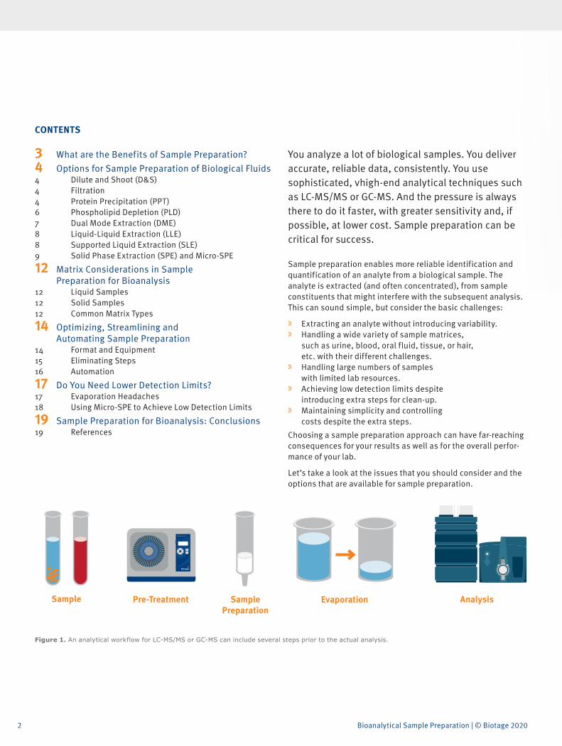
Figure 1. An analytical workflow for LC-MS/MS or GC-MS can include several steps prior to the actual analysis.
You analyze a lot of biological samples. You deliver accurate, reliable data, consistently. You use sophisticated, vhigh-end analytical techniques such as LC-MS/MS or GC-MS. And the pressure is always there to do it faster, with greater sensitivity and, if possible, at lower cost. Sample preparation can be critical for success.
Sample preparation enables more reliable identification and quantification of an analyte from a biological sample. The analyte is extracted (and often concentrated), from sample constituents that might interfere with the subsequent analysis. This can sound simple, but consider the basic challenges:
» Extracting an analyte without introducing variability. » Handling a wide variety of sample matrices,
such as urine, blood, oral fluid, tissue, or hair, etc. with their different challenges.
» Handling large numbers of samples with limited lab resources.
» Achieving low detection limits despite introducing extra steps for clean-up.
» Maintaining simplicity and controlling costs despite the extra steps.
Choosing a sample preparation approach can have far-reaching consequences for your results as well as for the overall perfor-mance of your lab.
Let’s take a look at the issues that you should consider and the options that are available for sample preparation.
CONTENTS
3 What are the Benefits of Sample Preparation?4 Options for Sample Preparation of Biological Fluids4 Dilute and Shoot (D&S)4 Filtration4 Protein Precipitation (PPT)6 Phospholipid Depletion (PLD)7 Dual Mode Extraction (DME)8 Liquid-Liquid Extraction (LLE)8 Supported Liquid Extraction (SLE)9 Solid Phase Extraction (SPE) and Micro-SPE
12 Matrix Considerations in Sample Preparation for Bioanalysis
12 Liquid Samples12 Solid Samples12 Common Matrix Types
14 Optimizing, Streamlining and Automating Sample Preparation
14 Format and Equipment15 Eliminating Steps16 Automation
17 Do You Need Lower Detection Limits?17 Evaporation Headaches18 Using Micro-SPE to Achieve Low Detection Limits
19 Sample Preparation for Bioanalysis: Conclusions19 References
3
What are the Benefits of Sample Preparation?
What are the Benefits of Sample Preparation?Sample preparation is often considered a necessary inconve-nience in the analytical process. You might want to disregard it as just extra work prior to the more glamorous analytical technique that delivers the result. Yet there are definite benefits you can gain from carefully chosen, appropriate, sample prep.
» Higher quality data. It makes good sense to ensure that samples are presented in a form that enables the best performance from sophisticated analytical equipment. This helps meet the demand for accurate and consistent results.
» Lower costs. By avoiding ‘dirty’ samples, you minimize downtime and maintenance of expensive equipment, as well as avoiding reruns.
» Better sensitivity. Clean samples, concentrated and without interfering matrix components, help you to achieve low detection limits.
» Greater robustness. By ensuring consistency in the treatment of samples applied to downstream analytical steps, you control variability (for example, protecting the subsequent analysis from extremes in matrix composition that can impact results through interference or masking of analytes).
However, sample prep does add to the complexity of the analytical workflow and, unfortunately, there is no ‘one size fits all’. Extra steps take time and an incorrect choice of method can introduce errors rather than reducing them. So, what are your options and how do you choose between them?
4 Bioanalytical Sample Preparation | © Biotage 2020
Options for Sample Preparation of Biological Fluids
Options for Sample Preparation of Biological Fluids
There is a whole range of sample preparation techniques and each one has its place. Loosely, methods can be divided into ‘matrix scavenging’, designed to remove specific matrix components, and ‘targeted extraction’ where the characteristics of the analyte of interest directly impact the choice of sample prep technique and, frequently, components within the chosen technique.
Matrix Scavenging Techniques tend to be quicker, cheaper and simpler, whilst they usually require little or no method development compared with targeted extraction. However, they might not provide sufficiently high recovery or concentration to provide satisfactory detection limits.
Targeted Extraction Techniques, such as supported liquid extraction (SLE) and solid phase extraction (SPE) can be tailored more effectively to exploit analyte characteristics. They are often more complex, require more method development, and can be more expensive per analyzed sample. However, this is compensated for by delivering cleaner extracts with higher recoveries and potentially higher analytical sensitivity (lower detection limits).
These techniques are applicable to liquid samples as well as solid or semi-solid biological samples (such as tissue, hair or fingernails). For solid and semi-solid samples, additional sample pre-treatment is normally required to transfer analytes into a liquid phase before clean-up.
Dilute and Shoot (D&S)Before we look at more complicated approaches to sample preparation, let us consider the simplest. There are situations where the sample can be diluted then injected directly into, for example, an LC-MS/MS instrument. This is most often exploited with urine samples, which can be simply diluted 1:10 with water or buffer.
Clearly, there will be a direct negative impact on the limit of detection, but this might be acceptable. Furthermore, because all matrix components (such as salts, urea, proteins
or phospholipids) are still present, they might reduce liquid chromatography (LC) column lifetime, build up on the mass spectrometer (MS) source, or suppress ionization of the target analyte (if co-eluting with analyte from the liquid chroma-tography column).
If you can live with reduced sensitivity, afford the potential instrument downtime, accept costs for replacing LC columns more frequently, and accept having to regularly clean the MS source, then D&S is itself cheap, fast, and simple.
FiltrationAnother simple technique is to pass the sample through a 0.45 or 0.2 µm filter. Clearly, this will remove particulates that could clog an LC column, but won’t remove dissolved matrix compo-nents that can interfere in other ways. Commonly, filtration will be followed by dilution then injection. This method is also typically applied to urine although it may be appropriate for other biofluids. As with D&S, it is simple, quick and relatively cheap.
Protein Precipitation (PPT)This is a matrix scavenging technique usually based on the familiar ‘acetonitrile crash’ approach. Simply add excess acetonitrile to the sample and either filter or centrifuge away the precipitated proteins.
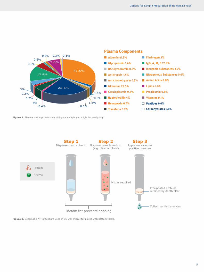
PPT is primarily used for blood, serum or plasma samples that carry a heavy protein load (Figure 2).
It is available in a 96-well microtiter plate format where precipi-tation occurs in the wells and the clear protein-free samples are collected after passage through bottom filters (induced by pressure or vacuum). This format is particularly suited to automated high-throughput assays, whereas using test tubes and a centrifuge is more appropriate for manual handling of small sample numbers (Figure 3).
PPT demands little method development, but it will not be suitable in all situations because it only removes proteins (not phospholipids) and doesn’t target any specific analyte. Nevertheless, the method is quick, simple and relatively inexpensive.
5
Options for Sample Preparation of Biological Fluids
Figure 2. Plasma is one protein-rich biological sample you might be analyzing1.
Figure 3. Schematic PPT procedure used in 96-well microtiter plates with bottom filters.
Albumin 41.5%
Plasma Components
Glycoprotein 1.4%
HS Glycoprotein 0.6%
Antitrypsin 1.5%
Antichymotrypsin 0.5%
Globulins 22.5%
Ceruloplasmin 0.4%
Haptoglobilin 4%
Hemopexin 0.7%
Transferin 0.2%
Fibrinogen 3%
IgG, A, M, D 12.8%
Inorganic Substances 3.5%
Nitrogenous Substances 0.6%
Amino Acids 0.8%
Lipids 0.8%
Prealbumin 0.8%
Vitamins 0.1%
Peptides 0.0%
Carbohydrates 0.0%
0.1%0.3%0.8%0.6%
3.5%
3%
0.2%
0.7%
4%0.4% 0.5%
1.5%
0.6%
1.4%
41.5%
22.5%
12.8%
5.8%
Protein Precipitation
Dispense crash solvent
Mix as required
Dispense sample matrix (e.g. plasma, blood)
Apply low vacuum/positive pressure
Collect purified analytes
Precipitated proteinsretained by depth filter
Protein
Analyte
Bottom frit prevents dripping
6 Bioanalytical Sample Preparation | © Biotage 2020
Options for Sample Preparation of Biological Fluids
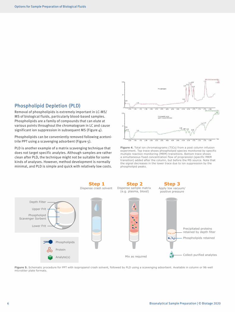
Phospholipid Depletion (PLD)Removal of phospholipids is extremely important in LC-MS/MS of biological fluids, particularly blood-based samples. Phospholipids are a family of compounds that can elute at various points throughout the chromatogram in LC and cause significant ion suppression in subsequent MS (Figure 4).
Phospholipids can be conveniently removed following acetoni-trile PPT using a scavenging adsorbent (Figure 5).
PLD is another example of a matrix scavenging technique that does not target specific analytes. Although samples are rather clean after PLD, the technique might not be suitable for some kinds of analyses. However, method development is normally minimal, and PLD is simple and quick with relatively low costs.
Figure 4. Total ion chromatograms (TICs) from a post column infusion experiment. Top trace shows phospholipid species monitored by specific multiple reaction monitoring (MRM) transitions. Bottom trace shows a simultaneous fixed concentration flow of propranolol (specific MRM transition) added after the column, but before the MS source. Note that the signal decreases in the lower trace due to ion suppression by the phospholipid peaks.
Dispense crash solvent
Mix as required
Dispense sample matrix (e.g. plasma, blood)
Apply low vacuum/positive pressure
Collect purified analytes
Phospholipids retained
Precipitated proteinsretained by depth filter
Upper Frit
PhospholipidScavenger Sorbent
Lower Frit
Depth Filter
Phospholipids
Protein
Analyte(s)
Figure 5. Schematic procedure for PPT with isopropanol crash solvent, followed by PLD using a scavenging adsorbent. Available in column or 96-well microtiter plate formats.
7
Options for Sample Preparation of Biological Fluids
ISOLUTE HYDRO DME+ with Hydrolysis step
Mix as required
Collect purified analytes
Add Acetonitrile EluteIn-Well
Hydrolysis
Matrix components
Analytes of interest
Hydrolysis enzyme
Apply low vacuum/positive pressure
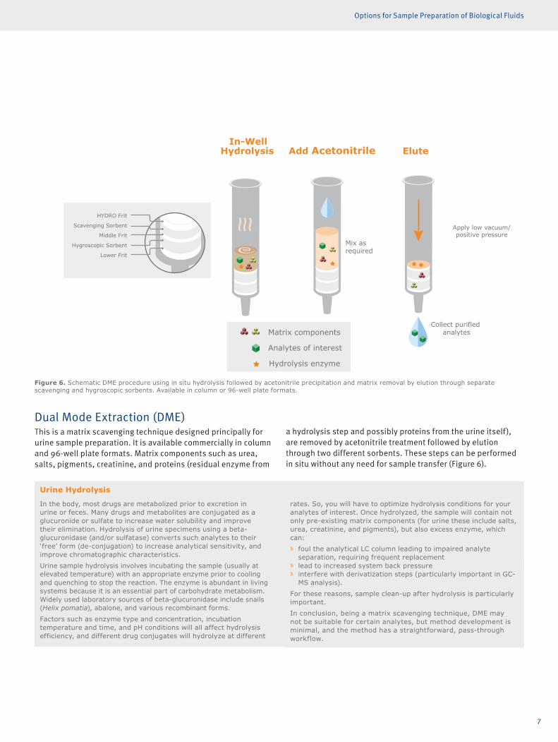
HYDRO Frit
Scavenging Sorbent
Middle Frit
Hygroscopic Sorbent
Lower Frit
Figure 6. Schematic DME procedure using in situ hydrolysis followed by acetonitrile precipitation and matrix removal by elution through separate scavenging and hygroscopic sorbents. Available in column or 96-well plate formats.
Urine Hydrolysis
In the body, most drugs are metabolized prior to excretion in urine or feces. Many drugs and metabolites are conjugated as a glucuronide or sulfate to increase water solubility and improve their elimination. Hydrolysis of urine specimens using a beta-glucuronidase (and/or sulfatase) converts such analytes to their ‘free’ form (de-conjugation) to increase analytical sensitivity, and improve chromatographic characteristics.Urine sample hydrolysis involves incubating the sample (usually at elevated temperature) with an appropriate enzyme prior to cooling and quenching to stop the reaction. The enzyme is abundant in living systems because it is an essential part of carbohydrate metabolism. Widely used laboratory sources of beta-glucuronidase include snails (Helix pomatia), abalone, and various recombinant forms. Factors such as enzyme type and concentration, incubation temperature and time, and pH conditions will all affect hydrolysis efficiency, and different drug conjugates will hydrolyze at different
rates. So, you will have to optimize hydrolysis conditions for your analytes of interest. Once hydrolyzed, the sample will contain not only pre-existing matrix components (for urine these include salts, urea, creatinine, and pigments), but also excess enzyme, which can: » foul the analytical LC column leading to impaired analyte
separation, requiring frequent replacement » lead to increased system back pressure » interfere with derivatization steps (particularly important in GC-
MS analysis).For these reasons, sample clean-up after hydrolysis is particularly important.In conclusion, being a matrix scavenging technique, DME may not be suitable for certain analytes, but method development is minimal, and the method has a straightforward, pass-through workflow.
Dual Mode Extraction (DME)This is a matrix scavenging technique designed principally for urine sample preparation. It is available commercially in column and 96-well plate formats. Matrix components such as urea, salts, pigments, creatinine, and proteins (residual enzyme from
a hydrolysis step and possibly proteins from the urine itself), are removed by acetonitrile treatment followed by elution through two different sorbents. These steps can be performed in situ without any need for sample transfer (Figure 6).
8 Bioanalytical Sample Preparation | © Biotage 2020
Options for Sample Preparation of Biological Fluids
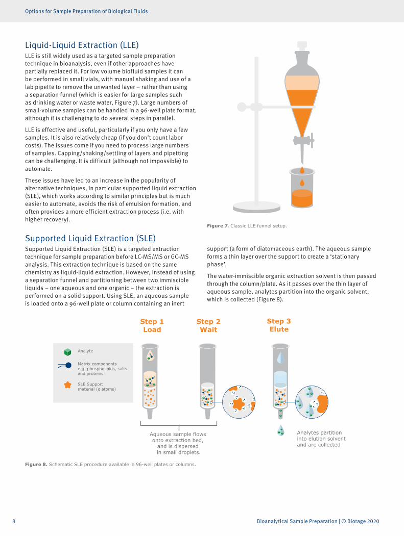
Liquid-Liquid Extraction (LLE)LLE is still widely used as a targeted sample preparation technique in bioanalysis, even if other approaches have partially replaced it. For low volume biofluid samples it can be performed in small vials, with manual shaking and use of a lab pipette to remove the unwanted layer – rather than using a separation funnel (which is easier for large samples such as drinking water or waste water, Figure 7). Large numbers of small-volume samples can be handled in a 96-well plate format, although it is challenging to do several steps in parallel.
LLE is effective and useful, particularly if you only have a few samples. It is also relatively cheap (if you don’t count labor costs). The issues come if you need to process large numbers of samples. Capping/shaking/settling of layers and pipetting can be challenging. It is difficult (although not impossible) to automate.
These issues have led to an increase in the popularity of alternative techniques, in particular supported liquid extraction (SLE), which works according to similar principles but is much easier to automate, avoids the risk of emulsion formation, and often provides a more efficient extraction process (i.e. with higher recovery).
Supported Liquid Extraction (SLE)Supported Liquid Extraction (SLE) is a targeted extraction technique for sample preparation before LC-MS/MS or GC-MS analysis. This extraction technique is based on the same chemistry as liquid-liquid extraction. However, instead of using a separation funnel and partitioning between two immiscible liquids – one aqueous and one organic – the extraction is performed on a solid support. Using SLE, an aqueous sample is loaded onto a 96-well plate or column containing an inert
support (a form of diatomaceous earth). The aqueous sample forms a thin layer over the support to create a ‘stationary phase’.
The water-immiscible organic extraction solvent is then passed through the column/plate. As it passes over the thin layer of aqueous sample, analytes partition into the organic solvent, which is collected (Figure 8).
Figure 7. Classic LLE funnel setup.
SLE+ Load Wait Elute
Analytes partition into elution solvent and are collected
Step 1Load
Step 2Wait
Step 3Elute
Aqueous sample flows onto extraction bed,
and is dispersed in small droplets.
Analyte
Matrix componentse.g. phospholipids, salts and proteins
SLE Support material (diatoms)
Figure 8. Schematic SLE procedure available in 96-well plates or columns.
9
Options for Sample Preparation of Biological Fluids
Clean-up is based on an analyte’s ability to partition into the water-immiscible organic extraction solvent. The method can be tailored to deliver high recovery of a specific analyte. Proteins, phospholipids, salts, pigments, etc., that might interfere with subsequent analyses tend not to be very soluble in the organic phase, so they are left behind in the aqueous phase on the column.
The outcome of SLE will always net an organic solvent, which one can direct inject or derivatize (for GC/MS analysis). If LC/MS analysis is used, then the solvent must be dried down and a compatible reconstitution solution must be employed, because LC-MS/MS normally uses reversed phase chromatography (RPC), which is incompatible with immiscible organic solvents.
Method development is required, but it is relatively simple. Key variables are:
» the pH of the sample (just like in LLE, you acidify to increase recovery of acidic analytes, or add base to increase recovery of basic ones)
» the choice of extraction solvent.
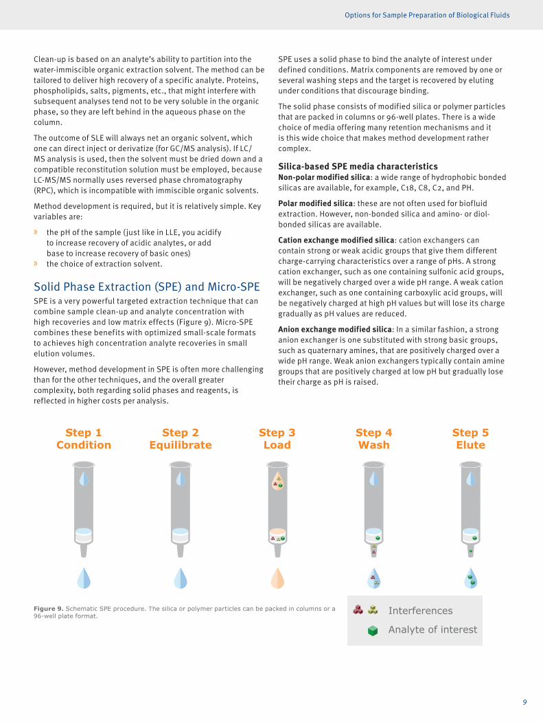
Solid Phase Extraction (SPE) and Micro-SPESPE is a very powerful targeted extraction technique that can combine sample clean-up and analyte concentration with high recoveries and low matrix effects (Figure 9). Micro-SPE combines these benefits with optimized small-scale formats to achieves high concentration analyte recoveries in small elution volumes.
However, method development in SPE is often more challenging than for the other techniques, and the overall greater complexity, both regarding solid phases and reagents, is reflected in higher costs per analysis.
SPE uses a solid phase to bind the analyte of interest under defined conditions. Matrix components are removed by one or several washing steps and the target is recovered by eluting under conditions that discourage binding.
The solid phase consists of modified silica or polymer particles that are packed in columns or 96-well plates. There is a wide choice of media offering many retention mechanisms and it is this wide choice that makes method development rather complex.
Silica-based SPE media characteristicsNon-polar modified silica: a wide range of hydrophobic bonded silicas are available, for example, C18, C8, C2, and PH.
Polar modified silica: these are not often used for biofluid extraction. However, non-bonded silica and amino- or diol-bonded silicas are available.
Cation exchange modified silica: cation exchangers can contain strong or weak acidic groups that give them different charge-carrying characteristics over a range of pHs. A strong cation exchanger, such as one containing sulfonic acid groups, will be negatively charged over a wide pH range. A weak cation exchanger, such as one containing carboxylic acid groups, will be negatively charged at high pH values but will lose its charge gradually as pH values are reduced.
Anion exchange modified silica: In a similar fashion, a strong anion exchanger is one substituted with strong basic groups, such as quaternary amines, that are positively charged over a wide pH range. Weak anion exchangers typically contain amine groups that are positively charged at low pH but gradually lose their charge as pH is raised.
Condition Equilibrate Load Wash Elute
Step 3Load
Step 4Wash
Step 2Equilibrate
Step 1Condition
Step 5Elute
Interferences
Analyte of interest
Figure 9. Schematic SPE procedure. The silica or polymer particles can be packed in columns or a 96-well plate format.
Metal Mitigation
Interferences
Analyte of interest
10 Bioanalytical Sample Preparation | © Biotage 2020
Options for Sample Preparation of Biological Fluids
Mixed-mode modified silica: These types of media combine non-polar or hydrophobic characteristics with anion- or cation exchangers. The hydrophobic- and charge-based retention mechanisms result in solid phases that can retain most drugs and other small molecules. They have proved very useful for SPE of biological fluids since the combined retention mechanisms together with strong washes and selective elution solvent choices offer highly effective removal of matrix components, such as phospholipids, and good recovery of the specific analyte of interest.
The most common mixed-mode silica types are cation exchangers with either C8 or C18 non-polar groups. They are widely used in forensic toxicology applications to extract basic drugs from urine samples.
Why use Polymeric SPE?Polymeric SPE phases are often described as being more robust and are increasingly chosen in favor of silica-based media for biofluid extraction. This is because of a couple of drawbacks common to silica-based media:
pH Stability: silica-based media are not very stable above pH 8 and should only be used at higher pHs (up to pH 10) for very short exposure times. In contrast, polymeric stationary phases, usually based on modified divinyl benzene (DVB) polymers, are stable over the pH range 1–14.
Conditioning: silica-based solid phases need to be ‘condi-tioned’ (usually by a wash with methanol) to ensure that the functional groups are active prior to use, and because of this, they can be sensitive to drying out. Polymeric SPE based on DVB are modified with polar monomers or functional groups to make them easily wettable without conditioning, and they are unaffected by drying out.
Polymer-based SPE media characteristicsPolymeric SPE particles consist of a backbone, typically a DVB modified with polar groups to make the polymer beads water wettable. These beads are then modified by substitution with ion exchange groups.
Mixed-mode polymeric SPE particles combine the charac-teristics of a non-polar (hydrophobic) polymer backbone (usually containing some polar/wettable groups) and an ionic functional group. In other words, they have mixed-mode binding characteristics, but this is achieved in a different way than with silica-based media.
A mixed-mode silica-based stationary phase tends to have lower hydrophobicity than the equivalent mixed-mode polymeric sorbent. Consequently, the non-polar retention mechanism provided by the silica-based C8 or C18 is generally less strong (less retentive) than that provided by a polymeric backbone. This can mean that cleaner extracts are possible from silica-based materials, because solvent washes can more effectively remove the weakly bound matrix interference components.
Polymeric SPE mediaTypically, polymeric SPE stationary phases are available in 5 types:
» Non-polar water wettable polymer: the primary retention mechanism is through non-polar interactions
» Mixed-mode - strong cation exchange + non-polar water wettable polymer
» Mixed-mode - weak cation exchange + non-polar water wettable polymer
» Mixed-mode - strong anion exchange + non-polar water wettable polymer
» Mixed-mode - weak anion exchange + non-polar water wettable polymer
Pore size considerationsThe high surface area of SPE phases makes it possible to extract analytes using small amounts of media. The particles used in SPE, whether silica- or polymer- based, are highly porous, giving them a high surface area.
The size of the pores can influence extract cleanliness, particu-larly regarding proteins. With wider pores, more protein can penetrate inside of the particles, giving high binding capacity not only for the analyte but also for protein components that can interfere with the subsequent analysis.
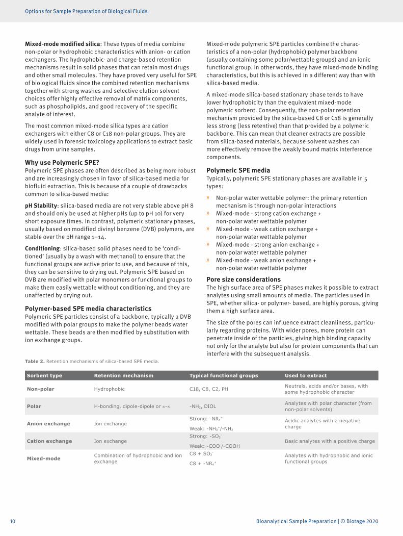
Table 2. Retention mechanisms of silica-based SPE media.
Sorbent type Retention mechanism Typical functional groups Used to extract
Non-polar Hydrophobic C18, C8, C2, PH Neutrals, acids and/or bases, with some hydrophobic character
Polar H-bonding, dipole-dipole or π-π -NH2, DIOL Analytes with polar character (from non-polar solvents)
Anion exchange Ion exchangeStrong: -NR4
+
Weak: -NH3+/-NH2
Acidic analytes with a negative charge
Cation exchange Ion exchangeStrong: -SO3
-
Weak: -COO-/-COOHBasic analytes with a positive charge
Mixed-mode Combination of hydrophobic and ion exchange
C8 + SO3-
C8 + -NR4+
Analytes with hydrophobic and ionic functional groups
11
Options for Sample Preparation of Biological Fluids
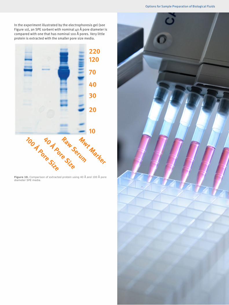
In the experiment illustrated by the electrophoresis gel (see Figure 10), an SPE sorbent with nominal 40 Å pore diameter is compared with one that has nominal 100 Å pores. Very little protein is extracted with the smaller pore size media.
Figure 10. Comparison of extracted protein using 40 Å and 100 Å pore diameter SPE media.
100 Å Pore Size
Mwt Marker
Raw Serum
40 Å Pore Size
10
20
30
40
70
120220
12 Bioanalytical Sample Preparation | © Biotage 2020
Matrix Considerations in Sample Preparation for Bioanalysis
Matrix Considerations in Sample Preparation for Bioanalysis
Liquid SamplesBiofluids are often diluted (usually 1:1 to 1:3, v:v) before the clean-up technique in order to:
» reduce viscosity so they flow through the extraction device more easily.
» adjust sample pH (using buffer) to improve the extraction or adapt for the subsequent extraction. This pH will vary depending upon the analyte, sample prep technique, and analytical technique.
Dilution in this way brings more consistency to samples that may vary considerably in viscosity — for example, urine samples can vary depending on how hydrated the patient is. Dilution will improve standardization, which is especially important when you are batch-processing or using 96-well plate formats.
Solid SamplesAll the sample preparation described, and the subsequent analytical techniques of LC-MS/MS or GC-MS, require liquid samples. Solids or semi solids must be pre-treated to get the analytes into the liquid state before clean-up. This is a very large area that we don’t cover here, but, for example, it can include:
» grinding and homogenization » solvent or buffer extraction » saponification to remove fats
Common Matrix TypesHere we briefly summarize the special issues related to a few specific biofluids.
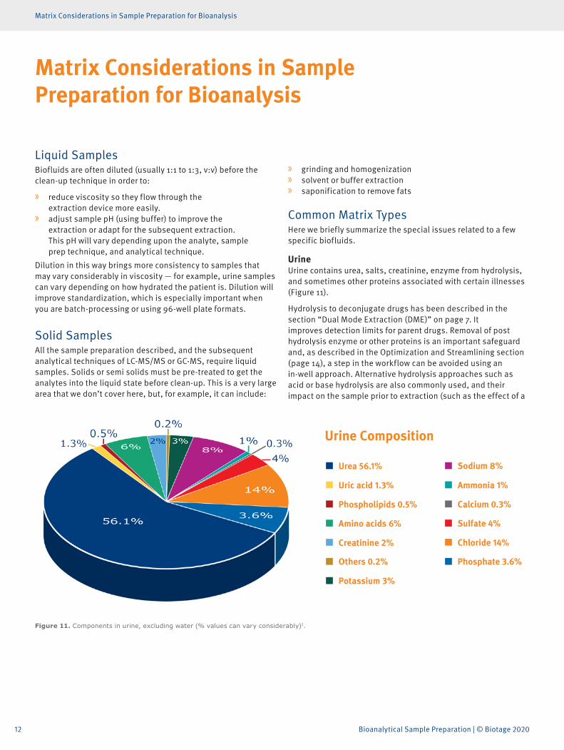
UrineUrine contains urea, salts, creatinine, enzyme from hydrolysis, and sometimes other proteins associated with certain illnesses (Figure 11).
Hydrolysis to deconjugate drugs has been described in the section “Dual Mode Extraction (DME)” on page 7. It improves detection limits for parent drugs. Removal of post hydrolysis enzyme or other proteins is an important safeguard and, as described in the Optimization and Streamlining section (page 14), a step in the workflow can be avoided using an in-well approach. Alternative hydrolysis approaches such as acid or base hydrolysis are also commonly used, and their impact on the sample prior to extraction (such as the effect of a
Urea 56.1%
Urine Composition
Uric acid 1.3%
Phospholipids 0.5%
Amino acids 6%
Creatinine 2%
Others 0.2%
Potassium 3%
Sodium 8%
Ammonia 1%
Calcium 0.3%
Sulfate 4%
Chloride 14%
Phosphate 3.6%
Figure 11. Components in urine, excluding water (% values can vary considerably)1.

13
Matrix Considerations in Sample Preparation for Bioanalysis
changed pH) needs to be considered when developing sample preparation methods.
Whole blood/serum/plasmaChallenges with these types of samples can be divided into:
» endogenous: including proteins, phospholipids, red blood cells
» exogenous: including dosing vehicles from therapeutic drug formulations
Lipid content can vary greatly depending on dietary differences and timing after meals.
Viscosity variations can also cause issues, for example related to vacuum manifolds, mentioned in the next section.
Some analytes, such as many drugs, are transported in blood bound to proteins and must be released prior to extraction using techniques such as SLE or SPE. Steps can include pH adjustment to denature the protein and addition of solvent to break the analyte – protein bond.
Oral fluid and associated collection devicesThings to consider here are if the sample is simply neat saliva or if it is collected with an oral fluid collection device that can introduce buffers and surfactants. These can impact on analyte recovery, or cause matrix effects in LC-MS/MS.
Others (hair/nails/meconium/tissue)These solid or semi-solid samples introduce another level of matrix complexity that we will not address in detail here.
Collection of hair and nails is a non-invasive operation which is seeing growing popularity in drugs-of-abuse (DoA) analysis and workplace testing because sampling can be done in ’public’ (unlike with urine).
Other examples are meconium, which is used for monitoring DoA exposure in the womb, and liver tissue which is particularly useful in forensic postmortem cases where blood or urine may not be available.
14 Bioanalytical Sample Preparation | © Biotage 2020
Optimizing, Streamlining and Automating Sample Preparation
Optimizing, Streamlining and Automating Sample Preparation
As in any method development process, the first thing you should do is consider your lab’s purpose, your goals, and the measurable objectives that define achievement and good performance.
Scenarios can be very different in research, forensic and clinical environments. Ask the questions:
» What is the ideal output from my lab? » What is the incoming sample load, in
qualitative and quantitative terms?
For example, incoming sample quality can be high and consis-tent, or low and variable.
Numbers of samples to be analyzed per day or week will steer the need for automation and the use of different formats and approaches.
Accuracy, precision and sensitivity are all important parameters for analytical work, but they might be prioritized differently depending upon the application. Normally you can’t have it all, and there will be a trade-off between simplicity, speed and cost.
Format and EquipmentFor many sample preparation methods, there is a basic choice between columns (or test tubes) and 96-well plates, the former allowing larger sample volumes, the latter offering greater opportunities for improved throughput and automation, with lower elution volumes and, usually, smaller sample volumes.
PPTIn devices with a bottom filter, flow can be induced by positive pressure or vacuum to remove the precipitate. Precipitate can also be spun down by centrifugation, although the workflow for multiple samples is difficult to automate.
LLE and SLELLE is a very hands-on procedure, with equipment varying from large, traditional separation funnels, through disposable capped tubes and micropipettes, to 96-well plates. In addition, LLE can often be replaced by SLE, which has greater potential for automation.
SPESPE is available in columns, standard 96-well plates- or micro-SPE plate formats. Your choice of column or 96-well plate format depends on the typical number of samples you want to process
per batch, the volume of sample to be extracted, and your overall lab throughput. Choosing a micro-SPE plate format may be governed by you having very small sample volumes, or by your need to achieve small and concentrated elution volumes – thereby gaining sensitivity in the subsequent analysis.
To induce flow, the simplest manual method is to connect an SPE column to a syringe barrel and, by depressing the barrel, force the liquid (solvent or sample) through the SPE column. This is cheap and effective, but not convenient for processing multiple samples.
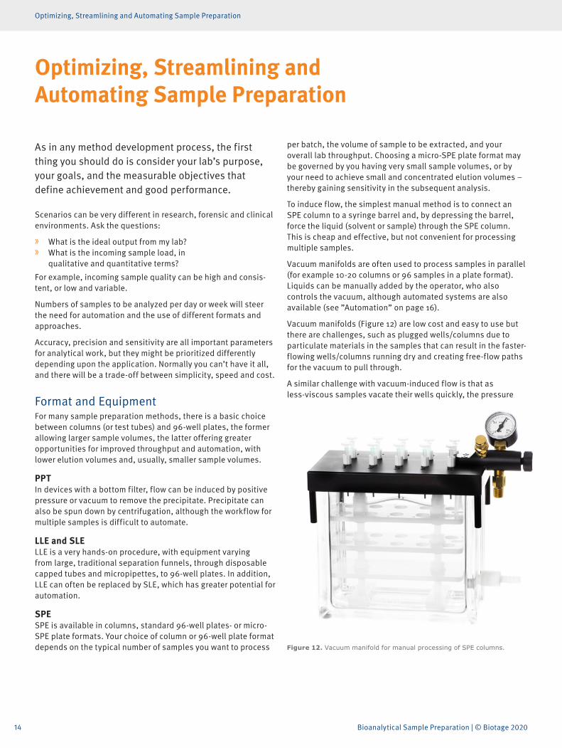
Vacuum manifolds are often used to process samples in parallel (for example 10-20 columns or 96 samples in a plate format). Liquids can be manually added by the operator, who also controls the vacuum, although automated systems are also available (see ”Automation” on page 16).
Vacuum manifolds (Figure 12) are low cost and easy to use but there are challenges, such as plugged wells/columns due to particulate materials in the samples that can result in the faster-flowing wells/columns running dry and creating free-flow paths for the vacuum to pull through.
A similar challenge with vacuum-induced flow is that as less-viscous samples vacate their wells quickly, the pressure
Figure 12. Vacuum manifold for manual processing of SPE columns.
15
Optimizing, Streamlining and Automating Sample Preparation
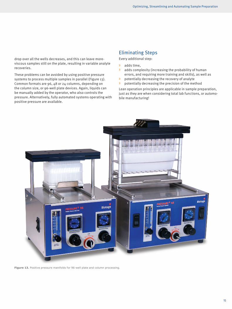
drop over all the wells decreases, and this can leave more-viscous samples still on the plate, resulting in variable analyte recoveries.
These problems can be avoided by using positive pressure systems to process multiple samples in parallel (Figure 13). Common formats are 96, 48 or 24 columns, depending on the column size, or 96-well plate devices. Again, liquids can be manually added by the operator, who also controls the pressure. Alternatively, fully automated systems operating with positive pressure are available.
Eliminating StepsEvery additional step:
» adds time, » adds complexity (increasing the probability of human
errors, and requiring more training and skills), as well as » potentially decreasing the recovery of analyte » potentially decreasing the precision of the method
Lean operation principles are applicable in sample preparation, just as they are when considering total lab functions, or automo-bile manufacturing!
Figure 13. Positive pressure manifolds for 96-well plate and column processing.
16 Bioanalytical Sample Preparation | © Biotage 2020
Optimizing, Streamlining and Automating Sample Preparation
Looking at table 1 again, we see that many sample preparation techniques can be steered to allow direct injection into the analytical system, without extra steps — the exceptions being LLE and SLE.
Also, evaporation and reconstitution can be avoided if sample prep can be achieved with SPE.
And, of course, if applicable, D&S and filtration require fewer steps than other sample preparation techniques, but they neither eliminate matrix effects nor ‘extract’ the analyte.
A smart example of step elimination is ‘in-well hydrolysis’ during DME or SPE, which eliminates a sample transfer step. This is achievable with the help of special hydrophobic frits that hold up the sample during hydrolysis but allow it to pass through when pressure is applied after hydrolysis. Hydrolysis efficiency is not affected by performing the hydrolysis in this way.
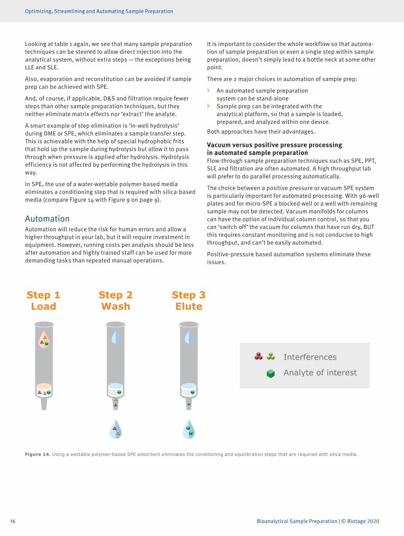
In SPE, the use of a water-wettable polymer-based media eliminates a conditioning step that is required with silica-based media (compare Figure 14 with Figure 9 on page 9).
AutomationAutomation will reduce the risk for human errors and allow a higher throughput in your lab, but it will require investment in equipment. However, running costs per analysis should be less after automation and highly trained staff can be used for more demanding tasks than repeated manual operations.
It is important to consider the whole workflow so that automa-tion of sample preparation or even a single step within sample preparation, doesn’t simply lead to a bottle neck at some other point.
There are 2 major choices in automation of sample prep:
» An automated sample preparation system can be stand-alone
» Sample prep can be integrated with the analytical platform, so that a sample is loaded, prepared, and analyzed within one device.
Both approaches have their advantages.
Vacuum versus positive pressure processing in automated sample preparationFlow-through sample preparation techniques such as SPE, PPT, SLE and filtration are often automated. A high throughput lab will prefer to do parallel processing automatically.
The choice between a positive pressure or vacuum SPE system is particularly important for automated processing. With 96-well plates and for micro-SPE a blocked well or a well with remaining sample may not be detected. Vacuum manifolds for columns can have the option of individual column control, so that you can ‘switch off’ the vacuum for columns that have run dry, BUT this requires constant monitoring and is not conducive to high throughput, and can’t be easily automated.
Positive-pressure based automation systems eliminate these issues.Load Wash Elute
Figure 14. Using a wettable polymer-based SPE adsorbent eliminates the conditioning and equilibration steps that are required with silica media.
17
Do You Need Lower Detection Limits?
Do You Need Lower Detection Limits?
Sample prep helps you clean-up biological samples before analysis, but it doesn’t automatically improve sensitivity. Sometimes, only small amounts of sample are available. High-sensitivity analysis by LC-MS/MS demands the injection of small volumes of sample extract. High concentrations of the analyte that is sometimes in short supply are needed to achieve low detection limits. So, the lower the extract volume the more concentrated the extract, and the lower the detection limit that can be achieved.
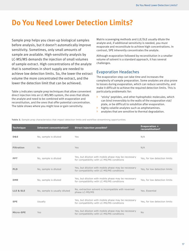
Table 3 indicates sample prep techniques that allow convenient direct injection into an LC-MS/MS system, the ones that dilute the analyte and need to be combined with evaporation and reconstitution, and the ones that offer potential concentration. The table shows where you might lose or gain sensitivity.
Matrix scavenging methods and LLE/SLE usually dilute the analyte and, if additional sensitivity is needed, you must evaporate and reconstitute to achieve high concentrations. In contrast, SPE inherently concentrates the analyte.
Although evaporation followed by reconstitution in a smaller volume of solvent is a standard approach, it has several challenges.
Evaporation HeadachesThe evaporation step can take time and increases the complexity of sample preparation. Some analytes are also prone to losses during evaporation, which can reduce sensitivity, and make it difficult to achieve the required detection limits. This is particularly problematic for:
» ‘sticky’ peptides, and other hydrophobic molecules, which can bind irreversibly to the walls of the evaporation vial/plate, or be difficult to solubilize after evaporation.
» highly volatile analytes such as amphetamines. » analytes that are sensitive to thermal degradation.
Table 3. Sample prep characteristics that impact detection limits and workflow streamlining opportunities.
Technique Inherent concentration? Direct injection possible? Evaporation + reconstitution?
D&S No, sample is diluted Yes N/A
Filtration No Yes N/A
PPT No, sample is diluted Yes, but dilution with mobile phase may be necessary for compatibility with LC-MS/MS conditions Yes, for low detection limits
PLD No, sample is diluted Yes, but dilution with mobile phase may be necessary for compatibility with LC-MS/MS conditions Yes, for low detection limits
DME No, sample is diluted Yes, but dilution with mobile phase may be necessary for compatibility with LC-MS/MS conditions Yes, for low detection limits
LLE & SLE No, sample is usually diluted No, extraction solvent is incompatible with reversed phase LC-MS/MS Yes. Essential
SPE Usually Yes, but dilution with mobile phase may be necessary for compatibility with LC-MS/MS conditions Yes, for low detection limits
Micro-SPE Yes Yes, but dilution with mobile phase may be necessary for compatibility with LC-MS/MS conditions No
18 Bioanalytical Sample Preparation | © Biotage 2020
Do You Need Lower Detection Limits?
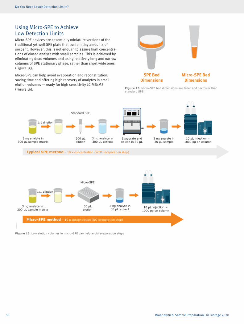
Using Micro-SPE to Achieve Low Detection LimitsMicro-SPE devices are essentially miniature versions of the traditional 96-well SPE plate that contain tiny amounts of sorbent. However, this is not enough to assure high concentra-tions of eluted analyte with small samples. This is achieved by eliminating dead volumes and using relatively long and narrow columns of SPE stationary phase, rather than short wide ones (Figure 15).
Micro-SPE can help avoid evaporation and reconstitution, saving time and offering high recovery of analytes in small elution volumes — ready for high sensitivity LC-MS/MS (Figure 16). Figure 15. Micro-SPE bed dimensions are taller and narrower than
standard SPE.
Micro-SPE Bed Dimensions
SPE Bed Dimensions
88888860
88888860
1:1 dilution
1:1 dilution
3 ng analyte in 300 µL sample matrix
3 ng analyte in 300 µL extract
3 ng analyte in 300 µL sample matrix
300 μL elution
30 μL elution
Evaporate and re-con in 30 µL
10 µL injection = 1000 pg on column
10 µL injection = 1000 pg on column
3 ng analyte in 30 µL sample
3 ng analyte in 30 µL extract
Standard SPE
Micro-SPE
biom
ark1
13.2
Typical SPE method – 10 x concentration (WITH evaporation step)
Micro-SPE method – 10 x concentration (NO evaporation step)
Figure 16. Low elution volumes in micro-SPE can help avoid evaporation steps
19
Sample Preparation for Bioanalysis: Conclusions
Sample Preparation for Bioanalysis: Conclusions
Sample preparation offers significant benefits if you are using sophisticated analytical techniques such as LC-MS/MS and GC-MS but finding the right solution to meet your needs can be complex. This brief overview hopefully illustrates the range of challenges and the choices available. You should speak with your peers and talk to manufacturers of sample preparation consumables and systems, who often have a deep understanding of the issues. Arrange to see various options in action, especially if you are thinking of high throughput situations and automation. Understand how equipment will operate in practice and note the difficulties as well as the benefits. In the end, your lab and your colleagues will benefit from a good choice of this ‘necessary inconvenience’.
References1. http://www.chromedia.org/chromedia?waxtrapp=wptlvLsHi
emBpdmBlIEcCKDnC
© Biotage 2020
Title Truncated if Neccessary... | Page 20
Literature Number: PPS621
© 2020 Biotage. All rights reserved. No material may be reproduced or published without the written permission of Biotage. Information in this document is subject to change without notice and does not represent any commitment from Biotage. E&OE. A list of all trademarks owned by Biotage AB is available at www.biotage.com/legal. Other product and company names mentioned herein may be trademarks or registered trademarks and/or service marks of their respective owners, and are used only for explanation and to the owners’ benefit, without intent to infringe.
EUROPEMain Office: +46 18 565900Toll Free: +800 18 565710Fax: +46 18 591922Order Tel: +46 18 565710Order Fax: +46 18 [email protected] Tel: +46 18 56 59 11Support Fax: + 46 18 56 57 [email protected]
NORTH & LATIN AMERICAMain Office: +1 704 654 4900Toll Free: +1 800 446 4752Fax: +1 704 654 4917Order Tel: +1 704 654 4900Order Fax: +1 434 296 [email protected] Tel: +1 800 446 4752Outside US: +1 704 654 [email protected]
JAPANTel: +81 3 5627 3123Fax: +81 3 5627 [email protected]@biotage.com
CHINATel: +86 21 68162810Fax: +86 21 [email protected]@biotage.com
KOREATel: +82 31 706 8500Fax: +82 31 706 [email protected]@biotage.com
INDIATel: +91 22 4005 [email protected]
Distributors in other regions are listed on www.biotage.com
Your Complete Partner for Effective Chemistry Biotage is a worldwide supplier of instruments and accessories designed to facilitate the work of scientists in life sciences. With our deep knowledge of the industry, academic contacts and in-house R&D teams, we can deliver the best solutions to your challenges. We take great pride in our flexibility and ability to meet our customer’s individual needs. With strong foundations in analytical, organic, process, and biomolecule chemistry, we can offer the widest range of solutions available on the market.


![Chapter 2 Sample preparation, and others1 Chapter 2 Sample preparation, and others [Summary of sample preparation method] Sample preparation is highly important to perform analytical](https://img.pdfslide.net/doc/110x75/5e9216dec3c2053e873640d8/chapter-2-sample-preparation-and-1-chapter-2-sample-preparation-and-others-summary.jpg)