Embed Size (px)
Citation preview
Biochemistry of cellular organellesLectures: 1. Membrane channels;
2. Membrane transporters;3. Soluble lipid/metabolite-transfer proteins;4. Mitochondria as cellular organelles;
Seminar: Isolation of subcellular organelles;5. Mitochondrial inheritance;6. Mitochondria in health and disease;7. Endoplasmic Reticulum (ER) and lipids;8. Structure and function of peroxisomes;
Seminar: Mitochondria in cellular life.
Dr. Vasily Antonenkov, Visiting professorDept. Biochemistry, Oulu UniversityOulu, Finland
Web site:
1
Kontinkangas, L101A
2
Seminar 2 – Mitochondria and other organelles
1. Uncommon lipid components of biological membranes: cardiolipin, sphingolipids, glycolipids, plasmalogenes - structureand function;
2. Membrane lipid rafts;
3. Lysosomes;
4. Mitochondrial dynamics (fusion/fission);
5. Plasma membrane – structure, function;
6. Intracellular lipid droplets as organelles;
7. Mitochondrial quality control.
Lecture 4: Mitochondria as cellular organelles
Lecture content:
• Compartments of eukaryotic cells;• Dynamic structure of the mitochondrion;• Functional role of different sub-compartments;• How to study mitochondria;• Role in ATP production;• Electron transfer chain;• Respiratory control;• ATP synthase.
3
4
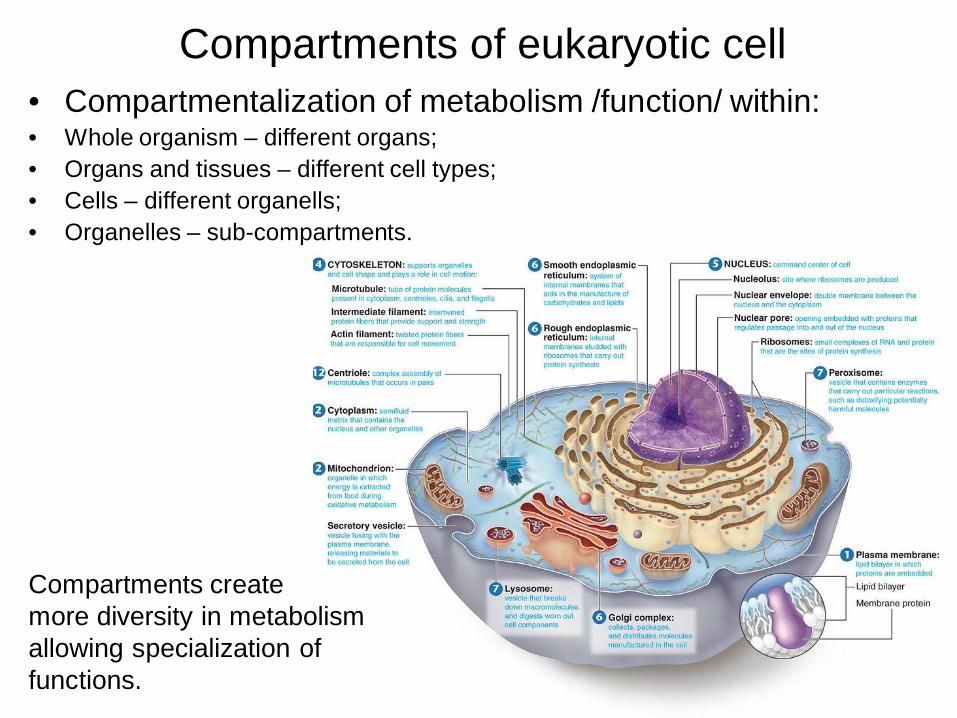
Compartments of eukaryotic cell• Compartmentalization of metabolism /function/ within:• Whole organism – different organs;• Organs and tissues – different cell types;• Cells – different organells;• Organelles – sub-compartments.
Compartments createmore diversity in metabolism allowing specialization of functions.
5
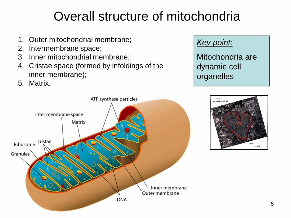
Overall structure of mitochondria
Key point:
Mitochondria are dynamic cell organelles
1. Outer mitochondrial membrane;2. Intermembrane space;3. Inner mitochondrial membrane;4. Cristae space (formed by infoldings of the
inner membrane);5. Matrix.
6
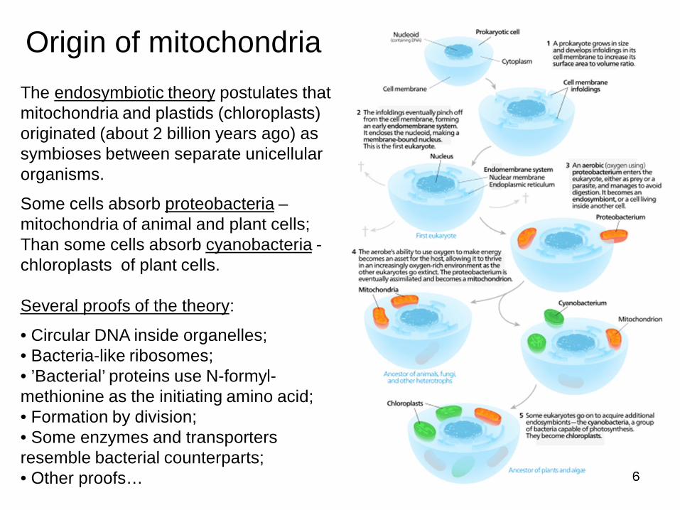
Origin of mitochondriaThe endosymbiotic theory postulates thatmitochondria and plastids (chloroplasts) originated (about 2 billion years ago) as symbioses between separate unicellular organisms.
Some cells absorb proteobacteria –mitochondria of animal and plant cells;Than some cells absorb cyanobacteria -chloroplasts of plant cells.
Several proofs of the theory:
• Circular DNA inside organelles;• Bacteria-like ribosomes;• ’Bacterial’ proteins use N-formyl-methionine as the initiating amino acid;• Formation by division;• Some enzymes and transporters resemble bacterial counterparts;• Other proofs…
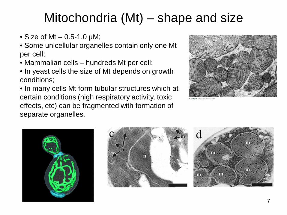
Mitochondria (Mt) – shape and size
7
• Size of Mt – 0.5-1.0 M;• Some unicellular organelles contain only one Mt per cell;• Mammalian cells – hundreds Mt per cell;• In yeast cells the size of Mt depends on growth conditions;• In many cells Mt form tubular structures which at certain conditions (high respiratory activity, toxic effects, etc) can be fragmented with formation of separate organelles.
8
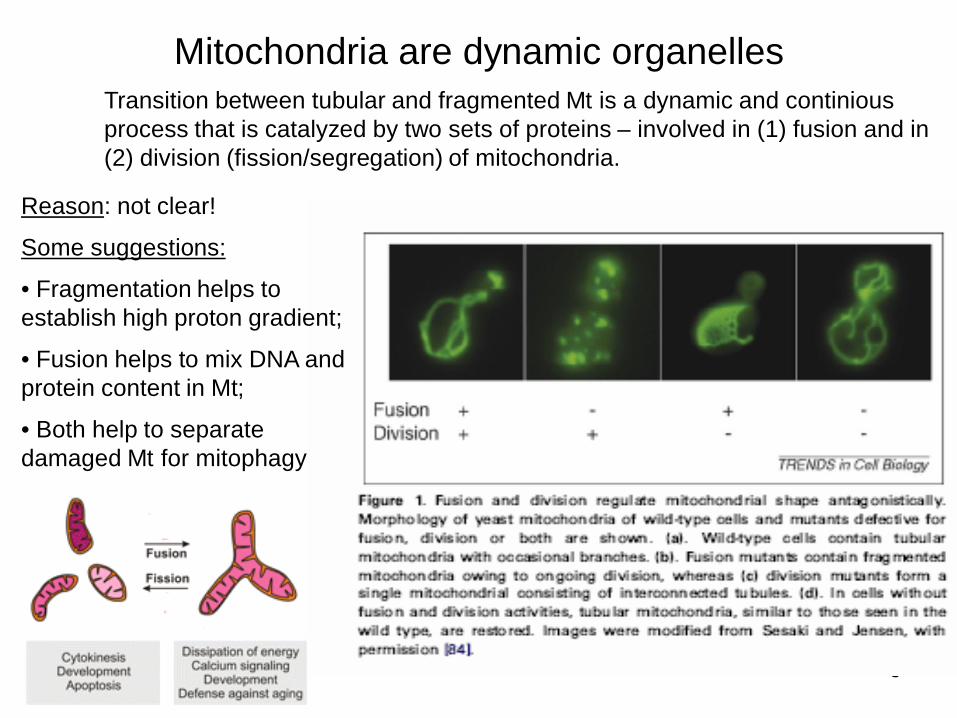
Mitochondria are dynamic organellesTransition between tubular and fragmented Mt is a dynamic and continious process that is catalyzed by two sets of proteins – involved in (1) fusion and in (2) division (fission/segregation) of mitochondria.
Reason: not clear!
Some suggestions:
• Fragmentation helps to establish high proton gradient;
• Fusion helps to mix DNA and protein content in Mt;
• Both help to separate damaged Mt for mitophagy
9
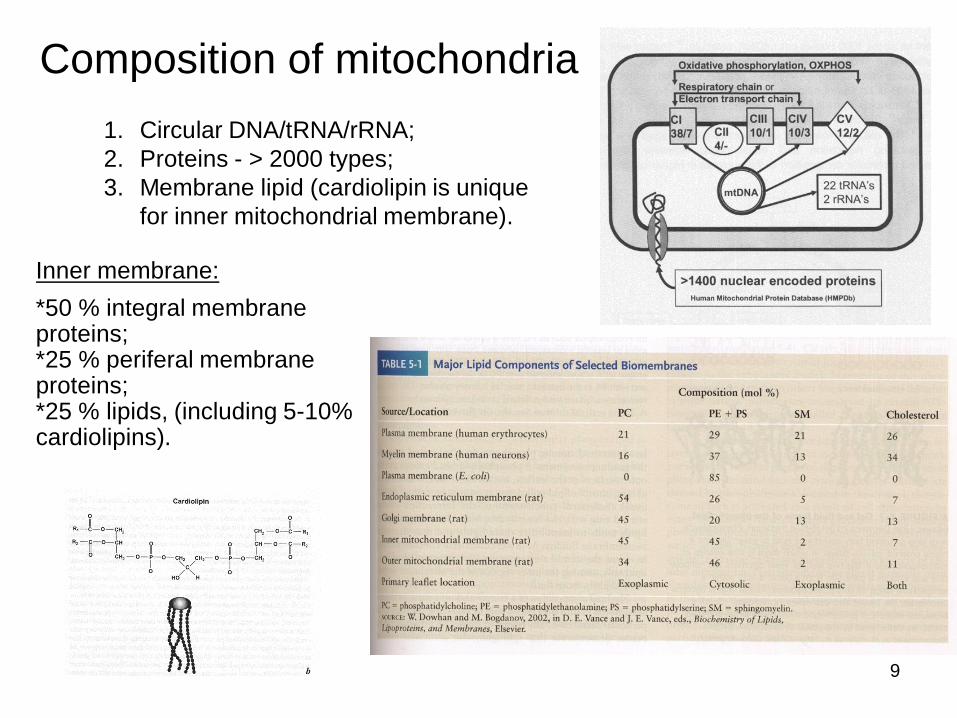
Composition of mitochondria1. Circular DNA/tRNA/rRNA;2. Proteins - > 2000 types;3. Membrane lipid (cardiolipin is unique
for inner mitochondrial membrane).
Inner membrane:*50 % integral membrane proteins;*25 % periferal membrane proteins;*25 % lipids, (including 5-10% cardiolipins).
10
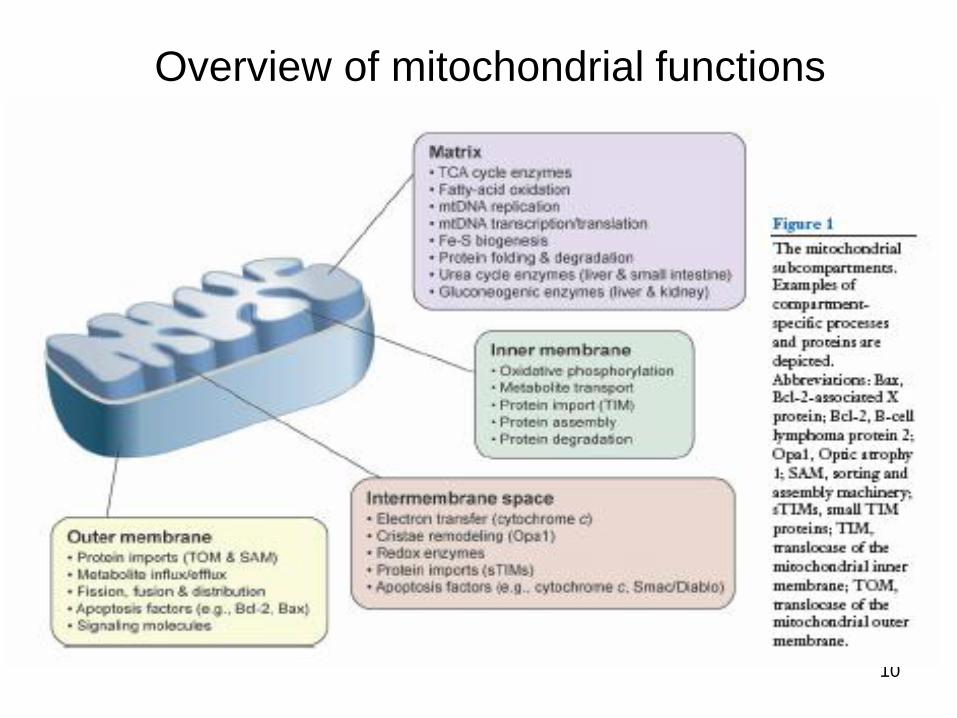
Overview of mitochondrial functions
11
How can we study mitochondria?
• Isolation of mitochondria and their components;
• Analysis of the composition of mitochondria;
• Imaging;
• Functional analysis:-In vivo, ex vivo (on tissue slides, cells, etc)-In vitro
12
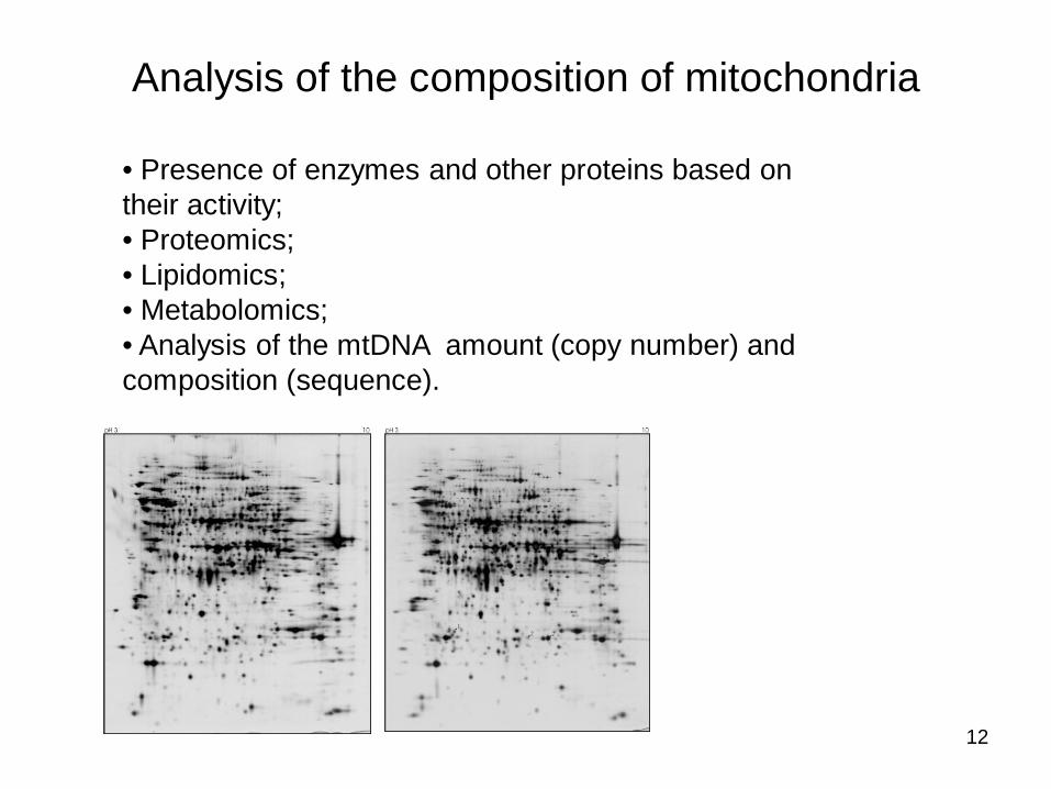
Analysis of the composition of mitochondria
• Presence of enzymes and other proteins based on their activity;• Proteomics;• Lipidomics;• Metabolomics;• Analysis of the mtDNA amount (copy number) and composition (sequence).
13
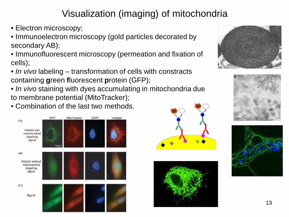
Visualization (imaging) of mitochondria• Electron microscopy;• Immunoelectron microscopy (gold particles decorated by secondary AB);• Immunofluorescent microscopy (permeation and fixation of cells);• In vivo labeling – transformation of cells with constracts containing green fluorescent protein (GFP);• In vivo staining with dyes accumulating in mitochondria due to membrane potential (MitoTracker);• Combination of the last two methods.
14
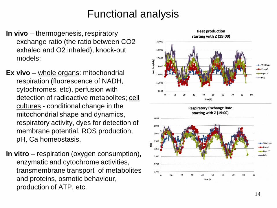
In vivo – thermogenesis, respiratory exchange ratio (the ratio between CO2 exhaled and O2 inhaled), knock-out models;
Ex vivo – whole organs: mitochondrial respiration (fluorescence of NADH, cytochromes, etc), perfusion with detection of radioactive metabolites; cell cultures - conditional change in the mitochondrial shape and dynamics, respiratory activity, dyes for detection of membrane potential, ROS production, pH, Ca homeostasis.
In vitro – respiration (oxygen consumption), enzymatic and cytochrome activities, transmembrane transport of metabolites and proteins, osmotic behaviour, production of ATP, etc.
Functional analysis
15
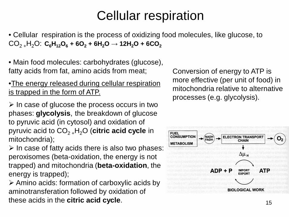
• Cellular respiration is the process of oxidizing food molecules, like glucose, to CO2 +H2O: C6H12O6 + 6O2 + 6H2O 12H2O + 6CO2
Cellular respiration
• Main food molecules: carbohydrates (glucose), fatty acids from fat, amino acids from meat;
•The energy released during cellular respiration is trapped in the form of ATP.
In case of glucose the process occurs in two phases: glycolysis, the breakdown of glucose to pyruvic acid (in cytosol) and oxidation of pyruvic acid to CO2 +H2O (citric acid cycle in mitochondria);
In case of fatty acids there is also two phases: peroxisomes (beta-oxidation, the energy is not trapped) and mitochondria (beta-oxidation, the energy is trapped);
Amino acids: formation of carboxylic acids by aminotransferation followed by oxidation of these acids in the citric acid cycle.
Conversion of energy to ATP is more effective (per unit of food) in mitochondria relative to alternative processes (e.g. glycolysis).
16
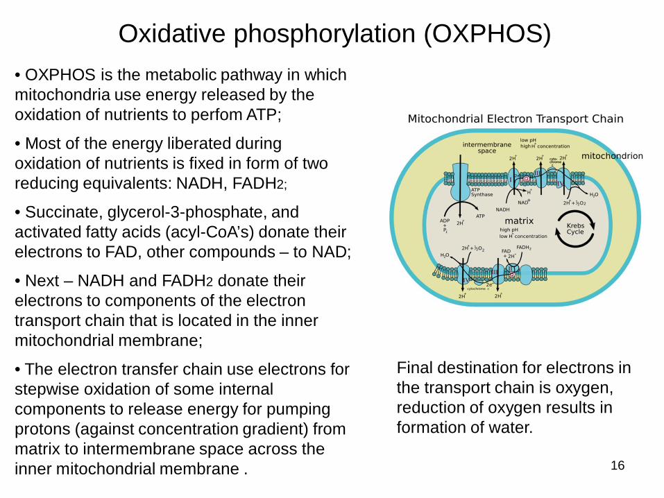
Oxidative phosphorylation (OXPHOS)• OXPHOS is the metabolic pathway in which mitochondria use energy released by the oxidation of nutrients to perfom ATP;
• Most of the energy liberated during oxidation of nutrients is fixed in form of two reducing equivalents: NADH, FADH2;
• Succinate, glycerol-3-phosphate, and activated fatty acids (acyl-CoA’s) donate their electrons to FAD, other compounds – to NAD;
• Next – NADH and FADH2 donate their electrons to components of the electron transport chain that is located in the inner mitochondrial membrane;
• The electron transfer chain use electrons for stepwise oxidation of some internal components to release energy for pumping protons (against concentration gradient) from matrix to intermembrane space across the inner mitochondrial membrane .
Final destination for electrons in the transport chain is oxygen, reduction of oxygen results in formation of water.
17
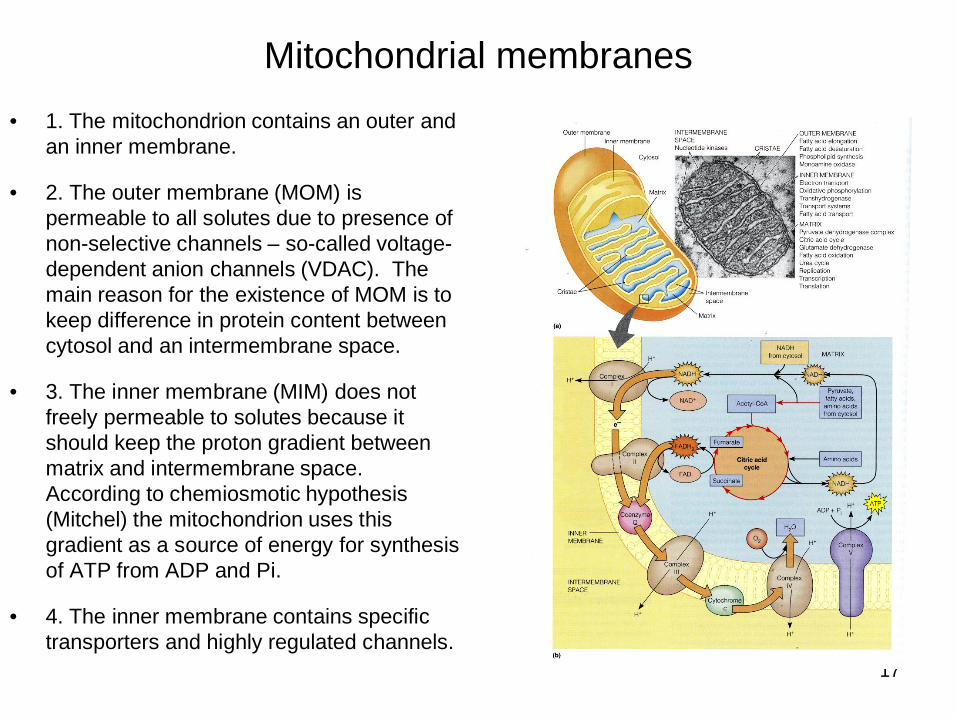
Mitochondrial membranes• 1. The mitochondrion contains an outer and
an inner membrane.
• 2. The outer membrane (MOM) is permeable to all solutes due to presence of non-selective channels – so-called voltage-dependent anion channels (VDAC). The main reason for the existence of MOM is to keep difference in protein content between cytosol and an intermembrane space.
• 3. The inner membrane (MIM) does not freely permeable to solutes because it should keep the proton gradient between matrix and intermembrane space. According to chemiosmotic hypothesis (Mitchel) the mitochondrion uses this gradient as a source of energy for synthesis of ATP from ADP and Pi.
• 4. The inner membrane contains specific transporters and highly regulated channels.
18
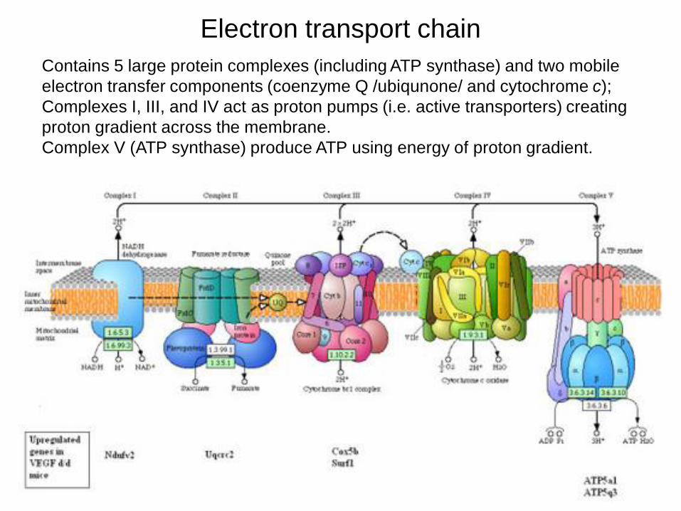
Electron transport chainContains 5 large protein complexes (including ATP synthase) and two mobile electron transfer components (coenzyme Q /ubiqunone/ and cytochrome c);Complexes I, III, and IV act as proton pumps (i.e. active transporters) creating proton gradient across the membrane. Complex V (ATP synthase) produce ATP using energy of proton gradient.
19
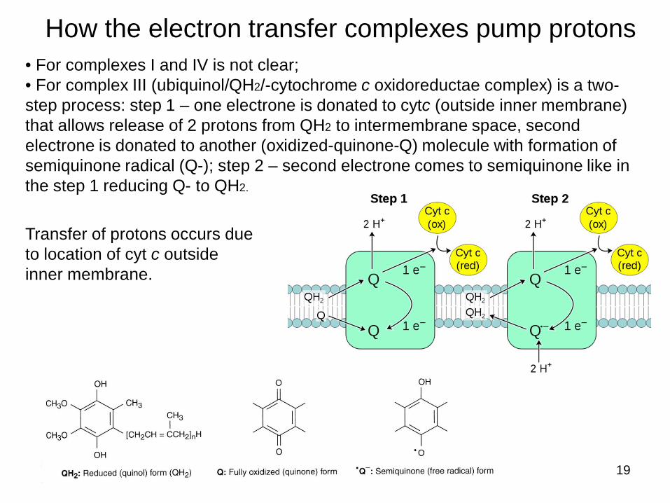
How the electron transfer complexes pump protons• For complexes I and IV is not clear;• For complex III (ubiquinol/QH2/-cytochrome c oxidoreductae complex) is a two-step process: step 1 – one electrone is donated to cytc (outside inner membrane) that allows release of 2 protons from QH2 to intermembrane space, second electrone is donated to another (oxidized-quinone-Q) molecule with formation of semiquinone radical (Q-); step 2 – second electrone comes to semiquinone like in the step 1 reducing Q- to QH2.
Transfer of protons occurs due to location of cyt c outside inner membrane.
20
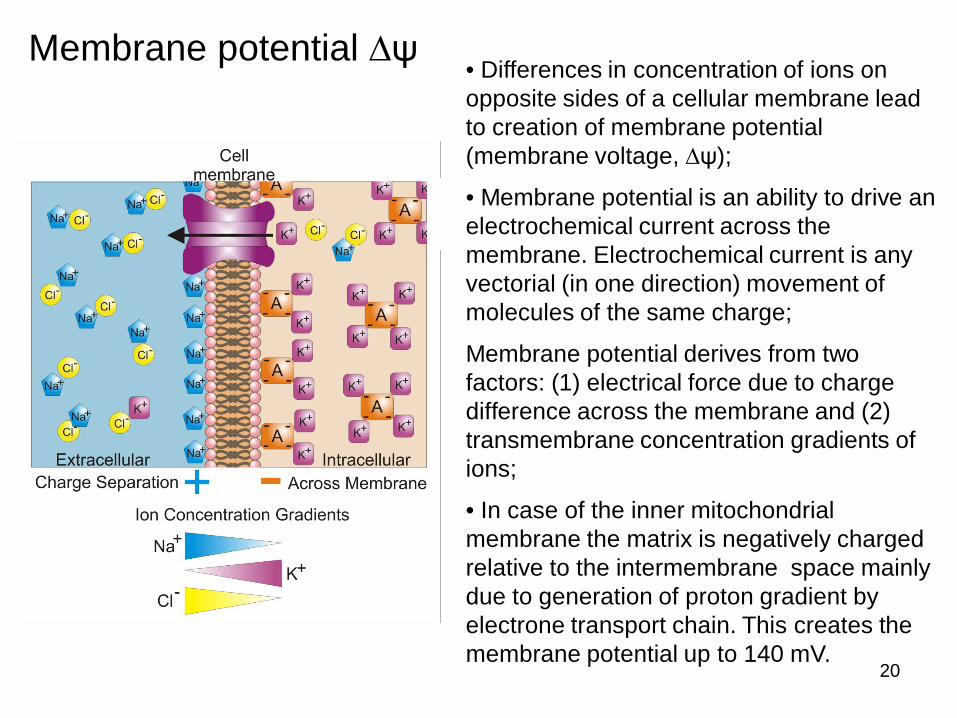
Membrane potential • Differences in concentration of ions on opposite sides of a cellular membrane lead to creation of membrane potential (membrane voltage, );
• Membrane potential is an ability to drive an electrochemical current across the membrane. Electrochemical current is any vectorial (in one direction) movement of molecules of the same charge;
Membrane potential derives from two factors: (1) electrical force due to charge difference across the membrane and (2) transmembrane concentration gradients of ions;
• In case of the inner mitochondrial membrane the matrix is negatively charged relative to the intermembrane space mainly due to generation of proton gradient by electrone transport chain. This creates the membrane potential up to 140 mV.
21
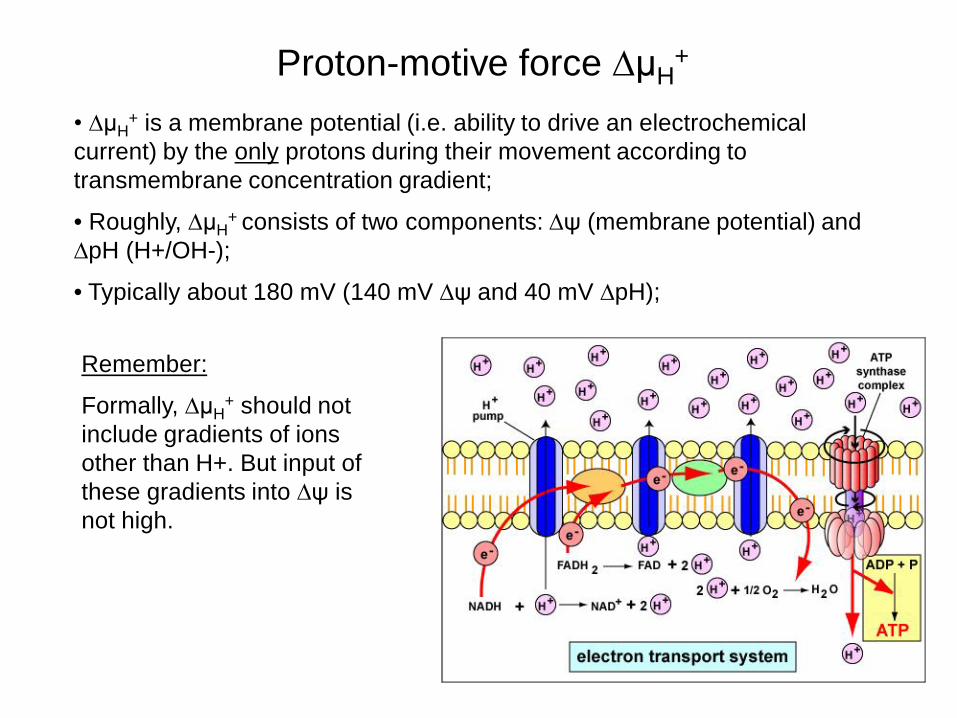
Proton-motive force H+
H+ is a membrane potential (i.e. ability to drive an electrochemical
current) by the only protons during their movement according to transmembrane concentration gradient;
• Roughly, H+ consists of two components: (membrane potential) and
pH (H+/OH-);
• Typically about 180 mV (140 mV and 40 mV pH);
Remember:
Formally, H+ should not
include gradients of ions other than H+. But input of these gradients into is not high.
22
Control of mitochondrial respiration
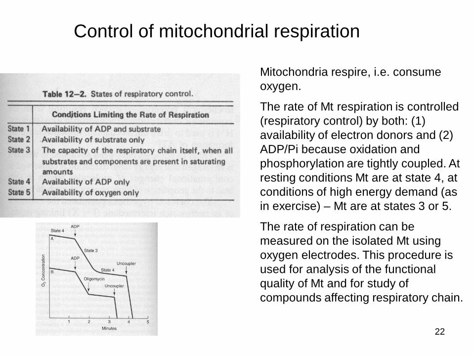
Mitochondria respire, i.e. consume oxygen.
The rate of Mt respiration is controlled (respiratory control) by both: (1) availability of electron donors and (2) ADP/Pi because oxidation and phosphorylation are tightly coupled. At resting conditions Mt are at state 4, at conditions of high energy demand (as in exercise) – Mt are at states 3 or 5.
The rate of respiration can be measured on the isolated Mt using oxygen electrodes. This procedure is used for analysis of the functional quality of Mt and for study of compounds affecting respiratory chain.
23
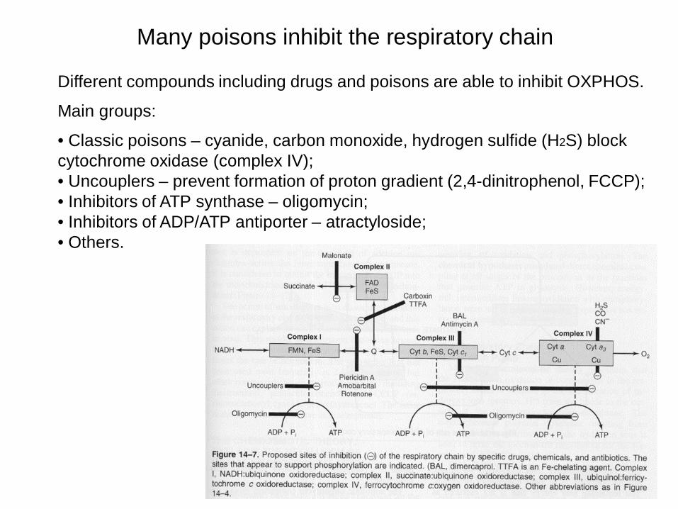
Many poisons inhibit the respiratory chain
Different compounds including drugs and poisons are able to inhibit OXPHOS.
Main groups:
• Classic poisons – cyanide, carbon monoxide, hydrogen sulfide (H2S) block cytochrome oxidase (complex IV);• Uncouplers – prevent formation of proton gradient (2,4-dinitrophenol, FCCP);• Inhibitors of ATP synthase – oligomycin;• Inhibitors of ADP/ATP antiporter – atractyloside;• Others.
24
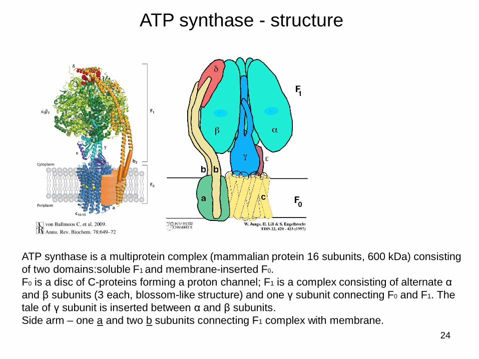
ATP synthase - structure
ATP synthase is a multiprotein complex (mammalian protein 16 subunits, 600 kDa) consisting of two domains:soluble F1 and membrane-inserted F0.F0 is a disc of C-proteins forming a proton channel; F1 is a complex consisting of alternate and subunits (3 each, blossom-like structure) and one subunit connecting F0 and F1. The tale of subunit is inserted between and subunits. Side arm – one a and two b subunits connecting F1 complex with membrane.
25
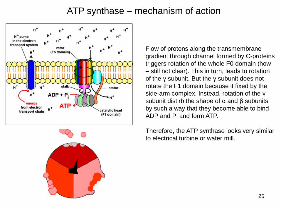
ATP synthase – mechanism of action
Flow of protons along the transmembrane gradient through channel formed by C-proteins triggers rotation of the whole F0 domain (how – still not clear). This in turn, leads to rotation of the subunit. But the subunit does not rotate the F1 domain because it fixed by the side-arm complex. Instead, rotation of the subunit distirb the shape of and subunits by such a way that they become able to bind ADP and Pi and form ATP.
Therefore, the ATP synthase looks very similar to electrical turbine or water mill.
26
Suggested questions
• What cellular components do you know? Their functional role in the cell;
• Overall structure of mitochondria, their shape and size;
• Origin of mitochondria;
• Dynamic behaviour of mitochondria;
• Composition of mitochondria, functional role of different subcompartments;
• Isolation of mitochondria, how to analyse the composition of mitochondria;
• Visualization (imaging) of mitochondria;
• Functional analysis of mitochondria – what is it and how to proceed it?
• Cellular respiration – what is it?
• Oxidative phosphorylation (OXPHOS) – what is it?
• Electron transport chain phunctional role and mechanism of action;
• Proton-motive force – what is it?
• Mitochondrial respiration, inhibitors of respiration;
• ATP synthase – structure and mechanism of action.