Embed Size (px)
Citation preview
Biochimica et Biophysica Acta 1822 (2012) 1896–1912
Contents lists available at SciVerse ScienceDirect
Biochimica et Biophysica Acta
j ourna l homepage: www.e lsev ie r .com/ locate /bbad is
Review
Molecular control of oogenesis☆
Flor Sánchez ⁎, Johan SmitzFollicle Biology Laboratory, Vrije Universiteit Brussel, Laarbeeklaan 101, 1090 Brussels, Belgium
☆ This article is part of a Special Issue entitled: Molecuductive Failure.⁎ Corresponding author. Tel.: +32 2 477 4645; fax: +
E-mail addresses: [email protected] (F. Sánchez),(J. Smitz).
0925-4439/$ – see front matter © 2012 Elsevier B.V. Aldoi:10.1016/j.bbadis.2012.05.013
a b s t r a c t
a r t i c l e i n f oArticle history:Received 17 January 2012Received in revised form 8 May 2012Accepted 13 May 2012Available online 24 May 2012
Keywords:Oocyte developmentFolliculogenesisGene expressionOocyte maturationCumulus–oocyte complex
Oogenesis is a complex process regulated by a vast number of intra- and extra-ovarian factors. Oogonia, whichoriginate from primordial germ cells, proliferate bymitosis and form primary oocytes that arrest at the prophasestage of the first meiotic division until they are fully-grown.Within primary oocytes, synthesis and accumulationof RNAs and proteins throughout oogenesis are essential for oocyte growth and maturation; and moreover,crucial for developing into a viable embryo after fertilization. Oocyte meiotic and developmental competenceis gained in a gradual and sequential manner during folliculogenesis and is related to the fact that the oocytegrows in interaction with its companion somatic cells. Communication between oocyte and its surroundinggranulosa cells is vital, both for oocyte development and for granulosa cells differentiation. Oocytes depend ondifferentiated cumulus cells, which provide them with nutrients and regulatory signals needed to promoteoocyte nuclear and cytoplasmic maturation and consequently the acquisition of developmental competence.Thepurpose of this article is to summarize recent knowledge on themolecular aspects of oogenesis and oocyte mat-uration, and the crucial role of cumulus–cell interactions, highlighting the valuable contribution of experimentalevidences obtained in animalmodels. This article is part of a Special Issue entitled: Molecular Genetics of HumanReproductive Failure.
© 2012 Elsevier B.V. All rights reserved.
1. Introduction
In the last decade, a substantial progress has beenmade in the eluci-dation of factors regulating oocyte and follicle growth and develop-ment, as well as oocyte maturation, through the study of the crucialroles of a large number of proteins expressed throughout oogenesis,in particular during the early stages of folliculogenesis. This has mainlybeen accomplished by assessing the lack of their gene products eitherby using knockout approaches and/or targeted deletion.
The application of molecular biology techniques for the quantifica-tion of gene expression, by using microarray analysis and real-timePCR technologies has provided new insights into the regulation ofmRNAs in a stage-dependent manner during folliculogenesis, bothin oocytes and cumulus cells. Likewise, proteomic approaches and arecent new genome-wide profiling of maternal mRNA in associationwith the polysome, have allowed to identify newly translated pro-teins during oocyte maturation, at a stage when transcription hasalready ceased.
In this sense, a huge progress in basic aspects of oocyte research hasbeen made at the molecular level. The discovery of many molecular
lar Genetics of Human Repro-
32 2 477 50 [email protected]
l rights reserved.
processes/pathways involved throughout oogenesis and ovulation,and the identification of potential markers of oocyte quality have beenaccomplished in the recent years.
The use of in vitro approaches in large animals, but mainly in themouse model, has also become relevant in providing valuable infor-mation that could not be obtained in human for ethical reasons.Making use of molecular biology techniques alongside with in vitropreantral follicle culture systems and/or in vitro oocyte maturation,for instance, has enabled the study of the influence of a number offactors (such as hormones, recombinant proteins, and/or growth fac-tors) on follicle development, survival, steroid production, oocytegrowth and maturation, and developmental potential. Moreover,granulosa cell culture and oocyte co-culture with granulosa cellshave become accepted models to study the interaction betweenoocytes and their surrounding somatic cells and to provide a focuson the potential of oocytes to regulate a variety of granulosa cellfunctions.
There has been an exponential increase in knowledge in the fieldof oocyte biology; the findings delivered from several studies mas-sively contribute to the understanding of the molecular basis of theacquisition of oocyte meiotic and developmental competence. Thepresent review has the ambition to provide information on the recentadvances in the field, emphasizing the key factors involved in the reg-ulation of oogenesis and oocyte maturation. A particular focus is pro-vided on the crucial role of the oocyte–cumulus cells interactions andthe oocyte control of granulosa cell function at different stages of fol-licle development and maturation.
1897F. Sánchez, J. Smitz / Biochimica et Biophysica Acta 1822 (2012) 1896–1912
2. Early stages of oogenesis and folliculogenesis
2.1. Primordial germ cells and follicle formation
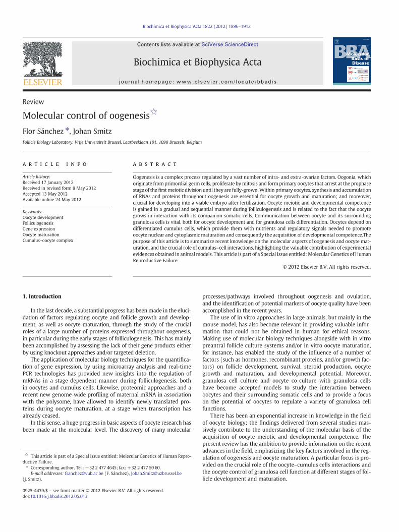
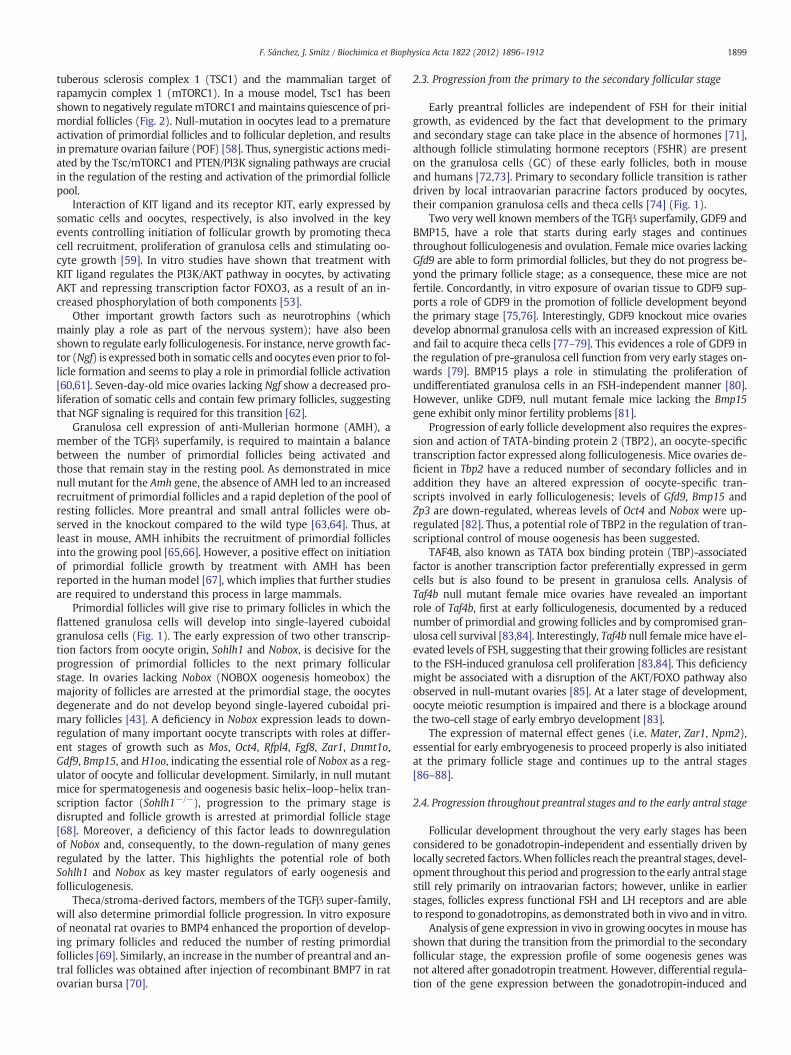
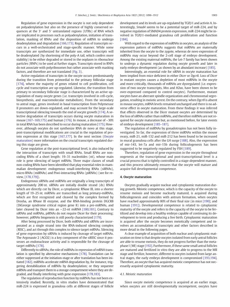
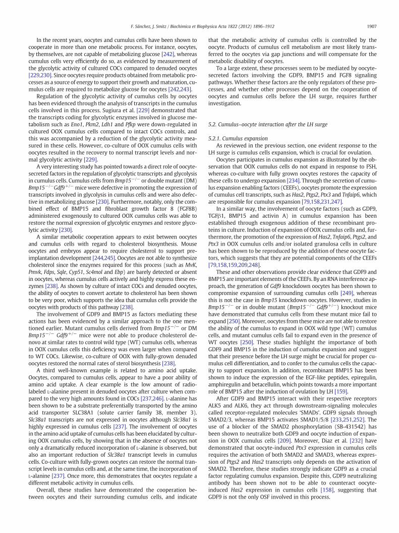
Oocytes originate from primordial germ cells (PGCs). Mouse PGCsoriginate as early as 7.5 days of embryonic development (E7.5) in theextra embryonic mesoderm and their development initially dependson signals derived from both the extra-embryonic ectoderm and visceralendoderm. Members of the TGFβ family such as bone morphogeneticproteins (BMPs), BMP4, BMP8b (ectodermorigin) and BMP2 (endodermorigin) are specific factors needed for PGC formation and regulation ofgene expression [1–3] (Fig. 1).
PGCs migrate to the genital ridge at about E10.5 in the mouse,where they proliferate by mitosis and give rise to oogonia. Migration,proliferation and colonization of PGCs to the developing gonads arecontrolled by many factors and depend as well on the interaction ofPGCs and their surrounding somatic cells. In vitro studies haveshown that BMP2 and BMP4 increase the number of mouse PGCs inculture [4,5], whereas Bmp7 mouse knockouts show a reduction inthe number of germ cells around this period [6]. Activin has alsobeen shown to increase the number of PGCs in human [7], althoughactivin inhibits PGC proliferation in mouse [8]. The role of germ cellderived transcription factors at this stage has also been demonstratedby using knockout approaches and conditional deletions in mice.These include factors such as BLIMP1 and PRDM14, which are criticalfor PGC proliferation and migration [9–11] as well as OCT4, NANOG[11–13], which are essential for PGC survival. Several factors seemto be required for PGC survival, such as FIGα (factor in the germline alpha), a factor responsible for the early expression of theglycoproteins that will form the zona pellucida [14], NANOS3(nanos homolog 3; Drosophila) and DND1 (dead end homolog 1),two RNA binding proteins that protect PGCs from undergoing apopto-sis, as well as the KIT/KIT ligand (KITL) pathway. Mutations in any ofthe genes coding for these factors lead to a deficiency in the formationof primordial follicles due to a depletion of germ cells [12–17].
Around the period of PGC migration into the genital ridges (E10.5)sex determination starts. Differentiation into ovaries seems to be the
Fig. 1. Representative figure of the factors involved in primordial germ cell (PGC) formatioblue), somatic/granulosa cells (in purple), germ cells (in red) or in both germ cell and granthe defined stages throughout folliculogenesis. Transcription factors involved are indicated wmation are indicated in black.
default pathway, since the XY genital ridges differentiate into testesunder the influence of the Y-linked gene Sry. The XX gonads have noSRY, and therefore they develop into ovaries [18]. As soon as PGCs areformed, the initially bipotential gonad will continue its differentiationmostly under the influence of somatic cell derived transcription factorsGATA4, FOXL2, LHX9, WT1, WNT4, and SF1 [19–22] (Fig. 1).
After colonization of the gonad (~E13.5), PGCs will undergo aphase of mitotic proliferation with an incomplete cytokinesis, leadingto the formation of ‘germ cell cysts’ or ‘germ cell nests’ [23]. Followingthis event and before follicle formation mitotic divisions stop andgerm cells initiate meiosis, become primary oocytes and commit tothe female program of development [24].
Meiosis initiate with a prophase stage, a complex phase which issubdivided into five stages: leptotene, zygotene, pachytene, diploteneand diakinesis.Within the first period of the prophase, a series of crucialevents occur, involving the pairing of homologous chromosomes, syn-apsis (close association between these chromosomes), and recombina-tion or ‘crossing over’ (exchange of genetic material). Subsequently,oocytes progress to the diplotene stage where they enter into a pro-longed resting phase called dictyate [25]. In mouse embryonic ovaries,initiation of the meiotic program is dependent on retinoic acid andStra8 (stimulated by retinoic acid gene 8) signaling. STRA8 is a cytoplas-mic factor expressed by female germ cells just prior to entering the pro-phase of first meiotic division [26], and in response to retinoic acid (RA)[27–29]. In females Stra8 is required for premeiotic DNA replication aswell as for meiotic prophase events (i.e. chromosome condensation, co-hesion, synapsis, and recombination) [30].
Oocytes remain at the dictyate stage of meiosis I throughout oogene-sis, until LH induces final oocyte maturation (in most mammals). Pro-phase events are vital for germ cell survival and meiotic progression,and errors occurring along this stage, as well as throughout the consecu-tive phases ofmeiosis, may originate and/or contribute to femalemeioticaneuploidies. Indeed, endocrine-disrupting chemicals, such as BisphenolA (BPA), have been shown to cause disturbances in spindle formation, tointerfere with microtubule polymerization and to induce multipolarspindles in mouse oocytes. In utero exposure to BPA appears to interferewith control of recombination in fetal prophase I oocytes and increasing
n, oogenesis and folliculogenesis. Ovarian factors produced by theca/stromal cells (inulosa cell (green), participate and regulate oocyte and follicle development at each ofith a star (*). Proteins from the extra embryonic ectoderm that participate in PGC for-
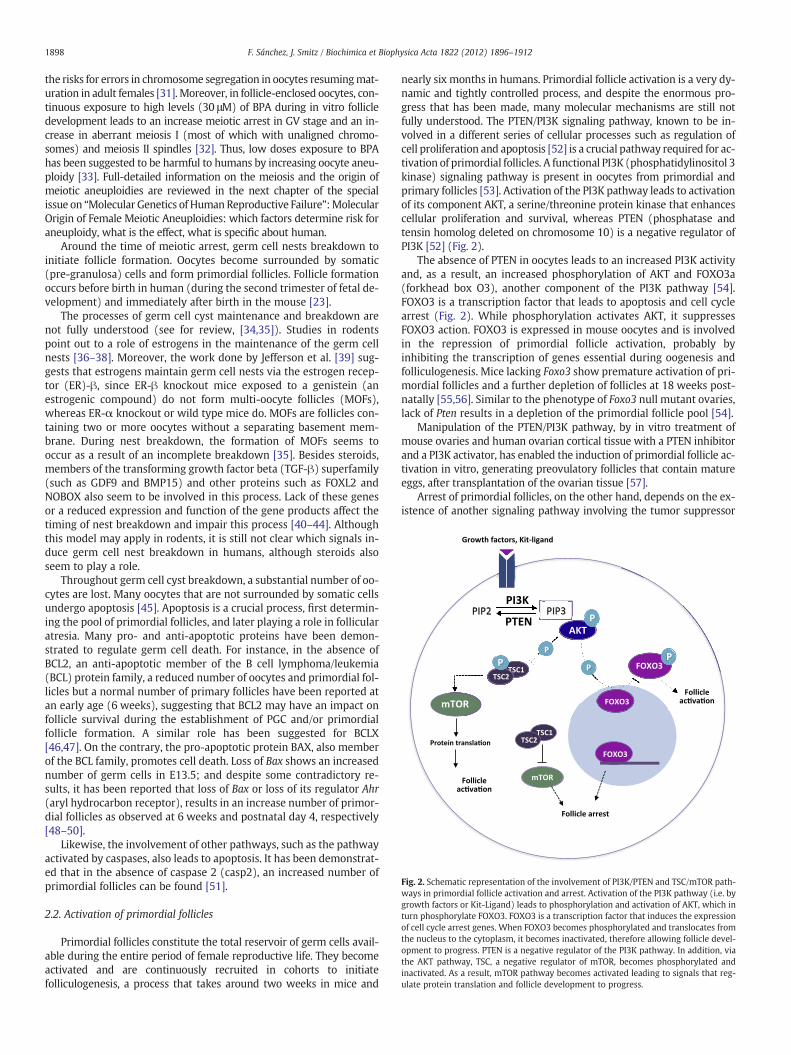
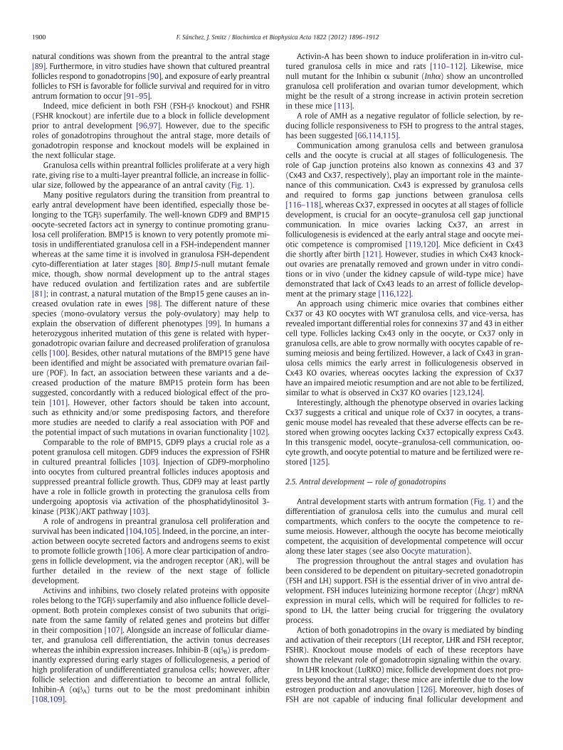
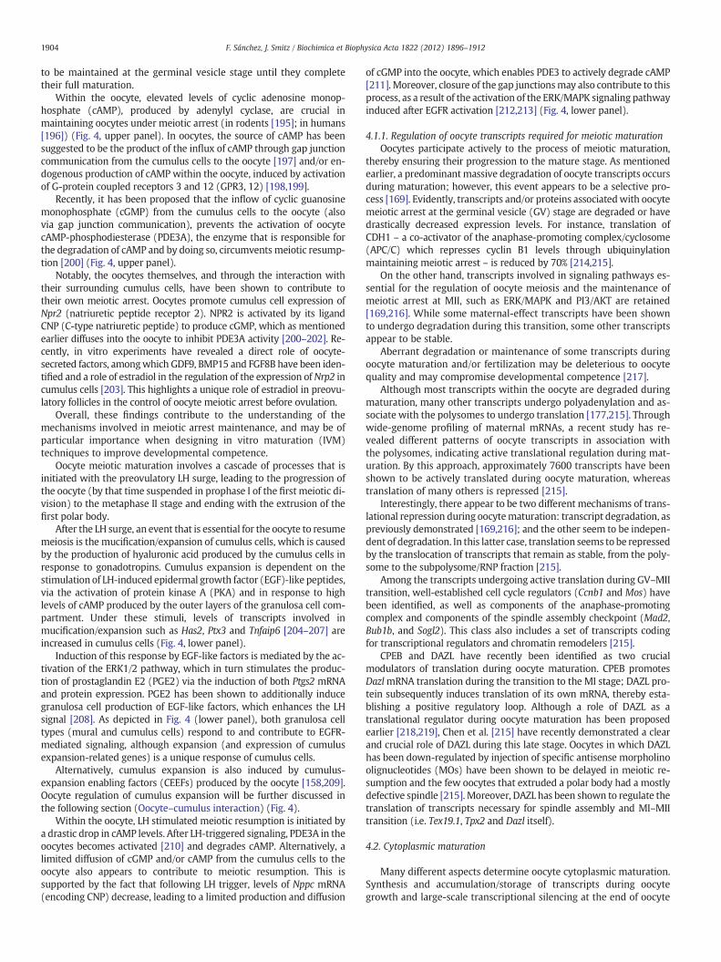
Fig. 2. Schematic representation of the involvement of PI3K/PTEN and TSC/mTOR path-ways in primordial follicle activation and arrest. Activation of the PI3K pathway (i.e. bygrowth factors or Kit-Ligand) leads to phosphorylation and activation of AKT, which inturn phosphorylate FOXO3. FOXO3 is a transcription factor that induces the expressionof cell cycle arrest genes. When FOXO3 becomes phosphorylated and translocates fromthe nucleus to the cytoplasm, it becomes inactivated, therefore allowing follicle devel-opment to progress. PTEN is a negative regulator of the PI3K pathway. In addition, viathe AKT pathway, TSC, a negative regulator of mTOR, becomes phosphorylated andinactivated. As a result, mTOR pathway becomes activated leading to signals that reg-ulate protein translation and follicle development to progress.
1898 F. Sánchez, J. Smitz / Biochimica et Biophysica Acta 1822 (2012) 1896–1912
the risks for errors in chromosome segregation in oocytes resumingmat-uration in adult females [31]. Moreover, in follicle-enclosed oocytes, con-tinuous exposure to high levels (30 μM) of BPA during in vitro follicledevelopment leads to an increase meiotic arrest in GV stage and an in-crease in aberrant meiosis I (most of which with unaligned chromo-somes) and meiosis II spindles [32]. Thus, low doses exposure to BPAhas been suggested to be harmful to humans by increasing oocyte aneu-ploidy [33]. Full-detailed information on the meiosis and the origin ofmeiotic aneuploidies are reviewed in the next chapter of the specialissue on “Molecular Genetics of Human Reproductive Failure”:MolecularOrigin of Female Meiotic Aneuploidies: which factors determine risk foraneuploidy, what is the effect, what is specific about human.
Around the time of meiotic arrest, germ cell nests breakdown toinitiate follicle formation. Oocytes become surrounded by somatic(pre-granulosa) cells and form primordial follicles. Follicle formationoccurs before birth in human (during the second trimester of fetal de-velopment) and immediately after birth in the mouse [23].
The processes of germ cell cyst maintenance and breakdown arenot fully understood (see for review, [34,35]). Studies in rodentspoint out to a role of estrogens in the maintenance of the germ cellnests [36–38]. Moreover, the work done by Jefferson et al. [39] sug-gests that estrogens maintain germ cell nests via the estrogen recep-tor (ER)-β, since ER-β knockout mice exposed to a genistein (anestrogenic compound) do not form multi-oocyte follicles (MOFs),whereas ER-α knockout or wild type mice do. MOFs are follicles con-taining two or more oocytes without a separating basement mem-brane. During nest breakdown, the formation of MOFs seems tooccur as a result of an incomplete breakdown [35]. Besides steroids,members of the transforming growth factor beta (TGF-β) superfamily(such as GDF9 and BMP15) and other proteins such as FOXL2 andNOBOX also seem to be involved in this process. Lack of these genesor a reduced expression and function of the gene products affect thetiming of nest breakdown and impair this process [40–44]. Althoughthis model may apply in rodents, it is still not clear which signals in-duce germ cell nest breakdown in humans, although steroids alsoseem to play a role.
Throughout germ cell cyst breakdown, a substantial number of oo-cytes are lost. Many oocytes that are not surrounded by somatic cellsundergo apoptosis [45]. Apoptosis is a crucial process, first determin-ing the pool of primordial follicles, and later playing a role in follicularatresia. Many pro- and anti-apoptotic proteins have been demon-strated to regulate germ cell death. For instance, in the absence ofBCL2, an anti-apoptotic member of the B cell lymphoma/leukemia(BCL) protein family, a reduced number of oocytes and primordial fol-licles but a normal number of primary follicles have been reported atan early age (6 weeks), suggesting that BCL2 may have an impact onfollicle survival during the establishment of PGC and/or primordialfollicle formation. A similar role has been suggested for BCLX[46,47]. On the contrary, the pro-apoptotic protein BAX, also memberof the BCL family, promotes cell death. Loss of Bax shows an increasednumber of germ cells in E13.5; and despite some contradictory re-sults, it has been reported that loss of Bax or loss of its regulator Ahr(aryl hydrocarbon receptor), results in an increase number of primor-dial follicles as observed at 6 weeks and postnatal day 4, respectively[48–50].
Likewise, the involvement of other pathways, such as the pathwayactivated by caspases, also leads to apoptosis. It has been demonstrat-ed that in the absence of caspase 2 (casp2), an increased number ofprimordial follicles can be found [51].
2.2. Activation of primordial follicles
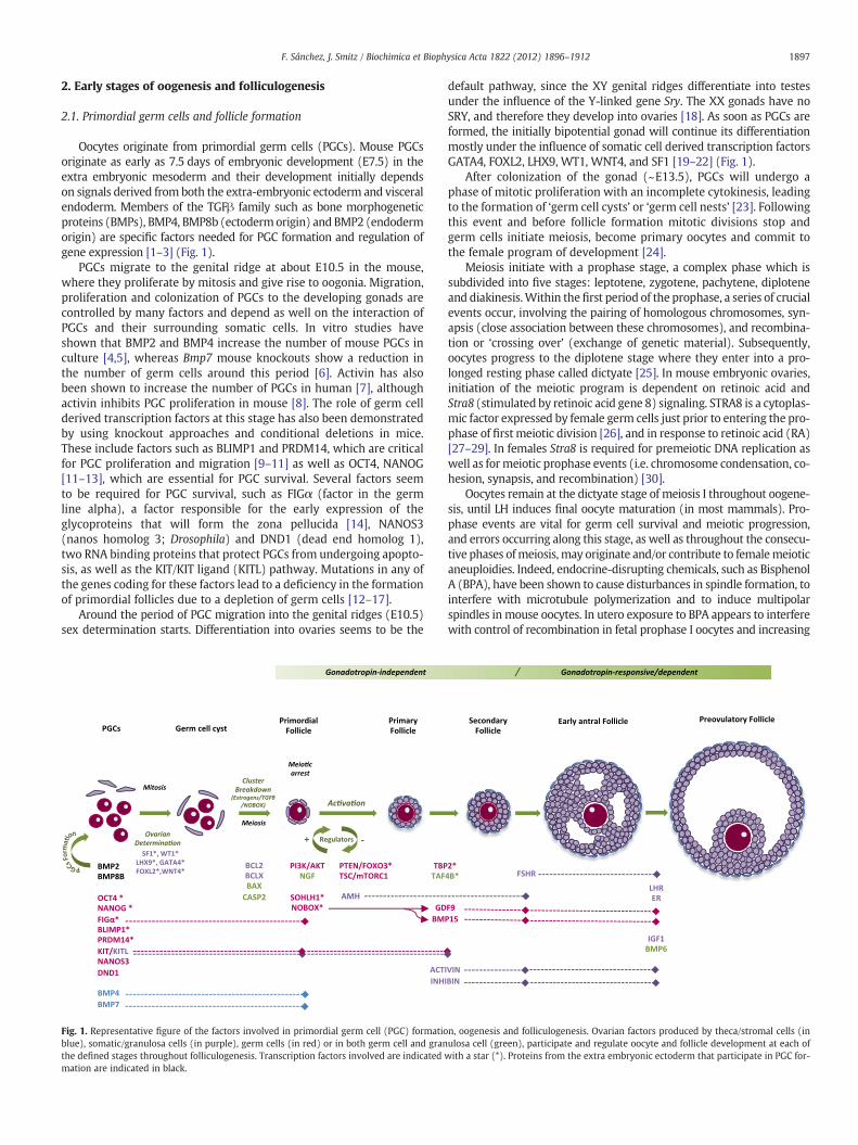
Primordial follicles constitute the total reservoir of germ cells avail-able during the entire period of female reproductive life. They becomeactivated and are continuously recruited in cohorts to initiatefolliculogenesis, a process that takes around two weeks in mice and
nearly six months in humans. Primordial follicle activation is a very dy-namic and tightly controlled process, and despite the enormous pro-gress that has been made, many molecular mechanisms are still notfully understood. The PTEN/PI3K signaling pathway, known to be in-volved in a different series of cellular processes such as regulation ofcell proliferation and apoptosis [52] is a crucial pathway required for ac-tivation of primordial follicles. A functional PI3K (phosphatidylinositol 3kinase) signaling pathway is present in oocytes from primordial andprimary follicles [53]. Activation of the PI3K pathway leads to activationof its component AKT, a serine/threonine protein kinase that enhancescellular proliferation and survival, whereas PTEN (phosphatase andtensin homolog deleted on chromosome 10) is a negative regulator ofPI3K [52] (Fig. 2).
The absence of PTEN in oocytes leads to an increased PI3K activityand, as a result, an increased phosphorylation of AKT and FOXO3a(forkhead box O3), another component of the PI3K pathway [54].FOXO3 is a transcription factor that leads to apoptosis and cell cyclearrest (Fig. 2). While phosphorylation activates AKT, it suppressesFOXO3 action. FOXO3 is expressed in mouse oocytes and is involvedin the repression of primordial follicle activation, probably byinhibiting the transcription of genes essential during oogenesis andfolliculogenesis. Mice lacking Foxo3 show premature activation of pri-mordial follicles and a further depletion of follicles at 18 weeks post-natally [55,56]. Similar to the phenotype of Foxo3 null mutant ovaries,lack of Pten results in a depletion of the primordial follicle pool [54].
Manipulation of the PTEN/PI3K pathway, by in vitro treatment ofmouse ovaries and human ovarian cortical tissue with a PTEN inhibitorand a PI3K activator, has enabled the induction of primordial follicle ac-tivation in vitro, generating preovulatory follicles that contain matureeggs, after transplantation of the ovarian tissue [57].
Arrest of primordial follicles, on the other hand, depends on the ex-istence of another signaling pathway involving the tumor suppressor
1899F. Sánchez, J. Smitz / Biochimica et Biophysica Acta 1822 (2012) 1896–1912
tuberous sclerosis complex 1 (TSC1) and the mammalian target ofrapamycin complex 1 (mTORC1). In a mouse model, Tsc1 has beenshown to negatively regulatemTORC1 andmaintains quiescence of pri-mordial follicles (Fig. 2). Null-mutation in oocytes lead to a prematureactivation of primordial follicles and to follicular depletion, and resultsin premature ovarian failure (POF) [58]. Thus, synergistic actions medi-ated by the Tsc/mTORC1 and PTEN/PI3K signaling pathways are crucialin the regulation of the resting and activation of the primordial folliclepool.
Interaction of KIT ligand and its receptor KIT, early expressed bysomatic cells and oocytes, respectively, is also involved in the keyevents controlling initiation of follicular growth by promoting thecacell recruitment, proliferation of granulosa cells and stimulating oo-cyte growth [59]. In vitro studies have shown that treatment withKIT ligand regulates the PI3K/AKT pathway in oocytes, by activatingAKT and repressing transcription factor FOXO3, as a result of an in-creased phosphorylation of both components [53].
Other important growth factors such as neurotrophins (whichmainly play a role as part of the nervous system); have also beenshown to regulate early folliculogenesis. For instance, nerve growth fac-tor (Ngf) is expressed both in somatic cells and oocytes even prior to fol-licle formation and seems to play a role in primordial follicle activation[60,61]. Seven-day-old mice ovaries lacking Ngf show a decreased pro-liferation of somatic cells and contain few primary follicles, suggestingthat NGF signaling is required for this transition [62].
Granulosa cell expression of anti-Mullerian hormone (AMH), amember of the TGFβ superfamily, is required to maintain a balancebetween the number of primordial follicles being activated andthose that remain stay in the resting pool. As demonstrated in micenull mutant for the Amh gene, the absence of AMH led to an increasedrecruitment of primordial follicles and a rapid depletion of the pool ofresting follicles. More preantral and small antral follicles were ob-served in the knockout compared to the wild type [63,64]. Thus, atleast in mouse, AMH inhibits the recruitment of primordial folliclesinto the growing pool [65,66]. However, a positive effect on initiationof primordial follicle growth by treatment with AMH has beenreported in the human model [67], which implies that further studiesare required to understand this process in large mammals.
Primordial follicles will give rise to primary follicles in which theflattened granulosa cells will develop into single-layered cuboidalgranulosa cells (Fig. 1). The early expression of two other transcrip-tion factors from oocyte origin, Sohlh1 and Nobox, is decisive for theprogression of primordial follicles to the next primary follicularstage. In ovaries lacking Nobox (NOBOX oogenesis homeobox) themajority of follicles are arrested at the primordial stage, the oocytesdegenerate and do not develop beyond single-layered cuboidal pri-mary follicles [43]. A deficiency in Nobox expression leads to down-regulation of many important oocyte transcripts with roles at differ-ent stages of growth such as Mos, Oct4, Rfpl4, Fgf8, Zar1, Dnmt1o,Gdf9, Bmp15, and H1oo, indicating the essential role of Nobox as a reg-ulator of oocyte and follicular development. Similarly, in null mutantmice for spermatogenesis and oogenesis basic helix–loop–helix tran-scription factor (Sohlh1−/−), progression to the primary stage isdisrupted and follicle growth is arrested at primordial follicle stage[68]. Moreover, a deficiency of this factor leads to downregulationof Nobox and, consequently, to the down-regulation of many genesregulated by the latter. This highlights the potential role of bothSohlh1 and Nobox as key master regulators of early oogenesis andfolliculogenesis.
Theca/stroma-derived factors, members of the TGFβ super-family,will also determine primordial follicle progression. In vitro exposureof neonatal rat ovaries to BMP4 enhanced the proportion of develop-ing primary follicles and reduced the number of resting primordialfollicles [69]. Similarly, an increase in the number of preantral and an-tral follicles was obtained after injection of recombinant BMP7 in ratovarian bursa [70].
2.3. Progression from the primary to the secondary follicular stage
Early preantral follicles are independent of FSH for their initialgrowth, as evidenced by the fact that development to the primaryand secondary stage can take place in the absence of hormones [71],although follicle stimulating hormone receptors (FSHR) are presenton the granulosa cells (GC) of these early follicles, both in mouseand humans [72,73]. Primary to secondary follicle transition is ratherdriven by local intraovarian paracrine factors produced by oocytes,their companion granulosa cells and theca cells [74] (Fig. 1).
Two very well known members of the TGFβ superfamily, GDF9 andBMP15, have a role that starts during early stages and continuesthroughout folliculogenesis and ovulation. Female mice ovaries lackingGfd9 are able to form primordial follicles, but they do not progress be-yond the primary follicle stage; as a consequence, these mice are notfertile. Concordantly, in vitro exposure of ovarian tissue to GDF9 sup-ports a role of GDF9 in the promotion of follicle development beyondthe primary stage [75,76]. Interestingly, GDF9 knockout mice ovariesdevelop abnormal granulosa cells with an increased expression of KitLand fail to acquire theca cells [77–79]. This evidences a role of GDF9 inthe regulation of pre-granulosa cell function from very early stages on-wards [79]. BMP15 plays a role in stimulating the proliferation ofundifferentiated granulosa cells in an FSH-independent manner [80].However, unlike GDF9, null mutant female mice lacking the Bmp15gene exhibit only minor fertility problems [81].
Progression of early follicle development also requires the expres-sion and action of TATA-binding protein 2 (TBP2), an oocyte-specifictranscription factor expressed along folliculogenesis. Mice ovaries de-ficient in Tbp2 have a reduced number of secondary follicles and inaddition they have an altered expression of oocyte-specific tran-scripts involved in early folliculogenesis; levels of Gfd9, Bmp15 andZp3 are down-regulated, whereas levels of Oct4 and Nobox were up-regulated [82]. Thus, a potential role of TBP2 in the regulation of tran-scriptional control of mouse oogenesis has been suggested.
TAF4B, also known as TATA box binding protein (TBP)-associatedfactor is another transcription factor preferentially expressed in germcells but is also found to be present in granulosa cells. Analysis ofTaf4b null mutant female mice ovaries have revealed an importantrole of Taf4b, first at early folliculogenesis, documented by a reducednumber of primordial and growing follicles and by compromised gran-ulosa cell survival [83,84]. Interestingly, Taf4b null female mice have el-evated levels of FSH, suggesting that their growing follicles are resistantto the FSH-induced granulosa cell proliferation [83,84]. This deficiencymight be associated with a disruption of the AKT/FOXO pathway alsoobserved in null-mutant ovaries [85]. At a later stage of development,oocyte meiotic resumption is impaired and there is a blockage aroundthe two-cell stage of early embryo development [83].
The expression of maternal effect genes (i.e. Mater, Zar1, Npm2),essential for early embryogenesis to proceed properly is also initiatedat the primary follicle stage and continues up to the antral stages[86–88].
2.4. Progression throughout preantral stages and to the early antral stage
Follicular development throughout the very early stages has beenconsidered to be gonadotropin-independent and essentially driven bylocally secreted factors.When follicles reach the preantral stages, devel-opment throughout this period and progression to the early antral stagestill rely primarily on intraovarian factors; however, unlike in earlierstages, follicles express functional FSH and LH receptors and are ableto respond to gonadotropins, as demonstrated both in vivo and in vitro.
Analysis of gene expression in vivo in growing oocytes inmouse hasshown that during the transition from the primordial to the secondaryfollicular stage, the expression profile of some oogenesis genes wasnot altered after gonadotropin treatment. However, differential regula-tion of the gene expression between the gonadotropin-induced and
1900 F. Sánchez, J. Smitz / Biochimica et Biophysica Acta 1822 (2012) 1896–1912
natural conditions was shown from the preantral to the antral stage[89]. Furthermore, in vitro studies have shown that cultured preantralfollicles respond to gonadotropins [90], and exposure of early preantralfollicles to FSH is favorable for follicle survival and required for in vitroantrum formation to occur [91–95].
Indeed, mice deficient in both FSH (FSH-β knockout) and FSHR(FSHR knockout) are infertile due to a block in follicle developmentprior to antral development [96,97]. However, due to the specificroles of gonadotropins throughout the antral stage, more details ofgonadotropin response and knockout models will be explained inthe next follicular stage.
Granulosa cells within preantral follicles proliferate at a very highrate, giving rise to a multi-layer preantral follicle, an increase in follic-ular size, followed by the appearance of an antral cavity (Fig. 1).
Many positive regulators during the transition from preantral toearly antral development have been identified, especially those be-longing to the TGFβ superfamily. The well-known GDF9 and BMP15oocyte-secreted factors act in synergy to continue promoting granu-losa cell proliferation. BMP15 is known to very potently promote mi-tosis in undifferentiated granulosa cell in a FSH-independent mannerwhereas at the same time it is involved in granulosa FSH-dependentcyto-differentiation at later stages [80]. Bmp15-null mutant femalemice, though, show normal development up to the antral stageshave reduced ovulation and fertilization rates and are subfertile[81]; in contrast, a natural mutation of the Bmp15 gene causes an in-creased ovulation rate in ewes [98]. The different nature of thesespecies (mono-ovulatory versus the poly-ovulatory) may help toexplain the observation of different phenotypes [99]. In humans aheterozygous inherited mutation of this gene is related with hyper-gonadotropic ovarian failure and decreased proliferation of granulosacells [100]. Besides, other natural mutations of the BMP15 gene havebeen identified and might be associated with premature ovarian fail-ure (POF). In fact, an association between these variants and a de-creased production of the mature BMP15 protein form has beensuggested, concordantly with a reduced biological effect of the pro-tein [101]. However, other factors should be taken into account,such as ethnicity and/or some predisposing factors, and thereforemore studies are needed to clarify a real association with POF andthe potential impact of such mutations in ovarian functionality [102].
Comparable to the role of BMP15, GDF9 plays a crucial role as apotent granulosa cell mitogen. GDF9 induces the expression of FSHRin cultured preantral follicles [103]. Injection of GDF9-morpholinointo oocytes from cultured preantral follicles induces apoptosis andsuppressed preantral follicle growth. Thus, GDF9 may at least partlyhave a role in follicle growth in protecting the granulosa cells fromundergoing apoptosis via activation of the phosphatidylinositol 3-kinase (PI3K)/AKT pathway [103].
A role of androgens in preantral granulosa cell proliferation andsurvival has been indicated [104,105]. Indeed, in the porcine, an inter-action between oocyte secreted factors and androgens seems to existto promote follicle growth [106]. A more clear participation of andro-gens in follicle development, via the androgen receptor (AR), will befurther detailed in the review of the next stage of follicledevelopment.
Activins and inhibins, two closely related proteins with oppositeroles belong to the TGFβ superfamily and also influence follicle devel-opment. Both protein complexes consist of two subunits that origi-nate from the same family of related genes and proteins but differin their composition [107]. Alongside an increase of follicular diame-ter, and granulosa cell differentiation, the activin tonus decreaseswhereas the inhibin expression increases. Inhibin-B (αβB) is predom-inantly expressed during early stages of folliculogenesis, a period ofhigh proliferation of undifferentiated granulosa cells; however, afterfollicle selection and differentiation to become an antral follicle,Inhibin-A (αβA) turns out to be the most predominant inhibin[108,109].
Activin-A has been shown to induce proliferation in in-vitro cul-tured granulosa cells in mice and rats [110–112]. Likewise, micenull mutant for the Inhibin α subunit (Inhα) show an uncontrolledgranulosa cell proliferation and ovarian tumor development, whichmight be the result of a strong increase in activin protein secretionin these mice [113].
A role of AMH as a negative regulator of follicle selection, by re-ducing follicle responsiveness to FSH to progress to the antral stages,has been suggested [66,114,115].
Communication among granulosa cells and between granulosacells and the oocyte is crucial at all stages of folliculogenesis. Therole of Gap junction proteins also known as connexins 43 and 37(Cx43 and Cx37, respectively), play an important role in the mainte-nance of this communication. Cx43 is expressed by granulosa cellsand required to forms gap junctions between granulosa cells[116–118], whereas Cx37, expressed in oocytes at all stages of follicledevelopment, is crucial for an oocyte–granulosa cell gap junctionalcommunication. In mice ovaries lacking Cx37, an arrest infolliculogenesis is evidenced at the early antral stage and oocyte mei-otic competence is compromised [119,120]. Mice deficient in Cx43die shortly after birth [121]. However, studies in which Cx43 knock-out ovaries are prenatally removed and grown under in vitro condi-tions or in vivo (under the kidney capsule of wild-type mice) havedemonstrated that lack of Cx43 leads to an arrest of follicle develop-ment at the primary stage [116,122].
An approach using chimeric mice ovaries that combines eitherCx37 or 43 KO oocytes with WT granulosa cells, and vice-versa, hasrevealed important differential roles for connexins 37 and 43 in eithercell type. Follicles lacking Cx43 only in the oocyte, or Cx37 only ingranulosa cells, are able to grow normally with oocytes capable of re-suming meiosis and being fertilized. However, a lack of Cx43 in gran-ulosa cells mimics the early arrest in folliculogenesis observed inCx43 KO ovaries, whereas oocytes lacking the expression of Cx37have an impaired meiotic resumption and are not able to be fertilized,similar to what is observed in Cx37 KO ovaries [123,124].
Interestingly, although the phenotype observed in ovaries lackingCx37 suggests a critical and unique role of Cx37 in oocytes, a trans-genic mouse model has revealed that these adverse effects can be re-stored when growing oocytes lacking Cx37 ectopically express Cx43.In this transgenic model, oocyte–granulosa-cell communication, oo-cyte growth, and oocyte potential to mature and be fertilized were re-stored [125].
2.5. Antral development — role of gonadotropins
Antral development starts with antrum formation (Fig. 1) and thedifferentiation of granulosa cells into the cumulus and mural cellcompartments, which confers to the oocyte the competence to re-sume meiosis. However, although the oocyte has become meioticallycompetent, the acquisition of developmental competence will occuralong these later stages (see also Oocyte maturation).
The progression throughout the antral stages and ovulation hasbeen considered to be dependent on pituitary-secreted gonadotropin(FSH and LH) support. FSH is the essential driver of in vivo antral de-velopment. FSH induces luteinizing hormone receptor (Lhcgr) mRNAexpression in mural cells, which will be required for follicles to re-spond to LH, the latter being crucial for triggering the ovulatoryprocess.
Action of both gonadotropins in the ovary is mediated by bindingand activation of their receptors (LH receptor, LHR and FSH receptor,FSHR). Knockout mouse models of each of these receptors haveshown the relevant role of gonadotropin signaling within the ovary.
In LHR knockout (LuRKO)mice, follicle development does not pro-gress beyond the antral stage; these mice are infertile due to the lowestrogen production and anovulation [126]. Moreover, high doses ofFSH are not capable of inducing final follicular development and
1901F. Sánchez, J. Smitz / Biochimica et Biophysica Acta 1822 (2012) 1896–1912
ovulation when LHR is not present [127]. Therefore, expression ofLHR is essential not only for ovulation but also for follicle maturationprior to ovulation.
Mice deficient in FSH (FSH-β knockouts) are infertile due to ablock of the follicle development prior to the antral stage. Withinthese follicles, granulosa cells express increased levels of FSH-R tran-scripts, but decreased levels of aromatase, and inhibin/activin sub-units. Moreover, they fail to express normal levels of LH receptors[97]. Fshr-null mutant female mice, in which all forms of the FSHRhave been eliminated (also known as FORKO — follitropin receptorknockout mice), display an atrophic uterus and are also infertile dueto a block in folliculogenesis before antral formation [96]. In thesemutants, the largest follicles observed have no more than four layersof granulosa cells. However, in spite of a normal expression of aroma-tase mRNA and protein in Fshr-null mutant mice, there is a completeloss of estrogen production by the ovary, leading to metabolic alter-ations such as obesity and skeletal abnormality (some of which canbe reversed by treatment with estradiol-17beta). This may suggestthat signaling through the FSHR may be involved in the activationof the aromatase enzyme [128].
Whether the block in follicle development in both FSH-β and FSHRknockouts is due to altered granulosa cell proliferation or due to in-creased apoptosis is not fully understood. Although the precise role ofcyclin D2 (a gene responsive to FSH and involved in granulosa cell pro-liferation) in this process has not been identified, FSH-β knockout micehave a modest decrease in the expression of cyclin D2, whereas this hasnot been clearly demonstrated in FORKO mice [96].
These studies are relevant for human reproduction since pheno-types similar to those reported in mice have been found in humanwith mutations in FSH-β and FSHR [129]. Moreover, a number of men-opausal symptoms inwomen, aswell as features of hypergonadotropic-hypogonadism that can be observed in infertile women are also mim-icked by the phenotype of FORKO mice.
By in vitro approaches, studies have demonstrated that in the ab-sence of FSH cultured follicles are arrested in their development, donot support antrum formation and exhibit apoptosis, all of which canbe prevented by FSH supplementation [91,92]. In spite of this, researchconducted by our group has demonstrated that once the antrum forma-tion is established, follicle development to the preovulatory stage underdecreased FSH concentrations induce a better cumulus cells differentia-tion [130,131]. Accordingly, low FSH levels applied in follicle cultureafter antrum formation appear to promote development of primate fol-licles and oocytes achieve larger diameters [95]. On the contrary, expo-sure to supraphysiologically high FSH levels during antral stages orthroughout development from preantral to antral stages in vitro per-turbs oocyte control of granulosa cell differentiation, as well as cumuluscell function, in mouse and primate follicle cultures [95,130]. Thus, al-though gonadotropins are essential during antral stages in vitro, adose-fine-tuning is critical to obtain an appropriate antral development.
Under the influence of gonadotropins, follicles synthesize steroidhormones such as androgens and estrogens, which contribute to fol-licular development, by inducing granulosa cell proliferation and dif-ferentiation via the androgen receptor (AR) and estrogen receptor(ER), respectively [109,132,133]. Through the well-known two-cell,two-gonadotropin model, theca cells produce androgens under influ-ence of LH stimuli, whereas granulosa cells produce estrogens usingandrogens as a substrate, under influence of FSH [108,134].
Androgens, have been shown to participate in granulosa cell pro-liferation and survival via the androgen receptor (AR). AR-deficientfemale mice are subfertile, and show a reduced number of antral fol-licles and ovulated oocytes, and a high rate of granulosa cells apopto-sis; they eventually develop premature ovarian failure [135,136].Most of the characteristics observed in these Ar-null mice seem tobe the result of a lack of AR expression more specifically in the gran-ulosa cells, as demonstrated by a model in which specific knockoutsfor the AR in either granulosa cells or oocytes were made. Therefore,
granulosa cell-specific AR appear to be essential for follicle develop-ment and survival [137].
Estradiol is the predominant estrogen in terms of estrogenic activ-ity. Indeed, one of the major functions of preovulatory granulosa cellsis the synthesis of estradiol. Inside the follicle, estradiol is producedvia the enzyme aromatase, and it enhances the response of granulosacells to the gonadotropins [138].
Knockout models for the two different estrogen receptors, ERαand ERβ have lead to the elucidation of the role of estrogens in follic-ular development. Knockout mice for the ERα, also known as ERKOmice, have increased levels of estradiol and LH, whereas FSH levelsare normal. Ovaries from ERKOmice show an arrest at the early antralstage and are infertile [139,140]; this demonstrates the essential roleof estradiol in gonadotropin-induced follicle differentiation. On thecontrary, in ERβ knockout (BERCO) mice follicles develop to the an-tral stages and these mice are fertile, but they have a diminished ovu-latory response to hCG stimuli [140–142].
Another approach involved the targeted disruption of the Cyp19(aromatase) gene in mice (ArKO), which causes a deficiency in estra-diol synthesis. These mice have elevated levels of circulating gonado-tropins and testosterone and are infertile. At adulthood, female ArKOmice ovaries may contain large antral follicles, but after one yearfolliculogenesis is severely affected and secondary follicles are no lon-ger found [143,144], which suggests an important role of estradiol infollicular development. Moreover, the ArKO phenotype seems to re-sult not only from the lack of estrogens but also from the high levelsof circulating gonadotropins, mainly LH [145].
Members of the insulin growth factor (IGF) family (i.e. IGF1, IGF2)also cooperate with gonadotropins to determine follicle selection andthe further progression through the antral stages. In rodents, IGF1 andIGF2 are the predominant forms in granulosa and theca cells, respec-tively, where they co-operate with FSH and LH action in each cell type[146]. Moreover, in IGF1 deficient mice, follicle development does notprogress beyond the small antral stage. In these mice there is reducedexpression of FSHr, and decreased synthesis of estradiol and reducedgranulosa cell proliferation [147,148].
Besides IGFs, follicular development throughout the antral stagesis clearly dependent on many other intraovarian factors. Similar toearlier stages, activins and inhibins produced by granulosa cells playessential paracrine roles by regulating the LH-induced androgen syn-thesis produced by theca cells [149–151], and therefore, they ensurethe estradiol supply. In mice lacking Acvr2b (the activin type-IIB re-ceptor), progression beyond the early antral stages fails [152]. Theactivin–inhibin system does not only regulate granulosa cell prolifer-ation but also granulosa cell differentiation and oocyte maturation,the latter being accelerated by activin A action [153–155].
Oocyte-secreted GDF9 and BMP15 also play a critical role during an-tral stages. GDF9 and BMP15 regulate cumulus cell function (more de-tailed explanation is given in the next section). As evidenced in rat andhuman, these oocyte factors alone, or in combination, attenuate FSH ef-fects and stimulate granulosa cell proliferation and differentiation.GDF9 suppresses FSH-stimulated estradiol secretion by repressing aro-matase activity and also suppresses FSH-induced LH receptor formationin differentiated cumulus cells. BMP15 is believed to reduce cumulus cellapoptosis and to suppress FSH receptor expression [79,156,157]. BothBMP15 and GDF9, together with BMP6, have been demonstrated to in-hibit FSH-induced progesterone production, which may be consideredimportant for follicle survival and prevention of premature luteinization.
In preovulatory follicles, a critical role of GDF9 and BMP15 in theinduction of cumulus mucification/expansion and the regulation ofgenes involved in this process has been demonstrated and will be fur-ther explained hereafter [79,158,159].
Other factors such as theca-derived factors BMP4 and BMP7 arepotential paracrine regulators of granulosa cell function. In the rat,these two factors attenuate FSH-induced progesterone secretionwhilst they enhance FSH-induced estradiol secretion [70,160].
1902 F. Sánchez, J. Smitz / Biochimica et Biophysica Acta 1822 (2012) 1896–1912
Finally, preovulatory follicles containing fully-grown oocytes areready to undergo ovulation, which is induced by the preovulatorysurge of gonadotropins. Ovulation is characterized by the rupture ofthe follicle wall and the release of the cumulus–oocyte complex; atthis time the oocyte has resumed meiosis and has progressed to themetaphase II stage of meiosis (Fig. 1) (oocyte maturation is describedin detail in the following sections).
After ovulation, granulosa and theca cells become luteal cells andare responsible for the production of estradiol and progesterone, thelatter is predominantly expressed in the corpus luteum [161].
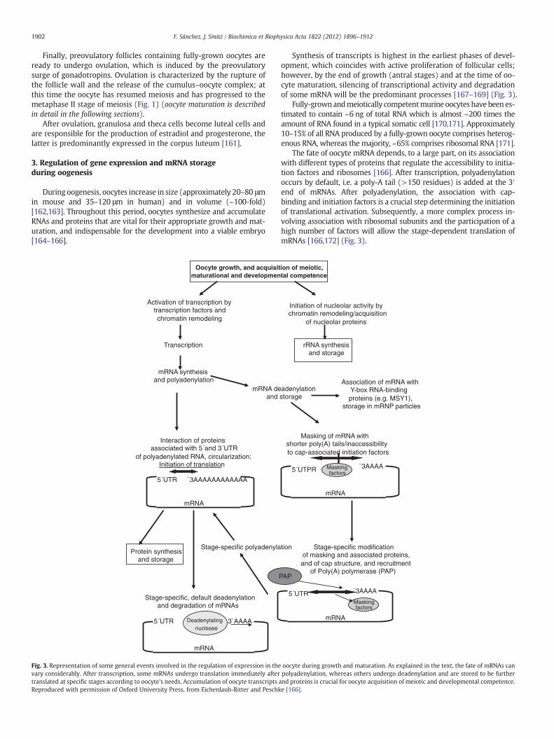
3. Regulation of gene expression and mRNA storageduring oogenesis
During oogenesis, oocytes increase in size (approximately 20–80 μmin mouse and 35–120 μm in human) and in volume (~100-fold)[162,163]. Throughout this period, oocytes synthesize and accumulateRNAs and proteins that are vital for their appropriate growth and mat-uration, and indispensable for the development into a viable embryo[164–166].
Activation of transcription by transcription factors andchromatin remodeling
Transcription
mRNA synthesis and polyadenylation
mRNA deand
Interaction of proteinsassociated with 5´and 3´UTR
of polyadenylated RNA, circularization:Initiation of translation
Oocyte growth, and acquisitmaturational and developmen
Stage-specific polyadenyla
Stage-specific, default deadenylationand degradation of mRNAs
Protein synthesisand storage
`3AAAAAAAAAAAA5´UTR
3`AAAA5´UTR Deadenylating nuclease
mRNA
mRNA
Fig. 3. Representation of some general events involved in the regulation of expression in thevary considerably. After transcription, some mRNAs undergo translation immediately aftertranslated at specific stages according to oocyte's needs. Accumulation of oocyte transcriptsReproduced with permission of Oxford University Press, from Eichenlaub-Ritter and Peschk
Synthesis of transcripts is highest in the earliest phases of devel-opment, which coincides with active proliferation of follicular cells;however, by the end of growth (antral stages) and at the time of oo-cyte maturation, silencing of transcriptional activity and degradationof some mRNA will be the predominant processes [167–169] (Fig. 3).
Fully-grownandmeiotically competentmurine oocytes have been es-timated to contain ~6 ng of total RNA which is almost ~200 times theamount of RNA found in a typical somatic cell [170,171]. Approximately10–15% of all RNA produced by a fully-grown oocyte comprises heterog-enous RNA, whereas the majority, ~65% comprises ribosomal RNA [171].
The fate of oocyte mRNA depends, to a large part, on its associationwith different types of proteins that regulate the accessibility to initia-tion factors and ribosomes [166]. After transcription, polyadenylationoccurs by default, i.e. a poly-A tail (>150 residues) is added at the 3′end of mRNAs. After polyadenylation, the association with cap-binding and initiation factors is a crucial step determining the initiationof translational activation. Subsequently, a more complex process in-volving association with ribosomal subunits and the participation of ahigh number of factors will allow the stage-dependent translation ofmRNAs [166,172] (Fig. 3).
Initiation of nucleolar activity bychromatin remodeling/acquisition
of nucleolar proteins
rRNA synthesis and storage
adenylationstorage
Masking of mRNA with shorter poly(A) tails/inaccessibility to cap-associated initiation factors
ion of meiotic, tal competence
Stage-specific modification of masking and associated proteins, and of cap structure, and recruitment
of Poly(A) polymerase (PAP)
tion
`3AAAA5´UTPR Maskingfactors
`3AAAA5´UTRMaskingfactors
PAP
Association of mRNA with Y-box RNA-binding proteins (e.g. MSY1),
storage in mRNP particles
mRNA
mRNA
oocyte during growth and maturation. As explained in the text, the fate of mRNAs canpolyadenylation, whereas others undergo deadenylation and are stored to be further
and proteins is crucial for oocyte acquisition of meiotic and developmental competence.e [166].
1903F. Sánchez, J. Smitz / Biochimica et Biophysica Acta 1822 (2012) 1896–1912
Regulation of gene expression in the oocyte is not only dependenton polyadenylation but also on the presence of highly conserved se-quences at the 3′ and 5′ untranslated regions (UTRs) of RNA whichare implicated in processes such as polyadenylation, initiation of trans-lation, masking of RNAs and the disposition of mRNA to undergodeadenylation and degradation [166,173]. Regulation of transcripts oc-curs in a well-orchestrated and stage-specific manner. While sometranscripts are synthesized for immediate use, other transcripts willbe deadenylated (by shortening of the poly-A tail, which confers morestability) to be either degraded or stored in the ooplasm in ribonuclearparticles (RNPs) to be used at further stages. Transcripts stored in RNPsdo not associate with polyribosomes but rather associate with maskingfactors, and therefore are not translated (Fig. 3).
Active regulation of transcripts in the oocyte occurs predominantlyduring the transition from primordial to the primary follicular stage[174], where the majority of genes related to cell proliferation, cellcycle and transcription are up-regulated. Likewise, the transition fromprimary to secondary follicular stage is characterized by an active up-regulation of many oocyte genes (i.e. transcripts involved in cell-cycle,biosynthesis, and macromolecular metabolism). From the secondaryto antral stage, genes involved in basal transcription from PolymeraseII promoters are down-regulated, and may account for the large-scaletranscriptional silencing towards the end of oocyte growth [174]. A se-lective degradation of transcripts occurs during oocyte maturation inmouse [167–169,175] and human [176]. In mouse, a decrease of ~30%in total RNA has been described to occur duringmaturation [175]. How-ever, although oocytes do not synthesize RNA de novo at this stage,post-transcriptional modifications are crucial in the regulation of pro-tein expression at this stage [167,177]. In the next section (Oocytematuration) more information on the crucial transcripts regulated dur-ing this stage are given.
Gene regulation at the post-transcriptional level, is also induced bythe interaction of transcripts with small RNAs. Small RNAs are non-coding RNAs of a short length: 19–31 nucleotides (nt), whose mainrole is gene silencing of target mRNAs. Three major classes of smallnon-coding RNAs have been identified that play essential roles inmam-malian development: endogenous small-interfering RNAs (siRNAs),micro RNAs (miRNAs) and Piwi-interacting RNAs (piRNAs) (see for re-view, [178,179]).
Endogenous siRNAs and miRNAs are originally a long transcripts ofapproximately 200 nt. siRNAs are initially double strand (ds) RNAswhich are directly cut by Dicer, a cytoplasmic RNase III, into a shorterlength of 19–25 nt. miRNAs are transcribed as long primary-miRNAs,which are first recognized and cleaved by the complex formed byDrosha, an RNase III enzyme, and the RNA-binding protein DGCR8(DiGeorge syndrome critical region gene 8) into a pre-miRNAs, andlater cleaved by Dicer into an ∼22-nt miRNA [180,181]. Contrary tosiRNAs and miRNAs, piRNAs do not require Dicer for their processing;however, piRNAs biogenesis is still poorly characterized [179].
After being processed by Dicer, both miRNAs and siRNAs are incor-porated as a single strand into the RNA-induced silencing complex(RISC), and act through this complex to silence target mRNAs. Silencingof gene expression by siRNAs is induced by cleavage of target mRNAs.The Argonaute 2 (AGO2) is a key component of the siRISC since it pos-sesses an endonuclease activity and is responsible for the cleavage oftarget mRNAs [178].
Differently to siRNAs, the role ofmiRNAs in repression ofmRNA trans-lation is specified by alternative mechanisms [178]. Translation can beeither suppressed at the initiation-stage or after translation has been ini-tiated [182]. miRNAs accelerate mRNA degradation by, for instance, trig-gering destabilization of mRNAs by deadenylation; or they sequestermRNAs and transport them to a storage compartmentwhere they are de-graded, and finally interfering with gene expression [178,183].
The regulation of reproduction function bymiRNAs has not been ex-tensively studied. Recently, in vitro studies have demonstrated thatmiR-224 is expressed in granulosa cells at different stages of follicle
development and its levels are up-regulated by TGFβ1 and activin A. In-terestingly, Smad4 seems to be a potential target of miR-224, and bynegative regulation of SMAD4 protein expression,miR-224might be in-volved in TGFβ1-mediated granulosa cell proliferation and function[184].
With regard to the expression ofmiRNAs in oocytes, an analysis of theexpression pattern of miRNAs suggests that miRNAs are maternallyinherited from the oocyte to the zygote, whereas de novo expression ofmiRNAs may occur beyond the 2-cell stage of embryo development.Among the existing maternal miRNAs, the Let-7 family has been shownto undergo a dynamic regulation during oocyte growth and later inearly embryo development (as shown by an abundant increase) [185].
Interestingly, an essential role for siRNA in oocyte maturation hasbeen implied from mice deficient in either Dicer or Dgcr8. Loss of Dicerin mutant oocytes causes a depletion of most miRNAs in the oocyteand more critically, thousands of mRNAs are dysregulated (i.e. expres-sion of two oocyte transcripts, Mos and H2ax, have been shown to beover-expressed compared to control oocytes). Furthermore, mutantoocytes have an aberrant spindle organization and chromosomal segre-gation, resulting inmeiotic arrest. On the contrary, whenDgcr8 is absentinmouse oocytes,mRNA levels remained unchanged and there is no ad-verse effect in oocyte maturation. From these findings it was inferredthat effects observed in the Dicer mutant oocytes are determined bythe loss of siRNAs rather thanmiRNAs, and thereforemiRNAs are not re-quired for oocyte maturation but, as mentioned before, for later eventsof embryo development [185–187].
The regulation of miRNAs by gonadotropins has not been fully in-vestigated. So far, the expression of three miRNAs within the mouseovary, miR-21, miR-132 and miR-212 has been found to be increasedin mural granulosa cells after the LH surge [188]. Also, the expressionof mir-143, let-7a and mir-15b during folliculogenesis has beensuggested to be negatively regulated by FSH [189].
Therefore, regulation of gene expression in the oocyte throughoutoogenesis at the transcriptional and post-transcriptional level is acrucial process that is tightly controlled in a stage-dependent manner,and this process ultimately ensures that the oocyte will mature andacquire full developmental competence.
4. Oocyte maturation
Oocytes gradually acquire nuclear and cytoplasmic maturation dur-ing growth. Meiotic competence, which is the capacity of the oocyte toresume meiosis and become nuclearly matured, is acquired duringfolliculogenesis and coincides with antrum formation, when oocyteshave reached approximately 80% of their final size (in mice [190]; andhuman [191]). Developmental competence is related to cytoplasmicmaturity of the oocyte and refers to the capacity of the oocyte to be fer-tilized and develop into a healthy embryo capable of continuing its de-velopment to term and producing a live birth. Cytoplasmic maturationis acquired after the oocyte becomes meiotically competent and in-volves an accumulation of transcripts and other factors described inmore detail in the following pages.
A clear example of acquisition of both nuclear and cytoplasmic mat-uration on time is that despite oocytes isolated from early antral folliclesare able to resumemeiosis, they do not progress further than themeta-phase I (MI) stage [192]. Furthermore, if these same small antral folliclesare matured and fertilized in vitro they are able to progress to the MIIstage and fertilize, however, different to oocytes isolated from large an-tral stages, the early embryo development is compromised [193,194].Therefore, an oocyte that has acquiredmeiotic competence has not nec-essarily acquired cytoplasmic maturity.
4.1. Meiotic maturation
Since oocyte meiotic competence is acquired at an earlier stage,when oocytes are still developmentally incompetent, oocytes have
1904 F. Sánchez, J. Smitz / Biochimica et Biophysica Acta 1822 (2012) 1896–1912
to be maintained at the germinal vesicle stage until they completetheir full maturation.
Within the oocyte, elevated levels of cyclic adenosine monop-hosphate (cAMP), produced by adenylyl cyclase, are crucial inmaintaining oocytes under meiotic arrest (in rodents [195]; in humans[196]) (Fig. 4, upper panel). In oocytes, the source of cAMP has beensuggested to be the product of the influx of cAMP through gap junctioncommunication from the cumulus cells to the oocyte [197] and/or en-dogenous production of cAMP within the oocyte, induced by activationof G-protein coupled receptors 3 and 12 (GPR3, 12) [198,199].
Recently, it has been proposed that the inflow of cyclic guanosinemonophosphate (cGMP) from the cumulus cells to the oocyte (alsovia gap junction communication), prevents the activation of oocytecAMP-phosphodiesterase (PDE3A), the enzyme that is responsible forthe degradation of cAMP and by doing so, circumvents meiotic resump-tion [200] (Fig. 4, upper panel).
Notably, the oocytes themselves, and through the interaction withtheir surrounding cumulus cells, have been shown to contribute totheir own meiotic arrest. Oocytes promote cumulus cell expression ofNpr2 (natriuretic peptide receptor 2). NPR2 is activated by its ligandCNP (C-type natriuretic peptide) to produce cGMP, which as mentionedearlier diffuses into the oocyte to inhibit PDE3A activity [200–202]. Re-cently, in vitro experiments have revealed a direct role of oocyte-secreted factors, amongwhich GDF9, BMP15 and FGF8B have been iden-tified and a role of estradiol in the regulation of the expression ofNrp2 incumulus cells [203]. This highlights a unique role of estradiol in preovu-latory follicles in the control of oocyte meiotic arrest before ovulation.
Overall, these findings contribute to the understanding of themechanisms involved in meiotic arrest maintenance, and may be ofparticular importance when designing in vitro maturation (IVM)techniques to improve developmental competence.
Oocyte meiotic maturation involves a cascade of processes that isinitiated with the preovulatory LH surge, leading to the progression ofthe oocyte (by that time suspended in prophase I of the first meiotic di-vision) to the metaphase II stage and ending with the extrusion of thefirst polar body.
After the LH surge, an event that is essential for the oocyte to resumemeiosis is the mucification/expansion of cumulus cells, which is causedby the production of hyaluronic acid produced by the cumulus cells inresponse to gonadotropins. Cumulus expansion is dependent on thestimulation of LH-induced epidermal growth factor (EGF)-like peptides,via the activation of protein kinase A (PKA) and in response to highlevels of cAMP produced by the outer layers of the granulosa cell com-partment. Under these stimuli, levels of transcripts involved inmucification/expansion such as Has2, Ptx3 and Tnfaip6 [204–207] areincreased in cumulus cells (Fig. 4, lower panel).
Induction of this response by EGF-like factors is mediated by the ac-tivation of the ERK1/2 pathway, which in turn stimulates the produc-tion of prostaglandin E2 (PGE2) via the induction of both Ptgs2 mRNAand protein expression. PGE2 has been shown to additionally inducegranulosa cell production of EGF-like factors, which enhances the LHsignal [208]. As depicted in Fig. 4 (lower panel), both granulosa celltypes (mural and cumulus cells) respond to and contribute to EGFR-mediated signaling, although expansion (and expression of cumulusexpansion-related genes) is a unique response of cumulus cells.
Alternatively, cumulus expansion is also induced by cumulus-expansion enabling factors (CEEFs) produced by the oocyte [158,209].Oocyte regulation of cumulus expansion will be further discussed inthe following section (Oocyte–cumulus interaction) (Fig. 4).
Within the oocyte, LH stimulated meiotic resumption is initiated bya drastic drop in cAMP levels. After LH-triggered signaling, PDE3A in theoocytes becomes activated [210] and degrades cAMP. Alternatively, alimited diffusion of cGMP and/or cAMP from the cumulus cells to theoocyte also appears to contribute to meiotic resumption. This issupported by the fact that following LH trigger, levels of Nppc mRNA(encoding CNP) decrease, leading to a limited production and diffusion
of cGMP into the oocyte, which enables PDE3 to actively degrade cAMP[211]. Moreover, closure of the gap junctionsmay also contribute to thisprocess, as a result of the activation of the ERK/MAPK signaling pathwayinduced after EGFR activation [212,213] (Fig. 4, lower panel).
4.1.1. Regulation of oocyte transcripts required for meiotic maturationOocytes participate actively to the process of meiotic maturation,
thereby ensuring their progression to the mature stage. As mentionedearlier, a predominantmassive degradation of oocyte transcripts occursduring maturation; however, this event appears to be a selective pro-cess [169]. Evidently, transcripts and/or proteins associatedwith oocytemeiotic arrest at the germinal vesicle (GV) stage are degraded or havedrastically decreased expression levels. For instance, translation ofCDH1 – a co-activator of the anaphase-promoting complex/cyclosome(APC/C) which represses cyclin B1 levels through ubiquinylationmaintaining meiotic arrest – is reduced by 70% [214,215].
On the other hand, transcripts involved in signaling pathways es-sential for the regulation of oocyte meiosis and the maintenance ofmeiotic arrest at MII, such as ERK/MAPK and PI3/AKT are retained[169,216]. While some maternal-effect transcripts have been shownto undergo degradation during this transition, some other transcriptsappear to be stable.
Aberrant degradation or maintenance of some transcripts duringoocyte maturation and/or fertilization may be deleterious to oocytequality and may compromise developmental competence [217].
Although most transcripts within the oocyte are degraded duringmaturation, many other transcripts undergo polyadenylation and as-sociate with the polysomes to undergo translation [177,215]. Throughwide-genome profiling of maternal mRNAs, a recent study has re-vealed different patterns of oocyte transcripts in association withthe polysomes, indicating active translational regulation during mat-uration. By this approach, approximately 7600 transcripts have beenshown to be actively translated during oocyte maturation, whereastranslation of many others is repressed [215].
Interestingly, there appear to be two different mechanisms of trans-lational repression during oocytematuration: transcript degradation, aspreviously demonstrated [169,216]; and the other seem to be indepen-dent of degradation. In this latter case, translation seems to be repressedby the translocation of transcripts that remain as stable, from the poly-some to the subpolysome/RNP fraction [215].
Among the transcripts undergoing active translation during GV–MIItransition, well-established cell cycle regulators (Ccnb1 and Mos) havebeen identified, as well as components of the anaphase-promotingcomplex and components of the spindle assembly checkpoint (Mad2,Bub1b, and Sogl2). This class also includes a set of transcripts codingfor transcriptional regulators and chromatin remodelers [215].
CPEB and DAZL have recently been identified as two crucialmodulators of translation during oocyte maturation. CPEB promotesDazlmRNA translation during the transition to the MI stage; DAZL pro-tein subsequently induces translation of its own mRNA, thereby esta-blishing a positive regulatory loop. Although a role of DAZL as atranslational regulator during oocyte maturation has been proposedearlier [218,219], Chen et al. [215] have recently demonstrated a clearand crucial role of DAZL during this late stage. Oocytes in which DAZLhas been down-regulated by injection of specific antisense morpholinoolignucleotides (MOs) have been shown to be delayed in meiotic re-sumption and the few oocytes that extruded a polar body had a mostlydefective spindle [215]. Moreover, DAZL has been shown to regulate thetranslation of transcripts necessary for spindle assembly and MI–MIItransition (i.e. Tex19.1, Tpx2 and Dazl itself).
4.2. Cytoplasmic maturation
Many different aspects determine oocyte cytoplasmic maturation.Synthesis and accumulation/storage of transcripts during oocytegrowth and large-scale transcriptional silencing at the end of oocyte
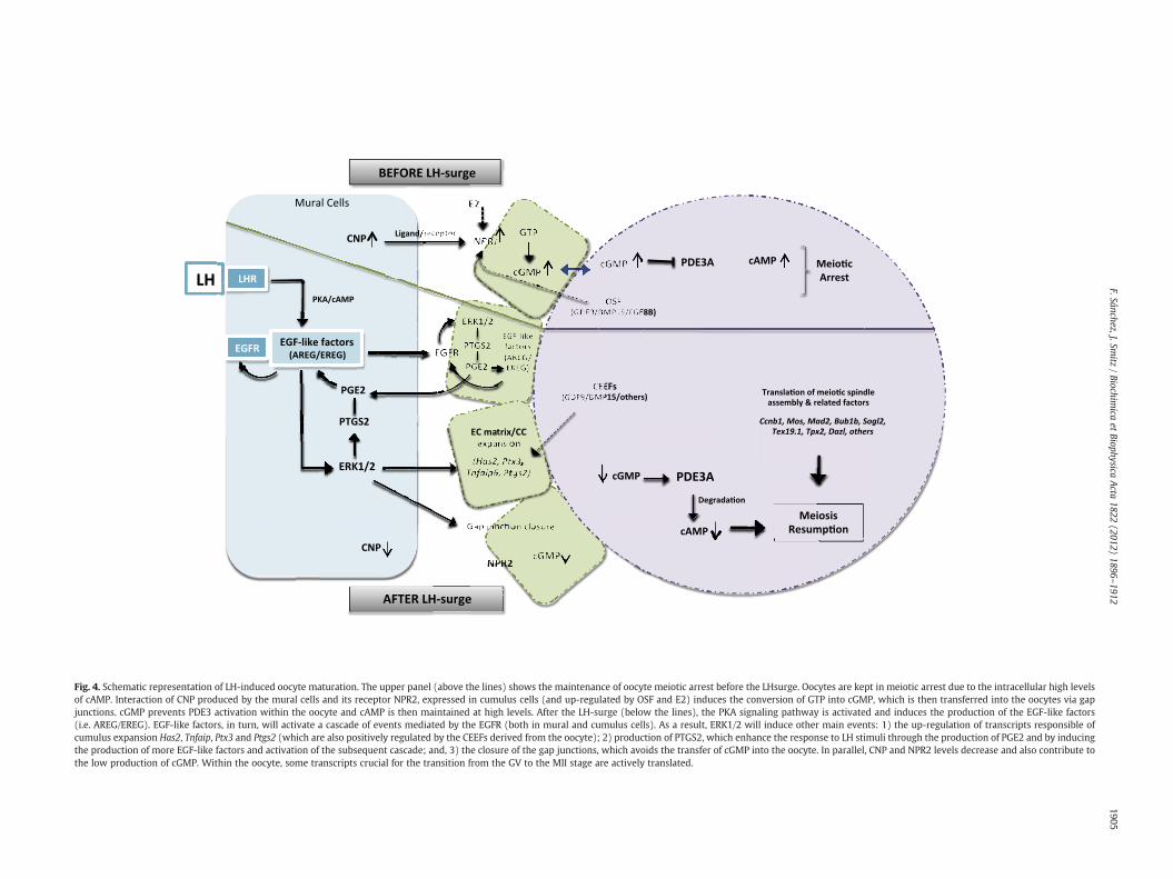
Fig. 4. Schematic representation of LH-induced oocyte maturation. The upper panel (above the lines) shows the maintenance of oocyte meiotic arrest before the LHsurge. Oocytes are kept in meiotic arrest due to the intracellular high levelsof cAMP. Interaction of CNP produced by the mural cells and its receptor NPR2, expressed in cumulus cells (and up-regulated by OSF and E2) induces the conversion of GTP into cGMP, which is then transferred into the oocytes via gapjunctions. cGMP prevents PDE3 activation within the oocyte and cAMP is then maintained at high levels. After the LH-surge (below the lines), the PKA signaling pathway is activated and induces the production of the EGF-like factors(i.e. AREG/EREG). EGF-like factors, in turn, will activate a cascade of events mediated by the EGFR (both in mural and cumulus cells). As a result, ERK1/2 will induce other main events: 1) the up-regulation of transcripts responsible ofcumulus expansion Has2, Tnfaip, Ptx3 and Ptgs2 (which are also positively regulated by the CEEFs derived from the oocyte); 2) production of PTGS2, which enhance the response to LH stimuli through the production of PGE2 and by inducingthe production of more EGF-like factors and activation of the subsequent cascade; and, 3) the closure of the gap junctions, which avoids the transfer of cGMP into the oocyte. In parallel, CNP and NPR2 levels decrease and also contribute tothe low production of cGMP. Within the oocyte, some transcripts crucial for the transition from the GV to the MII stage are actively translated.
1905F.Sánchez,J.Sm
itz/Biochim
icaet
BiophysicaActa
1822(2012)
1896–1912
1906 F. Sánchez, J. Smitz / Biochimica et Biophysica Acta 1822 (2012) 1896–1912
growth are essential to support early stages of embryo development(as reviewed in the previous section). Indeed, a contribution of granu-losa cells in oocyte global transcriptional silencing in pre-ovulatorymouse oocytes, and therefore in acquisition of oocyte cytoplasmicmaturation, has been suggested [220].
Global transcriptional silencing in the oocyte is associated with –
although not dependent on – another key event that also determinesoocyte developmental competence, chromatin condensation into thesurrounded nucleolus configuration [221]. Mattson and Albertini [222]initially demonstrated that the time of antrum formation in mouse co-incides with the appearance of two different oocyte chromatin configu-rations, the ‘surrounded nucleolus’ (SN) configuration, in which a ringof chromatin can be distinguished around the nucleolus, and the ‘non-surrounded nucleolus’ (NSN), in which the chromatin is dispersedthroughout the nucleolus. While NSN oocytes are transcriptionally ac-tive and have a poor developmental competence, due to a block at thetwo-cell stage, oocytes in the SN configuration are transcriptionally in-active and have a better developmental competence [223–225].
Furthermore, a microarray analysis has revealed that in-vitro ma-tured MII oocytes derived from either oocytes at the SN or NSN stageshowed different patterns of gene expression. OCT4 (or POU5F1), hasbeen shown to have an important role as a potent key regulator of mo-lecular events governing the establishment of developmental compe-tence of mouse oocytes [226]. Transcript levels of the transcriptionalregulator Oct4 and of Stella, an oocyte transcript regulated by OCT4,with an essential role during early embryo development, have beenfound to be downregulated, and their proteins have been shown to beabsent in antral and MII oocytes with a NSN configuration.
There is increasing evidence suggesting that factors present in oo-cytes at the SN stage but absent in the NSN configuration determine oo-cyte competence, as reported by Zuccotti et al. [226]. Their findingshave also been supported by results of micromanipulation experimentsof the germinal vesicle of fully grown oocytes (SN and NSN), whichstrongly indicate that factor(s) present in cytoplasm of MII SN oocytes(after GVBD occurs), determine developmental competence and arenot present in oocytes with the NSN configuration [227].
DAZL is another crucial protein not only essential for meiotic mat-uration but also required for acquisition of oocyte developmentalcompetence. Down-regulation of DAZL in GV oocytes does not onlycompromise meiosis progression but also fertilization. Moreover,down-regulation of maternal-derived DAZL in early zygotes causes ablock at the two-cell stage. Therefore, an important role of DAZL asa translational regulator in early embryo development and as a deter-minant of developmental competence has also been proposed [215].
5. Oocyte–cumulus cell interaction
In the recent years, remarkable progress has been made in thestudy of oocyte–granulosa cell interaction and of oocyte regulationof granulosa cell function [228–233].
In concert with the correct gene expression patterns in the oocyte,oocyte–granulosa cell interaction mediated by either paracrine sig-nals and/or by gap junctional communication from early stages of de-velopment determines the rate of follicle growth and differentiation.Furthermore, after differentiation, oocyte–cumulus cell interaction isessential to promote oocyte nuclear and cytoplasmic maturation,which determine the capacity of the oocyte to support early embryodevelopment [192,220,234].
Oocyte–granulosa cell interaction and oocyte control of granulosacell function are essential before and after LH surge. Indeed, in antral fol-licles, fully grown GV oocytes have been shown to be the most potentregulators of cumulus cell function [235]. Before the LH surge, oocytesdo not only influence granulosa cell proliferation [233,236] and differ-entiation [231,232], but very importantly, oocytes regulate metabolicactivity of cumulus cells within the COC (aminoacid uptake, glycolysisand cholesterol biosynthesis) [229,237,238]. After the LH surge, oocytes
regulate the expression of cumulus genes responsible for themucification/expansion process.
Overall, knowledge of oocyte control of the status of cumulus dif-ferentiation and function has been achieved by oocyte co-culture ex-periments, generation of knockout mice, and by molecular biologytechniques including quantification analysis of mRNAs and RNA regu-lation, for instance through RNA interference.
5.1. Cumulus–oocyte interaction before the LH surge
5.1.1. Regulation of cumulus cell differentiationAt the time of antrum formation, granulosa cells differentiate into
two compartments: the cumulus granulosa cells remain close to the oo-cyte, whereas the mural granulosa cells remain in the outer part of thefollicle. In vitro studies have provided evidences that within the follicle,mural and cumulus granulosa cells are regulated differently by the op-posing intrafollicular effects of gonadotropins (FSH) and oocytes, re-spectively, determining their phenotype and functionality [231,232].
Oocytes promote the expression of cumulus cells transcripts whereasthey suppressmural cell transcripts [79,231,232,239].When fully grownGV oocytes are microsurgically removed from the cumulus–oocytecomplexes (a procedure known as oocytectomy — OOX), cumulus cellstend to dedifferentiate, as shown by down-regulation of the expressionof cumulus cell markers (i.e. Amh, Slc38a1) [232,239] and, under thiscondition, FSH is capable to induce mural transcripts (i.e. Lhcgr,Cyp11a1) in these cells. Oocyte co-culture with OOX cumulus cells re-verts this effect.
As mentioned earlier, high FSH levels during follicle culture lead toan altered expression of cumulus transcripts, as evidenced by decreasedlevels ofAmh andhigh levels of functional Lhcgr [130]. This atypical phe-notype caused by high doses of FSH during culture is apparently alsoreflected in oocytes, which under this conditions express significantlyincreased levels of Gdf9 and Bmp15 mRNA compared to oocytes cul-tured under lower doses of FSH [130]. Given the regulation of Lhcgrand Amh by oocyte transcripts, the increased levels of Gdf9 and Bmp15mRNA are believed to reflect an oocyte response to compensate for ef-fects that occur in cumulus cells.
Oocytes have the potential to induce the expression of cumulus celltranscripts inmural cells, even in the presence of FSH. This clearly dem-onstrates that within the follicle, cumulus cell features are mainlyinfluenced by their close associationwith the oocyte, which can potent-ly buffer the continuous effects of FSH in order to maintain cumuluscells differentiated [232,240]. Interestingly, strong evidence suggeststhat these actions are mediated through the activation of SMAD2/3,thus identifying GDF9, which acts through this pathway, as one of theideal candidates among the oocyte-secreted factors exerting such ef-fects [232,233].
5.1.2. Metabolic cooperativityParacrine signals mediated by oocyte-secreted factors are probably
among the most essential, but not exclusive, mechanisms mediating cu-mulus–oocyte interactions. Gap junctions are highly specialized inter-cellular connections that facilitate communication between the oocyteand its surrounding cumulus cells. Notably, given that oocytes and cu-mulus cells are physically separated by the zona pellucida, gap-junctional communication can occur thanks to the specialized trans-zonal cytoplasmic projections (TZP) developed by cumulus cells. Theseprojections are able to penetrate through the zona pellucida reachingthe oocyte membrane and allow the formation of gap junctions [241].
Gap junctions play a crucial role in the bidirectional communica-tion between oocytes and cumulus cells, allowing the passage of mol-ecules of different types (i.e. aminoacids and metabolites such aspyruvate) from the cumulus to the oocyte [228]. Indeed expressionof gap junction proteins Cx43 and Cx37 in granulosa cells and oocytes,respectively, has been shown to be of vital importance in guarantee-ing oocyte and follicle development [123,124].
1907F. Sánchez, J. Smitz / Biochimica et Biophysica Acta 1822 (2012) 1896–1912
In the recent years, oocytes and cumulus cells have been shown tocooperate in more than one metabolic process. For instance, oocytes,by themselves, are not capable of metabolizing glucose [242], whereascumulus cells very efficiently do so, as evidenced by measurement ofthe glycolytic activity of cultured COCs compared to denuded oocytes[229,230]. Since oocytes require products obtained frommetabolic pro-cesses as a source of energy to support their growth andmaturation, cu-mulus cells are required to metabolize glucose for oocytes [242,243].
Regulation of the glycolytic activity of cumulus cells by oocyteshas been evidenced through the analysis of transcripts in the cumuluscells involved in this process. Sugiura et al. [229] demonstrated thatthe transcripts coding for glycolytic enzymes involved in glucose me-tabolism such as Eno1, Pkm2, Ldh1 and Pfkp were down-regulated incultured OOX cumulus cells compared to intact COCs controls, andthis was accompanied by a reduction of the glycolytic activity mea-sured in these cells. However, co-culture of OOX cumulus cells withoocytes resulted in the recovery to normal transcript levels and nor-mal glycolytic activity [229].
A very interesting study has pointed towards a direct role of oocyte-secreted factors in the regulation of glycolytic transcripts and glycolysisin cumulus cells. Cumulus cells from Bmp15−/− or doublemutant (DM)Bmp15−/−Gdf9+/− micewere defective in promoting the expression oftranscripts involved in glycolysis in cumulus cells and were also defec-tive in metabolizing glucose [230]. Furthermore, notably, only the com-bined effect of BMP15 and fibroblast growth factor 8 (FGF8B)administered exogenously to cultured OOX cumulus cells was able torestore the normal expression of glycolytic enzymes and restore glyco-lytic activity [230].
A similar metabolic cooperation appears to exist between oocytesand cumulus cells with regard to cholesterol biosynthesis. Mouseoocytes and embryos appear to require cholesterol to support pre-implantation development [244,245]. Oocytes are not able to synthesizecholesterol since the enzymes required for this process (such as MvK,Pmvk, Fdps, Sqle, Cyp51, Sc4mol and Ebp) are barely detected or absentin oocytes, whereas cumulus cells actively and highly express these en-zymes [238]. As shown by culture of intact COCs and denuded oocytes,the ability of oocytes to convert acetate to cholesterol has been shownto be very poor, which supports the idea that cumulus cells provide theoocytes with products of this pathway [238].
The involvement of GDF9 and BMP15 as factors mediating theseactions has been evidenced by a similar approach to the one men-tioned earlier. Mutant cumulus cells derived from Bmp15−/− or DMBmp15−/− Gdf9+/− mice were not able to produce cholesterol de-novo at similar rates to control wild type (WT) cumulus cells, whereasin OOX cumulus cells this deficiency was even larger when comparedto WT COCs. Likewise, co-culture of OOX with fully-grown denudedoocytes restored the normal rates of sterol biosynthesis [238].
A third well-known example is related to amino acid uptake.Oocytes, compared to cumulus cells, appear to have a poor ability ofamino acid uptake. A clear example is the low amount of radio-labeled L-alanine present in denuded oocytes after culture when com-pared to the very high amounts found in COCs [237,246]. L-alanine hasbeen shown to be a substrate preferentially transported by the aminoacid transporter SLC38A1 (solute carrier family 38, member 3).Slc38a1 transcripts are not expressed in oocytes although Slc38a1 ishighly expressed in cumulus cells [237]. The involvement of oocytesin the amino acid uptake of cumulus cells has been elucidated by cultur-ing OOX cumulus cells, by showing that in the absence of oocytes notonly a dramatically reduced incorporation of L-alanine is observed, butalso an important reduction of Slc38a1 transcript levels in cumuluscells. Co-culture with fully-grown oocytes can restore the normal tran-script levels in cumulus cells and, at the same time, the incorporation ofL-alanine [237]. Once more, this demonstrates that oocytes regulate adifferent metabolic activity in cumulus cells.
Overall, these studies have demonstrated the cooperation be-tween oocytes and their surrounding cumulus cells, and indicate
that the metabolic activity of cumulus cells is controlled by theoocyte. Products of cumulus cell metabolism are most likely trans-ferred to the oocytes via gap junctions and will compensate for themetabolic disability of oocytes.
To a large extent, these processes seem to be mediated by oocyte-secreted factors involving the GDF9, BMP15 and FGF8 signalingpathways. Whether these factors are the only regulators of these pro-cesses, and whether other processes depend on the cooperation ofoocytes and cumulus cells before the LH surge, requires furtherinvestigation.
5.2. Cumulus–oocyte interaction after the LH surge
5.2.1. Cumulus expansionAs reviewed in the previous section, one evident response to the
LH surge is cumulus cells expansion, which is crucial for ovulation.Oocytes participates in cumulus expansion as illustrated by the ob-
servation that OOX cumulus cells do not expand in response to FSH,whereas co-culture with fully grown oocytes restores the capacity ofthese cells to undergo expansion [234]. Through the secretion of cumu-lus expansion enabling factors (CEEFs), oocytes promote the expressionof cumulus cell transcripts, such as Has2, Ptgs2, Ptx3 and Tnfaip6, whichare responsible for cumulus expansion [79,158,231,247].
In a similar way, the involvement of oocyte factors (such as GDF9,TGFβ1, BMP15 and activin A) in cumulus expansion has beenestablished through exogenous addition of these recombinant pro-teins in culture. Induction of expansion of OOX cumulus cells and, fur-thermore, the promotion of the expression of Has2, Tnfaip6, Ptgs2, andPtx3 in OOX cumulus cells and/or isolated granulosa cells in culturehas been shown to be reproduced by the addition of these oocyte fac-tors, which suggests that they are potential components of the CEEFs[79,158,159,209,248].
These and other observations provide clear evidence that GDF9 andBMP15 are important elements of the CEEFs. By anRNA interference ap-proach, the generation of Gdf9 knockdown oocytes has been shown tocompromise expansion of surrounding cumulus cells [249], whereasthis is not the case in Bmp15 knockdown oocytes. However, studies inBmp15−/− or in double mutant (Bmp15−/− Gdf9+/−) knockout micehave demonstrated that cumulus cells from these mutant mice fail toexpand [250]. Moreover, oocytes from thesemice are not able to restorethe ability of the cumulus to expand in OOX wild type (WT) cumuluscells, and mutant cumulus cells fail to expand even in the presence ofWT oocytes [250]. These studies highlight the importance of bothGDF9 and BMP15 in the induction of cumulus expansion and suggestthat their presence before the LH surge might be crucial for proper cu-mulus cell differentiation, and to confer to the cumulus cells the capac-ity to support expansion. In addition, recombinant BMP15 has beenshown to induce the expression of the EGF-like peptides, epiregulin,amphiregulin and betacellulin, which points towards a more importantrole of BMP15 after the induction of ovulation by LH [159].
After GDF9 and BMP15 interact with their respective receptorsALK5 and ALK6, they act through downstream-signaling moleculescalled receptor-regulated molecules ‘SMADs’. GDF9 signals throughSMAD2/3, whereas BMP15 activates SMAD1/5/8 [233,251,252]. Theuse of a blocker of the SMAD2 phosphorylation (SB-431542) hasbeen shown to neutralize both GDF9 and oocyte induction of expan-sion in OOX cumulus cells [209]. Moreover, Diaz et al. [232] havedemonstrated that oocyte-induced Ptx3 expression in cumulus cellsrequires the activation of both SMAD2 and SMAD3, whereas expres-sion of Ptgs2 and Has2 transcripts only depends on the activation ofSMAD2. Therefore, these studies strongly indicate GDF9 as a crucialfactor regulating cumulus expansion. Despite this, GDF9 neutralizingantibody has been shown not to be able to counteract oocyte-induced Has2 expression in cumulus cells [158], suggesting thatGDF9 is not the only OSF involved in this process.
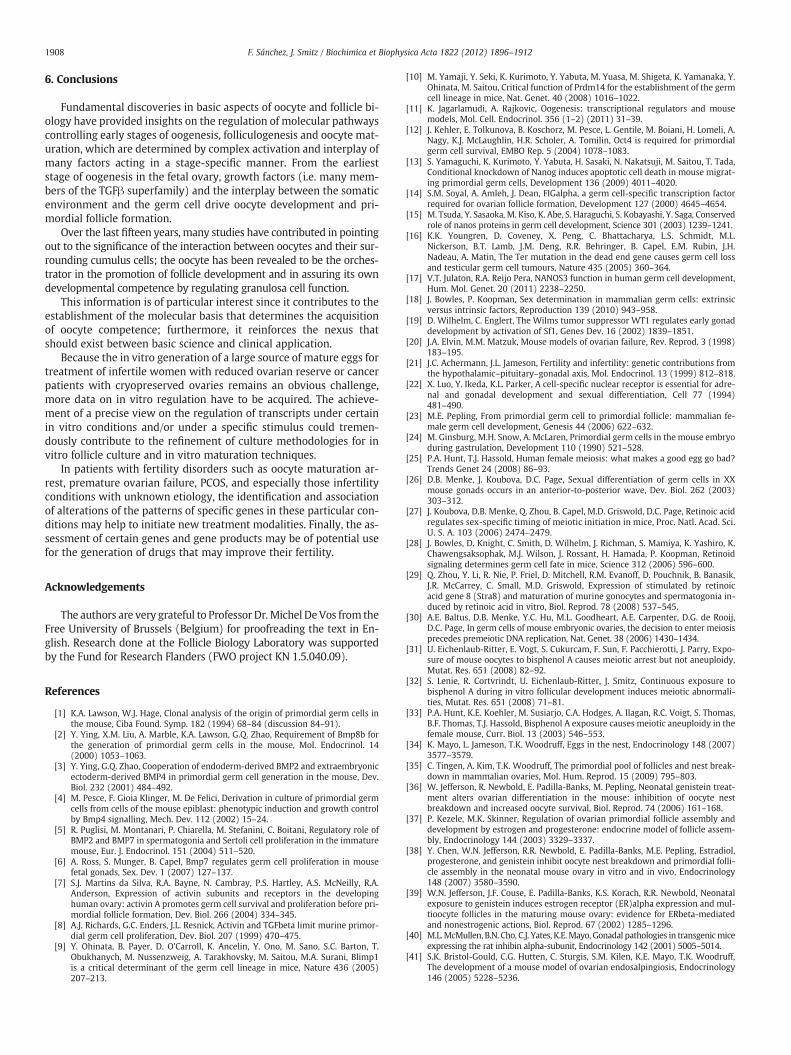
1908 F. Sánchez, J. Smitz / Biochimica et Biophysica Acta 1822 (2012) 1896–1912
6. Conclusions
Fundamental discoveries in basic aspects of oocyte and follicle bi-ology have provided insights on the regulation of molecular pathwayscontrolling early stages of oogenesis, folliculogenesis and oocyte mat-uration, which are determined by complex activation and interplay ofmany factors acting in a stage-specific manner. From the earlieststage of oogenesis in the fetal ovary, growth factors (i.e. many mem-bers of the TGFβ superfamily) and the interplay between the somaticenvironment and the germ cell drive oocyte development and pri-mordial follicle formation.
Over the last fifteen years, many studies have contributed in pointingout to the significance of the interaction between oocytes and their sur-rounding cumulus cells; the oocyte has been revealed to be the orches-trator in the promotion of follicle development and in assuring its owndevelopmental competence by regulating granulosa cell function.
This information is of particular interest since it contributes to theestablishment of the molecular basis that determines the acquisitionof oocyte competence; furthermore, it reinforces the nexus thatshould exist between basic science and clinical application.
Because the in vitro generation of a large source of mature eggs fortreatment of infertile women with reduced ovarian reserve or cancerpatients with cryopreserved ovaries remains an obvious challenge,more data on in vitro regulation have to be acquired. The achieve-ment of a precise view on the regulation of transcripts under certainin vitro conditions and/or under a specific stimulus could tremen-dously contribute to the refinement of culture methodologies for invitro follicle culture and in vitro maturation techniques.
In patients with fertility disorders such as oocyte maturation ar-rest, premature ovarian failure, PCOS, and especially those infertilityconditions with unknown etiology, the identification and associationof alterations of the patterns of specific genes in these particular con-ditions may help to initiate new treatment modalities. Finally, the as-sessment of certain genes and gene products may be of potential usefor the generation of drugs that may improve their fertility.
Acknowledgements
The authors are very grateful to Professor Dr.Michel DeVos from theFree University of Brussels (Belgium) for proofreading the text in En-glish. Research done at the Follicle Biology Laboratory was supportedby the Fund for Research Flanders (FWO project KN 1.5.040.09).
References
[1] K.A. Lawson, W.J. Hage, Clonal analysis of the origin of primordial germ cells inthe mouse, Ciba Found. Symp. 182 (1994) 68–84 (discussion 84–91).
[2] Y. Ying, X.M. Liu, A. Marble, K.A. Lawson, G.Q. Zhao, Requirement of Bmp8b forthe generation of primordial germ cells in the mouse, Mol. Endocrinol. 14(2000) 1053–1063.
[3] Y. Ying, G.Q. Zhao, Cooperation of endoderm-derived BMP2 and extraembryonicectoderm-derived BMP4 in primordial germ cell generation in the mouse, Dev.Biol. 232 (2001) 484–492.
[4] M. Pesce, F. Gioia Klinger, M. De Felici, Derivation in culture of primordial germcells from cells of the mouse epiblast: phenotypic induction and growth controlby Bmp4 signalling, Mech. Dev. 112 (2002) 15–24.
[5] R. Puglisi, M. Montanari, P. Chiarella, M. Stefanini, C. Boitani, Regulatory role ofBMP2 and BMP7 in spermatogonia and Sertoli cell proliferation in the immaturemouse, Eur. J. Endocrinol. 151 (2004) 511–520.
[6] A. Ross, S. Munger, B. Capel, Bmp7 regulates germ cell proliferation in mousefetal gonads, Sex. Dev. 1 (2007) 127–137.
[7] S.J. Martins da Silva, R.A. Bayne, N. Cambray, P.S. Hartley, A.S. McNeilly, R.A.Anderson, Expression of activin subunits and receptors in the developinghuman ovary: activin A promotes germ cell survival and proliferation before pri-mordial follicle formation, Dev. Biol. 266 (2004) 334–345.
[8] A.J. Richards, G.C. Enders, J.L. Resnick, Activin and TGFbeta limit murine primor-dial germ cell proliferation, Dev. Biol. 207 (1999) 470–475.
[9] Y. Ohinata, B. Payer, D. O'Carroll, K. Ancelin, Y. Ono, M. Sano, S.C. Barton, T.Obukhanych, M. Nussenzweig, A. Tarakhovsky, M. Saitou, M.A. Surani, Blimp1is a critical determinant of the germ cell lineage in mice, Nature 436 (2005)207–213.
[10] M. Yamaji, Y. Seki, K. Kurimoto, Y. Yabuta, M. Yuasa, M. Shigeta, K. Yamanaka, Y.Ohinata, M. Saitou, Critical function of Prdm14 for the establishment of the germcell lineage in mice, Nat. Genet. 40 (2008) 1016–1022.
[11] K. Jagarlamudi, A. Rajkovic, Oogenesis: transcriptional regulators and mousemodels, Mol. Cell. Endocrinol. 356 (1–2) (2011) 31–39.
[12] J. Kehler, E. Tolkunova, B. Koschorz, M. Pesce, L. Gentile, M. Boiani, H. Lomeli, A.Nagy, K.J. McLaughlin, H.R. Scholer, A. Tomilin, Oct4 is required for primordialgerm cell survival, EMBO Rep. 5 (2004) 1078–1083.
[13] S. Yamaguchi, K. Kurimoto, Y. Yabuta, H. Sasaki, N. Nakatsuji, M. Saitou, T. Tada,Conditional knockdown of Nanog induces apoptotic cell death in mouse migrat-ing primordial germ cells, Development 136 (2009) 4011–4020.
[14] S.M. Soyal, A. Amleh, J. Dean, FIGalpha, a germ cell-specific transcription factorrequired for ovarian follicle formation, Development 127 (2000) 4645–4654.
[15] M. Tsuda, Y. Sasaoka, M. Kiso, K. Abe, S. Haraguchi, S. Kobayashi, Y. Saga, Conservedrole of nanos proteins in germ cell development, Science 301 (2003) 1239–1241.
[16] K.K. Youngren, D. Coveney, X. Peng, C. Bhattacharya, L.S. Schmidt, M.L.Nickerson, B.T. Lamb, J.M. Deng, R.R. Behringer, B. Capel, E.M. Rubin, J.H.Nadeau, A. Matin, The Ter mutation in the dead end gene causes germ cell lossand testicular germ cell tumours, Nature 435 (2005) 360–364.
[17] V.T. Julaton, R.A. Reijo Pera, NANOS3 function in human germ cell development,Hum. Mol. Genet. 20 (2011) 2238–2250.
[18] J. Bowles, P. Koopman, Sex determination in mammalian germ cells: extrinsicversus intrinsic factors, Reproduction 139 (2010) 943–958.
[19] D. Wilhelm, C. Englert, The Wilms tumor suppressor WT1 regulates early gonaddevelopment by activation of Sf1, Genes Dev. 16 (2002) 1839–1851.
[20] J.A. Elvin, M.M. Matzuk, Mouse models of ovarian failure, Rev. Reprod. 3 (1998)183–195.
[21] J.C. Achermann, J.L. Jameson, Fertility and infertility: genetic contributions fromthe hypothalamic–pituitary–gonadal axis, Mol. Endocrinol. 13 (1999) 812–818.
[22] X. Luo, Y. Ikeda, K.L. Parker, A cell-specific nuclear receptor is essential for adre-nal and gonadal development and sexual differentiation, Cell 77 (1994)481–490.
[23] M.E. Pepling, From primordial germ cell to primordial follicle: mammalian fe-male germ cell development, Genesis 44 (2006) 622–632.
[24] M. Ginsburg, M.H. Snow, A. McLaren, Primordial germ cells in the mouse embryoduring gastrulation, Development 110 (1990) 521–528.
[25] P.A. Hunt, T.J. Hassold, Human female meiosis: what makes a good egg go bad?Trends Genet 24 (2008) 86–93.
[26] D.B. Menke, J. Koubova, D.C. Page, Sexual differentiation of germ cells in XXmouse gonads occurs in an anterior-to-posterior wave, Dev. Biol. 262 (2003)303–312.
[27] J. Koubova, D.B. Menke, Q. Zhou, B. Capel, M.D. Griswold, D.C. Page, Retinoic acidregulates sex-specific timing of meiotic initiation in mice, Proc. Natl. Acad. Sci.U. S. A. 103 (2006) 2474–2479.
[28] J. Bowles, D. Knight, C. Smith, D. Wilhelm, J. Richman, S. Mamiya, K. Yashiro, K.Chawengsaksophak, M.J. Wilson, J. Rossant, H. Hamada, P. Koopman, Retinoidsignaling determines germ cell fate in mice, Science 312 (2006) 596–600.
[29] Q. Zhou, Y. Li, R. Nie, P. Friel, D. Mitchell, R.M. Evanoff, D. Pouchnik, B. Banasik,J.R. McCarrey, C. Small, M.D. Griswold, Expression of stimulated by retinoicacid gene 8 (Stra8) and maturation of murine gonocytes and spermatogonia in-duced by retinoic acid in vitro, Biol. Reprod. 78 (2008) 537–545.
[30] A.E. Baltus, D.B. Menke, Y.C. Hu, M.L. Goodheart, A.E. Carpenter, D.G. de Rooij,D.C. Page, In germ cells of mouse embryonic ovaries, the decision to enter meiosisprecedes premeiotic DNA replication, Nat. Genet. 38 (2006) 1430–1434.
[31] U. Eichenlaub-Ritter, E. Vogt, S. Cukurcam, F. Sun, F. Pacchierotti, J. Parry, Expo-sure of mouse oocytes to bisphenol A causes meiotic arrest but not aneuploidy,Mutat. Res. 651 (2008) 82–92.
[32] S. Lenie, R. Cortvrindt, U. Eichenlaub-Ritter, J. Smitz, Continuous exposure tobisphenol A during in vitro follicular development induces meiotic abnormali-ties, Mutat. Res. 651 (2008) 71–81.
[33] P.A. Hunt, K.E. Koehler, M. Susiarjo, C.A. Hodges, A. Ilagan, R.C. Voigt, S. Thomas,B.F. Thomas, T.J. Hassold, Bisphenol A exposure causes meiotic aneuploidy in thefemale mouse, Curr. Biol. 13 (2003) 546–553.
[34] K. Mayo, L. Jameson, T.K. Woodruff, Eggs in the nest, Endocrinology 148 (2007)3577–3579.
[35] C. Tingen, A. Kim, T.K. Woodruff, The primordial pool of follicles and nest break-down in mammalian ovaries, Mol. Hum. Reprod. 15 (2009) 795–803.
[36] W. Jefferson, R. Newbold, E. Padilla-Banks, M. Pepling, Neonatal genistein treat-ment alters ovarian differentiation in the mouse: inhibition of oocyte nestbreakdown and increased oocyte survival, Biol. Reprod. 74 (2006) 161–168.
[37] P. Kezele, M.K. Skinner, Regulation of ovarian primordial follicle assembly anddevelopment by estrogen and progesterone: endocrine model of follicle assem-bly, Endocrinology 144 (2003) 3329–3337.
[38] Y. Chen, W.N. Jefferson, R.R. Newbold, E. Padilla-Banks, M.E. Pepling, Estradiol,progesterone, and genistein inhibit oocyte nest breakdown and primordial folli-cle assembly in the neonatal mouse ovary in vitro and in vivo, Endocrinology148 (2007) 3580–3590.
[39] W.N. Jefferson, J.F. Couse, E. Padilla-Banks, K.S. Korach, R.R. Newbold, Neonatalexposure to genistein induces estrogen receptor (ER)alpha expression and mul-tioocyte follicles in the maturing mouse ovary: evidence for ERbeta-mediatedand nonestrogenic actions, Biol. Reprod. 67 (2002) 1285–1296.
[40] M.L.McMullen, B.N. Cho, C.J. Yates, K.E.Mayo, Gonadal pathologies in transgenicmiceexpressing the rat inhibin alpha-subunit, Endocrinology 142 (2001) 5005–5014.
[41] S.K. Bristol-Gould, C.G. Hutten, C. Sturgis, S.M. Kilen, K.E. Mayo, T.K. Woodruff,The development of a mouse model of ovarian endosalpingiosis, Endocrinology146 (2005) 5228–5236.
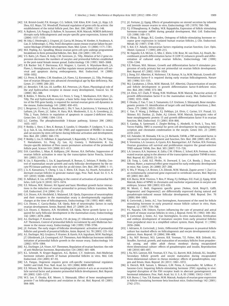
1909F. Sánchez, J. Smitz / Biochimica et Biophysica Acta 1822 (2012) 1896–1912
[42] S.K. Bristol-Gould, P.K. Kreeger, C.G. Selkirk, S.M. Kilen, R.W. Cook, J.L. Kipp, L.D.Shea, K.E. Mayo, T.K. Woodruff, Postnatal regulation of germ cells by activin: theestablishment of the initial follicle pool, Dev. Biol. 298 (2006) 132–148.
[43] A. Rajkovic, S.A. Pangas, D. Ballow, N. Suzumori, M.M. Matzuk, NOBOX deficiencydisrupts early folliculogenesis and oocyte-specific gene expression, Science 305(2004) 1157–1159.
[44] M. Uda, C. Ottolenghi, L. Crisponi, J.E. Garcia, M. Deiana,W. Kimber, A. Forabosco, A.Cao, D. Schlessinger, G. Pilia, Foxl2 disruption causes mouse ovarian failure by per-vasive blockage of follicle development, Hum. Mol. Genet. 13 (2004) 1171–1181.
[45] M.E. Pepling, A.C. Spradling, Mouse ovarian germ cell cysts undergo programmedbreakdown to form primordial follicles, Dev. Biol. 234 (2001) 339–351.
[46] V.S. Ratts, J.A. Flaws, R. Kolp, C.M. Sorenson, J.L. Tilly, Ablation of bcl-2 gene ex-pression decreases the numbers of oocytes and primordial follicles establishedin the post-natal female mouse gonad, Endocrinology 136 (1995) 3665–3668.
[47] E.B. Rucker 3rd, P. Dierisseau, K.U. Wagner, L. Garrett, A. Wynshaw-Boris, J.A.Flaws, L. Hennighausen, Bcl-x and Bax regulate mouse primordial germ cell sur-vival and apoptosis during embryogenesis, Mol. Endocrinol. 14 (2000)1038–1052.
[48] G.I. Perez, R. Robles, C.M. Knudson, J.A. Flaws, S.J. Korsmeyer, J.L. Tilly, Prolonga-tion of ovarian lifespan into advanced chronological age by Bax-deficiency, Nat.Genet. 21 (1999) 200–203.
[49] J.C. Benedict, T.M. Lin, I.K. Loeffler, R.E. Peterson, J.A. Flaws, Physiological role ofthe aryl hydrocarbon receptor in mouse ovary development, Toxicol. Sci. 56(2000) 382–388.
[50] R. Robles, Y. Morita, K.K. Mann, G.I. Perez, S. Yang, T. Matikainen, D.H. Sherr, J.L.Tilly, The aryl hydrocarbon receptor, a basic helix–loop–helix transcription fac-tor of the PAS gene family, is required for normal ovarian germ cell dynamics inthe mouse, Endocrinology 141 (2000) 450–453.
[51] L. Bergeron, G.I. Perez, G. Macdonald, L. Shi, Y. Sun, A. Jurisicova, S. Varmuza, K.E.Latham, J.A. Flaws, J.C. Salter, H. Hara, M.A. Moskowitz, E. Li, A. Greenberg, J.L.Tilly, J. Yuan, Defects in regulation of apoptosis in caspase-2-deficient mice,Genes Dev. 12 (1998) 1304–1314.
[52] L.C. Cantley, The phosphoinositide 3-kinase pathway, Science 296 (2002)1655–1657.
[53] P. Reddy, L. Shen, C. Ren, K. Boman, E. Lundin, U. Ottander, P. Lindgren, Y.-x. Liu,Q.-y. Sun, K. Liu, Activation of Akt (PKB) and suppression of FKHRL1 in mouseand rat oocytes by stem cell factor during follicular activation and development,Dev. Biol. 281 (2005) 160–170.
[54] P. Reddy, L. Liu, D. Adhikari, K. Jagarlamudi, S. Rajareddy, Y. Shen, C. Du, W. Tang,T. Hamalainen, S.L. Peng, Z.J. Lan, A.J. Cooney, I. Huhtaniemi, K. Liu,Oocyte-specific deletion of Pten causes premature activation of the primordialfollicle pool, Science 319 (2008) 611–613.
[55] D.H. Castrillon, L. Miao, R. Kollipara, J.W. Horner, R.A. DePinho, Suppression ofovarian follicle activation in mice by the transcription factor Foxo3a, Science301 (2003) 215–218.
[56] K. Liu, S. Rajareddy, L. Liu, K. Jagarlamudi, K. Boman, G. Selstam, P. Reddy, Con-trol of mammalian oocyte growth and early follicular development by the oo-cyte PI3 kinase pathway: new roles for an old timer, Dev. Biol. 299 (2006) 1–11.
[57] J. Li, K. Kawamura, Y. Cheng, S. Liu, C. Klein, E.K. Duan, A.J. Hsueh, Activation ofdormant ovarian follicles to generate mature eggs, Proc. Natl. Acad. Sci. U. S. A.107 (2010) 10280–10284.
[58] D. Adhikari, K. Liu, mTOR signaling in the control of activation of primordial fol-licles, Cell Cycle 9 (2010) 1673–1674.
[59] E.E. Nilsson, M.K. Skinner, Kit ligand and basic fibroblast growth factor interac-tions in the induction of ovarian primordial to primary follicle transition, Mol.Cell. Endocrinol. 214 (2004) 19–25.
[60] G.A. Dissen, A.N. Hirshfield, S. Malamed, S.R. Ojeda, Expression of neurotrophinsand their receptors in the mammalian ovary is developmentally regulated:changes at the time of folliculogenesis, Endocrinology 136 (1995) 4681–4692.
[61] G.A. Dissen, C. Garcia-Rudaz, S.R. Ojeda, Role of neurotrophic factors in earlyovarian development, Semin. Reprod. Med. 27 (2009) 24–31.
[62] G.A. Dissen, C. Romero, A.N. Hirshfield, S.R. Ojeda, Nerve growth factor is re-quired for early follicular development in the mammalian ovary, Endocrinology142 (2001) 2078–2086.
[63] A.L. Durlinger, P. Kramer, B. Karels, F.H. de Jong, J.T. Uilenbroek, J.A. Grootegoed,A.P. Themmen, Control of primordial follicle recruitment by anti-Mullerian hor-mone in the mouse ovary, Endocrinology 140 (1999) 5789–5796.
[64] J.E. Fortune, The early stages of follicular development: activation of primordialfollicles and growth of preantral follicles, Anim. Reprod. Sci. 78 (2003) 135–163.
[65] A.L. Durlinger, M.J. Gruijters, P. Kramer, B. Karels, H.A. Ingraham, M.W. Nachtigal,J.T. Uilenbroek, J.A. Grootegoed, A.P. Themmen, Anti-Mullerian hormone inhibitsinitiation of primordial follicle growth in the mouse ovary, Endocrinology 143(2002) 1076–1084.
[66] A.L. Durlinger, J.A. Visser, A.P. Themmen, Regulation of ovarian function: the roleof anti-Mullerian hormone, Reproduction 124 (2002) 601–609.
[67] K.L. Schmidt, N. Kryger-Baggesen, A.G. Byskov, C.Y. Andersen, Anti-Mullerianhormone initiates growth of human primordial follicles in vitro, Mol. Cell.Endocrinol. 234 (2005) 87–93.
[68] S.A. Pangas, Oogenesis requires germ cell-specific transcriptional regulatorsSohlh1 and Lhx8, Proc. Natl. Acad. Sci. 103 (2006) 8090–8095.
[69] E.E. Nilsson, M.K. Skinner, Bone morphogenetic protein-4 acts as an ovarian fol-licle survival factor and promotes primordial follicle development, Biol. Reprod.69 (2003) 1265–1272.
[70] W.S. Lee, F. Otsuka, R.K. Moore, S. Shimasaki, Effect of bone morphogeneticprotein-7 on folliculogenesis and ovulation in the rat, Biol. Reprod. 65 (2001)994–999.
[71] J.E. Fortune, J.J. Eppig, Effects of gonadotropins on steroid secretion by infantileand juvenile mouse ovaries in vitro, Endocrinology 105 (1979) 760–768.
[72] P.J. O'Shaughnessy, K. Dudley, W.R. Rajapaksha, Expression of follicle stimulatinghormone-receptor mRNA during gonadal development, Mol. Cell. Endocrinol.125 (1996) 169–175.
[73] K. Oktay, D. Briggs, R.G. Gosden, Ontogeny of follicle-stimulating hormone re-ceptor gene expression in isolated human ovarian follicles, J. Clin. Endocrinol.Metab. 82 (1997) 3748–3751.
[74] S. Kol, E.Y. Adashi, Intraovarian factors regulating ovarian function, Curr. Opin.Obstet. Gynecol. 7 (1995) 209–213.
[75] M. Hayashi, E.A. McGee, G. Min, C. Klein, U.M. Rose, M. van Duin, A.J. Hsueh, Re-combinant growth differentiation factor-9 (GDF-9) enhances growth and differ-entiation of cultured early ovarian follicles, Endocrinology 140 (1999)1236–1244.
[76] E.E. Nilsson, M.K. Skinner, Growth and differentiation factor-9 stimulates pro-gression of early primary but not primordial rat ovarian follicle development,Biol. Reprod. 67 (2002) 1018–1024.
[77] J. Dong, D.F. Albertini, K. Nishimori, T.R. Kumar, N. Lu, M.M. Matzuk, Growth dif-ferentiation factor-9 is required during early ovarian folliculogenesis, Nature383 (1996) 531–535.
[78] M.J. Carabatsos, J. Elvin, M.M. Matzuk, D.F. Albertini, Characterization of oocyteand follicle development in growth differentiation factor-9-deficient mice,Dev. Biol. 204 (1998) 373–384.
[79] J.A. Elvin, A.T. Clark, P. Wang, N.M. Wolfman, M.M. Matzuk, Paracrine actions ofgrowth differentiation factor-9 in the mammalian ovary, Mol. Endocrinol. 13(1999) 1035–1048.
[80] F. Otsuka, Z. Yao, T. Lee, S. Yamamoto, G.F. Erickson, S. Shimasaki, Bone morpho-genetic protein-15. Identification of target cells and biological functions, J. Biol.Chem. 275 (2000) 39523–39528.
[81] C. Yan, P. Wang, J. DeMayo, F.J. DeMayo, J.A. Elvin, C. Carino, S.V. Prasad, S.S.Skinner, B.S. Dunbar, J.L. Dube, A.J. Celeste, M.M. Matzuk, Synergistic roles ofbone morphogenetic protein 15 and growth differentiation factor 9 in ovarianfunction, Mol. Endocrinol. 15 (2001) 854–866.
[82] E. Gazdag, A. Santenard, C. Ziegler-Birling, G. Altobelli, O. Poch, L. Tora, M.E.Torres-Padilla, TBP2 is essential for germ cell development by regulating tran-scription and chromatin condensation in the oocyte, Genes Dev. 23 (2009)2210–2223.
[83] A.E. Falender, M. Shimada, Y.K. Lo, J.S. Richards, TAF4b, a TBP associated factor, isrequired for oocyte development and function, Dev. Biol. 288 (2005) 405–419.
[84] E. Voronina, L.A. Lovasco, A. Gyuris, R.A. Baumgartner, A.F. Parlow, R.N. Freiman,Ovarian granulosa cell survival and proliferation requires the gonad-selectiveTFIID subunit TAF4b, Dev. Biol. 303 (2007) 715–726.
[85] L.A. Lovasco, K.A. Seymour, K. Zafra, C.W. O'Brien, C. Schorl, R.N. Freiman, Accel-erated ovarian aging in the absence of the transcription regulator TAF4B in mice,Biol. Reprod. 82 (2010) 23–34.
[86] Z.B. Tong, L. Gold, K.E. Pfeifer, H. Dorward, E. Lee, C.A. Bondy, J. Dean, L.M.Nelson, Mater, a maternal effect gene required for early embryonic developmentin mice, Nat. Genet. 26 (2000) 267–268.
[87] X. Wu, P. Wang, C.A. Brown, C.A. Zilinski, M.M. Matzuk, Zygote arrest 1 (Zar1) isan evolutionarily conserved gene expressed in vertebrate ovaries, Biol. Reprod.69 (2003) 861–867.
[88] K.H. Burns, M.M. Viveiros, Y. Ren, P. Wang, F.J. DeMayo, D.E. Frail, J.J. Eppig, M.M.Matzuk, Roles of NPM2 in chromatin and nucleolar organization in oocytes andembryos, Science 300 (2003) 633–636.
[89] M. Monti, C. Redi, Oogenesis specific genes (Nobox, Oct4, Bmp15, Gdf9,Oogenesin1 and Oogenesin2) are differentially expressed during natural andgonadotropin-induced mouse follicular development, Mol. Reprod. Dev. 76(2009) 994–1003.
[90] R. Cortvrindt, J. Smitz, A.C. Van Steirteghem, Assessment of the need for folliclestimulating hormone in early preantral mouse follicle culture in vitro, Hum.Reprod. 12 (1997) 759–768.
[91] P.L. Nayudu, S.M. Osborn, Factors influencing the rate of preantral and antralgrowth of mouse ovarian follicles in vitro, J. Reprod. Fertil. 95 (1992) 349–362.
[92] R. Cortvrindt, J. Smitz, A.C. Van Steirteghem, In-vitro maturation, fertilizationand embryo development of immature oocytes from early preantral folliclesfrom prepuberal mice in a simplified culture system, Hum. Reprod. 11 (1996)2656–2666.
[93] I. Adriaens, R. Cortvrindt, J. Smitz, Differential FSH exposure in preantral follicleculture has marked effects on folliculogenesis and oocyte developmental com-petence, Hum. Reprod. 19 (2004) 398–408.
[94] J. Xu, M.P. Bernuci, M.S. Lawson, R.R. Yeoman, T.E. Fisher, M.B. Zelinski, R.L.Stouffer, Survival, growth, and maturation of secondary follicles from prepuber-tal, young, and older adult rhesus monkeys during encapsulatedthree-dimensional culture: effects of gonadotropins and insulin, Reproduction140 (2010) 685–697.
[95] J. Xu, M.S. Lawson, R.R. Yeoman, K.Y. Pau, S.L. Barrett, M.B. Zelinski, R.L. Stouffer,Secondary follicle growth and oocyte maturation during encapsulatedthree-dimensional culture in rhesus monkeys: effects of gonadotrophins, oxy-gen and fetuin, Hum. Reprod. 26 (2011) 1061–1072.
[96] A. Dierich, M.R. Sairam, L. Monaco, G.M. Fimia, A. Gansmuller, M. LeMeur, P.Sassone-Corsi, Impairing follicle-stimulating hormone (FSH) signaling in vivo:targeted disruption of the FSH receptor leads to aberrant gametogenesis andhormonal imbalance, Proc. Natl. Acad. Sci. U. S. A. 95 (1998) 13612–13617.
[97] K.H. Burns, C. Yan, T.R. Kumar, M.M. Matzuk, Analysis of ovarian gene expressionin follicle-stimulating hormone beta knockout mice, Endocrinology 142 (2001)2742–2751.
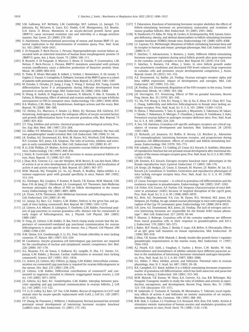
1910 F. Sánchez, J. Smitz / Biochimica et Biophysica Acta 1822 (2012) 1896–1912
[98] S.M. Galloway, K.P. McNatty, L.M. Cambridge, M.P. Laitinen, J.L. Juengel, T.S.Jokiranta, R.J. McLaren, K. Luiro, K.G. Dodds, G.W. Montgomery, A.E. Beattie,G.H. Davis, O. Ritvos, Mutations in an oocyte-derived growth factor gene(BMP15) cause increased ovulation rate and infertility in a dosage-sensitivemanner, Nat. Genet. 25 (2000) 279–283.
[99] O. Hashimoto, Posttranslational processing of mouse and human BMP-15: po-tential implication in the determination of ovulation quota, Proc. Natl. Acad.Sci. 102 (2005) 5426–5431.
[100] E. Di Pasquale, P. Beck-Peccoz, L. Persani, Hypergonadotropic ovarian failure as-sociated with an inherited mutation of human bone morphogenetic protein-15(BMP15) gene, Am. J. Hum. Genet. 75 (2004) 106–111.
[101] R. Rossetti, E. Di Pasquale, A. Marozzi, S. Bione, D. Toniolo, P. Grammatico, L.M.Nelson, P. Beck-Peccoz, L. Persani, BMP15 mutations associated with primaryovarian insufficiency cause a defective production of bioactive protein, Hum.Mutat. 30 (2009) 804–810.
[102] D. Tiotiu, B. Alvaro Mercadal, R. Imbert, J. Verbist, I. Demeestere, A. De Leener, Y.Englert, G. Vassart, S. Costagliola, A. Delbaere, Variants of the BMP15 gene in a cohortof patients with premature ovarian failure, Hum. Reprod. 25 (2010) 1581–1587.
[103] M. Orisaka, S. Orisaka, J.Y. Jiang, J. Craig, Y. Wang, F. Kotsuji, B.K. Tsang, Growthdifferentiation factor 9 is antiapoptotic during follicular development frompreantral to early antral stage, Mol. Endocrinol. 20 (2006) 2456–2468.
[104] H. Wang, K. Andoh, H. Hagiwara, L. Xiaowei, N. Kikuchi, Y. Abe, K. Yamada, R.Fatima, H. Mizunuma, Effect of adrenal and ovarian androgens on type 4 folliclesunresponsive to FSH in immature mice, Endocrinology 142 (2001) 4930–4936.
[105] K.A. Walters, C.M. Allan, D.J. Handelsman, Androgen actions and the ovary, Biol.Reprod. 78 (2008) 380–389.
[106] T.E. Hickey, D.L. Marrocco, F. Amato, L.J. Ritter, R.J. Norman, R.B. Gilchrist, D.T.Armstrong, Androgens augment the mitogenic effects of oocyte-secreted factorsand growth differentiation factor 9 on porcine granulosa cells, Biol. Reprod. 73(2005) 825–832.
[107] S.Y. Ying, Inhibins and activins: chemical properties and biological activity, Proc.Soc. Exp. Biol. Med. 186 (1987) 253–264.
[108] S.G. Hillier, P.F. Whitelaw, C.D. Smyth, Follicular oestrogen synthesis: the ‘two-cell,two-gonadotrophin’ model revisited, Mol. Cell. Endocrinol. 100 (1994) 51–54.
[109] J.K. Findlay, A.E. Drummond, K.L. Britt, M. Dyson, N.G. Wreford, D.M. Robertson,N.P. Groome, M.E. Jones, E.R. Simpson, The roles of activins, inhibins and estro-gen in early committed follicles, Mol. Cell. Endocrinol. 163 (2000) 81–87.
[110] R. Li, D.M. Phillips, J.P. Mather, Activin promotes ovarian follicle development invitro, Endocrinology 136 (1995) 849–856.
[111] J. Smitz, R. Cortvrindt, Inhibin A and B secretion in mouse preantral follicle cul-ture, Hum. Reprod. 13 (1998) 927–935.
[112] J. Zhao, M.A. Taverne, G.C. van der Weijden, M.M. Bevers, R. van den Hurk, Effectof activin A on in vitro development of rat preantral follicles and localization ofactivin A and activin receptor II, Biol. Reprod. 65 (2001) 967–977.
[113] M.M. Matzuk, M.J. Finegold, J.G. Su, A.J. Hsueh, A. Bradley, Alpha-inhibin is atumour-suppressor gene with gonadal specificity in mice, Nature 360 (1992)313–319.
[114] A.L. Durlinger, M.J. Gruijters, P. Kramer, B. Karels, T.R. Kumar, M.M. Matzuk, U.M.Rose, F.H. de Jong, J.T. Uilenbroek, J.A. Grootegoed, A.P. Themmen, Anti-Mullerianhormone attenuates the effects of FSH on follicle development in the mouseovary, Endocrinology 142 (2001) 4891–4899.
[115] J.A. Visser, A.P.N. Themmen, Anti-Müllerian hormone and folliculogenesis, Mol.Cell. Endocrinol. 234 (2005) 81–86.
[116] S.C. Juneja, K.J. Barr, G.C. Enders, G.M. Kidder, Defects in the germ line and go-nads of mice lacking connexin43, Biol. Reprod. 60 (1999) 1263–1270.
[117] J.E. Gittens, A.A. Mhawi, D. Lidington, Y. Ouellette, G.M. Kidder, Functional anal-ysis of gap junctions in ovarian granulosa cells: distinct role for connexin43 inearly stages of folliculogenesis, Am. J. Physiol. Cell Physiol. 284 (2003)C880–C887.
[118] D. Tong, J.E. Gittens, G.M. Kidder, D. Bai, Patch-clamp study reveals that the im-portance of connexin43-mediated gap junctional communication for ovarianfolliculogenesis is strain specific in the mouse, Am. J. Physiol. Cell Physiol. 290(2006) C290–C297.
[119] A.M. Simon, D.A. Goodenough, E. Li, D.L. Paul, Female infertility in mice lackingconnexin 37, Nature 385 (1997) 525–529.
[120] M. Carabatsos, Oocyte–granulosa cell heterologous gap junctions are requiredfor the coordination of nuclear and cytoplasmic meiotic competence, Dev. Biol.226 (2000) 167–179.
[121] A.G. Reaume, P.A. de Sousa, S. Kulkarni, B.L. Langille, D. Zhu, T.C. Davies, S.C.Juneja, G.M. Kidder, J. Rossant, Cardiac malformation in neonatal mice lackingconnexin43, Science 267 (1995) 1831–1834.
[122] C.L. Ackert, J.E. Gittens, M.J. O'Brien, J.J. Eppig, G.M. Kidder, Intercellular commu-nication via connexin43 gap junctions is required for ovarian folliculogenesis inthe mouse, Dev. Biol. 233 (2001) 258–270.
[123] J.E. Gittens, G.M. Kidder, Differential contributions of connexin37 and con-nexin43 to oogenesis revealed in chimeric reaggregated mouse ovaries, J. CellSci. 118 (2005) 5071–5078.
[124] J.E. Gittens, K.J. Barr, B.C. Vanderhyden, G.M. Kidder, Interplay between para-crine signaling and gap junctional communication in ovarian follicles, J. CellSci. 118 (2005) 113–122.
[125] T.Y. Li, D. Colley, K.J. Barr, S.P. Yee, G.M. Kidder, Rescue of oogenesis in Cx37-nullmutant mice by oocyte-specific replacement with Cx43, J. Cell Sci. 120 (2007)4117–4125.
[126] F.P. Zhang, M. Poutanen, J. Wilbertz, I. Huhtaniemi, Normal prenatal but arrestedpostnatal sexual development of luteinizing hormone receptor knockout(LuRKO) mice, Mol. Endocrinol. 15 (2001) 172–183.
[127] T. Pakarainen, Knockout of luteinizing hormone receptor abolishes the effects offollicle-stimulating hormone on preovulatory maturation and ovulation ofmouse graafian follicles, Mol. Endocrinol. 19 (2005) 2591–2602.
[128] N. Danilovich, P.S. Babu, W. Xing, M. Gerdes, H. Krishnamurthy, M.R. Sairam, Estro-gen deficiency, obesity, and skeletal abnormalities in follicle-stimulating hormonereceptor knockout (FORKO) female mice, Endocrinology 141 (2000) 4295–4308.
[129] L.C. Layman, P.G. McDonough, Mutations of follicle stimulating hormone-beta andits receptor in human and mouse: genotype/phenotype, Mol. Cell. Endocrinol. 161(2000) 9–17.
[130] F. Sánchez, T. Adriaenssens, S. Romero, J. Smitz, Different follicle-stimulatinghormone exposure regimens during antral follicle growth alter gene expressionin the cumulus–oocyte complex in mice, Biol. Reprod. 83 (2010) 514–524.
[131] F. Sánchez, S. Romero, F.K. Albuz, J. Smitz, In vitro follicle growth undernon-attachment conditions and decreased FSH levels reduces Lhcgr expressionin cumulus cells and promotes oocyte developmental competence, J. Assist.Reprod. Genet. 29 (2012) 141–152.
[132] A.E. Drummond, A.J. Baillie, J.K. Findlay, Ovarian estrogen receptor alpha andbeta mRNA expression: impact of development and estrogen, Mol. Cell.Endocrinol. 149 (1999) 153–161.
[133] J.K. Findlay, A.E. Drummond, Regulation of the FSH receptor in the ovary, TrendsEndocrinol. Metab. 10 (1999) 183–188.
[134] J.H. Dorrington, D.T. Armstrong, Effects of FSH on gonadal functions, RecentProg. Horm. Res. 35 (1979) 301–342.
[135] Y.C. Hu, P.H. Wang, S. Yeh, R.S. Wang, C. Xie, Q. Xu, X. Zhou, H.T. Chao, M.Y. Tsai,C. Chang, Subfertility and defective folliculogenesis in female mice lacking an-drogen receptor, Proc. Natl. Acad. Sci. U. S. A. 101 (2004) 11209–11214.
[136] H. Shiina, T. Matsumoto, T. Sato, K. Igarashi, J. Miyamoto, S. Takemasa, M. Sakari,I. Takada, T. Nakamura, D. Metzger, P. Chambon, J. Kanno, H. Yoshikawa, S. Kato,Premature ovarian failure in androgen receptor-deficient mice, Proc. Natl. Acad.Sci. U. S. A. 103 (2006) 224–229.
[137] A. Sen, S.R. Hammes, Granulosa cell-specific androgen receptors are critical reg-ulators of ovarian development and function, Mol. Endocrinol. 24 (2010)1393–1403.
[138] J.S. Richards, J.A. Jonassen, A.I. Rolfes, K. Kersey, L.E. Reichert Jr., Adenosine3′,5′-monophosphate, luteinizing hormone receptor, and progesterone duringgranulosa cell differentiation: effects of estradiol and follicle-stimulating hor-mone, Endocrinology 104 (1979) 765–773.
[139] D.B. Lubahn, J.S. Moyer, T.S. Golding, J.F. Couse, K.S. Korach, O. Smithies, Alterationof reproductive function but not prenatal sexual development after insertional dis-ruption of themouse estrogen receptor gene, Proc. Natl. Acad. Sci. U. S. A. 90 (1993)11162–11166.
[140] J.M. Emmen, K.S. Korach, Estrogen receptor knockout mice: phenotypes in thefemale reproductive tract, Gynecol. Endocrinol. 17 (2003) 169–176.
[141] J.H. Krege, J.B. Hodgin, J.F. Couse, E. Enmark, M. Warner, J.F. Mahler, M. Sar, K.S.Korach, J.A. Gustafsson, O. Smithies, Generation and reproductive phenotypes ofmice lacking estrogen receptor beta, Proc. Natl. Acad. Sci. U. S. A. 95 (1998)15677–15682.
[142] J.F. Couse, Estrogen receptor — is critical to granulosa cell differentiation and theovulatory response to gonadotropins, Endocrinology 146 (2005) 3247–3262.
[143] C.R. Fisher, K.H. Graves, A.F. Parlow, E.R. Simpson, Characterization of mice defi-cient in aromatase (ArKO) because of targeted disruption of the cyp19 gene,Proc. Natl. Acad. Sci. U. S. A. 95 (1998) 6965–6970.
[144] K.L. Britt, A.E. Drummond, V.A. Cox, M. Dyson, N.G. Wreford, M.E. Jones, E.R.Simpson, J.K. Findlay, An age-related ovarian phenotype in mice with targeted dis-ruption of the Cyp 19 (aromatase) gene, Endocrinology 141 (2000) 2614–2623.
[145] S.H. Liew, A.E. Drummond, M.E. Jones, J.K. Findlay, The lack of estrogen and ex-cess luteinizing hormone are responsible for the female ArKO mouse pheno-type☆, Mol. Cell. Endocrinol. 327 (2010) 56–64.
[146] F. Khamsi, S. Roberge, Granulosa cells of the cumulus oophorus are differentfrom mural granulosa cells in their response to gonadotrophins and IGF-I,J. Endocrinol. 170 (2001) 565–573.
[147] J. Baker, M.P. Hardy, J. Zhou, C. Bondy, F. Lupu, A.R. Bellve, A. Efstratiadis, Effectsof an Igf1 gene null mutation on mouse reproduction, Mol. Endocrinol. 10(1996) 903–918.
[148] J. Zhou, T.R. Kumar, M.M. Matzuk, C. Bondy, Insulin-like growth factor I regulatesgonadotropin responsiveness in the murine ovary, Mol. Endocrinol. 11 (1997)1924–1933.
[149] A.J. Hsueh, K.D. Dahl, J. Vaughan, E. Tucker, J. Rivier, C.W. Bardin, W. Vale,Heterodimers and homodimers of inhibin subunits have different paracrine ac-tion in the modulation of luteinizing hormone-stimulated androgen biosynthe-sis, Proc. Natl. Acad. Sci. U. S. A. 84 (1987) 5082–5086.
[150] S.G. Hillier, F. Miro, Inhibin, activin, and follistatin. Potential roles in ovarianphysiology, Ann. N. Y. Acad. Sci. 687 (1993) 29–38.
[151] B.K. Campbell, D.T. Baird, Inhibin A is a follicle stimulating hormone-responsivemarker of granulosa cell differentiation, which has both autocrine and paracrineactions in sheep, J. Endocrinol. 169 (2001) 333–345.
[152] M.M. Matzuk, T.R. Kumar, W. Shou, K.A. Coerver, A.L. Lau, R.R. Behringer, M.J.Finegold, Transgenic models to study the roles of inhibins and activins in repro-duction, oncogenesis, and development, Recent Prog. Horm. Res. 51 (1996)123–154 (discussion 155–157).
[153] M. Sadatsuki, O. Tsutsumi, R. Yamada, M. Muramatsu, Y. Taketani, Local regula-tory effects of activin A and follistatin on meiotic maturation of rat oocytes,Biochem. Biophys. Res. Commun. 196 (1993) 388–395.
[154] B.M. Alak, S. Coskun, C.I. Friedman, E.A. Kennard, M.H. Kim, D.B. Seifer, Activin Astimulates meiotic maturation of human oocytes and modulates granulosa cellsteroidogenesis in vitro, Fertil. Steril. 70 (1998) 1126–1130.
1911F. Sánchez, J. Smitz / Biochimica et Biophysica Acta 1822 (2012) 1896–1912
[155] B.M. Alak, G.D. Smith, T.K. Woodruff, R.L. Stouffer, D.P. Wolf, Enhancement ofprimate oocyte maturation and fertilization in vitro by inhibin A and activin A,Fertil. Steril. 66 (1996) 646–653.
[156] F. Otsuka, S. Yamamoto, G.F. Erickson, S. Shimasaki, Bone morphogeneticprotein-15 inhibits follicle-stimulating hormone (FSH) action by suppressingFSH receptor expression, J. Biol. Chem. 276 (2001) 11387–11392.
[157] U.A. Vitt, M. Hayashi, C. Klein, A.J. Hsueh, Growth differentiation factor-9 stimu-lates proliferation but suppresses the follicle-stimulating hormone-induced dif-ferentiation of cultured granulosa cells from small antral and preovulatory ratfollicles, Biol. Reprod. 62 (2000) 370–377.
[158] R.A. Dragovic, L.J. Ritter, S.J. Schulz, F. Amato, D.T. Armstrong, R.B. Gilchrist, Roleof oocyte-secreted growth differentiation factor 9 in the regulation of mouse cu-mulus expansion, Endocrinology 146 (2005) 2798–2806.
[159] O. Yoshino, H.E. McMahon, S. Sharma, S. Shimasaki, A unique preovulatory ex-pression pattern plays a key role in the physiological functions of BMP-15 inthe mouse, Proc. Natl. Acad. Sci. U. S. A. 103 (2006) 10678–10683.
[160] S. Shimasaki, R.J. Zachow, D. Li, H. Kim, S. Iemura, N. Ueno, K. Sampath, R.J.Chang, G.F. Erickson, A functional bone morphogenetic protein system in theovary, Proc. Natl. Acad. Sci. U. S. A. 96 (1999) 7282–7287.
[161] J. Kwintkiewicz, L.C. Giudice, The interplay of insulin-like growth factors, gonad-otropins, and endocrine disruptors in ovarian follicular development and func-tion, Semin. Reprod. Med. 27 (2009) 43–51.
[162] J.J. Eppig, M.J. O'Brien, Development in vitro of mouse oocytes from primordialfollicles, Biol. Reprod. 54 (1996) 197–207.
[163] H. Picton, D. Briggs, R. Gosden, The molecular basis of oocyte growth and devel-opment, Mol. Cell. Endocrinol. 145 (1998) 27–37.
[164] G.P. Moore, S. Lintern-Moore, Transcription of the mouse oocyte genome, Biol.Reprod. 18 (1978) 865–870.
[165] R. Bachvarova, Gene expression during oogenesis and oocyte development inmammals, Dev. Biol. 1 (1985) 453–524.
[166] U. Eichenlaub-Ritter, M. Peschke, Expression in in-vivo and in-vitro growing andmaturing oocytes: focus on regulation of expression at the translational level,Hum. Reprod. Update 8 (2002) 21–41.
[167] R. Bachvarova, V. De Leon, A. Johnson, G. Kaplan, B.V. Paynton, Changes in totalRNA, polyadenylated RNA, and actin mRNA during meiotic maturation of mouseoocytes, Dev. Biol. 108 (1985) 325–331.
[168] R. De La Fuente, M.M. Viveiros, K.H. Burns, E.Y. Adashi, M.M. Matzuk, J.J. Eppig,Major chromatin remodeling in the germinal vesicle (GV) of mammalianoocytes is dispensable for global transcriptional silencing but required for cen-tromeric heterochromatin function, Dev. Biol. 275 (2004) 447–458.
[169] Y.-Q. Su, K. Sugiura, Y. Woo, K. Wigglesworth, S. Kamdar, J. Affourtit, J.J. Eppig,Selective degradation of transcripts during meiotic maturation of mouseoocytes, Dev. Biol. 302 (2007) 104–117.
[170] A.L. Sternlicht, R.M. Schultz, Biochemical studies of mammalian oogenesis: ki-netics of accumulation of total and poly(A)-containing RNA during growth ofthe mouse oocyte, J. Exp. Zool. 215 (1981) 191–200.
[171] P.M. Wassarman, R.A. Kinloch, Gene expression during oogenesis in mice,Mutat. Res. 296 (1992) 3–15.
[172] C.H. de Moor, J.D. Richter, Translational control in vertebrate development,Int. Rev. Cytol. 203 (2001) 567–608.
[173] B. Oh, S. Hwang, J. McLaughlin, D. Solter, B.B. Knowles, Timely translation duringthe mouse oocyte-to-embryo transition, Development 127 (2000) 3795–3803.
[174] H. Pan, M. Obrien, K. Wigglesworth, J. Eppig, R. Schultz, Transcript profiling dur-ing mouse oocyte development and the effect of gonadotropin priming and de-velopment in vitro, Dev. Biol. 286 (2005) 493–506.
[175] B.V. Paynton, R. Rempel, R. Bachvarova, Changes in state of adenylation and timecourse of degradation of maternal mRNAs during oocyte maturation and earlyembryonic development in the mouse, Dev. Biol. 129 (1988) 304–314.
[176] G.M. Jones, D.S. Cram, B. Song, M.C. Magli, L. Gianaroli, O. Lacham-Kaplan, J.K.Findlay, G. Jenkin, A.O. Trounson, Gene expression profiling of humanoocytes following in vivo or in vitro maturation, Hum. Reprod. 23 (2008)1138–1144.
[177] B.V. Paynton, R. Bachvarova, Polyadenylation and deadenylation of maternalmRNAs during oocyte growth and maturation in the mouse, Mol. Reprod. Dev.37 (1994) 172–180.
[178] C.Y. Chu, T.M. Rana, Small RNAs: regulators and guardians of the genome, J. Cell.Physiol. 213 (2007) 412–419.
[179] N. Suh, R. Blelloch, Small RNAs in early mammalian development: from gametesto gastrulation, Development 138 (2011) 1653–1661.
[180] V.N. Kim, MicroRNA biogenesis: coordinated cropping and dicing, Nat. Rev. Mol.Cell Biol. 6 (2005) 376–385.
[181] T.M. Rana, Illuminating the silence: understanding the structure and function ofsmall RNAs, Nat. Rev. Mol. Cell Biol. 8 (2007) 23–36.
[182] R.S. Pillai, MicroRNA function: multiple mechanisms for a tiny RNA? RNA 11(2005) 1753–1761.
[183] L. Wu, J. Fan, J.G. Belasco, MicroRNAs direct rapid deadenylation of mRNA, Proc.Natl. Acad. Sci. U. S. A. 103 (2006) 4034–4039.
[184] G. Yao, M. Yin, J. Lian, H. Tian, L. Liu, X. Li, F. Sun, MicroRNA-224 is involved intransforming growth factor-mediated mouse granulosa cell proliferation andgranulosa cell function by targeting Smad4, Mol. Endocrinol. 24 (2010)540–551.
[185] F. Tang, M. Kaneda, D. O'Carroll, P. Hajkova, S.C. Barton, Y.A. Sun, C. Lee, A.Tarakhovsky, K. Lao, M.A. Surani, Maternal microRNAs are essential for mousezygotic development, Genes Dev. 21 (2007) 644–648.
[186] E.P. Murchison, P. Stein, Z. Xuan, H. Pan, M.Q. Zhang, R.M. Schultz, G.J. Hannon,Critical roles for Dicer in the female germline, Genes Dev. 21 (2007) 682–693.
[187] N. Suh, L. Baehner, F. Moltzahn, C. Melton, A. Shenoy, J. Chen, R. Blelloch, Micro-RNA function is globally suppressed in mouse oocytes and early embryos,Curr. Biol. 20 (2010) 271–277.
[188] S.D. Fiedler, M.Z. Carletti, X. Hong, L.K. Christenson, Hormonal regulation ofMicroRNA expression in periovulatory mouse mural granulosa cells, Biol.Reprod. 79 (2008) 1030–1037.
[189] N. Yao, C.L. Lu, J.J. Zhao, H.F. Xia, D.G. Sun, X.Q. Shi, C. Wang, D. Li, Y. Cui, X. Ma, Anetwork of miRNAs expressed in the ovary are regulated by FSH, Front. Biosci.14 (2009) 3239–3245.
[190] K. Szybek, In-vitro maturation of oocytes from sexually immature mice,J. Endocrinol. 54 (1972) 527–528.
[191] K.L. Durinzi, E.M. Saniga, S.E. Lanzendorf, The relationship between size andmaturation in vitro in the unstimulated human oocyte, Fertil. Steril. 63 (1995)404–406.
[192] J.J. Eppig, Oocyte control of ovarian follicular development and function in mam-mals, Reproduction 122 (2001) 829–838.
[193] J.J. Eppig, A.C. Schroeder, Capacity of mouse oocytes from preantral follicles toundergo embryogenesis and development to live young after growth, matura-tion, and fertilization in vitro, Biol. Reprod. 41 (1989) 268–276.
[194] A. Pavlok, A. Lucas-Hahn, H. Niemann, Fertilization and developmental compe-tence of bovine oocytes derived from different categories of antral follicles,Mol. Reprod. Dev. 31 (1992) 63–67.
[195] S.M. Downs, S.A. Daniel, E.A. Bornslaeger, P.C. Hoppe, J.J. Eppig, Maintenance ofmeiotic arrest in mouse oocytes by purines: modulation of cAMP levels andcAMP phosphodiesterase activity, Gamete Res. 23 (1989) 323–334.
[196] D. Nogueira, C. Albano, T. Adriaenssens, R. Cortvrindt, C. Bourgain, P. Devroey, J.Smitz, Human oocytes reversibly arrested in prophase I by phosphodiesterasetype 3 inhibitor in vitro, Biol. Reprod. 69 (2003) 1042–1052.
[197] S. Sela-Abramovich, I. Edry, D. Galiani, N. Nevo, N. Dekel, Disruption of gap junc-tional communication within the ovarian follicle induces oocyte maturation,Endocrinology 147 (2006) 2280–2286.
[198] L.M. Mehlmann, Oocyte-specific expression of Gpr3 is required for the mainte-nance of meiotic arrest in mouse oocytes, Dev. Biol. 288 (2005) 397–404.
[199] S. Vaccari, K. Horner, L.M. Mehlmann, M. Conti, Generation of mouse oocytes de-fective in cAMP synthesis and degradation: endogenous cyclic AMP is essentialfor meiotic arrest, Dev. Biol. 316 (2008) 124–134.
[200] R.P. Norris, W.J. Ratzan, M. Freudzon, L.M. Mehlmann, J. Krall, M.A. Movsesian, H.Wang, H. Ke, V.O. Nikolaev, L.A. Jaffe, Cyclic GMP from the surrounding somaticcells regulates cyclic AMP and meiosis in the mouse oocyte, Development 136(2009) 1869–1878.
[201] S. Vaccari, J.L. Weeks 2nd, M. Hsieh, F.S. Menniti, M. Conti, Cyclic GMP signalingis involved in the luteinizing hormone-dependent meiotic maturation of mouseoocytes, Biol. Reprod. 81 (2009) 595–604.
[202] M. Zhang, Y.Q. Su, K. Sugiura, G. Xia, J.J. Eppig, Granulosa cell ligand NPPC and itsreceptor NPR2 maintain meiotic arrest in mouse oocytes, Science 330 (2010)366–369.
[203] M. Zhang, Y.Q. Su, K. Sugiura, K. Wigglesworth, G. Xia, J.J. Eppig, Estradiol pro-motes and maintains cumulus cell expression of natriuretic peptide receptor 2(NPR2) and meiotic arrest in mouse oocytes in vitro, Endocrinology 152(2011) 4377–4385.
[204] S. Varani, J.A. Elvin, C. Yan, J. DeMayo, F.J. DeMayo, H.F. Horton, M.C. Byrne, M.M.Matzuk, Knockout of pentraxin 3, a downstream target of growth differentiationfactor-9, causes female subfertility, Mol. Endocrinol. 16 (2002) 1154–1167.
[205] C. Fulop, S. Szanto, D. Mukhopadhyay, T. Bardos, R.V. Kamath, M.S. Rugg, A.J. Day,A. Salustri, V.C. Hascall, T.T. Glant, K. Mikecz, Impaired cumulus mucification andfemale sterility in tumor necrosis factor-induced protein-6 deficient mice, De-velopment 130 (2003) 2253–2261.
[206] S.A. Ochsner, A.J. Day, M.S. Rugg, R.M. Breyer, R.H. Gomer, J.S. Richards,Disrupted function of tumor necrosis factor-alpha-stimulated gene 6 blocks cu-mulus cell–oocyte complex expansion, Endocrinology 144 (2003) 4376–4384.
[207] H. Ashkenazi, X. Cao, S. Motola, M. Popliker, M. Conti, A. Tsafriri, Epidermalgrowth factor family members: endogenous mediators of the ovulatory re-sponse, Endocrinology 146 (2005) 77–84.
[208] I. Ben-Ami, S. Freimann, L. Armon, A. Dantes, D. Strassburger, S. Friedler, A.Raziel, R. Seger, R. Ron-El, A. Amsterdam, PGE2 up-regulates EGF-like growthfactor biosynthesis in human granulosa cells: new insights into the coordinationbetween PGE2 and LH in ovulation, Mol. Hum. Reprod. 12 (2006) 593–599.
[209] R.A. Dragovic, L.J. Ritter, S.J. Schulz, F. Amato, J.G. Thompson, D.T. Armstrong, R.B.Gilchrist, Oocyte-secreted factor activation of SMAD 2/3 signaling enables initi-ation of mouse cumulus cell expansion, Biol. Reprod. 76 (2007) 848–857.
[210] F.J. Richard, A. Tsafriri, M. Conti, Role of phosphodiesterase type 3A in rat oocytematuration, Biol. Reprod. 65 (2001) 1444–1451.
[211] K. Kawamura, Y. Cheng, N. Kawamura, S. Takae, A. Okada, Y. Kawagoe, S.Mulders, Y. Terada, A.J. Hsueh, Pre-ovulatory LH/hCG surge decreases C-type na-triuretic peptide secretion by ovarian granulosa cells to promote meiotic re-sumption of pre-ovulatory oocytes, Hum. Reprod. 26 (2011) 3094–3101.
[212] S. Sela-Abramovich, E. Chorev, D. Galiani, N. Dekel, Mitogen-activated proteinkinase mediates luteinizing hormone-induced breakdown of communicationand oocyte maturation in rat ovarian follicles, Endocrinology 146 (2005)1236–1244.
[213] R.P. Norris, M. Freudzon, L.M. Mehlmann, A.E. Cowan, A.M. Simon, D.L. Paul, P.D.Lampe, L.A. Jaffe, Luteinizing hormone causes MAP kinase-dependent phosphor-ylation and closure of connexin 43 gap junctions in mouse ovarian follicles: oneof two paths to meiotic resumption, Development 135 (2008) 3229–3238.
[214] J.E. Holt, S.M. Tran, J.L. Stewart, K. Minahan, I. Garcia-Higuera, S. Moreno, K.T.Jones, The APC/C activator FZR1 coordinates the timing of meiotic resumption
1912 F. Sánchez, J. Smitz / Biochimica et Biophysica Acta 1822 (2012) 1896–1912
during prophase I arrest in mammalian oocytes, Development 138 (2011)905–913.
[215] J. Chen, C. Melton, N. Suh, J.S. Oh, K. Horner, F. Xie, C. Sette, R. Blelloch, M. Conti,Genome-wide analysis of translation reveals a critical role for deleted inazoospermia-like (Dazl) at the oocyte-to-zygote transition, Genes Dev. 25(2011) 755–766.
[216] X.S. Cui, X.Y. Li, X.J. Yin, I.K. Kong, J.J. Kang, N.H. Kim, Maternal gene transcriptionin mouse oocytes: genes implicated in oocyte maturation and fertilization,J. Reprod. Dev. 53 (2007) 405–418.
[217] Z. Alizadeh, S.-I. Kageyama, F. Aoki, Degradation of maternal mRNA in mouseembryos: selective degradation of specific mRNAs after fertilization, Mol.Reprod. Dev. 72 (2005) 281–290.
[218] B. Collier, B. Gorgoni, C. Loveridge, H.J. Cooke, N.K. Gray, The DAZL family pro-teins are PABP-binding proteins that regulate translation in germ cells, EMBOJ. 24 (2005) 2656–2666.
[219] G. Cauffman, H. Van de Velde, I. Liebaers, A. Van Steirteghem, DAZL expression inhuman oocytes, preimplantation embryos and embryonic stem cells, Mol. Hum.Reprod. 11 (2005) 405–411.
[220] R. De La Fuente, J.J. Eppig, Transcriptional activity of the mouse oocyte genome:companion granulosa cells modulate transcription and chromatin remodeling,Dev. Biol. 229 (2001) 224–236.
[221] E. Christians, M. Boiani, S. Garagna, C. Dessy, C.A. Redi, J.P. Renard, M. Zuccotti,Gene expression and chromatin organization during mouse oocyte growth,Dev. Biol. 207 (1999) 76–85.
[222] B.A. Mattson, D.F. Albertini, Oogenesis: chromatin and microtubule dynamicsduring meiotic prophase, Mol. Reprod. Dev. 25 (1990) 374–383.
[223] M. Zuccotti, P.G. Rossi, A. Martinez, S. Garagna, A. Forabosco, C.A. Redi, Meioticand developmental competence of mouse antral oocytes, Biol. Reprod. 58(1998) 700–704.
[224] C. Bouniol-Baly, L. Hamraoui, J. Guibert, N. Beaujean, M.S. Szollosi, P. Debey, Dif-ferential transcriptional activity associated with chromatin configuration in fullygrown mouse germinal vesicle oocytes, Biol. Reprod. 60 (1999) 580–587.
[225] A. Pesty, F. Miyara, P. Debey, B. Lefevre, C. Poirot, Multiparameter assessment ofmouse oogenesis during follicular growth in vitro, Mol. Hum. Reprod. 13 (2007)3–9.
[226] M. Zuccotti, V. Merico, L. Sacchi, M. Bellone, T.C. Brink, R. Bellazzi, M. Stefanelli, C.Redi, S. Garagna, J. Adjaye, Maternal Oct-4 is a potential key regulator of the de-velopmental competence of mouse oocytes, BMC Dev. Biol. 8 (2008) 97.
[227] A. Inoue, R. Nakajima, M. Nagata, F. Aoki, Contribution of the oocyte nucleus andcytoplasm to the determination of meiotic and developmental competence inmice, Hum. Reprod. 23 (2008) 1377–1384.
[228] Y.-Q. Su, K. Sugiura, J. Eppig, Mouse oocyte control of granulosa cell develop-ment and function: paracrine regulation of cumulus cell metabolism, Semin.Reprod. Med. 27 (2009) 032–042.
[229] K. Sugiura, F.L. Pendola, J.J. Eppig, Oocyte control of metabolic cooperativity be-tween oocytes and companion granulosa cells: energy metabolism, Dev. Biol.279 (2005) 20–30.
[230] K. Sugiura, Y.Q. Su, F.J. Diaz, S.A. Pangas, S. Sharma, K. Wigglesworth, M.J.O'Brien, M.M. Matzuk, S. Shimasaki, J.J. Eppig, Oocyte-derived BMP15 and FGFscooperate to promote glycolysis in cumulus cells, Development 134 (2007)2593–2603.
[231] F.J. Diaz, K. Wigglesworth, J.J. Eppig, Oocytes are required for the preantral gran-ulosa cell to cumulus cell transition in mice, Dev. Biol. 305 (2007) 300–311.
[232] F.J. Diaz, K. Wigglesworth, J.J. Eppig, Oocytes determine cumulus cell lineage inmouse ovarian follicles, J. Cell Sci. 120 (2007) 1330–1340.
[233] R.B. Gilchrist, Molecular basis of oocyte-paracrine signalling that promotes gran-ulosa cell proliferation, J. Cell Sci. 119 (2006) 3811–3821.
[234] R. Buccione, A.C. Schroeder, J.J. Eppig, Interactions between somatic cells andgerm cells throughout mammalian oogenesis, Biol. Reprod. 43 (1990) 543–547.
[235] R. Gilchrist, Oocyte–somatic cell interactions during follicle development inmammals, Anim. Reprod. Sci. 82–83 (2004) 431–446.
[236] B.C. Vanderhyden, E.E. Telfer, J.J. Eppig, Mouse oocytes promote proliferation ofgranulosa cells from preantral and antral follicles in vitro, Biol. Reprod. 46(1992) 1196–1204.
[237] J.J. Eppig, Mouse oocytes regulate metabolic cooperativity between granulosacells and oocytes: amino acid transport, Biol. Reprod. 73 (2005) 351–357.
[238] Y.Q. Su, K. Sugiura, K. Wigglesworth, M.J. O'Brien, J.P. Affourtit, S.A. Pangas, M.M.Matzuk, J.J. Eppig, Oocyte regulation of metabolic cooperativity between mousecumulus cells and oocytes: BMP15 and GDF9 control cholesterol biosynthesis incumulus cells, Development 135 (2008) 111–121.
[239] N. Salmon, Oocyte regulation of anti-Müllerian hormone expression in granu-losa cells during ovarian follicle development in mice, Dev. Biol. 266 (2004)201–208.
[240] M. Sasseville, L.J. Ritter, T.M. Nguyen, F. Liu, D.G. Mottershead, D.L. Russell, R.B.Gilchrist, Growth differentiation factor 9 signaling requires ERK1/2 activity inmouse granulosa and cumulus cells, J. Cell Sci. 123 (2010) 3166–3176.
[241] D.F. Albertini, C.M. Combelles, E. Benecchi, M.J. Carabatsos, Cellular basis forparacrine regulation of ovarian follicle development, Reproduction 121 (2001)647–653.
[242] J.D. Biggers, D.G. Whittingham, R.P. Donahue, The pattern of energy metabolismin the mouse oocyte and zygote, Proc. Natl. Acad. Sci. U. S. A. 58 (1967) 560–567.
[243] J.J. Eppig, Analysis of mouse oogenesis in vitro. Oocyte isolation and the utiliza-tion of exogenous energy sources by growing oocytes, J. Exp. Zool. 198 (1976)375–382.
[244] H.P. Pratt, Preimplantation mouse embryos synthesize membrane sterols, Dev.Biol. 89 (1982) 101–110.
[245] M. Comiskey, C.M. Warner, Spatio-temporal localization of membrane lipid raftsin mouse oocytes and cleaving preimplantation embryos, Dev. Biol. 303 (2007)727–739.
[246] R. Colonna, F. Mangia, Mechanisms of amino acid uptake in cumulus-enclosedmouse oocytes, Biol. Reprod. 28 (1983) 797–803.
[247] F.J. Diaz, M.J. O'Brien, K. Wigglesworth, J.J. Eppig, The preantral granulosa cell tocumulus cell transition in the mouse ovary: development of competence to un-dergo expansion, Dev. Biol. 299 (2006) 91–104.
[248] B.C. Vanderhyden, E.A. Macdonald, E. Nagyova, A. Dhawan, Evaluation of mem-bers of the TGFbeta superfamily as candidates for the oocyte factors that controlmouse cumulus expansion and steroidogenesis, Reproduction 61 (2003) 55–70.
[249] L.M. Gui, RNA interference evidence that growth differentiation factor-9 medi-ates oocyte regulation of cumulus expansion in mice, Biol. Reprod. 72 (2005)195–199.
[250] Y. Su, X.Wu, M. Obrien, F. Pendola, J. Denegre, M. Matzuk, J. Eppig, Synergistic rolesof BMP15 and GDF9 in the development and function of the oocyte–cumulus cellcomplex in mice: genetic evidence for an oocyte–granulosa cell regulatory loop,Dev. Biol. 276 (2004) 64–73.
[251] R.K. Moore, F. Otsuka, S. Shimasaki, Molecular basis of bone morphogeneticprotein-15 signaling in granulosa cells, J. Biol. Chem. 278 (2003) 304–310.
[252] S. Mazerbourg, C. Klein, J. Roh, N. Kaivo-Oja, D.G. Mottershead, O. Korchynskyi,O. Ritvos, A.J. Hsueh, Growth differentiation factor-9 signaling is mediated bythe type I receptor, activin receptor-like kinase 5, Mol. Endocrinol. 18 (2004)653–665.