Embed Size (px)
Citation preview
CHEMISTRY & CHEMICAL TECHNOLOGY
Vol. 6, No. 3, 2012 Chemistry
Elena Pekhtasheva1, Anatoly Neverov1, Stefan Kubica2
and Gennady Zaikov3
BIODEGRADATION AND BIODETERIORATION OF SOME NATURAL POLYMERS
1G.V. Plekhanov Russian Economic University 36, Stremyannyi way, 117997 Moscow, Russia; [email protected]
2Institut Inzynierii Materialow Polimerowych i Barwnikow 55, M. Sklodowskiej-Curie str., 87-100 Torun, Poland; [email protected]
3N.M Emanuel Institute of Biochemical Physics, Russian Academy of Sciences 4, Kosygin str., 119334 Moscow, Russia; [email protected]
Received: September 19, 2011 / Revised: October 19, 2011 / Accepted: May 17, 2012
Pekhtasheva E., Neverov A., Kubica S., Zaikov G., 2012
Abstract. This review is accumulating the information about biodamages and protection of textile materials and fibers. The contributors made a special emphasis on the problems of cotton fiber biodamaging, bast fiber biodamaging, biodamaging of artificial fibers, wool fiber biodamaging, change of structure and properties of wool fibers by microorganisms, and on the methods of textile material protection against damaging by microorganisms. The authors are also discussing the mechanism the natural polymer materials degradation. Keywords: biodegradation, biodeterioration, fiber, natural polymers, microorganisms.
1. Introduction
Materials produced from fibers and threads are classified as textile. These are fabrics, nonwoven materials, fur fabric, carpets and rugs, etc. [1].
Textile fibers are the main raw material of the textile industry. According to their origin, these fibers are divided into natural and chemical ones.
Natural fibers can be of plant (cotton, bast fibers), animal (wool, silk), or mineral (asbestos) origin.
Chemical fibers are produced from modified natural or synthetic high-molecular substances and are classified as artificial – the ones obtained by chemical processing of natural raw material, commonly cellulose (viscose, acetate), and synthetic – those obtained from synthetic polymers (nylon-6, polyester, acryl, PVC fibers, etc.).
Textile materials are damageable by micro-organisms, insects, rodents and other biodamaging agents.
Fibers and fabrics resistance to biodamages primarily depends upon the chemical nature of the fibers which they are made from. Most frequently we have to put up with microbiological damages of textile materials based on the natural fibers – cotton, linen, etc by saprophyte microflora. Chemical fibers and fabrics, especially synthetic ones, are of higher biologically resistance, but microorganisms-biodegraders can also adapt to them.
Textile material degradation by microorganisms depends on their wear rate, kind and origin, organic composition, temperature and humidity conditions, degree of aeration, etc.
With increased humidity and temperature and restricted air exchange microorganisms damage fibers and fabrics at different stages of their manufacture and application, starting from the primary processing of fibers including spinning, weaving, finishing and storage, transportation and operation of textile materials and articles made of them. Fiber and fabric biodamage intensity sharply increases when contacting with the soil and water, especially in the regions with warm and humid climate.
Textile materials are damageable by bacteria and microscopic fungi. Bacterial degradation of textile materials is more intensive than the fungal one. The damaging bacterium genera are: Cytophaga, Micrococcus, Bacterium, Bacillus, Cellulobacillus, Pseudomonas, and Sarcina. Among the fungi damaging textile materials in the air and in the soil the following are detected: Aspergillus, Penicillium, Alternaria, Cladosporium, Fusarium, Trichoderma, etc.
Annual losses due to microbiological damaging of fabrics reach hundreds of millions of dollars [2-10].
Elena Pekhtasheva et al.
264
Fiber and fabric biodamaging by microorganisms is usually accompanied by the mass and mechanical strength loss of the material as a result of, for example, fiber degradation by microorganism metabolites: enzymes, organic acids, etc.
Curious facts. The immediate future of the textile industry belongs to biotechnology. Even today suggestions on the synthesis of various polysaccharides using microbiological methods are made. These methods may be applied to synthesis of fiber-forming monomers and polymers.
Scientists have demonstrated possibilities of microbiological synthesis of some monomers to produce dicarboxylic acids, caprolactam, etc. Some kinds of fiber-forming polymers, polyethers, in particular, can also be obtained by microbiological synthesis.
Some kinds of fiber-forming polypeptides have already been obtained by the microbiological synthesis. In some cases, concentration of these products may reach 40 % of the biomass weight and they could be used as raw material for synthetic fibers. Studies in this direction are widely performed in many countries all over the world.
The impact of microorganisms on textile materials that causes their degradation is performed in at least two main ways (direct and indirect):
- fungi and bacteria use textile materials as the nutrient source (assimilation);
- textile materials are damaged by microorganism metabolism (degradation).
Biodamages of textile materials induced by microorganisms and their metabolites are manifested in coloring (occurrence of spots on textile materials or their coatings), defects (formation of bubbles on colored surfaces of textile materials), bond breaks in fibrous materials, penetration deep inside (penetration of microorganisms into the cavity of the natural fiber), deterioration of mechanical properties (e.g. strength at break reduction), mass loss, change of chemical properties (cellulose degradation by microorganisms), liberation of volatile substances, and changes of other properties.
It is known that when microorganisms completely consume one part of the substrate they then are able to liberate enzymes degrading other components of the culture medium. It has been found that degrading fiber components each group of microorganisms due to their physiological features decomposes some definite part of the fiber, damages it differently and in a different degree. It has been found that along with the enzymes, textile materials are also degraded by organic acids produced by microorganisms: lactic, gluconic, acetic, succinic, fu-maric, malic, citric, oxalic, etc. It is also found that enzymes and organic acids liberated by microorganisms continue degrading textile materials even after
microorganisms die. As noted, the content of cellulose, proteins, pectins and alcohol-soluble waxes increases with the fiber damage degree in it; pH increases, and concentration of water-soluble substances increases. This is probably explained by increased accumulation of metabolites and consumption of nutrients by microorganisms for their vital activity.
A typical feature of this damage is colouring (the occurrence of spots on textile materials or their coatings), the presence of honey dew, red violet or olive green spots which appear as the result of pigments that are produced by the microorganisms. As the microorganism pigment interacts with the fabric dye, spots of different hues and tints appear which cannot be removed by laundering or by hydrogen peroxide oxidation. They may sometimes be removed by heat treatment in sodium bisulfite solution. The occurrence of spotting on textile materials is usually accompanied by a strong musty odour.
Curious facts. Textile materials are also furnished by biotechnological methods based on the use of enzymes performing various physicochemical processes. Enzymes are used to improve sorption properties of cellulose fibers, to increase specific area and volume of fibers, and to remove pectin “companions” of cotton and linen cellulose. Enzymes also hydrolyze ether bonds on the surface of polyether fibers.
Cotton fabric dyeing technologies in the presence of enzymes improves coloristic parameters of prepared textile articles under softer conditions, which reduce pollutants in the sewage. For complete treatment of fabric surface, i.e. removal of surface fiber fibrilla, enzymes are applied. The enzymatic processing also decreases and even eliminates the adverse effect of long wool fiber puncturing. The application of enzymes to treatment of dyed tissues is industrially proven. This processing causes irregular bleaching that gives the articles a fashionable “worn” look.
Biotechnological methods may be used in finishing processes for textile materials, based on the use of enzymes performing various physicochemical processes. These methods are used mainly for operations such as cloth softening, boil-off and bleaching of cotton fabrics, and wool washing. The application of enzymes to treatment of dyed fabrics, to create a fashionable faded (‘stonewashed’) effect on jeans and other fabrics is industrially proven. They can also be used for biopolishing.
Temperature and humidity are the conditions promoting biodamaging of fibrous materials. The comparison of requirements to biological resistance of textile materials shall be based on the features of every type of fibers, among which mineral fiber are most biologically resistant.
Biodegradation and Biodeterioration of Some Natural Polymers
265
Different microorganism damaging rate for fabrics is due to their different structure. Thinner fabrics with lower surface density and higher porosity are subject to the greatest biodamaging, because these properties of the material provide large contact area for microorganisms and allow for their easy penetration deep into it. Thread bioresistance increases with the yarning rate.
2. Biodamaging and Protection of Textile Materials and Fibers
2.1. Cotton Fiber Biodamaging
Cotton fiber is a valuable raw material for the textile industry. Its technological value is due to a complex of properties that should be retained during harvesting, storage, primary and further processing to provide high quality of products. One of the factors providing retention of the primary fiber properties is its resistance to bacteria and fungi. This is tightly associated with the chemical and physical features of cotton fiber structures [11].
Ripe cotton fiber represents a unit extended plant cell shaped as a flattened tube with a corkscrew waviness. The upper fiber end is cone-shaped and dead-ended. The lower end attached to the seed is a torn open channel. The cotton fiber structure is formed during its maturation, when cellulose is biosynthesized and its macromolecules are regularly disposed. The basic elements of cotton fiber morphological structure are known to be the cuticle, primary wall, convoluted layer, secondary wall, and tertiary wall with the central channel [11-14]. The fiber surface is covered by a thin layer of wax substances – the primary wall (cuticle). This layer is a protective one and possesses rather high chemical resistance.
There is a primary wall under the cuticle consisting of a cellulose framework and fatty-wax-pectin substances. The upper layer of the primary wall is less densely packed as compared with the inner one, due to fiber surface expansion during its growth. Cellulose fibrils in the primary wall are not regularly oriented.
The primary layer covers the convoluted layer having the structure different from the secondary wall layers. It is more resistant to dissolution as compared with the main cellulose mass. The secondary cotton fabric wall is more homogeneous and contains the greatest amount of cellulose. It consists of densely packed, concurrently oriented cellulose fibrils composed in thin layers. The fibril layers are spiral-shaped twisted around the fiber axis. There are just a few micropores in this layer. The tertiary wall is an area adjacent to the fiber channel. Some authors think that the tertiary wall contains many pores
and consists of weakly ordered cellulose fibrils and plenty of protein admixtures, protoplasm, and pectin substances.
The fiber channel is filled with protoplasm residues, which are proteins, and contains various mineral salts and a complex of microelements. For the mature fiber, channel cross-section is 4–8 % of total cross-section.
Cotton fiber is formed during maturation. This includes not only cellulose biosynthesis but also ordering of the cellulose macromolecules shaped as chains and formed by repetitive units containing two β-D-glucose residues bound by glucosidic bonds.
Among all plant fibers cotton contains the maximum amount of cellulose (95–96 %).
The morphological structural unit of cellulose is a cluster of macromolecules – a fibril 1.0–-1.5 µm long and 8–15 nm thick, rather than an individual molecule. Cellulose fibers consist of fibril clusters uniformly oriented along the fiber or at some angle to it.
It is known that cellulose consists not only of crystalline areas – micelles where molecule chains are concurrently oriented and bound by intermolecular forces but also of amorphous areas.
Amorphous areas in the cellulose fiber are responsible for the finest capillaries formation, i.e. a “sub-microscopic” space inside the cellulose structure is formed. The presence of the sub-microscopic system of capillaries in the cellulose fibers is of paramount significance, because it is the channel of chemical reactions by which water-soluble reagents penetrate deep in the cellulose structure. More active hydroxyl groups interacting with various substances are also disposed here [15-18].
It is known that hydrogen bonds are present between hydroxyl groups of cellulose molecules in the crystalline areas. Hydroxyl groups of the amorphous area may occur free of weakly bound and, as a consequence, they are accessible for sorption. These hydroxyl groups represent active sorption centers able to attract water.
Among all plant fibers, cotton has the highest quantity of cellulose (95–96 %). Along with cellulose, the fibers contain some fatty, wax, and coloring mineral substances (4–5 %). Cellulose concomitant substances are disposed between macromolecule clusters and fibrils. Raw cotton contains mineral substances (K, Na, Ca, Mg) that promote mold growth and the microelements (Fe, Cu, Zn) stimulating the growth of microorganisms. Moreover, it contains sulfates, phosphorus, glucose, glycidols, and nitrogenous substances, which also stimulate the growth of microbes. The differences in their concentrations are one of the reasons for different aggressiveness of microorganisms in relation to the cotton fiber.
Elena Pekhtasheva et al.
266
The presence of cellulose, pectin, nitrogen-containing and other organic substances in the cotton fiber, as well as its hygroscopicity makes it a good culture medium for abundant microflora.
Cotton is infected by microorganisms during harvesting, transportation and storage. When machine harvested the raw cotton is clogged by various admixtures. It obtains multiple fractures of leaves and cotton-seed hulls with humidity higher than that of the fibers. Such admixtures create a humid macrozone around, where microorganisms intensively propagate [13]. Fiber humidity above 9 % is the favorable condition for cotton fiber degradation by microorganisms.
It has been found that cotton fiber damage rate directly in hulls may reach 42–59 %; hence, the fiber damage rate depends on a number of factors, e.g. cultivation conditions, harvesting period, type of selection, etc. Cotton fiber maturity is characterized by filling with cellulose. As the fiber becomes more mature, its strength, elasticity and coloring value increase.
Low quality cotton having higher humidity is damaged by microorganisms to a greater extent. The fifths quality fibers contain 3-5 times more microorganisms than the first quality fibers. When cotton hulls open the quantity of microorganisms sharply increases in them, because along with dust wind brings fungus spores and bacteria to the fibers.
Cotton is most seriously damaged during storage: in compartments with high humidity up to 24 % of cotton is damaged. Cotton storage in compact bales covered by tarpaulin is highly unsafe, especially after rains. For instance, after one and half months of such storage the fiber is damaged by 50 % or higher.
Cotton microflora remains active under conditions of the spinning industry. As a result, the initial damage degree of cotton significantly increases.
Curious facts. Cases of illness were observed for workers who were preparing cotton for spinning in textile factories in Ivanovskaya region. The reason was dust particles with microorganisms on them released into the air from biologically-contaminated cotton.
Under natural conditions, cotton products are widely used in contact with the soil (fabrics for tents) receiving damage both from the inside and the outside. The main role here is played by cellulose degrading bacteria and fungi.
It was considered over a number of years that the main role in cotton fiber damage is played by cellulose degrading microorganisms [13]. Not denying participation of cellulose degrading bacteria and fungi in damaging of the cotton fiber, it is noted that a group of bacteria with
yellow pigmented mucoid colonies representing epiphytic microflora always present on the cotton plant dominate in the process of the fiber degradation. Nonspore-forming epiphytic bacteria inhabiting in the cotton plants penetrate from their seeds into the fiber channel and begin developing there. Using chemical substances of the channel, these microorganisms then permeate into the sub-microscopic space of the tertiary wall primarily consuming pectins of the walls and proteins of the channels.
Enzymes and metabolites produced by microorganisms induce hydrolysis of cellulose macromolecules, increasing damage of internal areas of the fiber. Thus, the fiber delivered to processing factories may already be significantly damaged by microorganisms that inevitably affects production of raw yarn, fabric, etc.
135 Strains of fungi of different genus capable of damaging cotton fibers are currently determined [13]. It has been found that the population of phytopathogenic fungi is much lower than that of cellulose degraders: Chaetomium globosum, Aspergillus flavus, Aspergillus niger, Rhizopus nigricans, Trichothecium roseum. These species significantly deteriorate the raw cotton condition, which, in particular, sharply reduces spinning properties of the fiber.
It is also found that the following species of fungi are usually present on cotton fibers: Mucor (consumes water-soluble substances), Aspergillus and Penicillium (consumes insoluble compounds), Chaetomium, Trichoderma, etc. (degrade cellulose) [13]. This points to the fact that some species of mold fungi induce the real fiber decomposition that shall be distinguished from simple surface growth of microorganisms. For example, Mucor fungi incapable of inducing cellulose degradation may actively vegetate on the yarn finish [14]. Along with fungi, bacteria are always present on the raw cotton, most represented by Bacillus and Pseudomonas genus species.
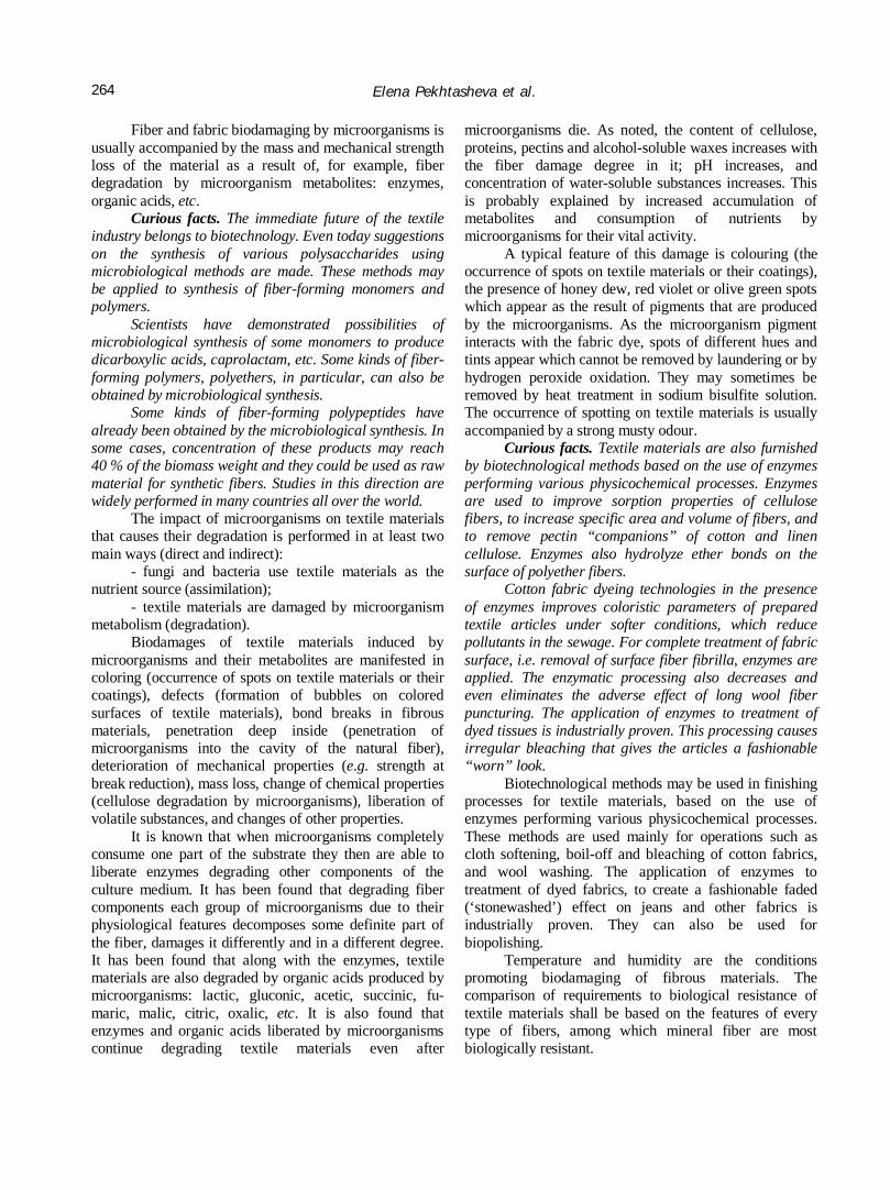
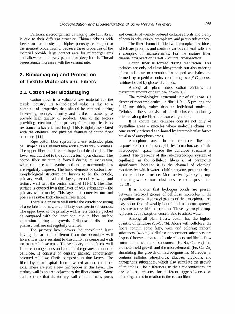
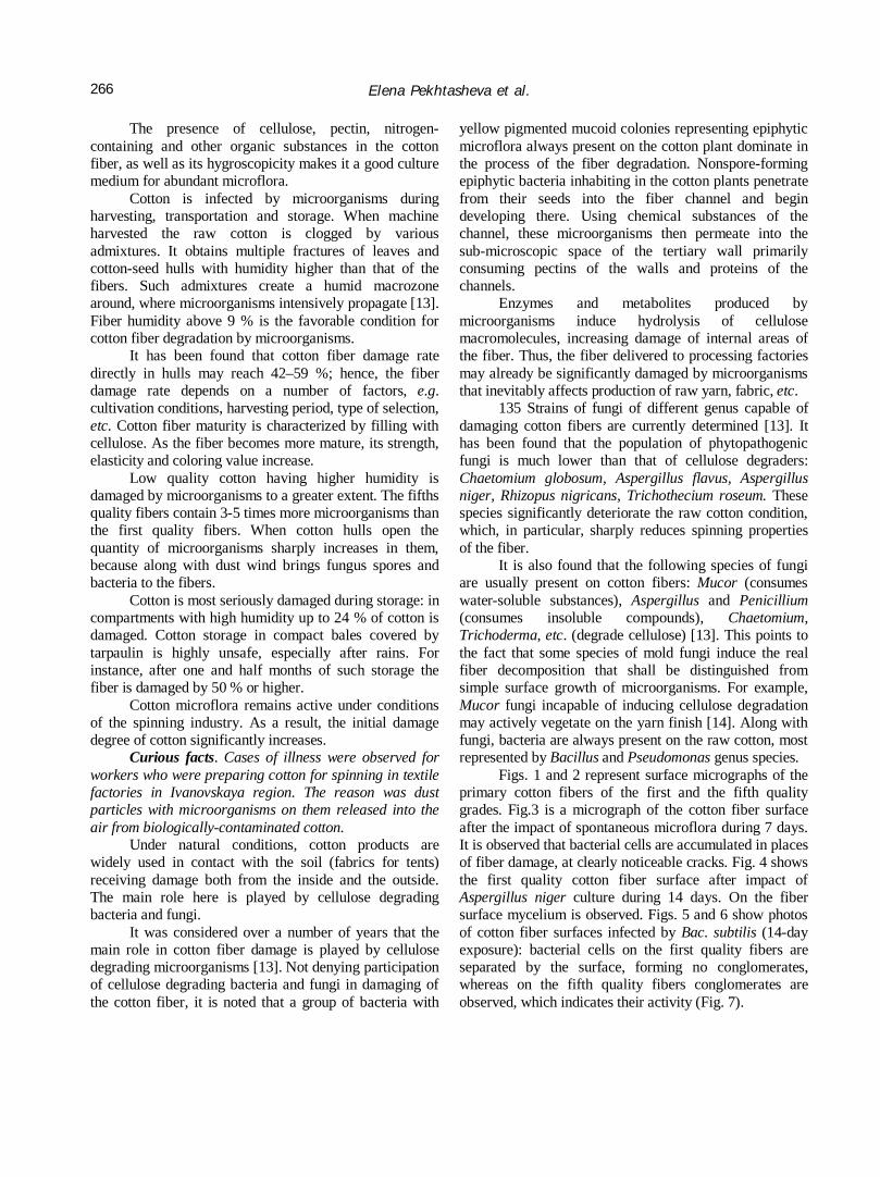
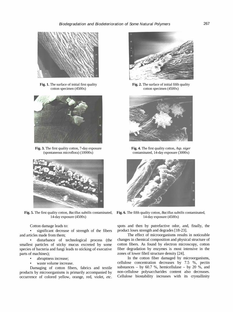
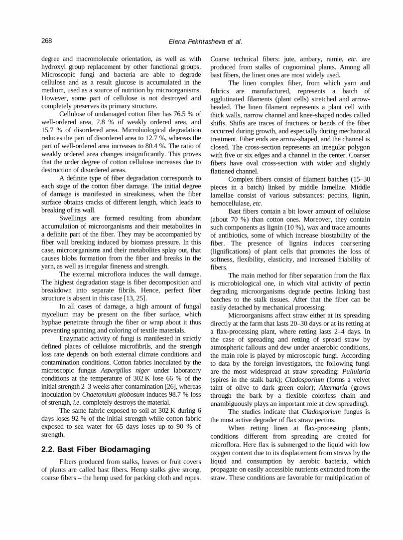
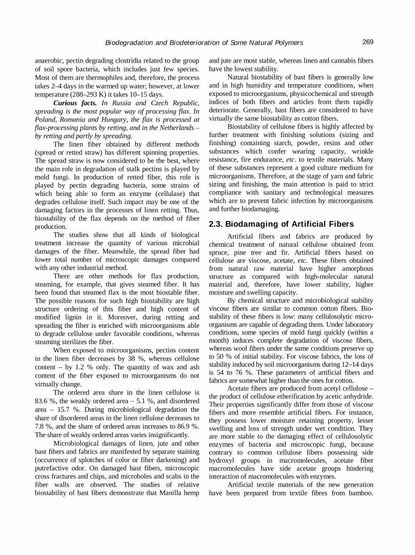
Figs. 1 and 2 represent surface micrographs of the primary cotton fibers of the first and the fifth quality grades. Fig.3 is a micrograph of the cotton fiber surface after the impact of spontaneous microflora during 7 days. It is observed that bacterial cells are accumulated in places of fiber damage, at clearly noticeable cracks. Fig. 4 shows the first quality cotton fiber surface after impact of Aspergillus niger culture during 14 days. On the fiber surface mycelium is observed. Figs. 5 and 6 show photos of cotton fiber surfaces infected by Bac. subtilis (14-day exposure): bacterial cells on the first quality fibers are separated by the surface, forming no conglomerates, whereas on the fifth quality fibers conglomerates are observed, which indicates their activity (Fig. 7).
Biodegradation and Biodeterioration of Some Natural Polymers
267
Fig. 1. The surface of initial first quality
cotton specimen (4500x) Fig. 2. The surface of initial fifth quality
cotton specimen (4500x)
Fig. 3. The first quality cotton, 7-day exposure (spontaneous microflora) (10000x)
Fig. 4. The first quality cotton, Asp. niger contaminated, 14-day exposure (3000x)
Fig. 5. The first quality cotton, Bacillus subtilis contaminated, 14-day exposure (4500x)
Fig. 6. The fifth quality cotton, Bacillus subtilis contaminated, 14-day exposure (4500x)
Cotton damage leads to: • significant decrease of strength of the fibers
and articles made from them; • disturbance of technological process (the
smallest particles of sticky mucus excreted by some species of bacteria and fungi leads to sticking of executive parts of machines);
• abruptness increase; • waste volume increase. Damaging of cotton fibers, fabrics and textile
products by microorganisms is primarily accompanied by occurrence of colored yellow, orange, red, violet, etc.
spots and then by putrefactive odor, and, finally, the product loses strength and degrades [18-23].
The effect of microorganisms results in noticeable changes in chemical composition and physical structure of cotton fibers. As found by electron microscopy, cotton fiber degradation by enzymes is most intensive in the zones of lower fibril structure density [24].
In the cotton fiber damaged by microorganisms, cellulose concentration decreases by 7.5 %, pectin substances – by 60.7 %, hemicellulase – by 20 %, and non-cellulose polysaccharides content also decreases. Cellulose biostability increases with its crystallinity
Elena Pekhtasheva et al.
268
degree and macromolecule orientation, as well as with hydroxyl group replacement by other functional groups. Microscopic fungi and bacteria are able to degrade cellulose and as a result glucose is accumulated in the medium, used as a source of nutrition by microorganisms. However, some part of cellulose is not destroyed and completely preserves its primary structure.
Cellulose of undamaged cotton fiber has 76.5 % of well-ordered area, 7.8 % of weakly ordered area, and 15.7 % of disordered area. Microbiological degradation reduces the part of disordered area to 12.7 %, whereas the part of well-ordered area increases to 80.4 %. The ratio of weakly ordered area changes insignificantly. This proves that the order degree of cotton cellulose increases due to destruction of disordered areas.
A definite type of fiber degradation corresponds to each stage of the cotton fiber damage. The initial degree of damage is manifested in streakiness, when the fiber surface obtains cracks of different length, which leads to breaking of its wall.
Swellings are formed resulting from abundant accumulation of microorganisms and their metabolites in a definite part of the fiber. They may be accompanied by fiber wall breaking induced by biomass pressure. In this case, microorganisms and their metabolites splay out, that causes blobs formation from the fiber and breaks in the yarn, as well as irregular fineness and strength.
The external microflora induces the wall damage. The highest degradation stage is fiber decomposition and breakdown into separate fibrils. Hence, perfect fiber structure is absent in this case [13, 25].
In all cases of damage, a high amount of fungal mycelium may be present on the fiber surface, which hyphae penetrate through the fiber or wrap about it thus preventing spinning and coloring of textile materials.
Enzymatic activity of fungi is manifested in strictly defined places of cellulose microfibrils, and the strength loss rate depends on both external climate conditions and contamination conditions. Cotton fabrics inoculated by the microscopic fungus Aspеrgillus niger under laboratory conditions at the temperature of 302 K lose 66 % of the initial strength 2–3 weeks after contamination [26], whereas inoculation by Chaetomium globosum induces 98.7 % loss of strength, i.e. completely destroys the material.
The same fabric exposed to soil at 302 K during 6 days loses 92 % of the initial strength while cotton fabric exposed to sea water for 65 days loses up to 90 % of strength.
2.2. Bast Fiber Biodamaging
Fibers produced from stalks, leaves or fruit covers of plants are called bast fibers. Hemp stalks give strong, coarse fibers – the hemp used for packing cloth and ropes.
Coarse technical fibers: jute, ambary, ramie, etc. are produced from stalks of cognominal plants. Among all bast fibers, the linen ones are most widely used.
The linen complex fiber, from which yarn and fabrics are manufactured, represents a batch of agglutinated filaments (plant cells) stretched and arrow-headed. The linen filament represents a plant cell with thick walls, narrow channel and knee-shaped nodes called shifts. Shifts are traces of fractures or bends of the fiber occurred during growth, and especially during mechanical treatment. Fiber ends are arrow-shaped, and the channel is closed. The cross-section represents an irregular polygon with five or six edges and a channel in the center. Coarser fibers have oval cross-section with wider and slightly flattened channel.
Complex fibers consist of filament batches (15–30 pieces in a batch) linked by middle lamellae. Middle lamellae consist of various substances: pectins, lignin, hemocellulase, etc.
Bast fibers contain a bit lower amount of cellulose (about 70 %) than cotton ones. Moreover, they contain such components as lignin (10 %), wax and trace amounts of antibiotics, some of which increase biostability of the fiber. The presence of lignins induces coarsening (lignifications) of plant cells that promotes the loss of softness, flexibility, elasticity, and increased friability of fibers.
The main method for fiber separation from the flax is microbiological one, in which vital activity of pectin degrading microorganisms degrade pectins linking bast batches to the stalk tissues. After that the fiber can be easily detached by mechanical processing.
Microorganisms affect straw either at its spreading directly at the farm that lasts 20–30 days or at its retting at a flax-processing plant, where retting lasts 2–4 days. In the case of spreading and retting of spread straw by atmospheric fallouts and dew under anaerobic conditions, the main role is played by microscopic fungi. According to data by the foreign investigators, the following fungi are the most widespread at straw spreading: Pullularia (spires in the stalk bark); Cladosporium (forms a velvet taint of olive to dark green color); Alternaria (grows through the bark by a flexible colorless chain and unambiguously plays an important role at dew spreading).
The studies indicate that Cladosporium fungus is the most active degrader of flax straw pectins.
When retting linen at flax-processing plants, conditions different from spreading are created for microflora. Here flax is submerged to the liquid with low oxygen content due to its displacement from straws by the liquid and consumption by aerobic bacteria, which propagate on easily accessible nutrients extracted from the straw. These conditions are favorable for multiplication of
Biodegradation and Biodeterioration of Some Natural Polymers
269
anaerobic, pectin degrading clostridia related to the group of soil spore bacteria, which includes just few species. Most of them are thermophiles and, therefore, the process takes 2–4 days in the warmed up water; however, at lower temperature (288–293 K) it takes 10–15 days.
Curious facts. In Russia and Czech Republic, spreading is the most popular way of processing flax. In Poland, Romania and Hungary, the flax is processed at flax-processing plants by retting, and in the Netherlands – by retting and partly by spreading.
The linen fiber obtained by different methods (spread or retted straw) has different spinning properties. The spread straw is now considered to be the best, where the main role in degradation of stalk pectins is played by mold fungi. In production of retted fiber, this role is played by pectin degrading bacteria, some strains of which being able to form an enzyme (cellulase) that degrades cellulose itself. Such impact may be one of the damaging factors in the processes of linen retting. Thus, biostability of the flax depends on the method of fiber production.
The studies show that all kinds of biological treatment increase the quantity of various microbial damages of the fiber. Meanwhile, the spread fiber had lower total number of microscopic damages compared with any other industrial method.
There are other methods for flax production, steaming, for example, that gives steamed fiber. It has been found that steamed flax is the most biostable fiber. The possible reasons for such high biostability are high structure ordering of this fiber and high content of modified lignin in it. Moreover, during retting and spreading the fiber is enriched with microorganisms able to degrade cellulose under favorable conditions, whereas steaming sterilizes the fiber.
When exposed to microorganisms, pectins content in the linen fiber decreases by 38 %, whereas cellulose content – by 1.2 % only. The quantity of wax and ash content of the fiber exposed to microorganisms do not virtually change.
The ordered area share in the linen cellulose is 83.6 %, the weakly ordered area – 5.1 %, and disordered area – 15.7 %. During microbiological degradation the share of disordered areas in the linen cellulose decreases to 7.8 %, and the share of ordered areas increases to 86.9 %. The share of weakly ordered areas varies insignificantly.
Microbiological damages of linen, jute and other bast fibers and fabrics are manifested by separate staining (occurrence of splotches of color or fiber darkening) and putrefactive odor. On damaged bast fibers, microscopic cross fractures and chips, and microholes and scabs in the fiber walls are observed. The studies of relative biostability of bast fibers demonstrate that Manilla hemp
and jute are most stable, whereas linen and cannabis fibers have the lowest stability.
Natural biostability of bast fibers is generally low and in high humidity and temperature conditions, when exposed to microorganisms, physicochemical and strength indices of both fibers and articles from them rapidly deteriorate. Generally, bast fibers are considered to have virtually the same biostability as cotton fibers.
Biostability of cellulose fibers is highly affected by further treatment with finishing solutions (sizing and finishing) containing starch, powder, resins and other substances which confer wearing capacity, wrinkle resistance, fire endurance, etc. to textile materials. Many of these substances represent a good culture medium for microorganisms. Therefore, at the stage of yarn and fabric sizing and finishing, the main attention is paid to strict compliance with sanitary and technological measures which are to prevent fabric infection by microorganisms and further biodamaging.
2.3. Biodamaging of Artificial Fibers Artificial fibers and fabrics are produced by
chemical treatment of natural cellulose obtained from spruce, pine tree and fir. Artificial fibers based on cellulose are viscose, acetate, etc. These fibers obtained from natural raw material have higher amorphous structure as compared with high-molecular natural material and, therefore, have lower stability, higher moisture and swelling capacity.
By chemical structure and microbiological stability viscose fibers are similar to common cotton fibers. Bio-stability of these fibers is low: many cellulosolytic micro-organisms are capable of degrading them. Under laboratory conditions, some species of mold fungi quickly (within a month) induces complete degradation of viscose fibers, whereas wool fibers under the same conditions preserve up to 50 % of initial stability. For viscose fabrics, the loss of stability induced by soil microorganisms during 12–14 days is 54 to 76 %. These parameters of artificial fibers and fabrics are somewhat higher than the ones for cotton.
Acetate fibers are produced from acetyl cellulose – the product of cellulose etherification by acetic anhydride. Their properties significantly differ from those of viscose fibers and more resemble artificial fibers. For instance, they possess lower moisture retaining property, lesser swelling and loss of strength under wet condition. They are more stable to the damaging effect of cellulosolytic enzymes of bacteria and microscopic fungi, because contrary to common cellulose fibers possessing side hydroxyl groups in macromolecules, acetate fiber macromolecules have side acetate groups hindering interaction of macromolecules with enzymes.
Artificial textile materials of the new generation have been prepared from textile fibres from bamboo.
Elena Pekhtasheva et al.
270
Bamboo possesses antimicrobial properties due to the presence of a substance known as ‘bamboo kun’ in the fibre. Bamboo fibres possess an extremely porous structure so they absorb much more water than cotton fibres. Clothes made from bamboo fibres absorb and evaporate sweat very quickly because of the presence of these pores and the high antimicrobial properties of bamboo prevent perspiration odour.
Various modified viscose fibers, micromodal and modal, for example, produced from beech were not studies for biostability. Information on biostability of artificial fibers produced from lactic casein, soybean protein, maize, peanut and corn is absent.
2.4. Wool Fiber Biodamaging
Wool is animal hair widely used in textile and light industry. The structure and chemical composition of the wool fiber significantly differ it from other types of fibers and shows great variety and heterogeneity of properties. Sheep, camel, goat and rabbit wool is used as the raw material.
After thorough cleaning, the wool fiber can be considered virtually consisting of a single protein – keratin. The wool contains the following elements (in %): carbon 50; hydrogen 6–7; nitrogen 15–21; oxygen 21–24; sulfur 2–5, and other elements.
The chemical feature of wool is high content of various amino acids. It is known that wool is a copolymer of, at least, 17 amino acids, whereas the most of synthetic fibers represent copolymers of two monomers.
Different content of amino acids in wool fibers promotes the features of their chemical properties. Of the great importance is the quantity of cystine containing virtually all sulfur, which is extremely important for the wool fiber properties. The higher sulfur content in the wool, the better its processing properties, the higher resistance to chemical and other impacts, and the higher the physico-mechanical properties.
Wool fiber layers, in turn, differ by the sulfur content: it is higher in the cortical layer than in the core.
Among all textile fibers, wool has the most complex structure.
The fine merino wool fiber consists of two layers: external flaky layer or cuticle and internal cortical layer – the cortex. Coarser fibers have the third layer – the core.
The cuticle consists of flattened cells overlapping one another (the flakes) and tightly linked to one another and the cortical layer inside.
Cuticular cells have a membrane, the so-called epicuticle, right around. It has been found that epicuticle gives about 2 % of the fiber mass. Cuticle cells limited by walls quite tightly adjoin one another, but, nevertheless,
there is a thin layer of intercellular protein substance between them, whose mass is 3-4% of the fiber mass.
The cortical layer, the cortex, is located under the cuticle and forms the main mass of the fiber and, consequently, defines basic physico-mechanical and many other properties of the wool. Cortex is composed of spindle-shaped cells connivent to one another. Protein substance is also located between the cells.
The cortical layer cells are composed of densely located cylindrical, thread-like macrofibrils of about 0.05–0.2 µm in diameter. Macrofibrils of the cortical layer are composed of microfibrils with the average diameter of 7–7.5 nm [26, 27].
Microfibrils, sometimes called the secondary agents, are composed of primary aggregates – protofibrils. Protofibril represents two or three twisted α-spiral chains. It is suggested [27, 28] that α-spirals are twisted due to periodic repetition of amino acid residues in the chain, hence, side radicals of the same spiral are disposed in the inner space of another α-spiral providing strong interaction, including for the account of hydrophobic bonds, because each seventh residue has a hydrophobic radical.
According to the data by the English investigator J.D. Leeder, the wool fiber can be considered as a collection of flaky and cortical cells bound by a cell membrane complex (CMC) which thus forms a uniform continuous phase in the keratine substance of the fiber. This intercellular cement can easily be chemically and microbiologically degraded, i.e. a δ-layer about 15 nm thick (CMC or intercellular cement) is located between cells filling in all gaps [29]. The studies show that the composition of intercellular material between flaky cells may differ from that of the material between cortical cells. In the cuticle-cuticle, cuticle-cortex and cortex-cortex complexes the intercellular “cement” has different chemical compositions.
Although the cell membrane complex gives only 6 % of the wool fiber mass, there are proofs that it causes the main effect on many properties of the fiber and fabric [29, 30]. For instance, a suggestion was made that CMC components may affect such mechanical properties, as wear resistance and torsion fatigue, as well as such chemical properties, as resistance to acids, proteolytic enzymes and chemical finishing agents.
The core layer is present in the fibers of coarser wool with the core cell content up to 15 %. Disposition and shape of the core layer cells significantly vary with respect to the fiber type. This layer can be continuous (along the whole fiber) or may be separated in sections. The cell carcass of the core layer is composed of protein similar to microfibril cortex protein.
Biodegradation and Biodeterioration of Some Natural Polymers
271
By its chemical composition, wool is a protein substance. The main substance forming wool is keratine – a complex protein containing much sulfur in contrast with other proteins. Keratine is produced during amino acid biosynthesis in the hair bag epidermis in the hide. Keratine structure represents a complex of high-molecular chain batches interacting both laterally and transversally [30-32]. Along with keratine, wool contains lower amounts of other substances.
Wool keratine reactivity is defined by its primary, secondary and tertiary structures, i.e. the structure of the main polypeptide chains, the nature of side radicals and the presence of cross bonds.
Among all amino acids, only cystine forms cross bonds; their presence considerably defines wool insolubility in many reagents. Cystine bond decomposition simplifies wool damaging by sunlight, oxidants and other agents. Cystine contains almost all sulfur present in the wool fibers. Sulfur is very important for the wool quality, because it improves chemical properties, strength and elasticity of fibers.
Along with general regularities in the structure of high-molecular compounds, fibers differ from one another by chemical composition, monomer structure, polyme-rization degree, orientation, intermolecular bond strength and type, etc., which defines different physico-mechanical and chemical properties of the fibers.
The main chemical component of wool – keratine – is nutrition for microorganisms. Microorganisms may not directly consume proteins. Therefore, they are only consumed by microbes having proteolytic enzymes – exoproteases that are excreted by cells to the environment. Wool damage may start already before sheepshearing, i.e. in the fleece, where favorable nutritive (sebaceous matters, wax, epithelium), temperature, aeration, and humidity conditions are formed.
Contrary to microorganisms damaging plant fibers, the wool microflora is versatile, generally represented by species typical of the soil and degrading plant residues. Initiated in the fleece, wool fiber damages are intensified during its storage, processing and transportation under unfavorable conditions. Specific epiphytic microflora typical of this particular fiber is always present on its surface. Representatives of this microflora excrete proteolyric enzymes (mostly pepsin), which induce hydrolytic keratine decay by polypeptide bonds to separate amino acids.
Wool is degraded in several stages: first, microorganisms destroy the flaky layer and then penetrate into the cortical layer of the fiber, although the cortical layer itself is not destroyed, because intercellular substance located between the cells is the culture medium. As a result, the fiber structure is disturbed: flakes and cells are not bound yet, the fiber cracks and decays.
The mechanism of wool fiber hydrolysis by microorganisms suggested by the American scientist E. Race represents a sequence of transformations: pro- teins–peptones–polypeptides–water + ammonia + carbo-xylic acids. The most active bacteria: Alkaligenes bookeri, Pseudomonas aeroginosa, Proteus vulgaris, Bacillus agri, B. mycoides, B. mesentericus, B. megatherium, B. subtilis, and microscopic fungi: Aspergillus, Alternaria, Cephalothecium, Dematium, Fusarium, Oospora, Penicillium, Trichoderma, were extracted from the wool fiber surface [33-39].
However, the dominant role in the wool degradation is played by bacteria. Fungi are less active in degrading wool. Consuming fat and dermal excretion, fungi create conditions for further vital activity of bacteria-degraders. The role of microscopic fungi may also be reduced to splitting the ends of fibers resulting from mechanical efforts of growing hyphae. Such splitting allows bacteria to penetrate into the fiber. Fungi weakly use wool as the source of carbon.
In 1960s, the data on the effect of fat and dirt present on the surface of unclean fibers on the wool biodamaging were published. It has been found that unclean wool is damaged much faster than the clean one. The presence of fats on unclean wool promotes fungal microflora development.
The activity of microbiological processes developing on the wool depends on mechanical damages of the fiber and preliminary processing of the wool.
It has been found that microorganism penetration may happen through fiber cuts or microcracks in the flaky layer. Cracks may be of different origins – mechanical, chemical, etc. It is also found that wool subject to intensive mechanical or chemical treatment is more easily degraded by microorganisms than the untreated one [39]. For instance, high activity of microorganisms during wool bleaching by hydrogen peroxide in the presence of alkaline agents and on wool washed in the alkaline medium was observed. When wool is treated in a weak acid medium, the activity of microorganisms is abruptly suppressed. This also takes place on the wool colored by chrome and metal-containing dyes. The middle activity of microorganisms is observed on the wool colored by acid dyes.
When impacted by microorganisms, structural changes in the wool are observed: flaky layer damages, its complete exfoliation, and lamination of the cortical layer.
Wool fiber damages can be reduced to several generalized types provided by their structural features:
• channeling and overgrowth – accumulation of bacteria or fungal hyphae and their metabolites on the fiber surface;
• flaky layer damage, local and spread; • cortical layer lamination to spindle-shaped cells; • spindle-shaped cell destruction.
Elena Pekhtasheva et al.
272
Along with the fiber structure damage, some bacteria and fungi decrease its quality by making wool dirty blue or green, which may not be removed by water or detergents. Splotches of color also occur on wool, for example, due to the impact of Pseudomonas aeruginosa bacteria; in this case, color depends on medium pH: green splotches occurred in a weakly alkaline medium, and in weak acid medium they are red. Green splotches may also be caused by development of Dermatophilus congolensis fungi. Black color of wool is provided by Pyronellaea glomerata fungi.
Thus, wool damage reduces its strength, increases waste quantity at combing and imparts undesirable blue, green or dirty color and putrefactive odor. However, wool is degraded by microorganisms slower than plant fibers.
2.4.1. Changes of structure and properties of wool fibers by microorganisms
To evaluate bacterial contamination of wool fibers, it is suggested to use an index proposed by A.I. Sapozh-nikova, which characterizes discoloration rate of resazurin solution, a weak organic dye and currently hydrogen acceptor. It is also indicator of both presence and activity of reductase enzyme [40].
The method is based on resazurin ability to lose color in the presence of reductase, which is micro-organisms’ metabolite, due to redox reaction proceeding. This enables judging about quantity of active microorganisms present in the studied objects by solution discoloration degree. Discoloration of the dye solution was evaluated both visually and spectrophotometrically by optical density value.
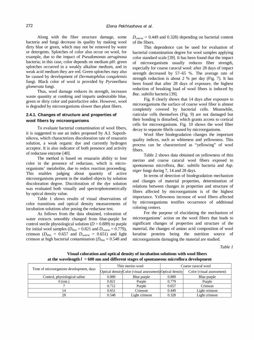
Table 1 shows results of visual observations of color transitions and optical density measurements of incubation solutions after posing the reductase test.
As follows from the data obtained, coloration of water extracts smoothly changed from blue-purple for control sterile physiological solution (D = 0.889) to purple for initial wool samples (Dthin = 0.821 and Dcoarse = 0.779), crimson (Dthin = 0.657 and Dcoarse = 0.651) and light crimson at high bacterial contamination (Dthin = 0.548 and
Dcoarse = 0.449 and 0.328) depending on bacterial content of the fibers.
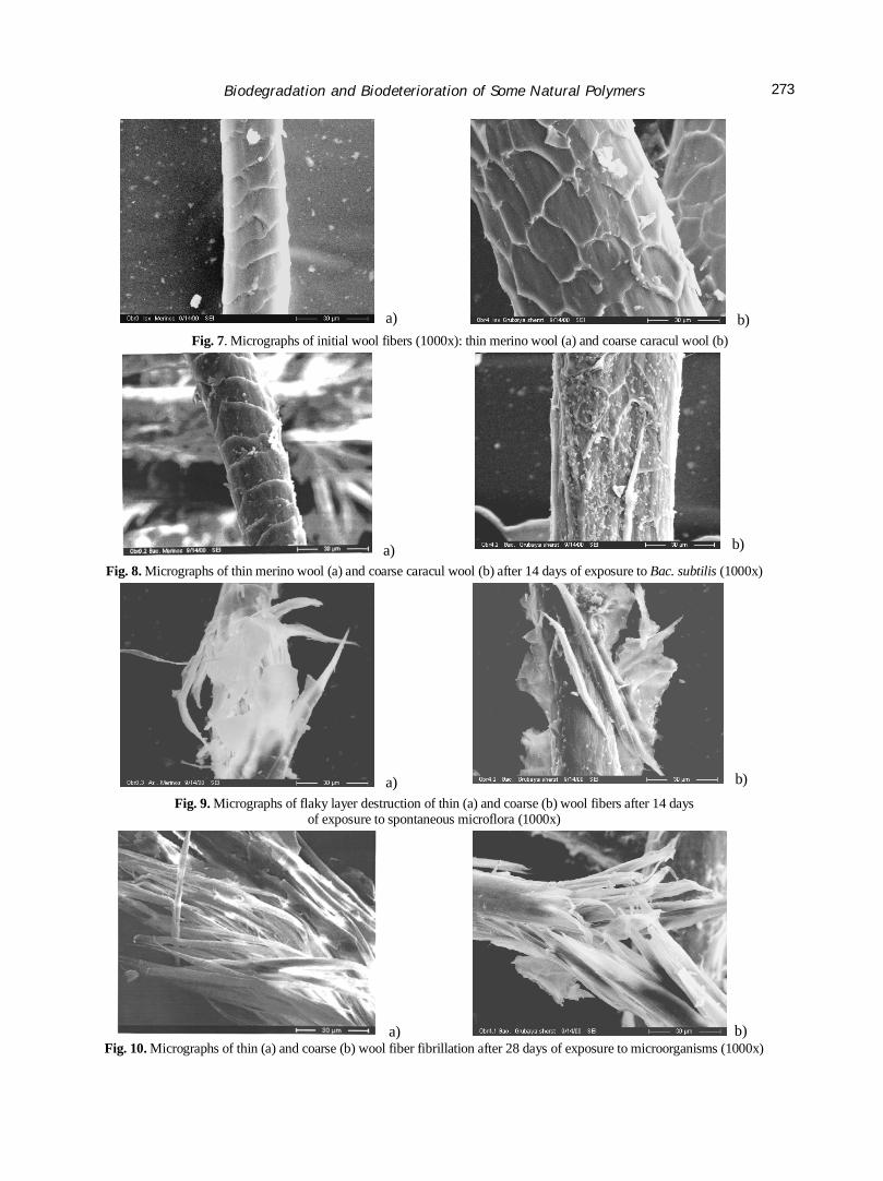
This dependence can be used for evaluation of bacterial contamination degree for wool samples applying color standard scale [39]. It has been found that the impact of microorganisms usually reduces fiber strength, especially for coarse caracul wool: after 28 days of impact strength decreased by 57–65 %. The average rate of strength reduction is about 2 % per day (Fig. 7). It has been found that after 28 days of exposure, the highest reduction of breaking load of wool fibers is induced by Bac. subtilis bacteria [39].
Fig. 8 clearly shows that 14 days after exposure to microorganisms the surface of coarse wool fiber is almost completely covered by bacterial cells. Meanwhile, cuticular cells themselves (Fig. 9) are not damaged but their bonding is disturbed, which grants access to cortical cells for microorganisms. Fig. 10 shows the wool fiber decay to separate fibrils caused by microorganisms.
Wool fiber biodegradation changes the important quality indices, such as whiteness and yellowness. This process can be characterized as “yellowing” of wool fibers.
Table 2 shows data obtained on yellowness of thin merino and coarse caracul wool fibers exposed to spontaneous microflora, Bac. subtilis bacteria and Asp. niger fungi during 7, 14 and 28 days.
In terms of detection of biodegradation mechanism and changes of material properties, determination of relations between changes in properties and structure of fibers affected by microorganisms is of the highest importance. Yellowness increase of wool fibers affected by microorganisms testifies occurrence of additional coloring centers.
For the purpose of elucidating the mechanism of microorganisms’ action on the wool fibers that leads to significant changes of properties and structure of the material, the changes of amino acid composition of wool keratine proteins being the nutrition source of microorganisms damaging the material are studied.
Table 1 Visual coloration and optical density of incubation solutions with wool fibers
at the wavelength λ = 600 nm and different stages of spontaneous microflora development Thin merino wool Coarse caracul wool
Time of microorganism development, days Optical density Color (visual assessment)Optical density Color (visual assessment)
Control, physiological saline 0.889 Blue purple 0.889 Blue purple 0 (init.) 0.821 Purple 0.779 Purple
7 0.712 Purple 0.657 Crimson 14 0.651 Crimson 0.449 Light crimson 28 0.548 Light crimson 0.328 Light crimson
Biodegradation and Biodeterioration of Some Natural Polymers
273
a) b) Fig. 7. Micrographs of initial wool fibers (1000x): thin merino wool (a) and coarse caracul wool (b)
a) b)
Fig. 8. Micrographs of thin merino wool (a) and coarse caracul wool (b) after 14 days of exposure to Bac. subtilis (1000x)
a) b) Fig. 9. Micrographs of flaky layer destruction of thin (a) and coarse (b) wool fibers after 14 days
of exposure to spontaneous microflora (1000x)
a) b) Fig. 10. Micrographs of thin (a) and coarse (b) wool fiber fibrillation after 28 days of exposure to microorganisms (1000x)
Elena Pekhtasheva et al.
274
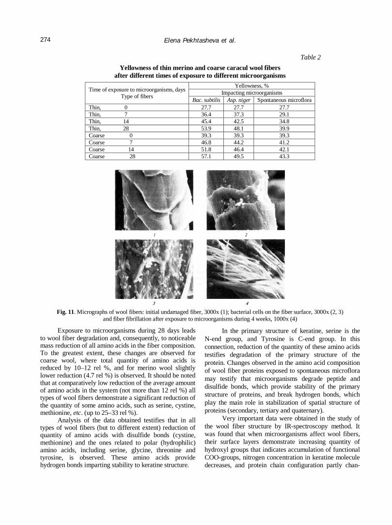
Table 2 Yellowness of thin merino and coarse caracul wool fibers
after different times of exposure to different microorganisms Yellowness, %
Impacting microorganisms Time of exposure to microorganisms, days Type of fibers Bac. subtilis Asp. niger Spontaneous microflora
Thin, 0 27.7 27.7 27.7 Thin, 7 36.4 37.3 29.1 Thin, 14 45.4 42.5 34.8 Thin, 28 53.9 48.1 39.9 Coarse 0 39.3 39.3 39.3 Coarse 7 46.8 44.2 41.2 Coarse 14 51.8 46.4 42.1 Coarse 28 57.1 49.5 43.3
Fig. 11. Micrographs of wool fibers: initial undamaged fiber, 3000x (1); bacterial cells on the fiber surface, 3000x (2, 3)
and fiber fibrillation after exposure to microorganisms during 4 weeks, 1000x (4)
Exposure to microorganisms during 28 days leads to wool fiber degradation and, consequently, to noticeable mass reduction of all amino acids in the fiber composition. To the greatest extent, these changes are observed for coarse wool, where total quantity of amino acids is reduced by 10–12 rel %, and for merino wool slightly lower reduction (4.7 rel %) is observed. It should be noted that at comparatively low reduction of the average amount of amino acids in the system (not more than 12 rel %) all types of wool fibers demonstrate a significant reduction of the quantity of some amino acids, such as serine, cystine, methionine, etc. (up to 25–33 rel %).
Analysis of the data obtained testifies that in all types of wool fibers (but to different extent) reduction of quantity of amino acids with disulfide bonds (cystine, methionine) and the ones related to polar (hydrophilic) amino acids, including serine, glycine, threonine and tyrosine, is observed. These amino acids provide hydrogen bonds imparting stability to keratine structure.
In the primary structure of keratine, serine is the N-end group, and Tyrosine is C-end group. In this connection, reduction of the quantity of these amino acids testifies degradation of the primary structure of the protein. Changes observed in the amino acid composition of wool fiber proteins exposed to spontaneous microflora may testify that microorganisms degrade peptide and disulfide bonds, which provide stability of the primary structure of proteins, and break hydrogen bonds, which play the main role in stabilization of spatial structure of proteins (secondary, tertiary and quaternary).
Very important data were obtained in the study of the wool fiber structure by IR-spectroscopy method. It was found that when microorganisms affect wool fibers, their surface layers demonstrate increasing quantity of hydroxyl groups that indicates accumulation of functional COO-groups, nitrogen concentration in keratine molecule decreases, and protein chain configuration partly chan-
Biodegradation and Biodeterioration of Some Natural Polymers
275
ges – β-configuration (stretched chains) transits to α-configuration (a spiral). This transition depends on α- and β-forms ratio in the initial fiber and is more significant for thin fibers, which mostly have β-configuration of chains in the initial fibers.
Thus, it has been found that microorganisms generally affect the cell membrane complex and degrade amino acids, such as cystine, methionine, serine, glycine, threonine, and tyrosine. Microorganisms reduce breaking load and causes “yellowing” of the wool fibers (Fig. 11).
2.5. Synthetic Fibers Biodamaging
Synthetic fibers are principally different from the natural and the artificial ones by structure and, being an alien substrate for microorganisms, are harder damaged by them. Since occurrence of synthetic fabrics in 1950s, it is suggested that they are “everlasting” and are not utilized by microorganisms. However, it has been found with time that, firstly, microorganisms although slower, but yet are capable of colonizing synthetic fabrics and utilizing their carbon in the course of development (i.e. causing biodamage), and secondly, there are both more and less microorganism resistant fabrics among the synthetic ones [41,42].
Among microorganisms damaging synthetic fibers, Trichoderma genus fungi are identified, at the initial stages developing due to lubricants and finishing agents without fiber damage and then wrap them with mycelium, loosen threads and, hence, reduce fabric strength.
When studying fabrics from nitrone, lavsan, and caprone it has been found that soil fungi and bacteria cause roughly the same effect on characteristics of these fabrics increasing the fiber swelling degree by 20–25 %, reducing strength by 10–15 % and elongation at break by 15–20 %.
Synthetic fibers represent a potential source of energy and nutrition for microorganisms. The ability of microorganisms to attach to surfaces of insoluble solids, then using them as the nutritive substrate, is well-known. Living cells of microorganisms have complex structure. On the surface of bacterial cells complexes of proteins, lipids and polysaccharides were found; they contains hydrophilic and hydrophobic areas, various functional groups and mosaic electric charge (at total negative charge of the cells).
The first stage of microorganism interaction with synthetic fibers can be rightfully considered in terms of the adhesion theory with provision for the features of structure and properties of microorganisms as a biological system.
The entire process of microorganism impact of the fiber can conditionally be divided into several stages: attachment to the fiber, growth and multiplication on it and its consumption as the nutrition and energy source [43, 44].
Enzymes excreted by bacteria act just in the vicinity of bacterial membrane. Being adsorbed onto the fiber, living cells attach to the surface and adapt to new living conditions. The ability to be adsorbed onto the surface of synthetic fibers is caused by:
• the features of chemical structure of the fibers. For instance, fibers adsorbing microorganisms are polyamide and polyvinyl alcohol ones; the fiber not adsorbing microorganisms is, for example, ftorin.
• physical structure of the fiber. For example, fibers with smaller linear density, with a lubricant on the surface absorb greater amount of microorganisms.
• the presence of electric charge on the surface, its value and sign. Positively charged chemical fibers adsorb virtually all bacteria, fibers having no electric charge adsorb the majority of bacteria, and negatively charged fibers do not adsorb bacteria.
Supermolecular structure also stipulates the possibility for microorganisms and their metabolites to diffuse inside the internal areas of the fiber. Microorganism assimilation of the fiber starts from the surface, and further degradation processes and their rate are determined by microphysical state of the fiber. Microorganism metabolite penetration into inner areas of the fiber and deep layers of a crystalline material is only possible in the presence of capillaries.
Chemical fiber damaging and degradation starting from the surface are, in many instances, promoted by defects like cracks, chips or hollows, which may occur in the course of fiber production and finishing.
Along with physical inhomogeneity, chemical inhomogeneity may promote biodegradation of synthetic fibers. Chemical inhomogeneity occurs during polymer synthesis and its thermal treatments, manifesting itself in different content of monomers and various end groups. The possibility for microorganism metabolites to penetrate inside the structure of synthetic fibers depends on the quantity and accessibility of functional end groups in the polymer, which are abundant in oligomers.
The ability of synthetic fibers to swell also enables penetration of biological agents inside low ordered areas of fibers and weakens intermolecular interactions, off-orientation of macromolecules, and degradation in the amorphous and crystalline zones. Structural changes result in reduction of strength properties of fibers.
According to theoretical statements synthetic fibers with the lower ordered structure and higher content of oligomers possess lower stability to microorganism impact than fibers with highly organized structure and lower content of low-molecular compounds. Thus, the most rapid occurrence and biodegradation of synthetic fibers are promoted by low ordering and low orientation of macromolecules in the fibers, their low density, low
Elena Pekhtasheva et al.
276
crystallinity and the presence of defects in macro- and microstructure of the fibers, pores and cavities in their internal zones.
Carbochain polymer based fibers are more resistant to microbiological damages. These polymers are: polyolefins, polyvinyl chloride, polyvinyl fluoride, polyacrylonitrile, polyvinyl alcohol. Fibers based on heterochain polymers: polyamide, polyether, polyure-thane, etc., are less bioresistant.
Comparative soil tests for biostability of artificial and synthetic fibers demonstrate that viscose fiber is completely destroyed on the 17th day of the tests; bacteria and fungi colonies occur on lavsan on the 20th day; caprone is overgrown by fungus mycelium on the 30th day. Chlorin and ftorlon have the highest biostability. The initial signs of their biodamage are only observed 3 months after the test initiation.
The studies of nitron, lavsan and capron fabric biostability have found that soil fungi and bacteria cause nearly equal influence on the parameters of these fabrics, increasing swelling degree of the fibers by 20–25 %, reducing strength by 10–15 % and elongation at break by 15–20 %. Meanwhile, nitron demonstrated higher biostability as compared with lavsan and capron.
2.5.1. Changes in structure and properties of polyamide fibers induced by microorganisms
In contrast with natural fibers, chemical fibers have no permanent and particular microflora. Therefore, the most widespread species of microorganisms possessing increased adaptability are the main biodegraders of these materials. Occurrence and progression of biological degra-dation of polyamide fibers is, in many instances, induced by their properties and properties of affecting microorganisms, and their species composition. Generally, the species of microorganisms degrading polyamide and other chemical fibers are determined by their operation conditions, which form microflora, and its adaptive abilities.
Polyamide fibers are most frequently used in mixtures with natural fibers. Natural fibers contain specific microflora on the surface and inside. Therefore, capron fibers mixed with cotton, wool or linen are affected by their microflora. It has been found [43-45] that capron fiber degradation by microorganisms obtained from wool is characterized as deep fiber decay; micro-organisms extracted from natural silk cause streakiness of capron fibers; microorganisms extracted from cotton cause fading and decomposition; and microorganisms extracted from linen cause fading, streakiness and decomposition.
The microorganism interaction with polyamide fibers is most fully studied in the works by I.A. Ermilova [43-45]. For the purpose of detecting bacteria-degraders of
polyamide fibers, microorganisms were extracted from fibers damaged in the medium of active sewage silt, soil, microflora of natural fibers and test-bacteria complex selected as degraders of polyamide materials. Capron fibers were inoculated by extracted cultures of microorganisms and types of damages were reproduced.
Polyamide fiber materials were natural nutritive and energy source for these microorganisms. Therefore, bacterial strains extracted from damaged fibers were different from the initial strains. It is proven that the existence on a new substrate has stipulated changes of intensity and direction of physiological-biochemical processes of bacterial cells, the change of their morphological and culture properties [43-45].
Extraction, cultivation and use of such adaptive strains are of both scientific and practical interest. Using bacterial strains adaptive to capron, polyamide production waste, warn products, toxic substances may be utilized. This allows obtaining of secondary raw materials and solving the problem of environmental protection.
It is proved experimentally [43-45] that polycapro-amide fibers possess high adsorbability, which value depends on the properties of impacting bacteria. For instance, gram-positive, especially spore-forming bacteria Bacillus subtilis, Bac. mesentericus (from 84.5 to 99.3 % of living cells), are most highly adsorbed, and adsorption of gram-positive bacteria varies significantly.
The extensive research of test-bacterium complex impact was performed [43-45]. These bacteria were chosen as degraders of polyamide fibers, along with microflora of active sewage silts, linen and jute microorganisms as degraders of a complex capron thread. It has been found that the test-bacterium complex injected by the author, after 7 months of exposure, increases biodegradation index to 1.27 that testifies to intensive degradation of the fiber microstructure. The highest degradation of complex polycaproamide (PCA) thread is caused by active sewage silt microorganisms.
High activity of silt microorganisms and test-bacteria is explained by the fact that they include bacterium strains Bacillus subtilis and Bac. mesentericus, which, according to the data by a number of authors [39], may induce full degradation of caprolactam to amino acids using it as the source of carbon and nitrogen.
Thus, adaptive forms of microorganisms induce the highest degradation of polyamide fibers.
Polyamide fibers are characterized by physical structure inhomogeneity, which occurs during processing and is associated with differences in crystallinity and orientation of macromolecules determining fiber accessi-bility for microorganisms and their metabolites pene-tration. The surface layer is damaged during orientational stretching and, consequently, has lower molecular alignment. That is why the surface layer is most
Biodegradation and Biodeterioration of Some Natural Polymers
277
intensively changed by microorganisms. The study of polyamide fiber macrostructure after exposure to microorganisms shows that streakiness and cover damage are the main damages of these fibers [43].
The studies of supermolecular structure of capron fiber surface show that after exposure to microorganisms the capron fiber cover becomes loose and uneven [39, 43]. Surface supermolecular structure degradation increases with the microbial impact: fibrils and their yarns become split and disaligned both laterally and transversely, multiple defects in the form of pores and cavities, and cracks of various depths are formed.
Chemical inhomogeneity of polyamide fibers also promotes changes in the fiber structure when exposed to microorganisms [46-50]. It has been found that the polyamide fiber degradation increases with low-molecular compound (LMC) content in them; meanwhile, at the same content of low-molecular compounds, thermally treated fibers were more biostable, as compared with untreated specimens [43, 44].
Along with the morphological characteristics which characterize biodamaging of the fibers functional features, such as strength decrease and increase of deformation properties of the fiber were detected. The greatest strength decrease (by 46.4 %) was observed for thermally untreated capron fiber containing 3.4 % LMC, and the smallest decrease (by 5 %) was observed for the fibers with 3.2 % LMC, thermally treated at the optimum time of 5 s [43, 44].
IR-spectroscopy method was applied to detect polycaproamide fiber damage by microorganisms [39]. It has been found that carboxyl and amide groups are accumulated during their biodegradation.
The change of various property indices of polyamide fibers also results from macro-, micro- and chemical changes.
Of interest are the studies of the microorganism effect on polyamide fabric quality [43, 44]. Fabrics (both bleached and colored) from capron monofilament were exposed to a set of test-cultures: Bac.subtilis, Ps.fluorecsens, Ps.herbicola, Bac.mesentericus. After 3–9 month of exposure, yellowness and dark spots occurred, coloring intensity decreased, and an odor appeared. The optical microscopy studies indicated that all fibers exposed to microorganisms, had damages typical of synthetic fibers – overgrowing, streakiness, bubbles, and wall damages. The increasing quantity of biodamages with time results in tensile strength reduction: by 6–8 % for capron fibers after 9 months, at inconsiderable change of relative elongation.
It all goes to show that development of micro-organisms on polyamide fibrous materials results in changes of fibers morphology, their molecular and
supermolecular structure and, as a consequence, reduction of strength properties, color change, and odor.
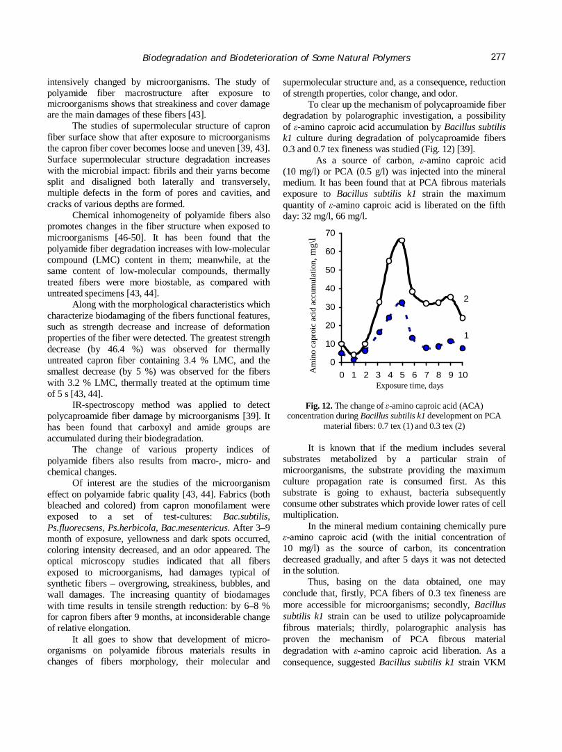
To clear up the mechanism of polycaproamide fiber degradation by polarographic investigation, a possibility of ε-amino caproic acid accumulation by Bacillus subtilis k1 culture during degradation of polycaproamide fibers 0.3 and 0.7 tex fineness was studied (Fig. 12) [39].
As a source of carbon, ε-amino caproic acid (10 mg/l) or PCA (0.5 g/l) was injected into the mineral medium. It has been found that at PCA fibrous materials exposure to Bacillus subtilis k1 strain the maximum quantity of ε-amino caproic acid is liberated on the fifth day: 32 mg/l, 66 mg/l.
1
2
0
10
20
30
40
50
60
70
0 1 2 3 4 5 6 7 8 9 10
мг/л
Fig. 12. The change of ε-amino caproic acid (ACA) concentration during Bacillus subtilis k1 development on PCA
material fibers: 0.7 tex (1) and 0.3 tex (2)
It is known that if the medium includes several substrates metabolized by a particular strain of microorganisms, the substrate providing the maximum culture propagation rate is consumed first. As this substrate is going to exhaust, bacteria subsequently consume other substrates which provide lower rates of cell multiplication.
In the mineral medium containing chemically pure ε-amino caproic acid (with the initial concentration of 10 mg/l) as the source of carbon, its concentration decreased gradually, and after 5 days it was not detected in the solution.
Thus, basing on the data obtained, one may conclude that, firstly, PCA fibers of 0.3 tex fineness are more accessible for microorganisms; secondly, Bacillus subtilis k1 strain can be used to utilize polycaproamide fibrous materials; thirdly, polarographic analysis has proven the mechanism of PCA fibrous material degradation with ε-amino caproic acid liberation. As a consequence, suggested Bacillus subtilis k1 strain VKM
Am
ino
capr
oic
acid
acc
umul
atio
n, m
g\l
Exposure time, days
Elena Pekhtasheva et al.
278
No.V-1676D degrades PCA fibrous materials at both macro- and microstructure levels, with ε-amino caproic acid formation.
3. Methods of Textile Material Protection against Damaging by Microorganisms
Imparting antimicrobial properties to textile materials pursues two main aims: protection of the objects contacting with textile materials against the actions of microorganisms and pathogenic microflora. In the first case we speak about imparting biostability to materials and, as a consequence, about passive protection. The second case concerns creation of conditions for preventive attack of a textile material on pathogenic bacteria and fungi to prevent their impact on the protected object [51].
The basic method of increasing biostability of textile materials is application of antimicrobial agents (biocides). The requirements to the “ideal” biocide are the following:
• efficacy against the most widespread microorga-nisms at minimal concentration and maximal action time;
• non-toxicity of applied concentrations for people;
• absence of color and odor; • low price and ease of application; • retaining of physicomechanical, hygienic and
other properties of the product; • compatibility with other finishing agents and
textile auxiliaries; • light stability and weatherability. Nearly every class of chemical compounds was
applied to impart antibacterial or antifungal properties to textile materials Today application of nanotechnologies, specifically injection of silver and iodine nanoparticles, to impart textile antimicrobial properties is of the greatest prospect.
At all times copper, silver, tin, mercury, etc. salts were used to protect fibrous materials against biodamages. Among these biocides, the most widespread are copper salts due to their low cost and comparatively low toxicity. The use of zinc salts is limited by their low biocide action, whereas mercury, tin and arsenic salts are highly toxic for humans [52-57]. However, there are organomercury preparations applied to synthetic and natural fibrous materials used as linings and shoe plates, widely advertised for antibacterial and antifungal finishing.
According to [52-57] by impregnation with a mixture of neomycin with tartaric, propionic, stearic, phthalic, and some other acids imparts bacteriostatic effect to textile materials. Acids were dissolved in water, methyl alcohol or butyl alcohol and were sprayed on the material.
The methods of imparting textile materials biostability can be divided into the following groups:
• impregnation by biocides, chemical and physical modification of fibers and threads, which then form a textile material;
• cloth impregnation by antimicrobial agent solutions of emulsions and its chemical modification;
• injection of antimicrobial agents into the binder (at nonwoven material manufacture by the chemical method);
• imparting antimicrobial properties to textile materials during their coloring and finishing;
• application of disinfectants during chemical cleaning or laundry of textile products.
However, impregnation of fibers and cloth does not provide firm attachment of reagents. As a result, the antimicrobial action of such materials is nondurable. The most effective methods of imparting biocide properties to textile materials are those providing chemical bond formation, i.e. chemical modification methods. Chemical modification methods for fibrous materials represent processing that leads to clathrate formation, e.g. injection of biologically active agents into spinning melts or solutions.
At the stage of capron polymerization, an antibacterial organotin compound (tributyltin oxide or hydroxide) is added, which retains the antibacterial effect after multiple laundries. Methods of imparting antimicrobial properties to textile materials by injection of nitrofuran compounds into spinning melts with their further fixing at molding in the fine structure of fibers similar to clathrates were designed.
There are data on imparting antimicrobial pro-perties to synthetic materials during oiling. Prior to drafting, fibers are treated by compounds based on oxy-quinoline derivatives, by aromatic amines or nitrofuran derivatives. Such fibers possess durable antimicrobial effect [58-60].
Nanotechnologies are actively applied in the light industry, allowing obtaining of materials with antimicrobial properties. The following directions can be singled out:
primarily, the use of textile nonfibers and threads in the materials;
secondly, the use of nanodispersions and nano-emulsions for textile finishing.
It may be said that nanotechnologies lead to a significant decrease of expenses at the main production stage, where consumption of raw materials and semiproducts is considerable. For nanoparticles imparting antimicrobial properties silver, copper, palladium, etc. particles are widely used. Silver is the natural antimicrobial agent, whose properties are intensified by the nanoscale of particles (the surface area sharply increases), so that such textile is able to kill multiple
Biodegradation and Biodeterioration of Some Natural Polymers
279
microorganisms and viruses. In this form silver also reduces necessity in fabric cleaning, eliminates sweat odor as a result of microorganism development on the human body during wearing.
Properties of materials designed with the use of silver nanoparticles, which prevent multiplication of various microorganisms, may be useful in medicine, for example. The examples are surgical retention sutures, bandages, plasters, surgical boots, medical masks, whites, skull-caps, towels, etc. Sports clothes and prophylactic socks with antimicrobial properties are already well-known.
Along with chemical fibers and threads, natural ones are treated by nanoparticles. For example, silver and palladium nanoparticles (5–20 nm in diameter) were synthesized in citric acid, which prevented their agglutination, and then natural fibers were dipped in the solution with these negatively charged particles. Nanoparticles imparted antibacterial properties and even the ability to purify air from pollutants and allergens to clothes and underwear.
When these products appeared at the world market, disputes about ecological properties and the influence of these technologies on the human organism have arisen. There are no accurate data yet how these developments may affect human organism. However, it should be noted that some specialists do not recommend everyday use of antibacterial socks, because these antibacterial properties affect the natural skin microflora.
Nanomaterials are primarily hazardous due to their microscopic size. Firstly, owing to small size they are chemically more active because of a great total area of the nanosubstance. As a result, low toxic substance may become extremely toxic. Secondly, chemical properties of the nanosubstance may significantly change due to mani-festations of quantum effects that, finally, may make a safe substance extremely hazardous. Thirdly, due to small size, nanoparticles freely permeate through cellular membranes damaging bioplasts and disturbing the cell operation.
Physical modification of fibers or threads is the direct change of their composition (without new chemical formations and transformations), structure (supermo-lecular and textile), properties, production technology, and processing. Modernization of the structure and increase of the fiber crystallinity degree induces biostability increase. However, in contrast with chemical modification, physical modification does not impart antimicrobial properties to the fibers, but may increase biostability.
It is by no means necessary for a textile materials to be produced completely from antimicrobial fibres. Even a small fraction of highly active antimicrobial fibre (e.g., one third or even one quarter) is able to provide sufficient biostability to the entire material. Studies showed that antimicrobial fibres were not only able to protect themselves against microorganism damage, but also capable of shielding plant fibres from their impact.
Manufacture of antimicrobial nonwoven materials by injection of active microcapsule ingredients into it is of interest. Microcapsules can contain solid particles of microdrops of antimicrobial substances liberated under particular conditions (e.g. by friction, pressure, dissolution of capsule coatings or their biodegradation).
Biostability of fibrous materials may be significantly affected by the dye selection. Dyes possessing antimicrobial activity on the fiber are known: salicylic acid derivatives capable of bonding copper, triphenylmethane, acridic, thiazonic, etc. For instance, chromium-containing dyes possess antibacterial action, but resistance to mold fungi is not imparted.
It is known that synthetic fibers dyed by dispersed pigments are more intensively degraded by microorganisms. It is suggested that these pigments make the fiber surface more accessible for bacteria and fungi. Single bath coloring and bioprotective finishing of textile materials are also applied. A combination of these processes is not only of theoretical interest, but is also perspective in terms of technology and economy.
Processing of textile materials by silicones also imparts antimicrobial properties to these clothes. Some authors state that textile material sizing by water repellents imparts them sufficient antimicrobial activity. Water repellency of materials may reduce the adverse impact of microorganisms, because the quantity of adsorbed moisture is reduced. However, hydrophobic finishing itself may not fully eliminate the adverse effect of microorganisms. Therefore, antimicrobial properties imparted to some textile materials during silicone finishing may be related to application of metal salts as catalysts, such as copper, chromium and aluminum.
Disinfectants, e.g. at laundry, may be applied by the customer himself. The method of sanitizing substance application for carpets, which is spraying or dispensing of a disinfectant on the surface of floor covers during operation is known. The acceptable disinfection level may be obtained during laundry of textile products by such detergents which may create residual fungal and bacteriostatic activity [58-60].
4. Conclusions
This article deals with the information about biodamages and protections of textile materials and fibers, cotton fibers biodegradation, bast fibers biodamages, degradation of some artificial fibers, wood fibers and synthetic fibers. The methods of textile material protections against of damaging by microorganisms and fungis are discussing. The contributors are recomending some stabilizers and biocides for protection of materials against of biodegradation, biodamages and biodeterioration.
Elena Pekhtasheva et al.
280
References [1] Pekhtasheva E.: [in:] Neverova A. (Ed.) Biopovregdeniya i Zachita Neprodovolstvennych Tovarov. Izd-vo Masterstvo, Moskwa 2002. [2] Emanuel N. and Buchachenko A.: Chemical Physics of Degradation and Stabilization of Polymers. VSP Int. Sci. Publ., Utrecht 1982. [3] Zaikov G., Buchachenko A. and Ivanov V.: Aging of Polymers, Polymer Blends and Polymer Composites. Vol. 1. Nova Science Publ., New York 2002. [4] Zaikov G., Buchachenko A. and Ivanov V.: Aging of Polymers, Polymer Blends and Polymer Composites. Vol. 2. Nova Science Publ., New York 2002. [5] Zaikov G., Buchachenko A. and Ivanov V.: Polymer Aging at the Cutting Adge. Nova Science Publ., New York 2002. [6] Gumargalieva K. and Zaikov G.: Biodegradation and Biodeterioration of Polymers. Kinetical Aspects. Nova Science Publishers, New York 1998. [7] Semenov S., Gumargalieva K. and Zaikov G.: About Biodegradation and Durability of Materials under the Effect of Microorganisms. VSP International Science Publ., Utrecht 2003. [8] Polishchuk A. and Zaikov G.: Multicomponent Transport in Polymer Systems. Gordon & Breach, New York 1996. [9] Moiseev Yu. and Zaikov G.: Chemical Resistance of Polymers in Reactive Media. Plenum Press, New York 1987. [10] Jimenez A. and Zaikov G.: Polymer Analysis and Degradation. Nova Science Publ., New York 2000. [11] Babaev D.: Khlopkovodstvo, 1985, 6, 14. [12] Ermilova I. and Semenova D.: Tekstil’naya Promyshlennost, 1999, 4, 13. [13] Ipatko L.: PhD thesis, Leningrad 1988. [14] Aminov Kh.: Khlopkovaya Promyshlennost, 1988, 6, 3. [15] Guban I., Voropaeva N. and Rashidova S.: Doklady Academii Nauk Yuzbek., 1988, 12, 48. [16] Chun D.: Text. Res. J., 1998, 68, 642. [17] Evans Е. and Mc Carthy B.: J. Soc. Dyers Colour., 1998, 114, 114. [18] Mangialardi Y., Lalor W. and Bassett D.: Text. Res. J., 1987, 57, 421. [19] Perkins H.: Text. Res. J., 1988, 58, 173. [20] Rakhmatov A.: Izuchenie Poverhnostnykh Svoistv i Kinetiki Razrushenia Khlopkovykh Volokon. Donish, Dushanbe 1971. [21] Xu B., Fang C. and Watson M.: Text. Res. J., 1998, 68, 779. [22] Bose R. and Ghose S.: Text. Res. J., 1969, 39, 982. [23] Kaplan A., Mandels M. and Greenberger N.: [in:] Walters A. et al (Eds.) Biodeterioration of Materials, V.2. 1972, 268-278. [24] Abu-Zeid A. and Abou-Zeid A.: Pakistan J. Sci., 1971, 23, 21. [25] Piven ’ T. and Khodyrev V.: Khimiya Drevesiny, 1988, 1, 100. [26] Fucumura T.: J. of Biochem., 1966, 59, 531. [27] Alexander P. and Hudson R.: Fizika i Khimiya Shersti. Khimiya, Moskwa 1985. [28] Novorodovskaya T. and Sadov S.: Khimiya i Khimicheskaya Technologiya Shersti. Lesprombytizdat, Moskwa 1986. [29] Wlochowicz A. and Pielesz A.: Prz. Wlok., 1997, 4, 4. [30] Leeder J.: Wool Science Rev., 1986, 63, 3. [31] Lewis J.: Microbial Biodeterioration [in:] Rose A. (Ed.) Economic Microbiology. Academic Press, London 1981, 81-130. [32] Mc Carthy B.: Int. Dyer, 1980, 164, 59. [33] Onions W.: Interscience, 1962, 41. [34] Mc Carthy B. and Greavest Ph.: Wool Sci. Rev., 1988, 65, 27. [35] Lewis J.: Wool Sci. Rev., 1973, 46, 17. [36] Lewis J.: Wool Sci. Rev., 1973, 47, 17. [37] Jain P. and Agrawal S.: Transactions of the Mycology Soc. of Japan, 1980, 21, 513.
[38] Espie S. and Manderson G.: J. Appl. Bacteriology, 1979, 47, 113. [39] Evans E. and Mc. Carthy B.: J. Soc. Dyers Colour., 1998, 114, 114. [40] Pekhtasheva E., Sapozhnikova A., Neverov A. and Sinitsin N.: Izvestiya Vuzov. Tekhnologiya Tekstil’noi Promyshlennosti, 2003, 2, 271. [41] Kato K. and Fukumura T.: Chem.and Industr., 1962, 23, 1146. [42] Huang S., Bell J. and Knox J.: Proc. of Third Int. Biodegradation Symposium (Kingston, USA). London Appl. Sci. Publ. LTD, 1975, 731. [43] Ermilova I.: DSc. thesis, Leningrad 1982. [44] Ermilova I.: Theoreticheskie i Practicheskie Osnovy Microbiologicheskoi Destrukzii Khimikheskich Volokon. Nauka, Moskwa 1991. [45]Ermilova I., Alekseeva L., Shamolina I. and Khokhlova V.: Tekstil’naya Promyshlennost’, 1981, 9, 55. [46] Watanabe T. and Miyazaki K.: Sen.-1 Gakkaishi, 1980, 36, 409. [47] Perepelkin K.: Struktura i Svoistva Volokon. Khimiya, Moskwa 1985. [48] Mankriff R.: [in:] Pakshver A. (Ed.) Khimicheskie Volokna. Legkaya Industriya, Moskwa 1964. [49] Kudryavtsev G., Nosov M. and Volokhina A.: Polyimidnye Volokna. Khimiya, Moskwa 1976. [50] Tager A.: Physico-Khimiya Polymerov. Khimiya, Moskwa 1978. [51] Wolf L.: Volokna Specialnogo Naznacheniya. Khimiya, Moskwa 1980. [52] Hamlyn P.: Textiles, 1983, 12, 73. [53] Hofman H.: Textiltechnik, 1986, 36, 30. [54] Vigo T. and Benjaminson M.: Textile Res. J., 1981, 51, 454. [55] Kozinda Z., Gorbacheva I., Suvorova E. and Sukhova L.: Metody Polucheniya Tekstilnyh Materialov so Spetsialnymi Svoistvami (Antimikrobnymi i Ognezaschitnymi). Legprombytizdat, Moskwa 1988. [56] McCarthy B.: Int. Biodeterioration, 1987, 23, 357. [57] Mc Carthy B.: Preservatives for Use in the Wool Textile Industry. [in:] Board R., Allwood M. and Bauks J. (Eds.) Preservatives in the Food, Pharmaceutical and Environmental Industries. Blackwell Scientific Publ., London 1987, 75-98. [58] Preservative Treatments for Textiles. Part I. British Standards Institution, London 1981. [59] Kalontarov I.: Svoistva i Metody Primeneniya Activnykh Pigmentov. Donish, Dushanbe 1970. [60] Emanuel N., Zaikov G. and Maizus Z.: Oxidation of Organic Compounds. Medium Effects in Radical Reactions. Pergamon Press, Oxford 1984.
БІОДЕСТРУКЦІЯ І БІОПОШКОДЖЕННЯ ДЕЯКИХ ПРИРОДНИХ ПОЛІМЕРІВ
Анотація. Наведено інформацію про біопошкодження і захист текстильних матеріалів та волокон. Особливу увагу приділено проблемам біопошкодження бавовняних, луб’яних, штучних, шерстяних волокон, зміні мікроорганізмами структури і характеристики шерстяних волокон, а також методам захисту текстильних матеріалів від впливу мікроорганізмів. Розглянуто механізм біопошкодження природних полімерів.
Ключові слова: біодеструкція, біопошкодження,
волокно, природні полімери, мікроорганізми.