Embed Size (px)
Citation preview
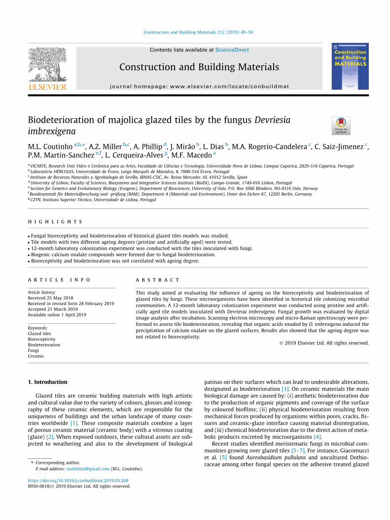
Construction and Building Materials 212 (2019) 49–56
Contents lists available at ScienceDirect
Construction and Building Materials
journal homepage: www.elsevier .com/locate /conbui ldmat
Biodeterioration of majolica glazed tiles by the fungus Devriesiaimbrexigena
https://doi.org/10.1016/j.conbuildmat.2019.03.2680950-0618/� 2019 Elsevier Ltd. All rights reserved.
⇑ Corresponding author.E-mail address: [email protected] (M.L. Coutinho).
M.L. Coutinho a,b,⇑, A.Z. Miller b,c, A. Phillip d, J. Mirão b, L. Dias b, M.A. Rogerio-Candelera c, C. Saiz-Jimenez c,P.M. Martin-Sanchez e,f, L. Cerqueira-Alves g, M.F. Macedo a
aVICARTE, Research Unit Vidro e Cerâmica para as Artes, Faculdade de Ciências e Tecnologia, Universidade Nova de Lisboa, Campus Caparica, 2829-516 Caparica, Portugalb Laboratório HÉRCULES, Universidade de Évora, Largo Marquês de Marialva, 8, 7000-554 Évora, Portugalc Instituto de Recursos Naturales y Agrobiologia de Sevilla, IRNAS-CSIC, Av. Reina Mercedes 10, 41012 Sevilla, SpaindUniversity of Lisbon, Faculty of Sciences, Biosystems and Integrative Sciences Institute (BioISI), Campo Grande, 1749-016 Lisbon, Portugale Section for Genetics and Evolutionary Biology (Evogene), Department of Biosciences, University of Oslo, P.O. Box 1066 Blindern, NO-0316 Oslo, NorwayfBundesanstalt für Materialforschung und -prüfung (BAM), Department 4 (Materials and Environment), Unter den Eichen 87, 12205 Berlin, GermanygC2TN, Instituto Superior Técnico, Universidade de Lisboa, Portugal
h i g h l i g h t s
� Fungal bioreceptivity and biodeterioration of historical glazed tiles models was studied.� Tile models with two different ageing degrees (pristine and artificially aged) were tested.� 12-month laboratory colonization experiment was conducted with the tiles inoculated with fungi.� Biogenic calcium oxalate compounds were formed due to fungal biodeterioration.� Bioreceptivity and biodeterioration was not correlated with ageing degree.
a r t i c l e i n f o
Article history:Received 25 May 2018Received in revised form 28 February 2019Accepted 21 March 2019Available online 1 April 2019
Keywords:Glazed tilesBioreceptivityBiodeteriorationFungiCeramic
a b s t r a c t
This study aimed at evaluating the influence of ageing on the bioreceptivity and biodeterioration ofglazed tiles by fungi. These microorganisms have been identified in historical tile colonizing microbialcommunities. A 12-month laboratory colonization experiment was conducted using pristine and artifi-cially aged tile models inoculated with Devriesia imbrexigena. Fungal growth was evaluated by digitalimage analysis after incubation. Scanning electron microscopy and micro-Raman spectroscopy were per-formed to assess tile biodeterioration, revealing that organic acids exuded by D. imbrexigena induced theprecipitation of calcium oxalate on the glazed surfaces. Results also showed that the ageing degree wasnot related to bioreceptivity.
� 2019 Elsevier Ltd. All rights reserved.
1. Introduction
Glazed tiles are ceramic building materials with high artisticand cultural value due to the variety of colours, glosses and iconog-raphy of these ceramic elements, which are responsible for theuniqueness of buildings and the urban landscape of many coun-tries worldwide [1]. These composite materials combine a layerof porous ceramic material (ceramic body) with a vitreous coating(glaze) [2]. When exposed outdoors, these cultural assets are sub-jected to weathering and also to the development of biological
patinas on their surfaces which can lead to undesirable alterations,designated as biodeterioration [3]. On ceramic materials the mainbiological damage are caused by: (i) aesthetic biodeterioration dueto the production of organic pigments and coverage of the surfaceby coloured biofilms; (ii) physical biodeterioration resulting frommechanical forces produced by organisms within pores, cracks, fis-sures and ceramic-glaze interface causing material disintegration,and (iii) chemical biodeterioration due to the direct action of meta-bolic products excreted by microorganisms [4].
Recent studies identified meristematic fungi in microbial com-munities growing over glazed tiles [5–7]. For instance, Giacomucciet al. [5] found Aureobasidium pullulans and uncultured Dothio-raceae among other fungal species on the adhesive treated glazed
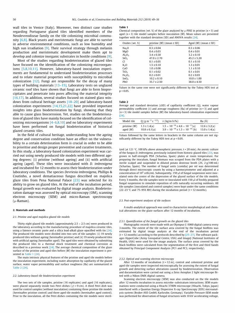
Table 1Chemical composition (wt. %) of the glaze analyzed by m-PIXE in pristine (n = 3) andaged (n = 3) tile model samples before inoculation (Bf). Mean values are presentedtogether with the standard deviation (SD) and ANOVA results [24].
Oxides (wt. %) pristine (Bf) (mean ± SD) Aged (Bf) (mean ± SD)
Na2O 0.3 ± 0.04 0.3 ± 0.06MgO 0.4 ± 0.03 0.4 ± 0.01Al2O3 3.4 ± 0.30 3.2 ± 0.10SiO2 47.0 ± 2.70 45.2 ± 5.20Cl 0.1 ± 0.05 0.1 ± 0.10K2O 1.5 ± 0.10 1.3 ± 0.01CaO 1.0 ± 0.08 1.1 ± 0.10TiO2 0.1 ± 0.02 0.1 ± 0.01Fe2O3 0.2 ± 0.01 0.2 ± 0.03SnO2 10.2 ± 0.10 10.0 ± 1.00PbO 35.7 ± 2.50 38.0 ± 4.50
Values in the same row were not significantly different by the Tukey HDS test atp < 0.05.
Table 2Average and standard deviation (±SD) of capillarity coefficient (Q), water vapourpermeability coefficient (d) and average roughness (Ra) of pristine (n = 3) and aged(n = 3) tile model samples before (Bf) the laboratory-based colonization experiment[24].
Model tile Q (g m�2 s�1/2) d (kg/m h Pa) Ra (Å)
pristine (Bf) 1.3 ± 1.4(a) 1.5 � 10�10 ± 4.6 � 10�11(a) 12.6 ± 3.2(a)aged (Bf) 10.8 ± 6.1(a) 3.9 � 10�10 ± 1.3 � 10�10 (b) 13.0 ± 1.4(a)
Values followed by the same letters in brackets in the same column are not sig-nificantly different by the Tukey HDS test at p < 0.05.
50 M.L. Coutinho et al. / Construction and Building Materials 212 (2019) 49–56
wall tiles in Venice (Italy). Moreover, two distinct case studiesregarding Portuguese glazed tiles identified members of theNeodevreodiseae family on the tile colonizing microbial commu-nity [6,8]. Black yeasts and meristematic fungi are able to survivein adverse environmental conditions, such as low humidity andhigh sun irradiation [9]. Their survival strategy through melaninproduction and meristematic development make them apt todevelop and colonize inorganic substrates in hostile conditions [9].
Most of the studies regarding biodeterioration of glazed tileshave focused on the identification of the colonizing microorgan-isms [5,8,10,11]. However, laboratory-based inoculation experi-ments are fundamental to understand biodeterioration processesand to relate material properties with susceptibility to microbialcolonization [12]. Fungi are responsible for the decay of manytypes of building materials [13–15]. Laboratory tests on unglazedceramic roof tiles have shown that fungi are able to form biopre-cipitates and penetrate into pores affecting the material integrity[16,17]. In addition, several studies focused on stained glass win-dows from cultural heritage assets [18–20] and laboratory-basedcolonization experiments [14,15,21,22] have provided importantinsights into glass biodeterioration by fungi, showing that theyable to cause glass biocorrosion. Yet, studies on the biodeteriora-tion of glazed tiles have mainly focused on the identification of col-onizing microorganisms [4–6,8,10] and no laboratory experimentshave been performed on fungal biodeterioration of historicalglazed ceramic tiles.
In the field of cultural heritage, understanding how the ageingdegree and conservation condition have an effect on the vulnera-bility to a certain deterioration form is crucial in order to be ableto prioritise and design proper preventive and curative treatments.In this study, a laboratory-based colonization experiment was car-ried out using newly produced tile models with two different age-ing degrees: (i) pristine (without ageing) and (ii) with artificialageing (aged). These tiles were inoculated with D. imbrexigenaand incubated for 12 months to assess their biodeterioration underlaboratory conditions. The species Devriesia imbrexigena, Phillips &Coutinho, a novel dematiaceous fungus described on majolicaglaze tiles from Pena National Palace [23] was selected for itsability to grow on glazed tiles. At the end of the incubation period,fungal growth was evaluated by digital image analysis. Biodeterio-ration damage was assessed by optical microscopy (OM), scanningelectron microscopy (SEM) and micro-Raman spectroscopy(m-Raman).
2. Materials and methods
2.1. Pristine and aged majolica glazed tile models
Thirty eight glazed tile models (approximately 2.5 � 2.5 cm) were produced inthe laboratory according to the manufacturing procedure of majolica ceramic tiles,using a faience ceramic paste and a silica lead-alkali glaze opacified with tin [24].The produced tile models were divided into two sets of tile samples: (i) 19 newlyproduced tiles without ageing (hereinafter pristine) and (ii) 19 newly produced tileswith artificial ageing (hereinafter aged). The aged tiles were obtained by submittingthe produced tiles to a thermal shock treatment and chemical corrosion asdescribed in a previous work [24]. The average chemical composition of the glazesurface of the pristine and aged tiles before (Bf) the inoculation experiment is pre-sented in Table 1 [24].
The main intrinsic physical features of the pristine and aged tile models beforethe inoculation experiment, including water absorption by capillarity of the glazedsurface, water vapor permeability and surface roughness (Ra) are summarized inTable 2 [24].
2.2. Laboratory-based tile biodeterioration experiment
The two sets of tile samples, pristine (16 replicates) and aged (16 replicates),were placed separately inside two Petri dishes (£ = 9 cm). A third Petri dish wasused for control samples (without inoculation) containing three pristine tile models(hereinafter pristine control) and three aged tile models (hereinafter aged control).Prior to the inoculation, all the Petri dishes containing the tile models were steril-
ized (at 121 �C, 100 kPa above atmospheric pressure, t = 20 min). An axenic cultureof the fungus D. imbrexigena, previously isolated from historic glazed tiles [23], wasgrown in half-strength PDA (Scharlau, Spain) plates at room temperature. Forpreparing the inoculum, fungal biomass was scraped from the PDA plates with asterile scalpel and suspended in diluted potato dextrose broth (2%, 2 g/100 mL)(Scharlau, Spain). The number of fungal units (conidia) per ml was determinedthrough direct microscopic counts by using a haemocytometer and adjusted at aconcentration of 105 cells/mL. Subsequently, 150 ml of fungal suspension were inoc-ulated onto the centre of the depression of the glazed surface of the tile models.Every 3 months, the tile samples were re-inoculated with 150 ml of freshly preparedfungal suspension to simulate reposition of cells naturally occurring outdoors. Alltile samples (inoculated and control samples) were kept under the same conditions(22–23 �C and 75–95% RH) during the incubation period (t = 12 months).
2.3. Post-experiment analyses of tile surfaces
A multi-analytical approach was used to characterize morphological and chem-ical alterations on the glaze surfaces after 12 months of incubation.
2.3.1. Quantification of the fungal growth on the glazed tilesPhotographic records were made with an Olympus C-5060 digital camera every
3 months. The extent of the tile surface area covered by the fungal biofilms wasestimated by digital image analysis at the end of the incubation period(t = 12 months) according to the protocols described by [25–27]. The software pack-ages HyperCube (Army Geospatial Centre, USA) and ImageJ (National Institutes ofHealth, USA) were used for the image analysis. The surface areas covered by theblack biofilms were calculated from the segmentation of the first and third bandsobtained by Principal Component Analysis (PC1 and PC3, respectively).
2.3.2. Optical and scanning electron microscopyAfter 12 months of incubation (t = 12 m), control and colonized pristine and
aged tile samples were inspected microscopically for assessing the extent of fungalgrowth and detecting surface alterations caused by biodeterioration. Observationand documentation were carried out using a Zeiss Axioplan 2 light microscope fit-ted with a Nikon DMX digital camera.
Scanning electron microscopy (SEM) was also conducted on the tile modelsafter 12 months incubation to assess microbe–substratum interactions. SEM exam-inations were conducted using a Hitachi 3700N microscope (Hitachi, Tokyo, Japan)interfaced with a Quantax Energy Dispersive X-ray Spectroscopy (EDS) microanal-ysis system (Bruker AXS GmbH, Karlsruhe, Germany). Variable Pressure-SEM modewas performed for observation of fungal structures with 10 kV accelerating voltage,
M.L. Coutinho et al. / Construction and Building Materials 212 (2019) 49–56 51
10 mm working distance and 30 Pa. The operating conditions for EDS analysis were20 kV accelerating voltage, 10–12 mm working distance and 120 mA emissioncurrent.
In addition, the colonized tile samples were observed after cleaning with a cot-ton swab embedded in a 1:1 water-ethanol solution to assess corrosion patterns,crystalline compounds and leached elements. For comparison purposes, the non-inoculated control samples were also analysed. Prior to SEM observations, the tilesamples were directly mounted on sample stubs and sputter-coated withgold/palladium.
2.3.3. Micro-Raman spectroscopyMicro-Raman spectroscopy (l-Raman) was carried out for characterizing crys-
tals formed on the surface of the glaze using a Labram 300 Jobin Yvon spectrometer,equipped with a He-Ne laser of 17mWpower operating at 632.8 nm and also a solidstate external laser of 50 mW power operating at 514.5 nm. Spectra were recordedas an extended scan. The laser beam was focused either with a 10�, 50� or a 100�Olympus objective lens. The laser power at the surface of the samples varied withthe aid of a set of neutral density filters (optical densities 0.3, 0.6, 1 and 2). For com-parison purposes, spectra of whellite (CaC2O4�H2O) (Fluka) were also obtained andanalyzed.
3. Results and discussion
3.1. Fungal growth
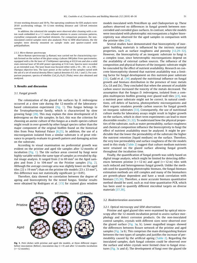
The colonization of the glazed tile surfaces by D. imbrexigenaoccurred at a slow rate during the 12 months of the laboratory-based colonization experiment (Fig. 1). This fungus belongs tothe Teratosphaeriaceae family, which is characterized by slowgrowing fungi [28]. This may explain the slow development of D.imbrexigena on the tile samples. In fact, this was the criterion forchoosing an axenic culture of this fungus as a multi-species culturemight result in over-growth by other fungal species rather than themajor component of the original biofilm found on the historicaltiles from Pena National Palace [8,23]. In addition, the use of amicroorganism isolated from a similar substrate is of great rele-vance to properly evaluate its growth pattern and damaging actionon the substrate.
According to visual examination no preferential growth wasevident on the pristine and aged tile samples after 12 months ofincubation (Fig. 1). The tile surface areas covered by fungi werehighly variable within the same type of samples as revealed by dig-ital image analysis. It ranged from 2 to 69 mm2 on the Aged sam-ples and from 2 to 104 mm2 on the Pristine samples (Fig. 2).Although the average coverage area was slightly lower on the agedtiles (22 ± 19 mm2) than on the pristine tile models (23 ± 33 mm2),this difference was not statistically significant (p > 0.05).
Therefore, data showed no correlation between the degree ofageing and bioreceptivity for the tested fungus. Similar resultswere obtained by Rodrigues et al. [15] for stained glass window
Fig. 1. Petri dishes with pristine and aged tile models, at three different stages:before inoculation (Before), inoculation day (t = 0) and after 12 months incubation(t = 12 months).
models inoculated with Penicillium sp. and Cladosporium sp. Theseauthors observed no differences in fungal growth between non-corroded and corroded glass surfaces. In contrast, when tile modelswere inoculated with phototrophic microorganisms a higher biore-ceptivity was observed for the aged samples in comparison withthe pristine tiles [24].
Several studies have demonstrated that bioreceptivity of inor-ganic building materials is influenced by the intrinsic materialproperties, such as surface roughness and porosity [12,29–32].However, the bioreceptivity of an inorganic substrate to fungi isa complex issue, since heterotrophic microorganisms depend onthe availability of external carbon sources. The influence of thecomposition and physical features of the inorganic substrate mightbe concealed by the effect of nutrient availability. Research on con-crete bioreceptivity showed that nutrient availability was a limit-ing factor for fungal development on this nutrient-poor substrate[33]. Gadd et al. [34] analysed the nutritional influence on fungalgrowth and biomass distribution in the presence of toxic metals(Cu, Cd and Zn). They concluded that when the amount of availablecarbon source increased the toxicity of the metals decreased. Theassumption that the fungus D. imbrexigena, isolated from a com-plex multispecies biofilm growing over glazed wall tiles [23], wasa nutrient poor substrate might not be correct. In natural condi-tions, cell debris of bacteria, photosynthetic microorganisms andtheir organic exudates provide carbon sources for fungal growthon inorganic substrates [33]. Consequently, the use of nutritionalculture media for laboratory assays might result in higher growthon the surfaces, which in short term experiments can lead to morediscernible results [31,32]. To understand how the physical proper-ties of the substrate, such as water permeability, surface roughnessand chemical composition influence the bioreceptivity to fungi theeffect of nutrient availability must be analysed. It might be pre-dictable that the lower the permeability of the substrate the higherthe nutrient retention (liquid medium) on the surface. Therefore,the very low permeability and surface roughness of the tile modelsused in this study (Table 2) suggest that culture medium nutrientswere retained on the glazed surface allowing fungal growththroughout the incubation time.
Finally, the quantification of fungal growth was solely based ondigital image analysis, which might be limited for detecting differ-ences between pristine (t = 12 m) and aged (t = 12 m) tiles withsuch reduced and heterogeneous fungal growth. Unlike the meth-ods used for quantifying phototrophic biomass, the fungal-biomassestimation methods are still complex and many of the biomarkersare growth-phase dependent and have a weak correlation withbiomass [35,36]. Therefore, a more accurate biomass quantitativemethod should be used, such as real-time quantitative PCR, whichhas been used to quantify different microbial targets on diversematerials [37,38].
3.2. Biodeterioration assessment
3.2.1. Optical microscopy and SEM observationsPristine and aged glazed tiles were examined by optical micro-
scopy after the 12-month incubation period to assess surface mor-phology and detect corrosion products. On the non-inoculatedcontrol samples, crystals with different sizes were observed overthe glazed surface (Fig. 3a, b). Lower magnified images showedthe differences between fissure network of the pristine and agedsamples (Fig. 3a b). This comprises the main distinguishing featurebetween the two types of samples and justifies the increase of per-meability caused by the artificial ageing (Table 2). Regarding theinoculated samples, dark fungal colonies could be observed overthe surface and white crystals were formed close to fungal struc-tures (Fig. 3c and d). The development of fungi over the glazed tile
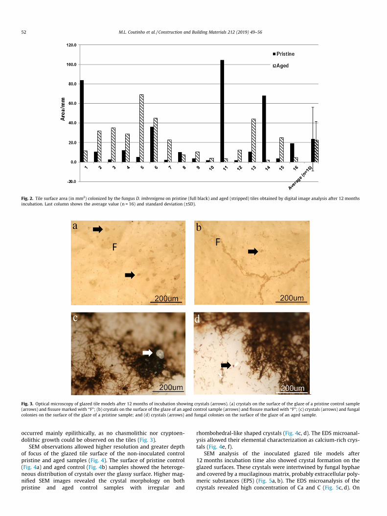
Fig. 2. Tile surface area (in mm2) colonized by the fungus D. imbrexigena on pristine (full black) and aged (stripped) tiles obtained by digital image analysis after 12 monthsincubation. Last column shows the average value (n = 16) and standard deviation (±SD).
Fig. 3. Optical microscopy of glazed tile models after 12 months of incubation showing crystals (arrows). (a) crystals on the surface of the glaze of a pristine control sample(arrows) and fissure marked with ‘‘F”; (b) crystals on the surface of the glaze of an aged control sample (arrows) and fissure marked with ‘‘F”; (c) crystals (arrows) and fungalcolonies on the surface of the glaze of a pristine sample; and (d) crystals (arrows) and fungal colonies on the surface of the glaze of an aged sample.
52 M.L. Coutinho et al. / Construction and Building Materials 212 (2019) 49–56
occurred mainly epilithically, as no chasmolithic nor cryptoen-dolithic growth could be observed on the tiles (Fig. 3).
SEM observations allowed higher resolution and greater depthof focus of the glazed tile surface of the non-inoculated controlpristine and aged samples (Fig. 4). The surface of pristine control(Fig. 4a) and aged control (Fig. 4b) samples showed the heteroge-neous distribution of crystals over the glassy surface. Higher mag-nified SEM images revealed the crystal morphology on bothpristine and aged control samples with irregular and
rhombohedral-like shaped crystals (Fig. 4c, d). The EDS microanal-ysis allowed their elemental characterization as calcium-rich crys-tals (Fig. 4e, f).
SEM analysis of the inoculated glazed tile models after12 months incubation time also showed crystal formation on theglazed surfaces. These crystals were intertwined by fungal hyphaeand covered by a mucilaginous matrix, probably extracellular poly-meric substances (EPS) (Fig. 5a, b). The EDS microanalysis of thecrystals revealed high concentration of Ca and C (Fig. 5c, d). On
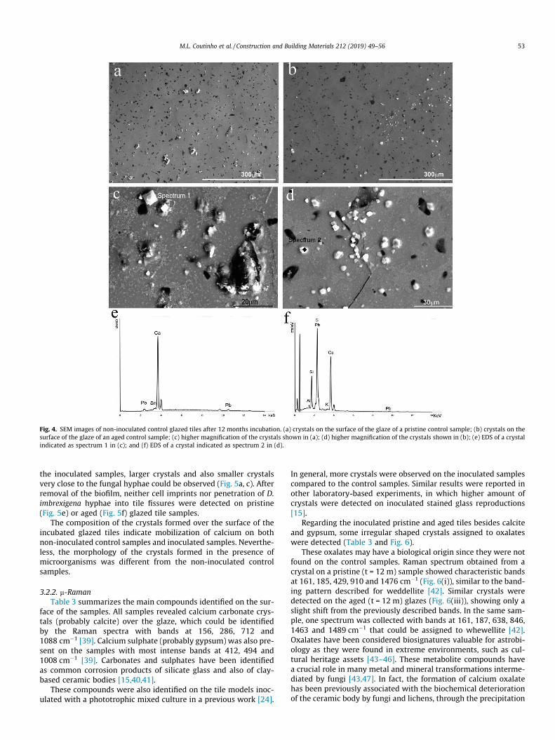
Fig. 4. SEM images of non-inoculated control glazed tiles after 12 months incubation. (a) crystals on the surface of the glaze of a pristine control sample; (b) crystals on thesurface of the glaze of an aged control sample; (c) higher magnification of the crystals shown in (a); (d) higher magnification of the crystals shown in (b); (e) EDS of a crystalindicated as spectrum 1 in (c); and (f) EDS of a crystal indicated as spectrum 2 in (d).
M.L. Coutinho et al. / Construction and Building Materials 212 (2019) 49–56 53
the inoculated samples, larger crystals and also smaller crystalsvery close to the fungal hyphae could be observed (Fig. 5a, c). Afterremoval of the biofilm, neither cell imprints nor penetration of D.imbrexigena hyphae into tile fissures were detected on pristine(Fig. 5e) or aged (Fig. 5f) glazed tile samples.
The composition of the crystals formed over the surface of theincubated glazed tiles indicate mobilization of calcium on bothnon-inoculated control samples and inoculated samples. Neverthe-less, the morphology of the crystals formed in the presence ofmicroorganisms was different from the non-inoculated controlsamples.
3.2.2. m-RamanTable 3 summarizes the main compounds identified on the sur-
face of the samples. All samples revealed calcium carbonate crys-tals (probably calcite) over the glaze, which could be identifiedby the Raman spectra with bands at 156, 286, 712 and1088 cm�1 [39]. Calcium sulphate (probably gypsum) was also pre-sent on the samples with most intense bands at 412, 494 and1008 cm�1 [39]. Carbonates and sulphates have been identifiedas common corrosion products of silicate glass and also of clay-based ceramic bodies [15,40,41].
These compounds were also identified on the tile models inoc-ulated with a phototrophic mixed culture in a previous work [24].
In general, more crystals were observed on the inoculated samplescompared to the control samples. Similar results were reported inother laboratory-based experiments, in which higher amount ofcrystals were detected on inoculated stained glass reproductions[15].
Regarding the inoculated pristine and aged tiles besides calciteand gypsum, some irregular shaped crystals assigned to oxalateswere detected (Table 3 and Fig. 6).
These oxalates may have a biological origin since they were notfound on the control samples. Raman spectrum obtained from acrystal on a pristine (t = 12 m) sample showed characteristic bandsat 161, 185, 429, 910 and 1476 cm�1 (Fig. 6(i)), similar to the band-ing pattern described for weddellite [42]. Similar crystals weredetected on the aged (t = 12 m) glazes (Fig. 6(iii)), showing only aslight shift from the previously described bands. In the same sam-ple, one spectrum was collected with bands at 161, 187, 638, 846,1463 and 1489 cm�1 that could be assigned to whewellite [42].Oxalates have been considered biosignatures valuable for astrobi-ology as they were found in extreme environments, such as cul-tural heritage assets [43–46]. These metabolite compounds havea crucial role in many metal and mineral transformations interme-diated by fungi [43,47]. In fact, the formation of calcium oxalatehas been previously associated with the biochemical deteriorationof the ceramic body by fungi and lichens, through the precipitation
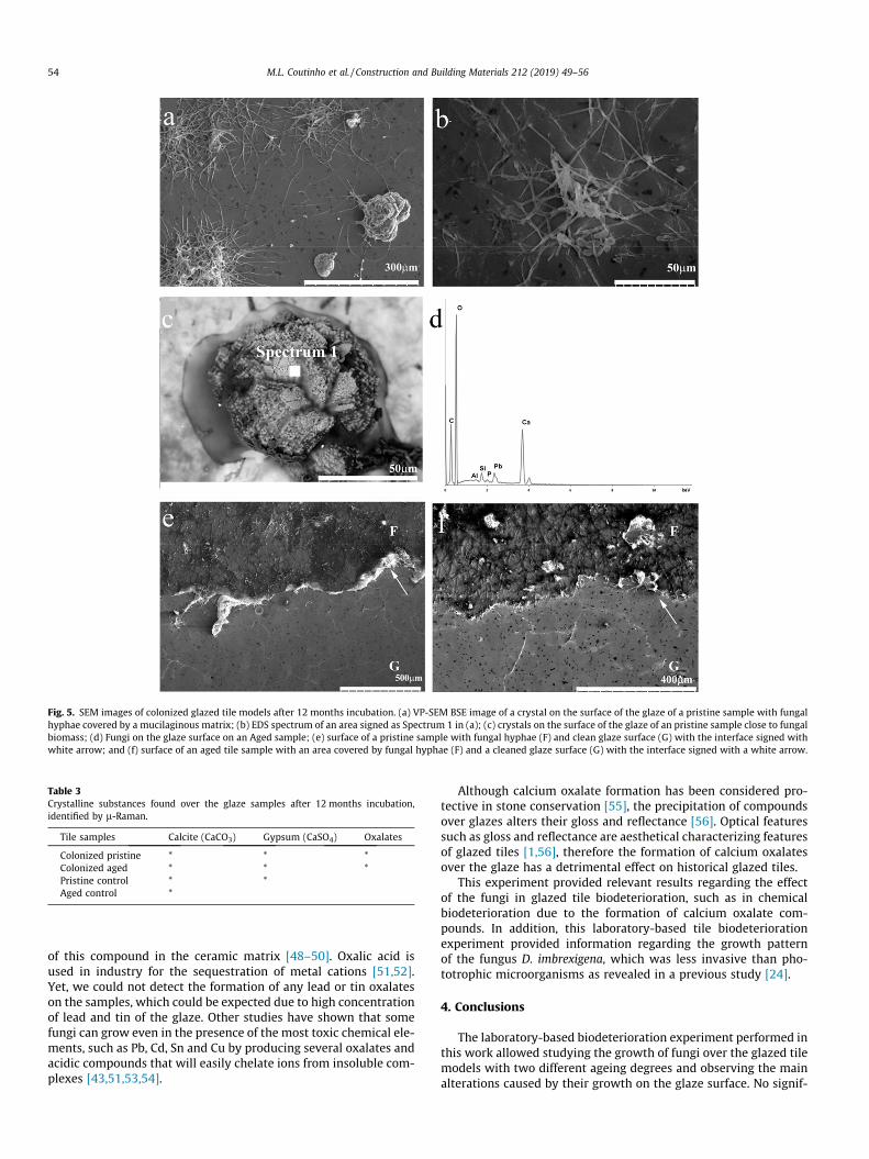
Fig. 5. SEM images of colonized glazed tile models after 12 months incubation. (a) VP-SEM BSE image of a crystal on the surface of the glaze of a pristine sample with fungalhyphae covered by a mucilaginous matrix; (b) EDS spectrum of an area signed as Spectrum 1 in (a); (c) crystals on the surface of the glaze of an pristine sample close to fungalbiomass; (d) Fungi on the glaze surface on an Aged sample; (e) surface of a pristine sample with fungal hyphae (F) and clean glaze surface (G) with the interface signed withwhite arrow; and (f) surface of an aged tile sample with an area covered by fungal hyphae (F) and a cleaned glaze surface (G) with the interface signed with a white arrow.
Table 3Crystalline substances found over the glaze samples after 12 months incubation,identified by l-Raman.
Tile samples Calcite (CaCO3) Gypsum (CaSO4) Oxalates
Colonized pristine * * *Colonized aged * * *Pristine control * *Aged control *
54 M.L. Coutinho et al. / Construction and Building Materials 212 (2019) 49–56
of this compound in the ceramic matrix [48–50]. Oxalic acid isused in industry for the sequestration of metal cations [51,52].Yet, we could not detect the formation of any lead or tin oxalateson the samples, which could be expected due to high concentrationof lead and tin of the glaze. Other studies have shown that somefungi can grow even in the presence of the most toxic chemical ele-ments, such as Pb, Cd, Sn and Cu by producing several oxalates andacidic compounds that will easily chelate ions from insoluble com-plexes [43,51,53,54].
Although calcium oxalate formation has been considered pro-tective in stone conservation [55], the precipitation of compoundsover glazes alters their gloss and reflectance [56]. Optical featuressuch as gloss and reflectance are aesthetical characterizing featuresof glazed tiles [1,56], therefore the formation of calcium oxalatesover the glaze has a detrimental effect on historical glazed tiles.
This experiment provided relevant results regarding the effectof the fungi in glazed tile biodeterioration, such as in chemicalbiodeterioration due to the formation of calcium oxalate com-pounds. In addition, this laboratory-based tile biodeteriorationexperiment provided information regarding the growth patternof the fungus D. imbrexigena, which was less invasive than pho-totrophic microorganisms as revealed in a previous study [24].
4. Conclusions
The laboratory-based biodeterioration experiment performed inthis work allowed studying the growth of fungi over the glazed tilemodels with two different ageing degrees and observing the mainalterations caused by their growth on the glaze surface. No signif-
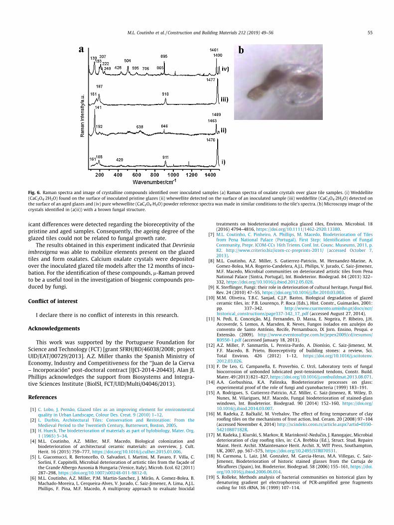
Fig. 6. Raman spectra and image of crystalline compounds identified over inoculated samples (a) Raman spectra of oxalate crystals over glaze tile samples. (i) Weddellite(CaC2O4�2H2O) found on the surface of inoculated pristine glazes (ii) whewellite detected on the surface of an inoculated sample (iii) weddellite (CaC2O4�2H2O) detected onthe surface of an aged glazes and (iv) pure whewellite (CaC2O4�H2O) powder reference spectra was made in similar conditions to the tile’s spectra. (b) Microscopy image of thecrystals identified in (a(ii)) with a brown fungal structure.
M.L. Coutinho et al. / Construction and Building Materials 212 (2019) 49–56 55
icant differences were detected regarding the bioreceptivity of thepristine and aged samples. Consequently, the ageing degree of theglazed tiles could not be related to fungal growth rate.
The results obtained in this experiment indicated that Devriesiaimbrexigena was able to mobilize elements present on the glazedtiles and form oxalates. Calcium oxalate crystals were depositedover the inoculated glazed tile models after the 12 months of incu-bation. For the identification of these compounds, m-Raman provedto be a useful tool in the investigation of biogenic compounds pro-duced by fungi.
Conflict of interest
I declare there is no conflict of interests in this research.
Acknowledgements
This work was supported by the Portuguese Foundation forScience and Technology (FCT) [grant SFRH/BD/46038/2008; projectUID/EAT/00729/2013]. A.Z. Miller thanks the Spanish Ministry ofEconomy, Industry and Competitiveness for the ‘‘Juan de la Cierva– Incorporación” post-doctoral contract [IJCI-2014-20443]. Alan JLPhillips acknowledges the support from Biosystems and Integra-tive Sciences Institute (BioISI, FCT/UID/Multi/04046/2013).
References
[1] C. Lobo, J. Pernão, Glazed tiles as an improving element for environmentalquality in Urban Landscape, Colour Des. Creat. 9 (2010) 1–12.
[2] L. Durbin, Architectural Tiles: Conservation and Restoration: From theMedieval Period to the Twentieth Century, Butterwort, Boston, 2005.
[3] H. Hueck, The biodeterioration of materials as part of hylobiology, Mater. Org.1 (1965) 5–34.
[4] M.L. Coutinho, A.Z. Miller, M.F. Macedo, Biological colonization andbiodeterioration of architectural ceramic materials: an overview, J. Cult.Herit. 16 (2015) 759–777, https://doi.org/10.1016/j.culher.2015.01.006.
[5] L. Giacomucci, R. Bertoncello, O. Salvadori, I. Martini, M. Favaro, F. Villa, C.Sorlini, F. Cappitelli, Microbial deterioration of artistic tiles from the façade ofthe Grande Albergo Ausonia & Hungaria (Venice, Italy), Microb. Ecol. 62 (2011)287–298, https://doi.org/10.1007/s00248-011-9812-0.
[6] M.L. Coutinho, A.Z. Miller, P.M. Martin-Sanchez, J. Mirão, A. Gomez-Bolea, B.Machado-Moreira, L. Cerqueira-Alves, V. Jurado, C. Saiz-Jimenez, A. Lima, A.J.L.Phillips, F. Pina, M.F. Macedo, A multiproxy approach to evaluate biocidal
treatments on biodeteriorated majolica glazed tiles, Environ. Microbiol. 18(2016) 4794–4816, https://doi.org/10.1111/1462-2920.13380.
[7] M.L. Coutinho, C. Pinheiro, A. Phillips, M. Macedo, Biodeterioration of Tilesfrom Pena National Palace (Portugal). First Step: Identification of FungalCommunity, Prepr. ICOM-CCs 16th Trienn. Conf. Int. Counc. Museums, 2011, p.82. http://www.criterio.biz/icom-cc-preprints-2011/ (accessed October 7,2013).
[8] M.L. Coutinho, A.Z. Miller, S. Gutierrez-Patricio, M. Hernandez-Marine, A.Gomez-Bolea, M.A. Rogerio-Candelera, A.J.L. Philips, V. Jurado, C. Saiz-Jimenez,M.F. Macedo, Microbial communities on deteriorated artistic tiles from PenaNational Palace (Sintra, Portugal), Int. Biodeterior. Biodegrad. 84 (2013) 322–332, https://doi.org/10.1016/j.ibiod.2012.05.028.
[9] K. Sterflinger, Fungi: their role in deterioration of cultural heritage, Fungal Biol.Rev. 24 (2010) 47–55, https://doi.org/10.1016/j.fbr.2010.03.003.
[10] M.M. Oliveira, T.B.C. Sanjad, C.J.P. Bastos, Biological degradation of glazedceramic tiles, in: P.B. Lourenço, P. Roca (Eds.), Hist. Constr., Guimarães, 2001:pp. 337–342. http://www.csarmento.uminho.pt/docs/ncr/historical_constructions/page337-342_17_pdf (accessed August 27, 2014).
[11] N. Pedi, E. Conceição, M.J. Fernandes, D. Massa, E. Nogeira, P. Ribeiro, J.H.Arcoverde, S. Lemos, A. Marsden, R. Neves, Fungos isolados em azulejos doconvento de Santo António, Recife, Pernambuco, IX Jorn. Ensino, Pesqui. eExtensão. (2009). http://www.eventosufrpe.com.br/jepex2009/cd/resumos/R0550-1.pdf (accessed January 18, 2013).
[12] A.Z. Miller, P. Sanmartín, L. Pereira-Pardo, A. Dionísio, C. Saiz-Jimenez, M.F.F. Macedo, B. Prieto, Bioreceptivity of building stones: a review, Sci.Total Environ. 426 (2012) 1–12, https://doi.org/10.1016/j.scitotenv.2012.03.026.
[13] F. De Leo, G. Campanella, E. Proverbio, C. Urzì, Laboratory tests of fungalbiocorrosion of unbonded lubricated post-tensioned tendons, Constr. Build.Mater. 49 (2013) 821–827, https://doi.org/10.1016/j.conbuildmat.2013.08.071.
[14] A.A. Gorbushina, K.A. Palinska, Biodeteriorative processes on glass:experimental proof of the role of fungi and cyanobacteria (1999) 183–191.
[15] A. Rodrigues, S. Gutierrez-Patricio, A.Z. Miller, C. Saiz-Jimenez, R. Wiley, D.Nunes, M. Vilarigues, M.F. Macedo, Fungal biodeterioration of stained-glasswindows, Int. Biodeterior. Biodegrad. 90 (2014) 152–160, https://doi.org/10.1016/j.ibiod.2014.03.007.
[16] M. Radeka, Z. Backalic, M. Vrebalov, The effect of firing temperature of clayroofing tiles on the mechanisms of frost action, Ind. Ceram. 20 (2008) 97–104(accessed November 4, 2014) http://scindeks.ceon.rs/article.aspx?artid=0350-54210807182R.
[17] M. Radeka, J. Kiurski, S. Markov, R. Marinkovic-Neducin, J. Ranogajec, Microbialdeterioration of clay roofing tiles, in: C.A. Brebbia (Ed.), Struct. Stud. RepairsMaint. Herit. Archit. XMaintenance Herit. Archit. X, WIT Press, Southampton,UK, 2007, pp. 567–575, https://doi.org/10.2495/STR070531.
[18] N. Carmona, L. Laiz, J.M. Gonzalez, M. Garcia-Heras, M.A. Villegas, C. Saiz-Jimenez, Biodeterioration of historic stained glasses from the Cartuja deMiraflores (Spain), Int. Biodeterior. Biodegrad. 58 (2006) 155–161, https://doi.org/10.1016/j.ibiod.2006.06.014.
[19] S. Rolleke, Methods analysis of bacterial communities on historical glass bydenaturing gradient gel electrophoresis of PCR-amplified gene fragmentscoding for 16S rRNA, 36 (1999) 107–114.
56 M.L. Coutinho et al. / Construction and Building Materials 212 (2019) 49–56
[20] C. Schabereiter-Gurtner, G. Piñar, W. Lubitz, S. Rölleke, An advanced molecularstrategy to identify bacterial communities on art objects, J. Microbiol. Methods45 (2001) 77–87. http://www.ncbi.nlm.nih.gov/pubmed/11311392.
[21] M. Bartosik, Z. Zakowska, K. Cedzinska, K. Rozniakowski, Biodeterioration ofoptical glass induced by lubricants used in optical instruments technology, Pol.J. Microbiol. 59 (2010) 295–300. http://www.ncbi.nlm.nih.gov/pubmed/21466048.
[22] M.A. Shirakawa, V.M. John, A. Mocelin, R. Zilles, S.H. Toma, K. Araki, H.E. Toma,A.C. Thomaz, C.C. Gaylarde, Effect of silver nanoparticle and TiO2 coatings onbiofilm formation on four types of modern glass, Int. Biodeterior. Biodegrad.108 (2016) 175–180, https://doi.org/10.1016/j.ibiod.2015.12.025.
[23] P.W. Crous, R.G. Shivas, M.J. Wingfield, B.A. Summerell, A.Y. Rossman, J.L.Alves, G.C. Adams, R.W. Barreto, A. Bell, M.L. Coutinho, S.L. Flory, G. Gates, K.R.Grice, G.E.St.J. Hardy, N.M. Kleczewski, L. Lombard, C.M.O. Longa, G. Louis-Seize, F. Macedo, D.P. Mahoney, G. Maresi, P.M. Martin-Sanchez, L. Marvanová,A.M. Minnis, L.N. Morgado, M.E. Noordeloos, A.J.L. Phillips, W. Quaedvlieg, P.G.Ryan, C. Saiz-Jimenez, K.A. Seifert, W.J. Swart, Y.P. Tan, J.B. Tanney, P.Q. Thu, S.I.R. Videira, D.M. Walker, J.Z. Groenewald, Fungal Planet description sheets:128–153, Persoonia 29 (2012) 146–201, https://doi.org/10.3767/003158512X661589.
[24] M.L. Coutinho, A.Z. Miller, M.A. Rogerio-Candelera, J. Mirão, L. Cerqueira Alves,J.P. Veiga, H. Águas, S. Pereira, A. Lyubchyk, M.F. Macedo, An integratedapproach for assessing the bioreceptivity of glazed tiles to phototrophicmicroorganisms, Biofouling 32 (2016) 243–259, https://doi.org/10.1080/08927014.2015.1135242.
[25] A.Z. Miller, M.Á. Rogerio-candelera, M.F. Macedo, C. Saiz, Microalgae asbiodeteriogens of stone cultural heritage: qualitative and quantitativeresearch by non-contact techniques, Microalgae Biotechnol. Microbiol.Energy (2011) 345–358. http://hdl.handle.net/10261/56993 (accessed May16, 2018).
[26] A.Z. Miller, M.A. Rogerio-Candelera, L. Laiz, J. Wierzchos, C. Ascaso, M.A.Sequeira-Braga, M. Hernández-Mariné, A. Maurício, A. Dionisio, M.F.F. Macedo,C. Saiz-Jimenez, M.A. Sequeira Braga, M. Hernández-Mariné, A. Maurício, A.Dionísio, M.F. Macedo, C. Saiz-Jimenez, Laboratory-induced endolithic growthin calcarenites: biodeteriorating potential assessment, Microb. Ecol. 60 (2010)55–68, https://doi.org/10.1007/s00248-010-9666-x.
[27] M.A. Rogerio-Candelera, V. Jurado, L. Laiz, C. Saiz-Jimenez, Laboratory andin situ assays of digital image analysis based protocols for biodeteriorated rockand mural paintings recording, J. Archaeol. Sci. 38 (2011) 2571–2578, https://doi.org/10.1016/j.jas.2011.04.020.
[28] E. Egidi, G.S. Hoog, D. Isola, S. Onofri, W. Quaedvlieg, M. Vries, G.J.M. Verkley, J.B. Stielow, L. Zucconi, L. Selbmann, Phylogeny and taxonomy of meristematicrock-inhabiting black fungi in the Dothideomycetes based on multi-locusphylogenies, Fungal Divers. 65 (2014) 127–165, https://doi.org/10.1007/s13225-013-0277-y.
[29] M. D’Orazio, G. Cursio, L. Graziani, L. Aquilanti, A. Osimani, F. Clementi, C.Yéprémian, V. Lariccia, S. Amoroso, Effects of water absorption and surfaceroughness on the bioreceptivity of ETICS compared to clay bricks, Build.Environ. 77 (2014) 20–28, https://doi.org/10.1016/j.buildenv.2014.03.018.
[30] M. Korkanç, A. Savran, Impact of the surface roughness of stones used inhistorical buildings on biodeterioration, Constr. Build. Mater. 80 (2015) 279–294, https://doi.org/10.1016/J.CONBUILDMAT.2015.01.073.
[31] M.F. Gazulla, E. Sánchez, J.M. González, M.C. Portillo, M. Orduña, Relationshipbetween certain ceramic roofing tile characteristics and biodeterioration, J.Eur. Ceram. Soc. 31 (2011) 2753–2761, https://doi.org/10.1016/j.jeurceramsoc.2011.07.023.
[32] V. Ferrándiz-Mas, T. Bond, Z. Zhang, J. Melchiorri, C.R. Cheeseman, Optimisingthe bioreceptivity of porous glass tiles based on colonization by the algaChlorella vulgaris, Sci. Total Environ. 563–564 (2016) 71–80, https://doi.org/10.1016/j.scitotenv.2016.04.023.
[33] D.J. Giannantonio, J.C. Kurth, K.E. Kurtis, P.A. Sobecky, Effects of concreteproperties and nutrients on fungal colonization and fouling, Int. Biodeterior.Biodegrad. 63 (2009) 252–259, https://doi.org/10.1016/j.ibiod.2008.10.002.
[34] G.M. Gadd, L. Ramsay, J.W. Crawford, K. Ritz, Nutritional influence on fungalcolony growth and biomass distribution in response to toxic metals, FEMSMicrobiol. Lett. 204 (2001) 311–316, https://doi.org/10.1016/S0378-1097(01)00399-8.
[35] M. Miller, A. Palojärvi, A. Rangger, M. Reeslev, A. Kjøller, A. Paloja, The use offluorogenic substrates to measure fungal presence and activity in soil the useof fluorogenic substrates to measure fungal presence and activity in soil, 64(1998).
[36] K. Suberkropp, M.O. Gessner, E. Chauvet, Comparison of ATP and ergosterol asindicators of fungal biomass associated with decomposing leaves in streams,
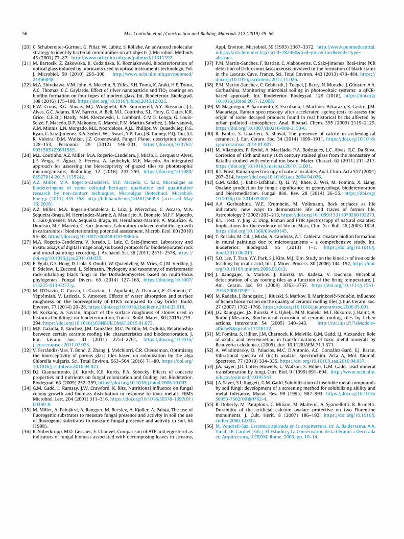
Appl. Environ. Microbiol. 59 (1993) 3367–3372. http://www.pubmedcentral.nih.gov/articlerender.fcgi?artid=182460&tool=pmcentrez&rendertype=abstract.
[37] P.M. Martin-Sanchez, F. Bastian, C. Alabouvette, C. Saiz-Jimenez, Real-time PCRdetection of Ochroconis lascauxensis involved in the formation of black stainsin the Lascaux Cave, France, Sci. Total Environ. 443 (2013) 478–484, https://doi.org/10.1016/j.scitotenv.2012.11.026.
[38] P.M. Martin-Sanchez, C. Gebhardt, J. Toepel, J. Barry, N. Munzke, J. Günster, A.A.Gorbushina, Monitoring microbial soiling in photovoltaic systems: a qPCR-based approach, Int. Biodeterior. Biodegrad. 129 (2018), https://doi.org/10.1016/j.ibiod.2017.12.008.
[39] M. Maguregui, A. Sarmiento, R. Escribano, I. Martinez-Arkarazo, K. Castro, J.M.Madariaga, Raman spectroscopy after accelerated ageing tests to assess theorigin of some decayed products found in real historical bricks affected byurban polluted atmospheres, Anal. Bioanal. Chem. 395 (2009) 2119–2129,https://doi.org/10.1007/s00216-009-3153-6.
[40] B. Fabbri, S. Gualtieri, S. Shoval, The presence of calcite in archeologicalceramics, J. Eur. Ceram. Soc. 34 (2014) 1899–1911, https://doi.org/10.1016/j.jeurceramsoc.2014.01.007.
[41] M. Vilarigues, P. Redol, A. MacHado, P.A. Rodrigues, L.C. Alves, R.C. Da Silva,Corrosion of 15th and early 16th century stained glass from the monastery ofBatalha studied with external ion beam, Mater. Charact. 62 (2011) 211–217,https://doi.org/10.1016/j.matchar.2010.12.001.
[42] R.L. Frost, Raman spectroscopy of natural oxalates, Anal. Chim. Acta 517 (2004)207–214, https://doi.org/10.1016/j.aca.2004.04.036.
[43] G.M. Gadd, J. Bahri-Esfahani, Q. Li, Y.J. Rhee, Z. Wei, M. Fomina, X. Liang,Oxalate production by fungi: significance in geomycology, biodeteriorationand bioremediation, Fungal Biol. Rev. 28 (2014) 36–55, https://doi.org/10.1016/j.fbr.2014.05.001.
[44] A.A. Gorbushina, W.E. Krumbein, M. Volkmann, Rock surfaces as lifeindicators: new ways to demonstrate life and traces of former life,Astrobiology 2 (2002) 203–213, https://doi.org/10.1089/15311070260192273.
[45] R.L. Frost, Y. Jing, Z. Ding, Raman and FTIR spectroscopy of natural oxalates:Implications for the evidence of life on Mars, Chin. Sci. Bull. 48 (2003) 1844,https://doi.org/10.1360/03wd0145.
[46] T. Rosado, M. Gil, J. Mirão, A. Candeias, A.T. Caldeira, Oxalate biofilm formationin mural paintings due to microorganisms – a comprehensive study, Int.Biodeterior. Biodegrad. 85 (2013) 1–7, https://doi.org/10.1016/j.ibiod.2013.06.013.
[47] S.O. Lee, T. Tran, Y.Y. Park, S.J. Kim, M.J. Kim, Study on the kinetics of iron oxideleaching by oxalic acid, Int. J. Miner. Process. 80 (2006) 144–152, https://doi.org/10.1016/j.minpro.2006.03.012.
[48] J. Ranogajec, S. Markov, J. Kiurski, M. Radeka, V. Ducman, Microbialdeterioration of clay roofing tiles as a function of the firing temperature, J.Am. Ceram. Soc. 91 (2008) 3762–3767, https://doi.org/10.1111/j.1551-2916.2008.02691.x.
[49] M. Radeka, J. Ranogajec, J. Kiurski, S. Markov, R. Marinkovic-Neducin, Influenceof lichen biocorrosion on the quality of ceramic roofing tiles, J. Eur. Ceram. Soc.27 (2007) 1763–1766, https://doi.org/10.1016/j.jeurceramsoc.2006.05.001.
[50] J.G. Ranogajec, J.S. Kiurski, A.L. Ujhelji, M.M. Radeka, M.T. Bokorov, J. Balint, A.Borbelj-Mesaros, Biochemical corrosion of ceramic roofing tiles by lichenactions, Interceram 54 (2005) 340–343. http://cat.inist.fr/?aModele=afficheN&cpsidt=17126132.
[51] M. Fomina, S. Hillier, J.M. Charnock, K. Melville, G.M. Gadd, I.J. Alexander, Roleof oxalic acid overexcretion in transformations of toxic metal minerals byBeauveria caledonica, (2005). doi: 10.1128/AEM.71.1.371.
[52] A. Wladimirsky, D. Palacios, M.C. D’Antonio, A.C. González-Baró, E.J. Baran,Vibrational spectra of tin(II) oxalate, Spectrochim. Acta A, Mol. Biomol.Spectrosc. 77 (2010) 334–335, https://doi.org/10.1016/j.saa.2010.04.037.
[53] J.A. Sayer, J.D. Cotter-Howells, C. Watson, S. Hillier, G.M. Gadd, Lead mineraltransformation by fungi, Curr. Biol. 9 (1999) 691–694. http://www.ncbi.nlm.nih.gov/pubmed/10395543.
[54] J.A. Sayer, S.L. Raggett, G.M. Gadd, Solubilization of insoluble metal compoundsby soil fungi: development of a screening method for solubilizing ability andmetal tolerance, Mycol. Res. 99 (1995) 987–993, https://doi.org/10.1016/S0953-7562(09)80762-4.
[55] B. Doherty, M. Pamplona, C. Miliani, M. Matteini, A. Sgamellotti, B. Brunetti,Durability of the artificial calcium oxalate protective on two Florentinemonuments, J. Cult. Herit. 8 (2007) 186–192, https://doi.org/10.1016/j.culher.2006.12.002.
[56] M. Vendrell-Saz, Ceramica aplicada en la arquitectura, in: A. Balderrama, A.A.Vidal, I.B. Cardiel (Eds.), El Estudio y La Conservation de la Cerámica Decoradaen Arquitectura, ICCROM, Rome, 2003, pp. 10–14.