Embed Size (px)
Citation preview
August 2014⎪Vol. 24⎪No. 8
J. Microbiol. Biotechnol. (2014), 24(8), 1015–1025http://dx.doi.org/10.4014/jmb.1310.10099 Research Article jmbReview
Biofilm Formation and Indole-3-Acetic Acid Production by TwoRhizospheric Unicellular CyanobacteriaMehboob Ahmed1,2*, Lucas J. Stal2,3, and Shahida Hasnain1
1Department of Microbiology and Molecular Genetics, University of the Punjab, Lahore-54590, Pakistan2Department of Marine Microbiology, Royal Netherlands Institute for Sea Research (NIOZ), NL-4400 AC Yerseke, The Netherlands3Department of Aquatic Microbiology, IBED, University of Amsterdam, 1090 GE Amsterdam, The Netherlands
Introduction
Agricultural soil provides an excellent ecological niche
for microbial diversity governed by the soil and type of
vegetation [14]. In particular, the rhizosphere, soil close to
plant roots, is considered to be an environment rich in
substrates for microbes, particularly for those that are
associated with the roots of plants, such as various
heterotrophic bacteria and fungi, as well as phototrophs
like cyanobacteria and microalgae [9]. Rhizospheric
microbes actively respond to the variety of metabolites
released by plant roots. These microbes along with their
metabolites also interact with plant roots in a variety of
beneficial, harmful, and harmless ways [32, 33]. These
interactions can control not only plant growth and
development but also alter the plant's susceptibility to disease
and abiotic stress [33]. In return, the plants modify the
rhizospheric environment by providing the microorganisms
with substrate through root exudates [28]. Plants secrete
more than 40% of their photosynthates in the form of root
Received: October 29, 2013
Revised: February 8, 2014
Accepted: April 7, 2014
First published online
April 7, 2014
*Corresponding author
Phone: +92-42-35952811 Ext 827;
Fax: +92-42-9230481;
E-mail: [email protected]
pISSN 1017-7825, eISSN 1738-8872
Copyright© 2014 by
The Korean Society for Microbiology
and Biotechnology
Microorganisms that live in the rhizosphere play a pivotal role in the functioning and
maintenance of soil ecosystems. The study of rhizospheric cyanobacteria has been hampered
by the difficulty to culture and maintain them in the laboratory. The present work investigated
the production of the plant hormone indole-3-acetic acid (IAA) and the potential of biofilm
formation on the rhizoplane of pea plants by two cyanobacterial strains, isolated from rice
rhizosphere. The unicellular cyanobacteria Chroococcidiopsis sp. MMG-5 and Synechocystis sp.
MMG-8 that were isolated from a rice rhizosphere, were investigated. Production of IAA by
Chroococcidiopsis sp. MMG-5 and Synechocystis sp. MMG-8 was measured under experimental
conditions (pH and light). The bioactivity of the cyanobacterial auxin was demonstrated
through the alteration of the rooting pattern of Pisum sativum seedlings. The increase in the
concentration of L-tryptophan and the time that this amino acid was present in the medium
resulted in a significant enhancement of the synthesis of IAA (r > 0.900 at p = 0.01). There was
also a significant correlation between the concentration of IAA in the supernatant of the
cyanobacteria cultures and the root length and number of the pea seedlings. Observations
made by confocal laser scanning microscopy revealed the presence of cyanobacteria on the
surface of the roots and also provided evidence for the penetration of the cyanobacteria in the
endorhizosphere. We show that the synthesis of IAA by Chroococcidiopsis sp. MMG-5 and
Synechocystis sp. MMG-8 occurs under different environmental conditions and that the auxin
is important for the development of the seedling roots and for establishing an intimate
symbiosis between cyanobacteria and host plants.
Keywords: Auxin, bioassay, Chroococcidiopsis sp., cyanobacterial biofilms, indole-3-acetic acid,
Pisum sativum, plant hormone, Synechocystis sp.
1016 Ahmed et al.
J. Microbiol. Biotechnol.
exudates that can be actively released from the root
(including mucilage) and passively released (diffusive
compounds) owing to osmotic differences between the soil
solution and the cell [33, 42]. Soil microbes are sensitive to
changes in the environmental conditions that prevail in the
rhizosphere. Cyanobacteria are well-known for their
capacity to produce a variety of secondary metabolites of
which many are infochemicals [22]. Many cyanobacterial
secondary metabolites influence plant growth and
development [24]. Cyanobacteria have been reported to
give a benefit to plants by producing growth-promoting
substances (resembling auxin and giberellin), vitamins,
polysaccharides, and antibacterial and antifungal compounds.
Production of phytohormones is not limited to plants.
Certain microbes, including cyanobacteria, produce all
known phytohormones [42, 44]. The capacity of microbes
to synthesize secondary metabolites such as auxins varies
with minor fluctuations of environmental conditions in the
surroundings [10]. The microbes adapt to the rhizospheric
conditions to sustain their growth and maintain their
metabolic activity [7]. Auxin-producing cyanobacteria have
been reported to stimulate plant growth in vitro [24], in a
hydroponics system [31], as well as under field conditions
[23]. Surprisingly, although cyanobacteria are phototrophic
organisms, they are able to inhabit and live in the dark
rhizosphere. Root colonization of cyanobacteria is not
limited to the ectorhizoshpere (the soil immediately adjacent
to the root) and rhizoplane (root epidermal and mucilage
layer) but also extends to the endorhizosphere (root tissue
including the endodermis and cortical layers) [2]. Some
cyanobacteria penetrate the root cells and grow intracellularly
[38]. The effectiveness of rhizospheric microbe at promoting
growth depends on its density in the rhizosphere, and once
a critical microbial density is reached then the biofilm as a
whole acts as a plant growth-promoting unit [33]. Furthermore,
owing to their extracellular polymeric coating, cyanobacteria
have the ability to attach to solid substrates or to other
organisms and form biofilms [26]. Pisum sativum is a dicot,
commonly used as an experimental plant in many
microbiological, physiological, genetical, and ecological
studies [41, 43]. Its seedlings with hairy roots are used in
bioassays with phytohormones [5, 19]. Thus, cyanobacteria
that have the ability to produce auxin and form biofilms on
roots may be interesting for biotechnological application in
agriculture. The purpose of this study was to assess the
capability of two unicellular rhizospheric cyanobacteria to
form biofilms on seedling roots and to synthesize the plant
hormone auxin under relevant environmental conditions
(such as low pH and less light).
Materials and Methods
Strains and Growth Conditions
The unicellular cyanobacteria Chroococcidiopsis sp. MMG-5 and
Synechocystis sp. MMG-8 have been isolated from rice rhizosphere
in the agricultural fields of the University of Punjab, Lahore,
Pakistan (31°30’13’’ N , 74°17’47.05’’ E), in September 2006, using
standard cyanobacterial isolation and purification techniques [2].
The isolates were characterized by 16S rRNA gene sequencing
technique, and the GenBank accession numbers are FJ839355 and
FJ839359, respectively. Axenic cultures were maintained in BG-11 [39]
medium, kept under constant conditions with light (~150 µE/ m2/s)
provided by four 18W fluorescent tubes at a 16:8 light:dark cycle
and the temperature maintained at 26 ± 2°C.
Morphology
The cyanobacteria were observed by light microscopy and
confocal laser scanning microscopy (CLSM) following the methods
described previously [2]. The cyanobacteria were dispersed in
warm liquid agarose (1% (w/v)) on an object glass and covered
with a cover slip and observed with a light microscope (Zeiss
Axiophot, Oberkochen, Germany) using differential interference
contrast (DIC) microscopy. CLSM was performed using a TCS-NT
microscope (Leica, Heidelberg, Germany) equipped with an Argon-
Krypton laser. Autofluorescence from cyanobacterial chlorophyll a
and phycobiliproteins was used for observation without staining.
The organisms were excited by wavelength 488 nm, and emitted
wavelengths were collected with a 590 nm long-pass filter.
Determination of Chlorophyll a Concentration
For the estimation of growth, chlorophyll a was determined
following the method of Tandeau de Marsac and Houmard [46].
Harvesting of the cyanobacteria was done by centrifugation at
10,000 ×g for 10 min at 4oC. The pellets were extracted by 80%
methanol for 2 h in the dark at 4°C. The extract was centrifuged at
10,000 ×g for 10 min at 4oC, and the absorbance of the supernatant
was measured spectrophotometrically at 665 nm against 80%
methanol. The chlorophyll content (µg/ml) was calculated from
the absorption at 665 nm (OD665nm) × 13.9.
Extraction of IAA
Released and intracellular auxins were extracted from the spent
culture media and from the cyanobacterial cells, respectively, as
reported previously [3, 42]. Briefly, 3-week-old cultures (incubated
with 1,000 µg/ml of L-tryptophan supplementation) were harvested
by centrifugation at 10,000 ×g for 20 min at 4°C. For estimation of
extracellular IAA, the pH of the supernatant was adjusted to 2.8
(using 1.0 M HCl) and extracted three times with three volumes of
ethyl acetate. The extracts were evaporated at 37°C using a rotary
evaporator (Heidolph LABOROTA, Cole-Parmer, IL, USA) and
the aqueous fraction during extraction was adjusted to pH 7.0
(using 1.0 N NaOH) and extracted three times with water-
saturated n-butanol, followed by drying in a rotary evaporator.
Biofilm Formation and IAA Production by Cyanobacteria 1017
August 2014⎪Vol. 24⎪No. 8
The extracts obtained were filtered through membrane filters
(Millipore, 0.45 µm). For intracellular IAA, the cyanobacterial cells
were homogenized in liquid nitrogen using a mortar and pestle.
IAA was extracted overnight at 4°C in 80% methanol containing
10 mg/l butylated hydroxytoluene as the antioxidant. The methanolic
fraction was centrifuged at 5,000 ×g for 10 min at 4°C, followed by
filtering through filters (nominal pore size 1.2 µm; GF/C, Whatman).
The filtrate was partitioned with ethyl acetate and water-saturated
n-butanol as described above. The organic phases were dried
under a stream of nitrogen gas and the residues were re-dissolved
in methanol. All extracts were stored at -18°C and assayed within
48 h.
Determination of Concentration of IAA
IAA was measured by the Salkowski colorimetric method [18].
Cyanobacteria were grown in BG-11 medium supplemented with
L-tryptophan (500 µg/ml). After 18 days of incubation, the cultures
were harvested by centrifugation at 10,000 ×g for 10 min at 4°C.
The spent medium was filter-sterilized (Millipore filter, 0.45 µm)
and mixed with two parts of Salkowski reagent (1 ml of 0.05 M
FeCl3 mixed with 50 ml of 35% HClO4), and then incubated for
30 min in the dark at room temperature. The appearance of red
color indicated the presence of an IAA. The absorption was
measured spectrophotometrically at 535 nm against a control of
1 ml culture medium and 2 ml of Salkowski reagent. A standard
curve was made from a concentration series of indole-3-acetic acid
(3-indoleacetic acid; Sigma-Aldrich) solutions. All measurements
were done in triplicate.
Effect of L-Tryptophan on IAA Production
IAA was determined in the culture medium at regular intervals
during 5 weeks as described previously [3]. Each strain of
cyanobacterium was inoculated in six flasks containing 100 ml of
BG-11 medium supplemented with L-tryptophan (250, 500, 750,
1,000, 1,250, and 1,500 µg/ml). Another flask was used as the
control and was not inoculated. Every week, cells from one flask
were harvested for the estimation of cyanobacterial growth
(chlorophyll a) and for the measurement of released IAA. Each
experiment was performed in triplicate.
Effect of pH on IAA Exudation
The effect of pH on the synthesis and exudation of IAA by
Chroococcidiopsis sp. MMG-5 and Synechocystis sp. MMG-8 was
measured. For each of the two strains, five flasks were inoculated,
containing 100 ml of BG-11 medium supplemented with 500 µg/
ml L-tryptophan but adjusted to different pH values (5, 6, 7, 8, and
9) by adding HCl or NaOH. After three weeks of growth, the
cultures were harvested and IAA was measured [3].
Effect of Light-Dark Regime on IAA Production
Chroococcidiopsis sp. MMG-5 and Synechocystis sp. MMG-8 were
inoculated into 100 ml of BG-11 medium supplemented with
500 µg/ml L-tryptophan. Cultures were grown for three weeks
under four light-dark regimes: 16:8, 8:16, 24:0, and 0:24 h. Cultures
were harvested and IAA was measured from the spent media [3].
Effect of Nitrate on IAA Production
The strains MMG-5 and MMG-8 were grown in BG-11 medium
supplemented with 500 µg/ml L-tryptophan in two sets. The first
set contained 1.5 g/l NaNO3, whereas the second set lacked
NaNO3. Cultures were harvested after three weeks of growth and
IAA was measured in the spent media.
Quantification of IAA by GC-MS
Chroococcidiopsis sp. MMG-5 and Synechocystis sp. MMG-8 were
grown for three weeks in BG-11 medium supplemented with
500 µg/ml L-tryptophan, after which 5 µg/ml of 5-methoxy-
indole-3-acetic acid (5-Me-IAA; Sigma-Aldrich) was added to the
cultures as an internal standard. Both intracellular and extracellular
IAAs were extracted and analyzed by GC-MS following the
method of Gutierrez et al. [20].
Root Bioassay
The IAA bioactivity of both cyanobacterial strains was
demonstrated by assessing the effect of culture supernatant on the
root growth of pea (P. sativum var. Climax), as described
previously [1, 3]. Pea seeds were obtained from the Punjab Seed
Corporation, Lahore, Pakistan. Seeds were surface sterilized by
washing them for 5 min in 0.1% HgCl2 followed by repeated
washing with sterile distilled water. Subsequently, the seeds (7-
8 seeds) were placed in Petri dishes containing two layers of
Whatman filter paper, which served to retain the moisture. Cultures
of strains MMG-5 and MMG-8 incubated with 1,000 µg/ml of
L-tryptophan were centrifuged at 10,000 ×g for 10 min in sterile
tubes. Filter-sterilized (Millipore filter, 0.45 µm) supernatant (1,
2.5, and 5 ml) was added to Petri dishes along with MilliQ water
(Millipore Corporation, MA, USA) to make a total volume of
10 ml. The Petri dishes were kept in the dark for germination of
the pea seeds. After germination the dishes were transferred to a
16:8 light:dark (200 µE/m2/s) regime. The number and length of
the roots of the seedlings were determined in triplicate after
10 days. These measurements were repeated with supplementation
of filter-sterilized supernatant of the cyanobacteria from cultures
of different age (1-6 weeks). IAA was measured colorimetrically
with the Salkawski reagent from the spent media at each
measurement.
Biofilm Formation
Pea seeds were germinated in Petri dishes under axenic conditions
for 10 days (16:8 h light:dark period, and light intensity of
200 µE/m2/s). Subsequently, the seedlings were suspended in
50 ml tubes with their roots immersed in 25 ml of 10-fold diluted
BG11 medium. Three-week-old cyanobacteria cultures were
harvested by centrifugation at 10,000 ×g for 10 min and suspended
in 10 ml of sterile MilliQ water (Millipore Corporation, MA, USA).
Cyanobacteria were added to the suspension of seedling roots to a
1018 Ahmed et al.
J. Microbiol. Biotechnol.
final amount of 2 µg chl-a/ml. Tubes were incubated at 25°C at a
16:8 h light-dark cycle (200 µE/m2/s light intensity) for 7 days to
allow for biofilm formation on the seedling roots. Subsequently,
the seedling roots were excised, and loosely attached cyanobacterial
cells were removed by washing with sterile MilliQ water by
squeeze bottle. The presence of cyanobacterial biofilm was observed
by CLSM [2].
Confocal Laser Scanning Microscopy
CLSM of the roots and the image analysis were done as
previously described [2, 4]. The root cells were stained overnight
with the fluorescent dye 5-(4,6-dichlorotriazinyl) aminofluores
(DTAF; Cat. # D-16; Invitrogen Corporations, USA) (1 mM).
Excess stain was removed by washing twice with phosphate
buffer saline, pH 8 (1×), followed by two washings with 0.1 M
carbonate buffer, pH 9. CLSM was performed using a TCS-NT
microscope (Leica, Heidelberg, Germany) equipped with an
Argon-Krypton laser. For simultaneous imaging of emission
fluorescence from DTAF and autofluorescence of chlorophyll a
and phycobiliproteins, root sections were excited by 488 nm.
Emitted wavelengths from DTAF were collected using band-pass
filter 530/30 and autofluorescence with a 590-nm-long pass filter.
Images were obtained from the same field at different depths. To
visualize the cyanobacterial colonization, the root surface was
observed, and a stack of images was generated to a depth of 10-
20 µm. The acquired images were analyzed with Leica TCS NT/
SP scanware (ver. 1.6.587) software. Overlaid images were generated
by the outputs of two channels and a maximum projection
algorithm was applied. All figures were produced and edited
with Adobe Photoshop CS3 ver. 10.0.1.
Statistical Analysis
Data were statistically analyzed using the IBM SPSS personal
computer statistical package (ver. 20; SPSS Inc, Chicago, IL, USA).
The Student’s t-test was performed to measure significant difference
between the intra- and extracellular IAA pools (p < 0.05). Analysis
of variance (ANOVA) was performed, and the means were
separated using Duncan’s multiple range test (p < 0.05). The degree
of association of IAA with precursor or time was determined by
Pearson’s correlation (p < 0.05 and 0.01).
Results
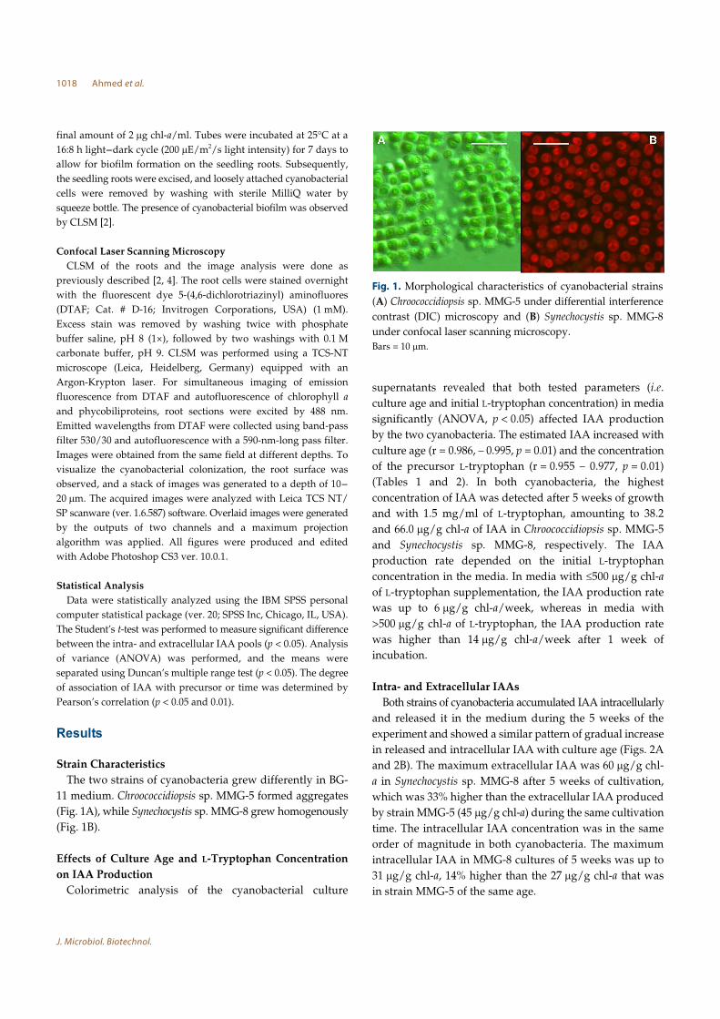
Strain Characteristics
The two strains of cyanobacteria grew differently in BG-
11 medium. Chroococcidiopsis sp. MMG-5 formed aggregates
(Fig. 1A), while Synechocystis sp. MMG-8 grew homogenously
(Fig. 1B).
Effects of Culture Age and L-Tryptophan Concentration
on IAA Production
Colorimetric analysis of the cyanobacterial culture
supernatants revealed that both tested parameters (i.e.
culture age and initial L-tryptophan concentration) in media
significantly (ANOVA, p < 0.05) affected IAA production
by the two cyanobacteria. The estimated IAA increased with
culture age (r = 0.986, – 0.995, p = 0.01) and the concentration
of the precursor L-tryptophan (r = 0.955 – 0.977, p = 0.01)
(Tables 1 and 2). In both cyanobacteria, the highest
concentration of IAA was detected after 5 weeks of growth
and with 1.5 mg/ml of L-tryptophan, amounting to 38.2
and 66.0 µg/g chl-a of IAA in Chroococcidiopsis sp. MMG-5
and Synechocystis sp. MMG-8, respectively. The IAA
production rate depended on the initial L-tryptophan
concentration in the media. In media with ≤500 µg/g chl-a
of L-tryptophan supplementation, the IAA production rate
was up to 6 µg/g chl-a/week, whereas in media with
>500 µg/g chl-a of L-tryptophan, the IAA production rate
was higher than 14 µg/g chl-a/week after 1 week of
incubation.
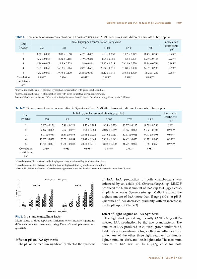
Intra- and Extracellular IAAs
Both strains of cyanobacteria accumulated IAA intracellularly
and released it in the medium during the 5 weeks of the
experiment and showed a similar pattern of gradual increase
in released and intracellular IAA with culture age (Figs. 2A
and 2B). The maximum extracellular IAA was 60 µg/g chl-
a in Synechocystis sp. MMG-8 after 5 weeks of cultivation,
which was 33% higher than the extracellular IAA produced
by strain MMG-5 (45 µg/g chl-a) during the same cultivation
time. The intracellular IAA concentration was in the same
order of magnitude in both cyanobacteria. The maximum
intracellular IAA in MMG-8 cultures of 5 weeks was up to
31 µg/g chl-a, 14% higher than the 27 µg/g chl-a that was
in strain MMG-5 of the same age.
Fig. 1. Morphological characteristics of cyanobacterial strains
(A) Chroococcidiopsis sp. MMG-5 under differential interference
contrast (DIC) microscopy and (B) Synechocystis sp. MMG-8
under confocal laser scanning microscopy.
Bars = 10 µm.
Biofilm Formation and IAA Production by Cyanobacteria 1019
August 2014⎪Vol. 24⎪No. 8
Effect of pH on IAA Synthesis
The pH of the medium significantly affected the synthesis
of IAA. IAA production in both cyanobacteria was
enhanced by an acidic pH. Chroococcidiopsis sp. MMG-5
produced the highest amount of IAA (up to 43 µg/g chl-a)
at pH 6, whereas Synechocystis sp. MMG-8 exuded the
highest amount of IAA (more than 45 µg/g chl-a) at pH 5.
Quantities of IAA decreased gradually with an increase in
media pH up to 9 (Table 3).
Effect of Light Regime on IAA Synthesis
The light:dark period significantly (ANOVA, p < 0.05)
affected IAA production by the two cyanobacteria. The
amount of IAA produced in cultures grown under 8:16 h
light:dark was significantly higher than in cultures grown
under any of the other three light regimes (continuous
light, continuous dark, and 16:8 h light:dark). The maximum
amount of IAA was up to 40 µg/g chl-a for both
Table 1. Time course of auxin concentration in Chroococcidiopsis sp. MMG-5 cultures with different amounts of tryptophan.
Time
(weeks)
Initial tryptophan concentration (µg/g chl-a) Correlation
coefficients
(r)a250 500 750 1,000 1,250 1,500
1 1.58 ± 0.055 3.87 ± 0.058 4.92 ± 0.085 9.68 ± 0.155 11.7 ± 0.179 11.43 ± 0.149 0.965**
2 3.67 ± 0.053 8.32 ± 0.165 11.9 ± 0.281 13.8 ± 0.381 15.3 ± 0.505 17.69 ± 0.455 0.977**
3 4.86 ± 0.073 14.3 ± 0.228 18 ± 0.464 22.95 ± 0.518 23.22 ± 0.720 28.94 ± 0.736 0.965**
4 5.81 ± 0.065 16.12 ± 0.264 23.2 ± 0.608 28.57 ± 0.815 31.88 ± 0.908 32.99 ± 0.980 0.959**
5 7.37 ± 0.060 19.75 ± 0.170 25.65 ± 0.530 34.42 ± 1.116 35.68 ± 1.390 38.2 ± 1.289 0.955**
Correlation
coefficients
(r)b
0.991** 0.986** 0.987** 0.995** 0.990** 0.986**
aCorrelation coefficients (r) of initial tryptophan concentration with given incubation time.bCorrelation coefficients (r) of incubation time with given initial tryptophan concentration.
Mean ± SE of three replicates. **Correlation is significant at the 0.01 level; *Correlation is significant at the 0.05 level.
Table 2. Time course of auxin concentration in Synechocystis sp. MMG-8 cultures with different amounts of tryptophan.
Time
(Weeks)
Initial tryptophan concentration (µg/g chl-a) Correlation
coefficients
(r)a250 500 750 1,000 1250 1,500
1 3.87 ± 0.136 5.48 ± 0.121 8.53 ± 0.205 9.24 ± 0.223 13.27 ± 0.115 14.38 ± 0.256 0.912*
2 7.66 ± 0.066 9.77 ± 0.078 16.4 ± 0.088 20.09 ± 0.049 23.96 ± 0.056 28.57 ± 0.102 0.995**
3 9.77 ± 0.057 16.54 ± 0.033 20.83 ± 0.032 22.85 ± 0.033 32.07 ± 0.045 37.97 ± 0.093 0.987**
4 13.27 ± 0.052 23.52 ± 0.034 28.47 ± 0.045 35.18 ± 0.041 44.42 ± 0.033 60.27 ± 0.083 0.983**
5 14.52 ± 0.063 28.38 ± 0.033 34.14 ± 0.011 38.22 ± 0.000 48.77 ± 0.000 66 ± 0.066 0.977**
Correlation
coefficients
(r) b
0.989** 0.983** 0.991** 0.980** 0.992** 0.987**
aCorrelation coefficients (r) of initial tryptophan concentration with given incubation time.bCorrelation coefficients (r) of incubation time with given initial tryptophan concentration.
Mean ± SE of three replicates. **Correlation is significant at the 0.01 level; *Correlation is significant at the 0.05 level.
Fig. 2. Intra- and extracellular IAAs.
Mean values of three replicates. Different letters indicate significant
difference between treatments, using Duncan’s multiple range test
(p = 0.05).
1020 Ahmed et al.
J. Microbiol. Biotechnol.
Chroococcidiopsis sp. MMG-5 and Synechocystis sp. MMG-8
grown under 8:16 h light:dark. There was a marginal
difference between the production of IAA by cultures
grown under 16:8 h light:dark and cultures grown under
continuous light. The cyanobacteria did not grow in the
dark and, consequently, no IAA was produced (Table 3).
Effect of Nitrate on IAA Synthesis
Chroococcidiopsis sp. MMG-5 could grow without combined
nitrogen and produced IAA under these conditions. IAA
production was significantly higher (ANOVA, p < 0.05)
(28%) in medium containing nitrate. Synechocystis sp.
MMG-8 was unable to grow without combined nitrogen
(Table 3).
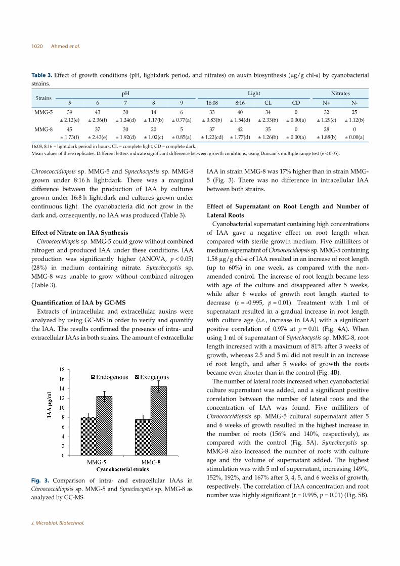
Quantification of IAA by GC-MS
Extracts of intracellular and extracellular auxins were
analyzed by using GC-MS in order to verify and quantify
the IAA. The results confirmed the presence of intra- and
extracellular IAAs in both strains. The amount of extracellular
IAA in strain MMG-8 was 17% higher than in strain MMG-
5 (Fig. 3). There was no difference in intracellular IAA
between both strains.
Effect of Supernatant on Root Length and Number of
Lateral Roots
Cyanobacterial supernatant containing high concentrations
of IAA gave a negative effect on root length when
compared with sterile growth medium. Five milliliters of
medium supernatant of Chroococcidiopsis sp. MMG-5 containing
1.58 µg/g chl-a of IAA resulted in an increase of root length
(up to 60%) in one week, as compared with the non-
amended control. The increase of root length became less
with age of the culture and disappeared after 5 weeks,
while after 6 weeks of growth root length started to
decrease (r = -0.995, p = 0.01). Treatment with 1 ml of
supernatant resulted in a gradual increase in root length
with culture age (i.e., increase in IAA) with a significant
positive correlation of 0.974 at p = 0.01 (Fig. 4A). When
using 1 ml of supernatant of Synechocystis sp. MMG-8, root
length increased with a maximum of 81% after 3 weeks of
growth, whereas 2.5 and 5 ml did not result in an increase
of root length, and after 5 weeks of growth the roots
became even shorter than in the control (Fig. 4B).
The number of lateral roots increased when cyanobacterial
culture supernatant was added, and a significant positive
correlation between the number of lateral roots and the
concentration of IAA was found. Five milliliters of
Chroococcidiopsis sp. MMG-5 cultural supernatant after 5
and 6 weeks of growth resulted in the highest increase in
the number of roots (156% and 140%, respectively), as
compared with the control (Fig. 5A). Synechocystis sp.
MMG-8 also increased the number of roots with culture
age and the volume of supernatant added. The highest
stimulation was with 5 ml of supernatant, increasing 149%,
152%, 192%, and 167% after 3, 4, 5, and 6 weeks of growth,
respectively. The correlation of IAA concentration and root
number was highly significant (r = 0.995, p = 0.01) (Fig. 5B).
Table 3. Effect of growth conditions (pH, light:dark period, and nitrates) on auxin biosynthesis (µg/g chl-a) by cyanobacterial
strains.
StrainspH Light Nitrates
5 6 7 8 9 16:08 8:16 CL CD N+ N-
MMG-5 39
± 2.12(e)
43
± 2.36(f)
30
± 1.24(d)
14
± 1.17(b)
6
± 0.77(a)
33
± 0.83(b)
40
± 1.54(d)
34
± 2.33(b)
0
± 0.00(a)
32
± 1.29(c)
25
± 1.12(b)
MMG-8 45
± 1.73(f)
37
± 2.43(e)
30
± 1.92(d)
20
± 1.02(c)
5
± 0.85(a)
37
± 1.22(cd)
42
± 1.77(d)
35
± 1.26(b)
0
± 0.00(a)
28
± 1.88(b)
0
± 0.00(a)
16:08, 8:16 = light:dark period in hours; CL = complete light; CD = complete dark.
Mean values of three replicates. Different letters indicate significant difference between growth conditions, using Duncan’s multiple range test (p < 0.05).
Fig. 3. Comparison of intra- and extracellular IAAs in
Chroococcidiopsis sp. MMG-5 and Synechocystis sp. MMG-8 as
analyzed by GC-MS.

Biofilm Formation and IAA Production by Cyanobacteria 1021
August 2014⎪Vol. 24⎪No. 8
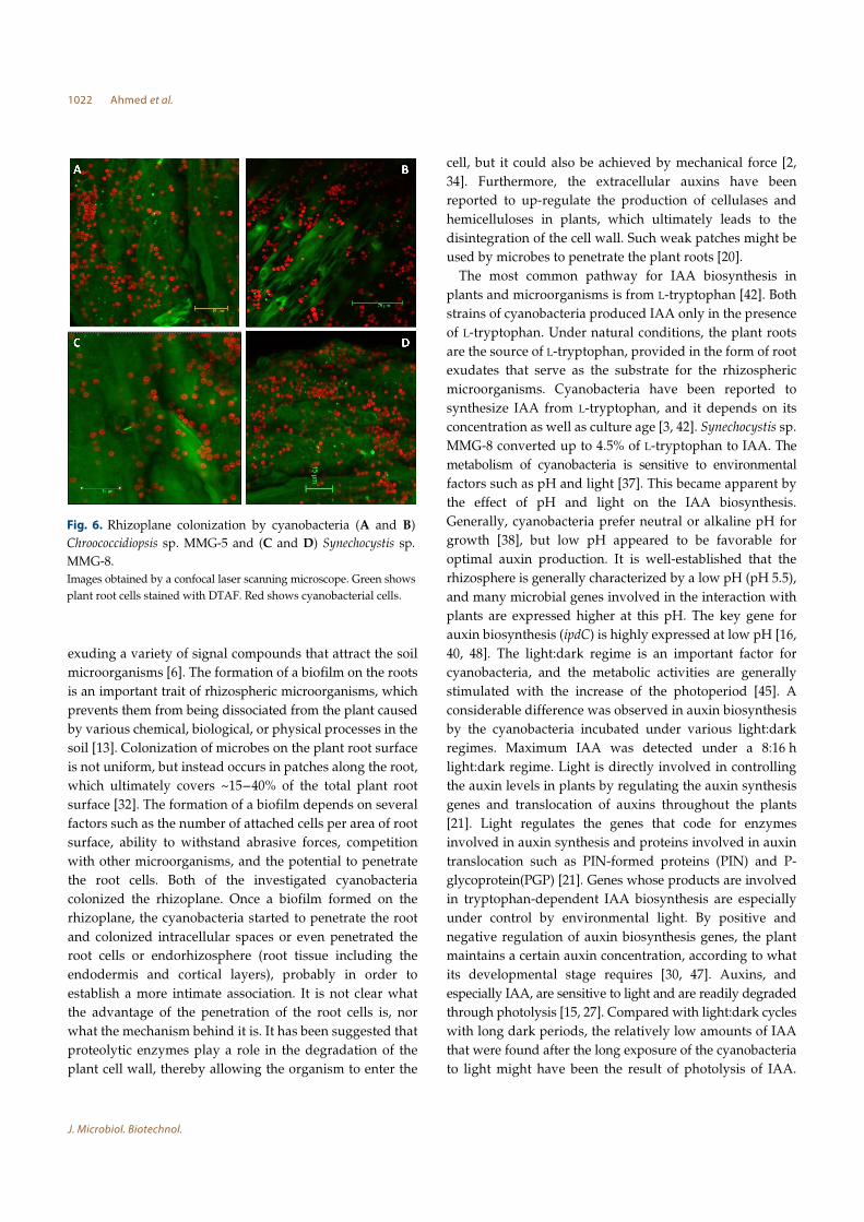
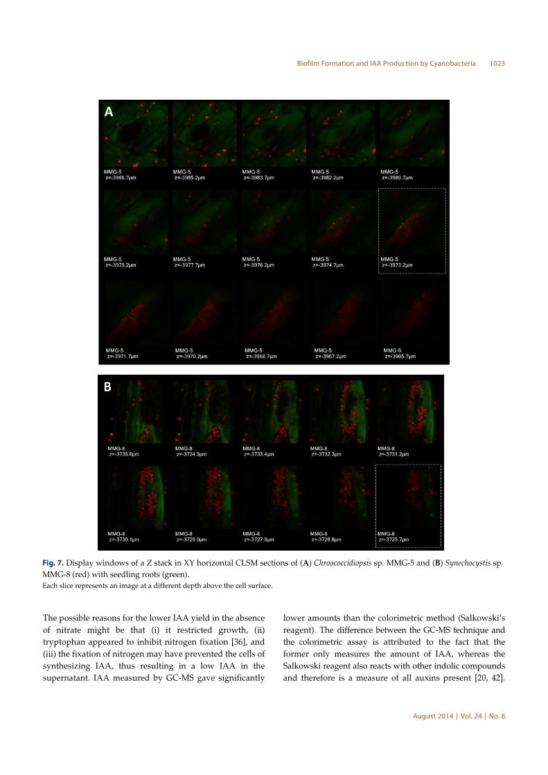
Formation of Biofilms on the Rhizoplane of Pisum sativum
Seedlings
The cyanobacteria showed ability to colonize the rhizoplane.
Chroococcidiopsis sp. MMG-5 extensively colonized the root
surface and concentrated in the grooves between adjacent
surface cells (Figs. 6A and 6B). Synechocystis sp. MMG-8
was scattered on the root surface but sometimes formed
colonies (Figs. 6C and 6D). Furthermore, the orthogonal
sectioning by CLSM illustrates how the cyanobacteria
invaded the epidermis and the cortex region, because they
appeared inside the root cells or underneath the outermost
layer. Chroococcidiopsis sp. MMG-5 was mainly present at a
depth of 10-15 µm inside the root cells (Fig. 7A).
Synechocystis penetrated the plant cells to a depth of 5-
7 µm (Fig. 7B).
Discussion
This study shows the ability of rhizospheric unicellular
cyanobacteria to produce IAA and to colonize the roots,
forming a thick biofilm. These two traits are considered to
be pivotal for rhizospheric microbes, and any microorganism
possessing these abilities should be successful in agricultural
ecosystems [35].
Cyanobacteria have the ability to attach to surfaces
through their extracellular polysaccharides. Such cyanobacteria
are considered as important colonizers of surfaces and
initiate the development of biofilms [17]. In order to allow
the establishment of an intimate association of rhizospheric
microorganisms, the plant root surface acts as the substrate
to which microbes attach. The roots assist in this process by
Fig. 4. Effect of the volume of spent media (5, 2.5, and 1 ml) of auxin-producing cyanobacteria on the root length of P. sativum.
The histograms represent auxin content of strains. (A) Chroococcidiopsis sp. MMG-5 and (B) Synechocystis sp. MMG-8. Mean of three replicates;
Correlation coefficient (r) between IAA and root length in different treatments (values beneath the legends). (**Correlation is significant at the 0.01
level; *Correlation is significant at the 0.05 level).
Fig. 5. Effect of the volume of spent media (5, 2.5, and 1ml) of auxin-producing cyanobacteria on root number of P. sativum.
The histograms represent auxin content of the cyanobacteria. (A) Synechocystis sp. MMG-8 and (B) Chroococcidiopsis sp. MMG-5. Mean of three
replicates; Correlation coefficient (r) between IAA and number of roots in different treatments (values beneath the legends). (**Correlation is
significant at the 0.01 level; *Correlation is significant at the 0.05 level).
1022 Ahmed et al.
J. Microbiol. Biotechnol.
exuding a variety of signal compounds that attract the soil
microorganisms [6]. The formation of a biofilm on the roots
is an important trait of rhizospheric microorganisms, which
prevents them from being dissociated from the plant caused
by various chemical, biological, or physical processes in the
soil [13]. Colonization of microbes on the plant root surface
is not uniform, but instead occurs in patches along the root,
which ultimately covers ~15-40% of the total plant root
surface [32]. The formation of a biofilm depends on several
factors such as the number of attached cells per area of root
surface, ability to withstand abrasive forces, competition
with other microorganisms, and the potential to penetrate
the root cells. Both of the investigated cyanobacteria
colonized the rhizoplane. Once a biofilm formed on the
rhizoplane, the cyanobacteria started to penetrate the root
and colonized intracellular spaces or even penetrated the
root cells or endorhizosphere (root tissue including the
endodermis and cortical layers), probably in order to
establish a more intimate association. It is not clear what
the advantage of the penetration of the root cells is, nor
what the mechanism behind it is. It has been suggested that
proteolytic enzymes play a role in the degradation of the
plant cell wall, thereby allowing the organism to enter the
cell, but it could also be achieved by mechanical force [2,
34]. Furthermore, the extracellular auxins have been
reported to up-regulate the production of cellulases and
hemicelluloses in plants, which ultimately leads to the
disintegration of the cell wall. Such weak patches might be
used by microbes to penetrate the plant roots [20].
The most common pathway for IAA biosynthesis in
plants and microorganisms is from L-tryptophan [42]. Both
strains of cyanobacteria produced IAA only in the presence
of L-tryptophan. Under natural conditions, the plant roots
are the source of L-tryptophan, provided in the form of root
exudates that serve as the substrate for the rhizospheric
microorganisms. Cyanobacteria have been reported to
synthesize IAA from L-tryptophan, and it depends on its
concentration as well as culture age [3, 42]. Synechocystis sp.
MMG-8 converted up to 4.5% of L-tryptophan to IAA. The
metabolism of cyanobacteria is sensitive to environmental
factors such as pH and light [37]. This became apparent by
the effect of pH and light on the IAA biosynthesis.
Generally, cyanobacteria prefer neutral or alkaline pH for
growth [38], but low pH appeared to be favorable for
optimal auxin production. It is well-established that the
rhizosphere is generally characterized by a low pH (pH 5.5),
and many microbial genes involved in the interaction with
plants are expressed higher at this pH. The key gene for
auxin biosynthesis (ipdC) is highly expressed at low pH [16,
40, 48]. The light:dark regime is an important factor for
cyanobacteria, and the metabolic activities are generally
stimulated with the increase of the photoperiod [45]. A
considerable difference was observed in auxin biosynthesis
by the cyanobacteria incubated under various light:dark
regimes. Maximum IAA was detected under a 8:16 h
light:dark regime. Light is directly involved in controlling
the auxin levels in plants by regulating the auxin synthesis
genes and translocation of auxins throughout the plants
[21]. Light regulates the genes that code for enzymes
involved in auxin synthesis and proteins involved in auxin
translocation such as PIN-formed proteins (PIN) and P-
glycoprotein(PGP) [21]. Genes whose products are involved
in tryptophan-dependent IAA biosynthesis are especially
under control by environmental light. By positive and
negative regulation of auxin biosynthesis genes, the plant
maintains a certain auxin concentration, according to what
its developmental stage requires [30, 47]. Auxins, and
especially IAA, are sensitive to light and are readily degraded
through photolysis [15, 27]. Compared with light:dark cycles
with long dark periods, the relatively low amounts of IAA
that were found after the long exposure of the cyanobacteria
to light might have been the result of photolysis of IAA.
Fig. 6. Rhizoplane colonization by cyanobacteria (A and B)
Chroococcidiopsis sp. MMG-5 and (C and D) Synechocystis sp.
MMG-8.
Images obtained by a confocal laser scanning microscope. Green shows
plant root cells stained with DTAF. Red shows cyanobacterial cells.
Biofilm Formation and IAA Production by Cyanobacteria 1023
August 2014⎪Vol. 24⎪No. 8
The possible reasons for the lower IAA yield in the absence
of nitrate might be that (i) it restricted growth, (ii)
tryptophan appeared to inhibit nitrogen fixation [36], and
(iii) the fixation of nitrogen may have prevented the cells of
synthesizing IAA, thus resulting in a low IAA in the
supernatant. IAA measured by GC-MS gave significantly
lower amounts than the colorimetric method (Salkowski’s
reagent). The difference between the GC-MS technique and
the colorimetric assay is attributed to the fact that the
former only measures the amount of IAA, whereas the
Salkowski reagent also reacts with other indolic compounds
and therefore is a measure of all auxins present [20, 42].
Fig. 7. Display windows of a Z stack in XY horizontal CLSM sections of (A) Chroococcidiopsis sp. MMG-5 and (B) Synechocystis sp.
MMG-8 (red) with seedling roots (green).
Each slice represents an image at a different depth above the cell surface.
1024 Ahmed et al.
J. Microbiol. Biotechnol.
Hence, both methods do not measure the same, and the
results indicate that other auxins than IAA are produced
and may be involved in the observed effects on the root
development.
Like other phytohormones, auxins are a group of important
signaling molecules involved in the interaction between
rhizospheric microbes and plants that affect plants even at
very low concentrations (10-5–10-6 M) [11, 25]. Roots are the
most sensitive parts of plants with respect to the effect of
auxins. Hence, root length and the number of lateral roots
have been used as a bioassay for auxins, particularly for
IAA [8]. Different volumes of sterile supernatant from the
growth medium of cyanobacterial cultures of different age
were used in the root bioassay in order to detect IAA-like
activity. Pea (P. sativum) was used as the test plant. The
concentration of secreted IAA in the culture supernatant
increased with the culture age. A gradual change in the
root parameters (root length and number of lateral roots)
was observed with the concentration of IAA. The
concentration of auxin in or near roots is critical because a
slight change results in a change in root length. On the one
hand, IAA in low concentration causes an increase in root
length; on the other hand, above a critical concentration,
auxin inhibits the growth of the root or even causes a
decrease of its length. This threshold concentration varies
between different plants [12, 29]. Highly significant correlations
were found between the concentration of IAA in the
supernatant and the root parameters (root length and
number of lateral roots). This could indicate an involvement
of cyanobacterial IAA in the regulation of root growth [3].
Thus, the rhizospheric cyanobacteria showed useful traits
(auxin synthesis and biofilm formation that possibly lead
to intense root colonization) in vitro, as they do in nature.
This ability of rhizospheric cyanobacteria can be exploited
for agriculture.
Acknowledgments
The Higher Education Commission of Pakistan is
acknowledged for funding the visit of Mehboob Ahmed to
the Netherlands Institute of Ecology (NIOO-KNAW)
(IRSIP No.1-8 HECHRD 2007 923). We thank Mrs. Anita
Wijnholds for assistance with confocal laser scanning
microscopy.
References
1. Ahmed M. 2011. Cyanobacterial secondary metabolites: their
impact on plant growth and fertility status of soil. Ph.D.
Thesis. University of the Punjab, Lahore, Pakistan.
2. Ahmed M, Stal LJ, Hasnain S. 2010. Association of non-
heterocystous cyanobacteria with crop plants. Plant Soil 336:
363-375.
3. Ahmed M, Stal LJ, Hasnain S. 2010. Production of indole-3-
acetic acid by the cyanobacterium Arthrospira platensis strain
MMG-9. J. Microbiol. Biotechnol. 20: 1259-1265.
4. Ahmed M, Stal LJ, Hasnain S. 2011. DTAF: an efficient
probe to study cyanobacterial-plant interaction using confocal
laser scanning microscopy (CLSM). J. Ind. Microbiol. Biotechnol.
38: 249-255.
5. Amzallag GN, Vaisman J. 2006. Influence of brassinosteroids
on initiation of the root gravitropic response in Pisum
sativum seedlings. Biol. Plant 50: 283-286.
6. Badri DV, Weir TL, van der Lelie D, Vivanco JM. 2009.
Rhizosphere chemical dialogues: plant–microbe interactions.
Curr. Opin. Biotechnol. 20: 642-650.
7. Bais HP, Weir TL, Perry LG, Gilroy S, Vivanco JM. 2006.
The role of root exudates in rhizosphere interactions with
plants and other organisms. Annu. Rev. Plant Biol. 57: 233-
266.
8. Barazani O, Friedman J. 1999. Is IAA the major root growth
factor secreted from plant-growth-mediating bacteria? J.
Chem. Ecol. 25: 2397-2406.
9. Berendsen RL, Pieterse CM, Bakker PA. 2012. The rhizosphere
microbiome and plant health. Trends Plant Sci. 17: 478-486.
10. Bergman B, Zheng WW, Klint J, Ran L. 2008. On the origin
of plants and relations to contemporary cyanobacterial-plant
symbioses. Plant Biotechnol. 25: 213-220.
11. Bladergroen MR, Spaink HP. 1998. Genes and signal
molecules involved in the rhizobia-leguminoseae symbiosis.
Curr. Opin. Plant Biol. 1: 353-359.
12. Callis J. 2005. Plant biology: auxin action. Nature 435: 436-
437.
13. Davies K, Whitbread R. 1989. Factors affecting the colonisation
of a root system by fluorescent pseudomonads: the effects
of water, temperature and soil microflora. Plant Soil 116:
247-256.
14. Dey R, Pal KK, Tilak KVBR. 2012. Influence of soil and
plant types on diversity of rhizobacteria. Proc. Natl. Acad.
Sci. India B Biol. Sci. 82: 341-352.
15. Dunlap JR, Robacker KM. 1988. Nutrient salts promote
light-induced degradation of indole-3-acetic-acid in tissue-
culture media. Plant Physiol. 88: 379-382.
16. Fierer N, Jackson RB. 2006. The diversity and biogeography
of soil bacterial communities. Proc. Natl. Acad. Sci. USA 103:
626-631.
17. Gaudes A, Sabater S, Vilalta E, Munoz I. 2006. The
nematode community in cyanobacterial biofilms in the river
Llobregat, Spain. Nematology 8: 909-919.
18. Glickmann E, Dessaux Y. 1995. A critical examination of the
specificity of the Salkowski reagent for indolic compounds
produced by phytopathogenic bacteria. Appl. Environ. Microbiol.
Biofilm Formation and IAA Production by Cyanobacteria 1025
August 2014⎪Vol. 24⎪No. 8
61: 793-796.
19. Guevara E, Jiménez VM, Herrera J, Bangerth F. 2008. Effect
of hydrogen cyanamide on the endogenous hormonal content
of pea seedlings (Pisum sativum L.). Braz. J. Plant Physiol. 20:
159-163.
20. Gutierrez CK, Matsui GY, Lincoln DE, Lovell CR. 2009.
Production of the phytohormone indole-3-acetic acid by
estuarine species of the genus Vibrio. Appl. Environ. Microbiol.
75: 2253-2258.
21. Halliday KJ, Martinez-Garcia JF, Josse EM. 2009. Integration
of light and auxin signaling. Cold Spring Harb. Perspect. Biol.
1: 1-11.
22. Höckelmann C, Jüttner F. 2004. Volatile organic compound
(VOC) analysis and sources of limonene, cyclohexanone and
straight chain aldehydes in axenic cultures of Calothrix and
Plectonema. Water Sci. Technol. 49: 47.
23. Hussain A, Hasnain S. 2011. Phytostimulation and
biofertilization in wheat by cyanobacteria. J. Ind. Microbiol.
Biotechnol. 38: 85-92.
24. Hussain A, Hasnain S. 2012. Comparative assessment of the
efficacy of bacterial and cyanobacterial phytohormones in
plant tissue culture. World J. Microbiol. Biotechnol. 28: 1459-
1466.
25. Khalid A, Arshad M, Zahir ZA. 2006. Phytohormones:
microbial production and applications, pp. 207-220. In
Uphoff N, Ball AS, Fernandes E, Herren H, Husson O,
Laing M, et al. (eds.). Biological Approaches to Sustainable Soil
Systems. Taylor & Francis/CRC, Boca Raton, Florida
26. Klock J-H, Wieland A, Seifert R, Michaelis W. 2007.
Extracellular polymeric substances (EPS) from cyanobacterial
mats: characterisation and isolation method optimisation.
Mar. Biol. 152: 1077-1085.
27. Kusaka N, Maisch J, Nick P, Hayashi K, Nozaki H. 2009.
Manipulation of intracellular auxin in a single cell by light
with esterase-resistant caged auxins. Chembiochem 10: 2195-
2202.
28. Lambers H, Mougel C, Jaillard B, Hinsinger P. 2009. Plant-
microbe-soil interactions in the rhizosphere: an evolutionary
perspective. Plant Soil 321: 83-115.
29. Lambers H, Pons TL, Chapin FS. 2008. Plant Physiological
Ecology. Springer, New York.
30. Lau S, Jurgens G, De Smet I. 2008. The evolving complexity
of the auxin pathway. Plant Cell 20: 1738-1746.
31. Mazhar S, Cohen JD, Hasnain S. 2013. Auxin producing
non-heterocystous Cyanobacteria and their impact on the
growth and endogenous auxin homeostasis of wheat. J.
Basic Microbiol. 53: 996-1003.
32. McNear Jr DH. 2013. The rhizosphere — roots, soil and
everything in between. Nature Educ. Knowledge 4: 1.
33. Morgan JAW, Bending GD, White PJ. 2005. Biological costs
and benefits to plant–microbe interactions in the rhizosphere.
J. Exp. Bot. 56: 1729-1739.
34. Okon Y, Vanderleyden J. 1997. Root-associated Azospirillum
species can stimulate plants. ASM News 63: 366-370.
35. Ortiz-Castro R, Contreras-Cornejo HA, Macias-Rodriguez L,
Lopez-Bucio J. 2009. The role of microbial signals in plant
growth and development. Plant Signal Behav. 4: 701-712.
36. Pain R, Duggan PS, Adams DG. 2000. Creation of a
tryptophan auxotroph to study the role of tryptophan in
heterocyst development. Abstracts of the 10th International
Symposium on Phototrophic Prokaryotes, Barcelona, Spain,
August 26-31, 2000.
37. Pentecost A, Whitton BA. 2012. Subaerial Cyanobacteria, pp.
291-316. In Whitton BA (ed.). Ecology of Cyanobacteria II:
Their Diversity in Space and Time. Springer, The Netherlands
38. Prasanna R, Jaiswal P, Nayak S, Sood A, Kaushik BD. 2009.
Cyanobacterial diversity in the rhizosphere of rice and its
ecological significance. Indian J. Microbiol. 49: 89-97.
39. Rippka R, Herdman M. 1992. Pasteur Culture Collection of
Cyanobacteria: Catalogue and Taxonomic Handbook. I. Catalogue
of Strains, Institut Pasteur, Paris.
40. Ryu RJ, Patten CL. 2008. Aromatic amino acid-dependent
expression of indole-3-pyruvate decarboxylase is regulated
by TyrR in Enterobacter cloacae UW5. J. Bacteriol. 190: 7200-
7208.
41. Sabrine H, Afif H, Mohamed B, Hamadi B, Maria H. 2010.
Effects of cadmium and copper on pollen germination and
fruit set in pea (Pisum sativum L.). Sci. Horticult. 125: 551-
555.
42. Sergeeva E, Liaimer A, Bergman B. 2002. Evidence for
production of the phytohormone indole-3-acetic acid by
cyanobacteria. Planta 215: 229-238.
43. Smýkal P, Aubert G, Burstin J, Coyne CJ, Ellis NT, Flavell
AJ, et al. 2012. Pea (Pisum sativum L.) in the genomic era.
Agronomy 2: 74-115.
44. Spaepen S, Vanderleyden J, Remans R. 2007. Indole-3-acetic
acid in microbial and microorganism-plant signaling. FEMS
Microbiol. Rev. 31: 425-448.
45. Tabei Y, Okada K, Makita N, Tsuzuki M. 2009. Light-
induced gene expression of fructose 1,6-bisphosphate aldolase
during heterotrophic growth in a cyanobacterium, Synechocystis
sp. PCC 6803. FEBS J. 276: 187-198.
46. Tandeau de Marsac N, Houmard J. 1988. Complementary
chromatic adaptation: physiological conditions and action
spectra. Methods Enzymol 167: 318-328.
47. Tao Y, Ferrer JL, Ljung K, Pojer F, Hong F, Long JA, et al.
2008. Rapid synthesis of auxin via a new tryptophan-
dependent pathway is required for shade avoidance in
plants. Cell 133: 164-176.
48. Yuan ZC, Liu P, Saenkham P, Kerr K, Nester EW. 2008.
Transcriptome profiling and functional analysis of Agrobacterium
tumefaciens reveals a general conserved response to acidic
conditions (pH 5.5) and a complex acid-mediated signaling
involved in Agrobacterium-plant interactions. J. Bacteriol. 190:
494-507.


![RiceGROWTH-REGULATINGFACTOR7Modulates Plant … · RiceGROWTH-REGULATINGFACTOR7Modulates Plant Architecture through Regulating GA and Indole-3-Acetic Acid Metabolism1[OPEN] Yunping](https://img.pdfslide.net/doc/110x75/606f2571d2b9b9029938beb9/ricegrowth-regulatingfactor7modulates-plant-ricegrowth-regulatingfactor7modulates.jpg)


![Auxin Biosynthesis: Are the Indole-3-Acetic Acid and ... · Auxin Biosynthesis: Are the Indole-3-Acetic Acid and Phenylacetic Acid Biosynthesis Pathways Mirror Images?1[OPEN] Sam](https://img.pdfslide.net/doc/110x75/5b9eb03a09d3f2ab0b8c6e3b/auxin-biosynthesis-are-the-indole-3-acetic-acid-and-auxin-biosynthesis.jpg)