Embed Size (px)
Citation preview
© Endeavour College of Natural Health endeavour.edu.au 1
BIOH111
oCell Module
oTissue Module
o Integumentary system
oSkeletal system
oMuscle system
oNervous system
oEndocrine system
© Endeavour College of Natural Health endeavour.edu.au 2
Textbook and required/recommended
readings
o Principles of anatomy and physiology. Tortora et al; 14th
edition: Chapter 5
© Endeavour College of Natural Health endeavour.edu.au 3
BIOH111 – INTEGUMENTARY
SYSTEM MODULE
o Session 6 (Lectures 9 and 10) – Structure and
function of the skin and accessory structures
BIOH111Lectures 9 and 10
Structure of the skin and accessory
structures
Department of Bioscience
endeavour.edu.au
© Endeavour College of Natural Health endeavour.edu.au 5
Preparation for this session
o Complete any missing concepts and linking words from
Session 5
o Consider epithelial membrane called cutaneous
membrane – what is it composed of and where can it be
found?
© Endeavour College of Natural Health endeavour.edu.au 6
ObjectivesLecture 9:
Structure of skin
• Define and describe the structure of each skin layer
Accessory skin structures
• Define and describe the structure of each accessory skin structure
Lecture 10:
Functions of skin
• Define and describe the function the skin
Wound healing
• Describe process of the epidermal wound healing
• Describe the process of deep wound healing
• Compare epidermal and deep wound healing
Burns
• Describe types of burns and damages to the skin layer
• Describe method for measuring the extent of burns

© Endeavour College of Natural Health endeavour.edu.au 7
INTEGUMENTARY SYSTEM
FUNCTIONS o Thermoregulation
o Blood reservoir
o Protection
o Cutaneous sensations
o Synthesis of Vitamin D
o Excretion
© Endeavour College of Natural Health endeavour.edu.au 8
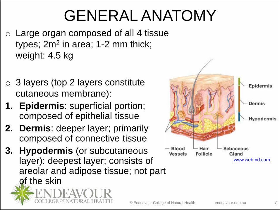
GENERAL ANATOMY
www.webmd.com
o Large organ composed of all 4 tissue
types; 2m2 in area; 1-2 mm thick;
weight: 4.5 kg
o 3 layers (top 2 layers constitute
cutaneous membrane):
1. Epidermis: superficial portion; composed of epithelial tissue
2. Dermis: deeper layer; primarily composed of connective tissue
3. Hypodermis (or subcutaneous layer): deepest layer; consists of areolar and adipose tissue; not part of the skin
© Endeavour College of Natural Health endeavour.edu.au 9
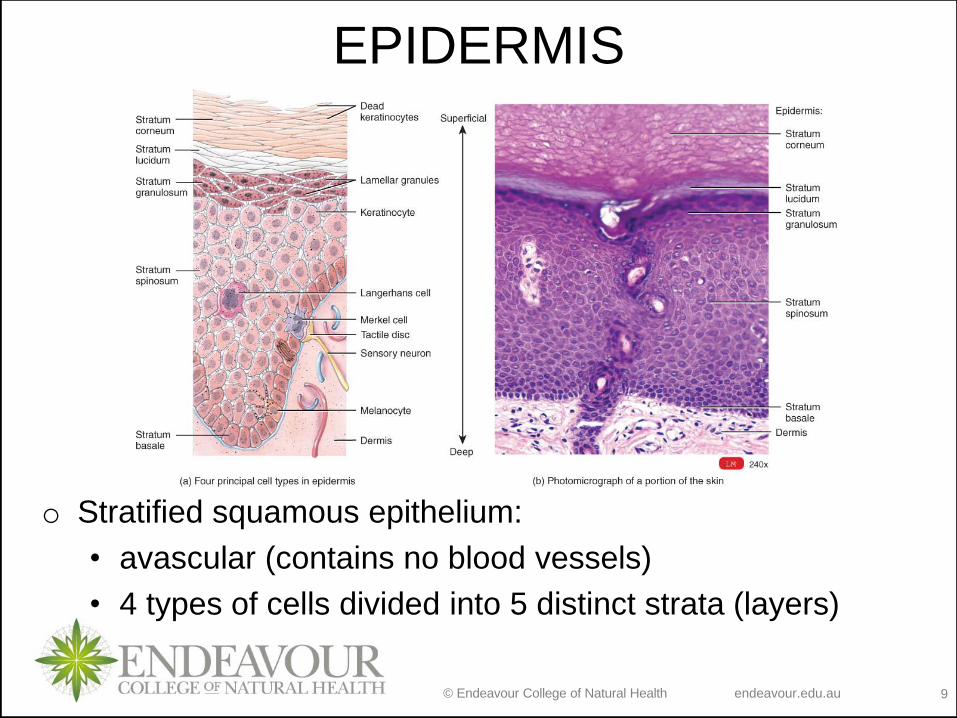
EPIDERMIS
o Stratified squamous epithelium:
• avascular (contains no blood vessels)
• 4 types of cells divided into 5 distinct strata (layers)
© Endeavour College of Natural Health endeavour.edu.au 10
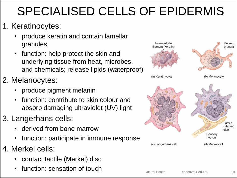
SPECIALISED CELLS OF EPIDERMIS1. Keratinocytes:
• produce keratin and contain lamellar
granules
• function: help protect the skin and
underlying tissue from heat, microbes,
and chemicals; release lipids (waterproof)
2. Melanocytes:
• produce pigment melanin
• function: contribute to skin colour and
absorb damaging ultraviolet (UV) light
3. Langerhans cells:
• derived from bone marrow
• function: participate in immune response
4. Merkel cells:
• contact tactile (Merkel) disc
• function: sensation of touch
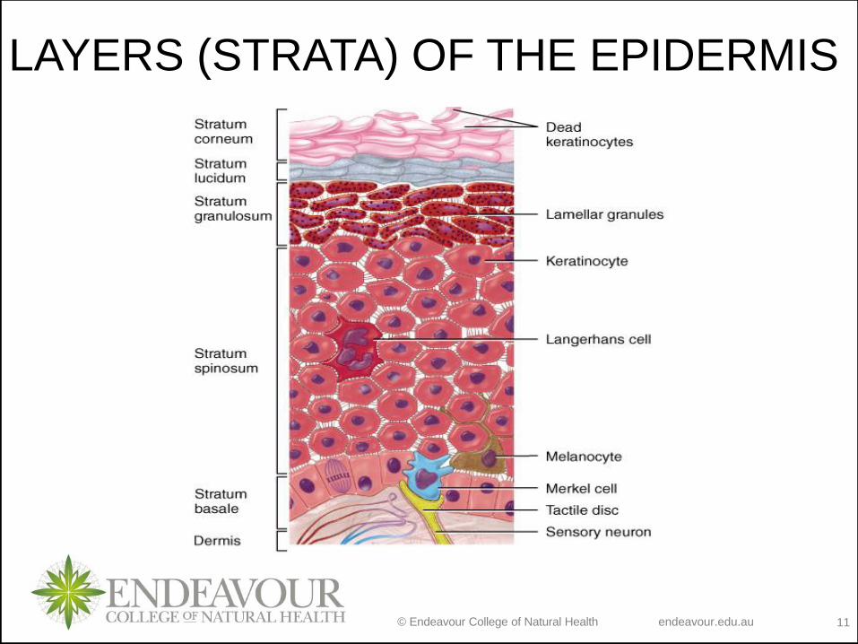
© Endeavour College of Natural Health endeavour.edu.au 11
LAYERS (STRATA) OF THE EPIDERMIS
© Endeavour College of Natural Health endeavour.edu.au 12
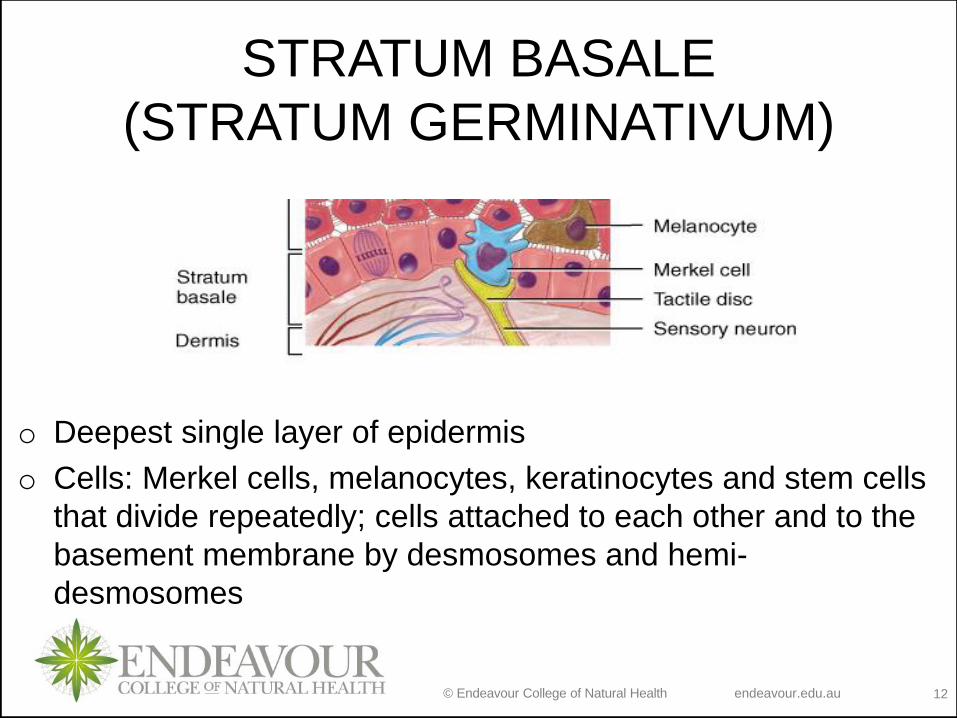
STRATUM BASALE
(STRATUM GERMINATIVUM)
o Deepest single layer of epidermis
o Cells: Merkel cells, melanocytes, keratinocytes and stem cells
that divide repeatedly; cells attached to each other and to the
basement membrane by desmosomes and hemi-
desmosomes
© Endeavour College of Natural Health endeavour.edu.au 13
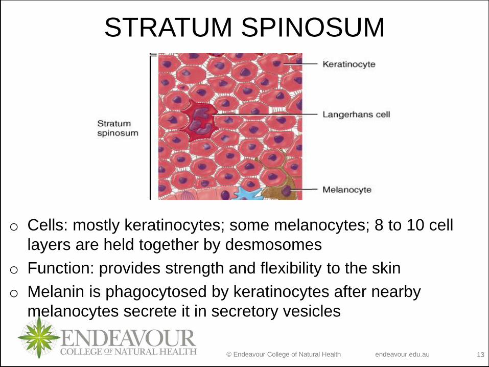
STRATUM SPINOSUM
o Cells: mostly keratinocytes; some melanocytes; 8 to 10 cell
layers are held together by desmosomes
o Function: provides strength and flexibility to the skin
o Melanin is phagocytosed by keratinocytes after nearby
melanocytes secrete it in secretory vesicles
© Endeavour College of Natural Health endeavour.edu.au 14
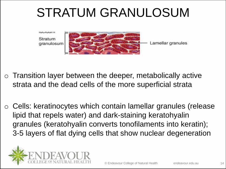
STRATUM GRANULOSUM
o Transition layer between the deeper, metabolically active
strata and the dead cells of the more superficial strata
o Cells: keratinocytes which contain lamellar granules (release
lipid that repels water) and dark-staining keratohyalin
granules (keratohyalin converts tonofilaments into keratin);
3-5 layers of flat dying cells that show nuclear degeneration
© Endeavour College of Natural Health endeavour.edu.au 15
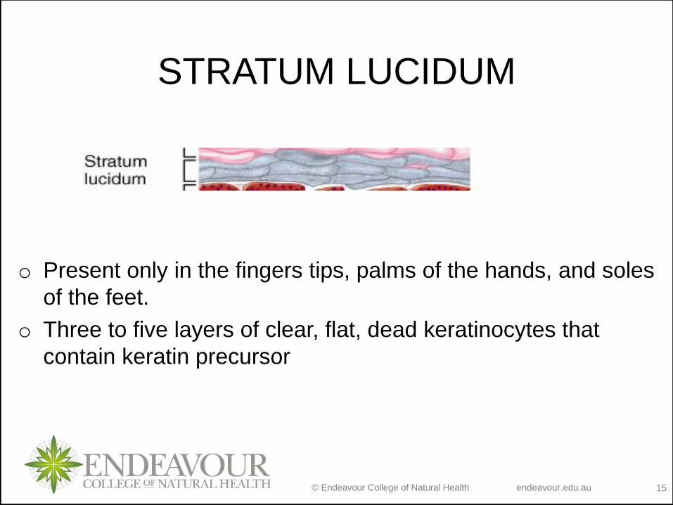
STRATUM LUCIDUM
o Present only in the fingers tips, palms of the hands, and soles
of the feet.
o Three to five layers of clear, flat, dead keratinocytes that
contain keratin precursor
© Endeavour College of Natural Health endeavour.edu.au 16
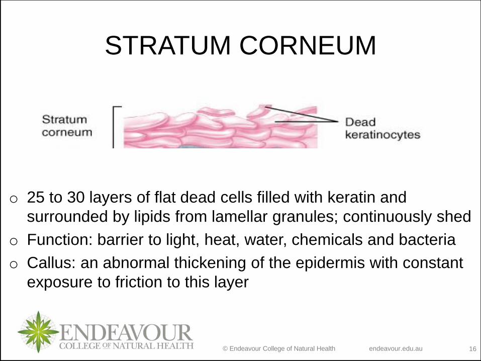
STRATUM CORNEUM
o 25 to 30 layers of flat dead cells filled with keratin and
surrounded by lipids from lamellar granules; continuously shed
o Function: barrier to light, heat, water, chemicals and bacteria
o Callus: an abnormal thickening of the epidermis with constant
exposure to friction to this layer
© Endeavour College of Natural Health endeavour.edu.au 17
Ep
ith
elia
l d
iffe
ren
tia
tio
n
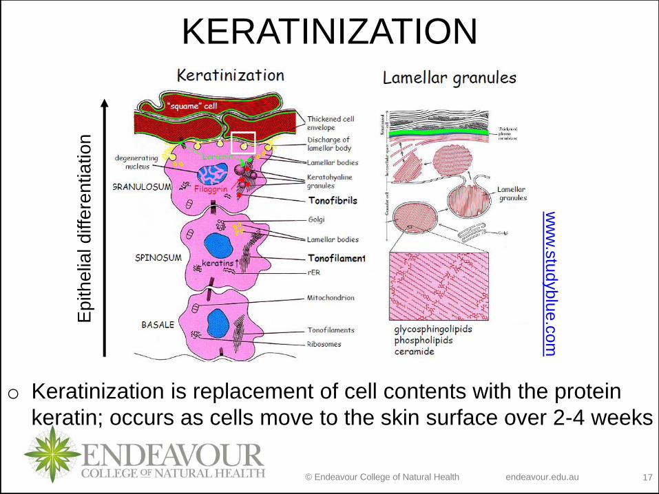
o Keratinization is replacement of cell contents with the protein
keratin; occurs as cells move to the skin surface over 2-4 weeks
KERATINIZATION
ww
w.s
tudyblu
e.c
om
© Endeavour College of Natural Health endeavour.edu.au 18
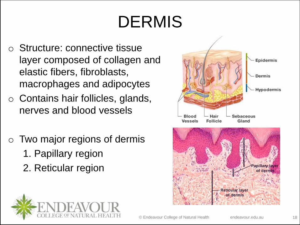
DERMIS
o Structure: connective tissue
layer composed of collagen and
elastic fibers, fibroblasts,
macrophages and adipocytes
o Contains hair follicles, glands,
nerves and blood vessels
o Two major regions of dermis
1. Papillary region
2. Reticular region
© Endeavour College of Natural Health endeavour.edu.au 19
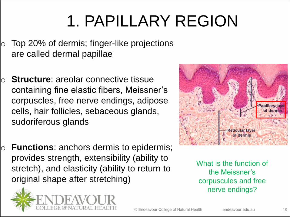
1. PAPILLARY REGION
o Top 20% of dermis; finger-like projections
are called dermal papillae
o Structure: areolar connective tissue
containing fine elastic fibers, Meissner’s
corpuscles, free nerve endings, adipose
cells, hair follicles, sebaceous glands,
sudoriferous glands
o Functions: anchors dermis to epidermis;
provides strength, extensibility (ability to
stretch), and elasticity (ability to return to
original shape after stretching)
What is the function of
the Meissner’s
corpuscules and free
nerve endings?
© Endeavour College of Natural Health endeavour.edu.au 20
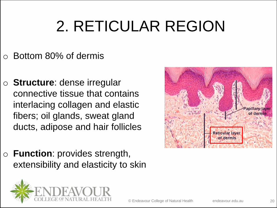
2. RETICULAR REGION
o Bottom 80% of dermis
o Structure: dense irregular
connective tissue that contains
interlacing collagen and elastic
fibers; oil glands, sweat gland
ducts, adipose and hair follicles
o Function: provides strength,
extensibility and elasticity to skin
© Endeavour College of Natural Health endeavour.edu.au 21
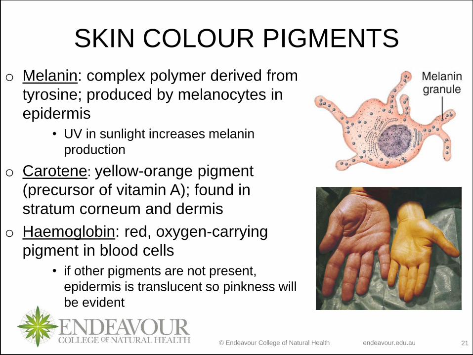
SKIN COLOUR PIGMENTS
o Melanin: complex polymer derived from
tyrosine; produced by melanocytes in
epidermis
• UV in sunlight increases melanin
production
o Carotene: yellow-orange pigment
(precursor of vitamin A); found in
stratum corneum and dermis
o Haemoglobin: red, oxygen-carrying
pigment in blood cells
• if other pigments are not present,
epidermis is translucent so pinkness will
be evident
© Endeavour College of Natural Health endeavour.edu.au 22
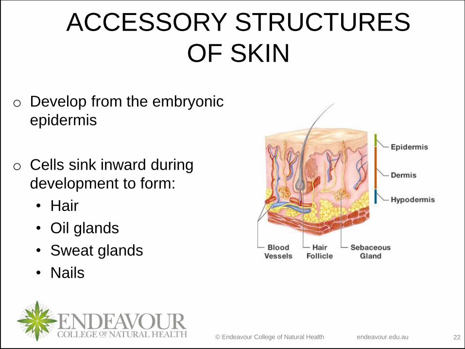
ACCESSORY STRUCTURES
OF SKIN
o Develop from the embryonic
epidermis
o Cells sink inward during
development to form:
• Hair
• Oil glands
• Sweat glands
• Nails
© Endeavour College of Natural Health endeavour.edu.au 23
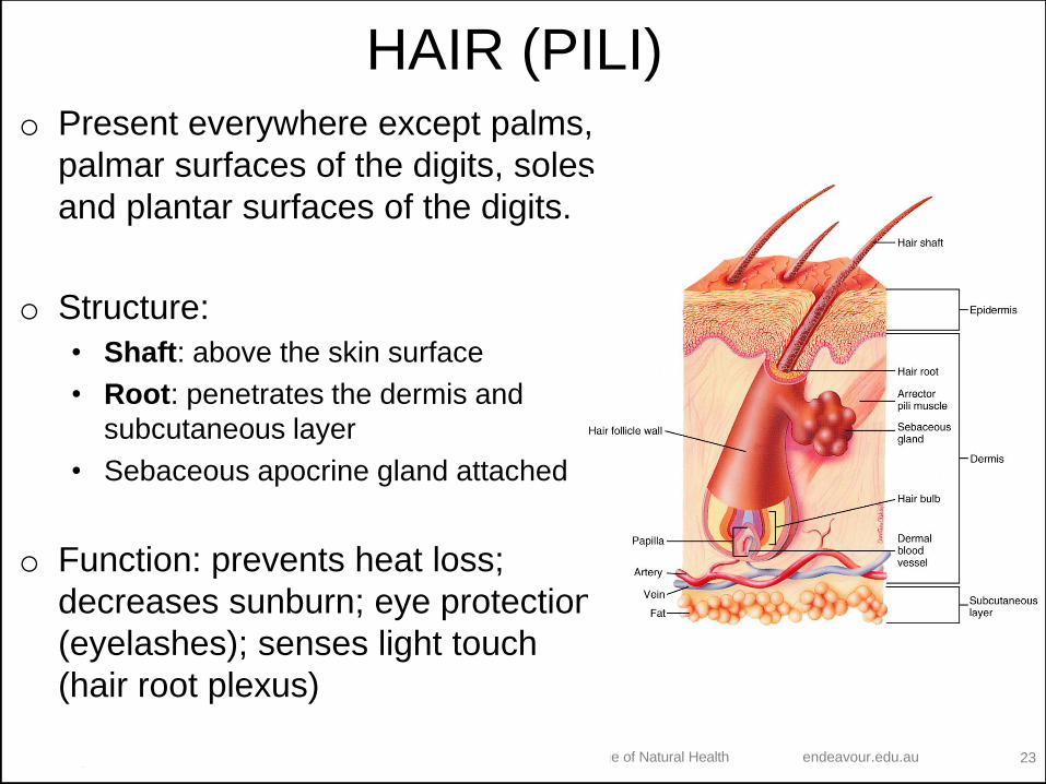
HAIR (PILI)o Present everywhere except palms,
palmar surfaces of the digits, soles
and plantar surfaces of the digits.
o Structure:
• Shaft: above the skin surface
• Root: penetrates the dermis and
subcutaneous layer
• Sebaceous apocrine gland attached
o Function: prevents heat loss;
decreases sunburn; eye protection
(eyelashes); senses light touch
(hair root plexus)
© Endeavour College of Natural Health endeavour.edu.au 24
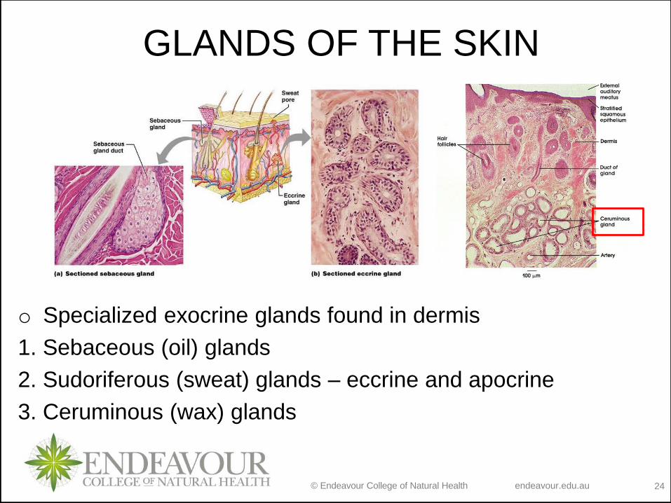
GLANDS OF THE SKIN
o Specialized exocrine glands found in dermis
1. Sebaceous (oil) glands
2. Sudoriferous (sweat) glands – eccrine and apocrine
3. Ceruminous (wax) glands
© Endeavour College of Natural Health endeavour.edu.au 25
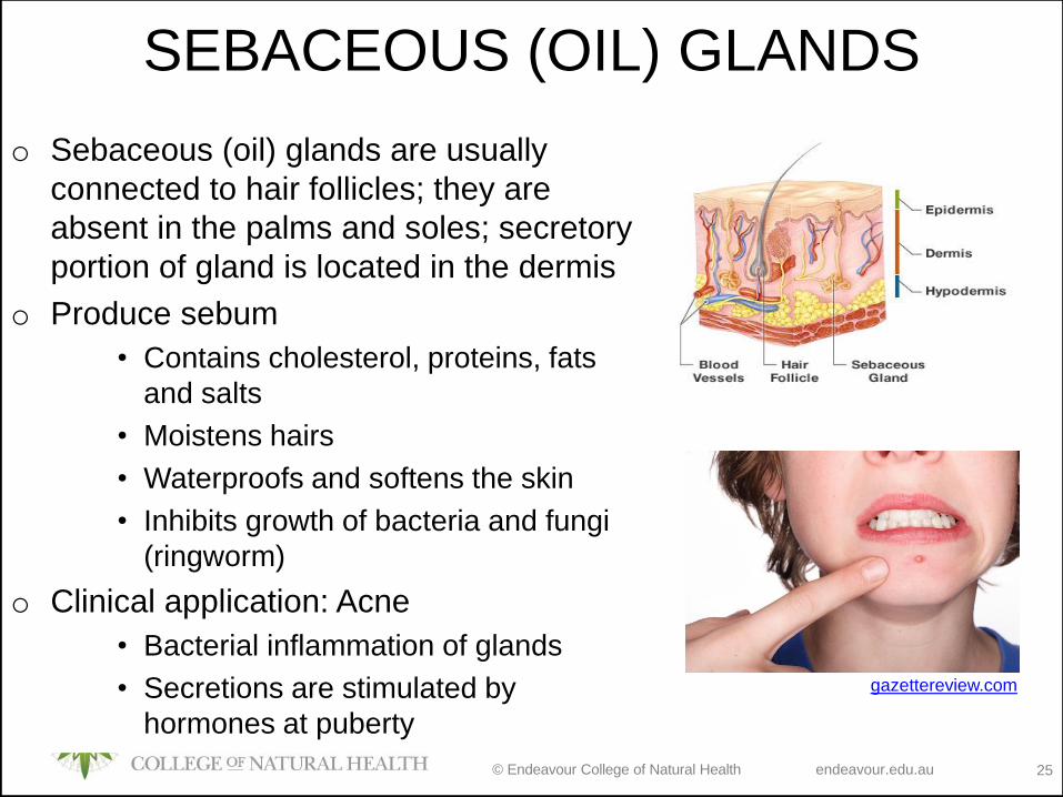
SEBACEOUS (OIL) GLANDS
o Sebaceous (oil) glands are usually
connected to hair follicles; they are
absent in the palms and soles; secretory
portion of gland is located in the dermis
o Produce sebum
• Contains cholesterol, proteins, fats
and salts
• Moistens hairs
• Waterproofs and softens the skin
• Inhibits growth of bacteria and fungi
(ringworm)
o Clinical application: Acne
• Bacterial inflammation of glands
• Secretions are stimulated by
hormones at puberty
gazettereview.com
© Endeavour College of Natural Health endeavour.edu.au 26
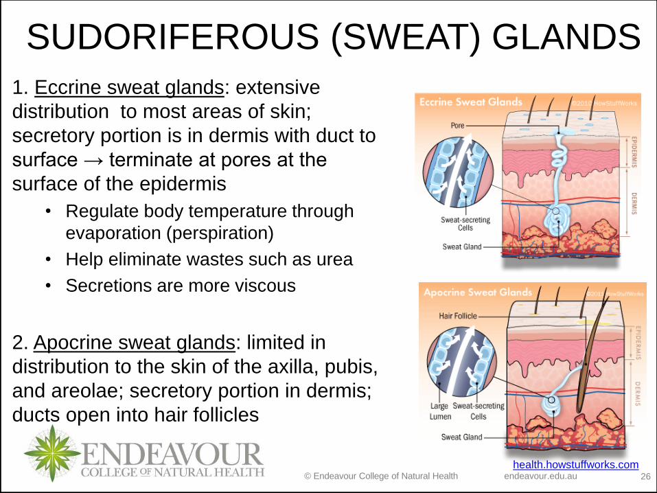
SUDORIFEROUS (SWEAT) GLANDS
1. Eccrine sweat glands: extensive
distribution to most areas of skin;
secretory portion is in dermis with duct to
surface → terminate at pores at the
surface of the epidermis
• Regulate body temperature through
evaporation (perspiration)
• Help eliminate wastes such as urea
• Secretions are more viscous
2. Apocrine sweat glands: limited in
distribution to the skin of the axilla, pubis,
and areolae; secretory portion in dermis;
ducts open into hair follicles
health.howstuffworks.com
© Endeavour College of Natural Health endeavour.edu.au 27
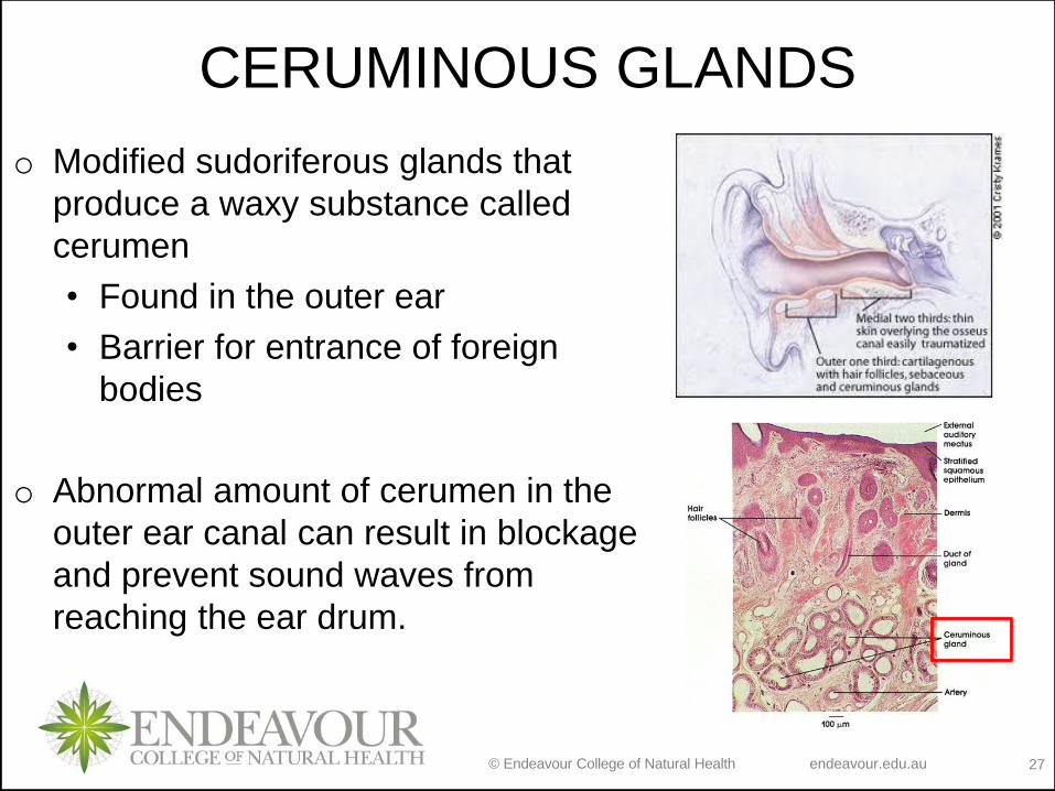
CERUMINOUS GLANDS
o Modified sudoriferous glands that
produce a waxy substance called
cerumen
• Found in the outer ear
• Barrier for entrance of foreign
bodies
o Abnormal amount of cerumen in the
outer ear canal can result in blockage
and prevent sound waves from
reaching the ear drum.
© Endeavour College of Natural Health endeavour.edu.au 28
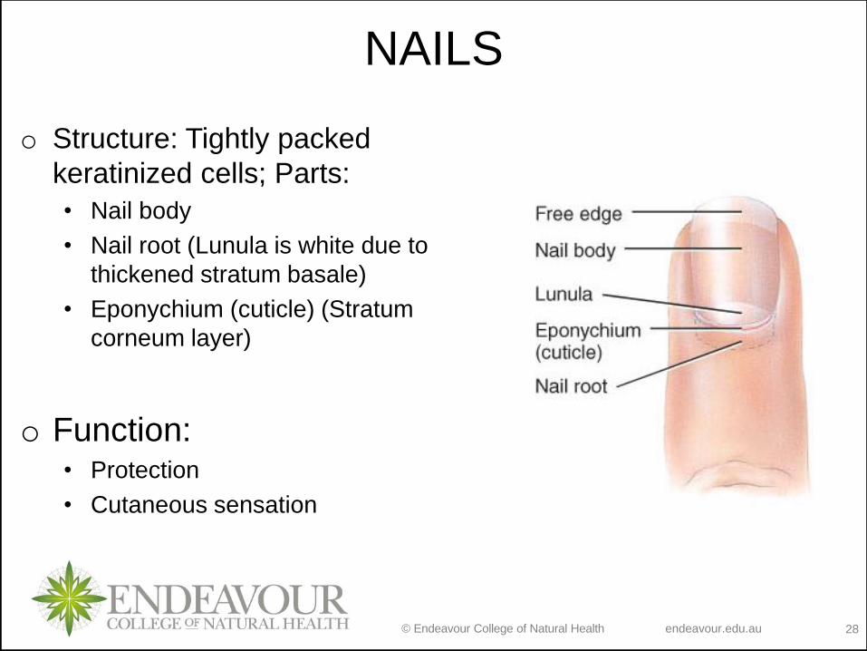
NAILS
o Structure: Tightly packed
keratinized cells; Parts:
• Nail body
• Nail root (Lunula is white due to
thickened stratum basale)
• Eponychium (cuticle) (Stratum
corneum layer)
o Function: • Protection
• Cutaneous sensation
© Endeavour College of Natural Health endeavour.edu.au 29
ObjectivesLecture 9:
Structure of skin
• Define and describe the structure of each skin layer
Accessory skin structures
• Define and describe the structure of each accessory skin structure
Lecture 10:
Functions of skin
• Define and describe the function the skin
Wound healing
• Describe process of the epidermal wound healing
• Describe the process of deep wound healing
• Compare epidermal and deep wound healing
Burns
• Describe types of burns and damages to the skin layer
• Describe method for measuring the extent of burns
© Endeavour College of Natural Health endeavour.edu.au 30
INTEGUMENTARY SYSTEM
FUNCTIONS o Thermoregulation
o Blood reservoir
o Protection
o Cutaneous sensations
o Synthesis of Vitamin D
o Excretion
© Endeavour College of Natural Health endeavour.edu.au 31
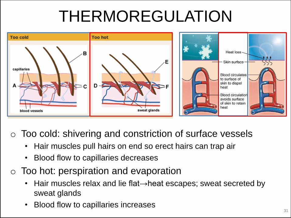
THERMOREGULATION
o Too cold: shivering and constriction of surface vessels
• Hair muscles pull hairs on end so erect hairs can trap air
• Blood flow to capillaries decreases
o Too hot: perspiration and evaporation
• Hair muscles relax and lie flat→heat escapes; sweat secreted by
sweat glands
• Blood flow to capillaries increases
© Endeavour College of Natural Health endeavour.edu.au 32
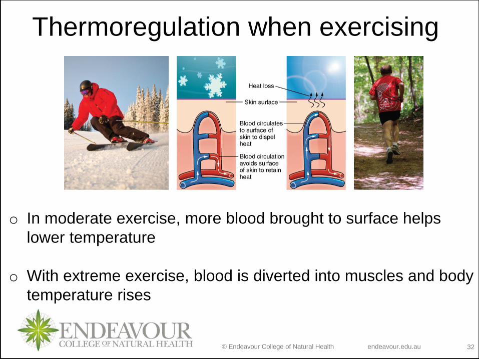
Thermoregulation when exercising
o In moderate exercise, more blood brought to surface helps
lower temperature
o With extreme exercise, blood is diverted into muscles and body
temperature rises
© Endeavour College of Natural Health endeavour.edu.au 33
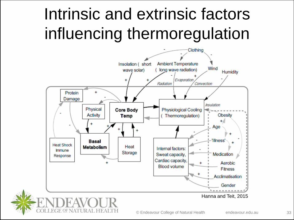
Intrinsic and extrinsic factors
influencing thermoregulation
Hanna and Teit, 2015
© Endeavour College of Natural Health endeavour.edu.au 34
OTHER FUNCTIONS OF SKINo Blood reservoir
• extensive network of blood vessels
o Protection - physical, chemical and biological barriers
• tight cell junctions prevent bacterial invasion
• lipids released decrease evaporation
• pigment protects somewhat against UV light
• Langerhans cells alert immune system
o Cutaneous sensations
• Touch, pressure, vibration, tickle, heat, cold, and pain arise
in the skin
© Endeavour College of Natural Health endeavour.edu.au 35
OTHER FUNCTIONS OF SKIN
o Synthesis of Vitamin D
• Necessary vitamin for absorption of calcium from food in
the gastrointestinal tract
• activation of a precursor molecule in the skin by UV
light→blood→enzymes in the liver and kidneys modify the
activated molecule to produce calcitriol (the most active
form of vitamin D)
o Excretion
• 400 mL of water/day, small amounts salt, CO2, ammonia
and urea
© Endeavour College of Natural Health endeavour.edu.au 36
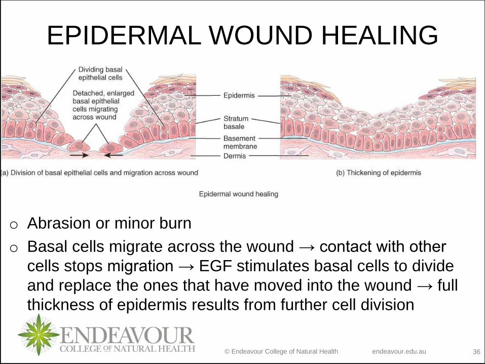
EPIDERMAL WOUND HEALING
o Abrasion or minor burn
o Basal cells migrate across the wound → contact with other
cells stops migration → EGF stimulates basal cells to divide
and replace the ones that have moved into the wound → full
thickness of epidermis results from further cell division
© Endeavour College of Natural Health endeavour.edu.au 37
DEEP WOUND HEALING
o When an injury extends to tissues deep to the epidermis, the
repair process is more complex than epidermal healing and
scar formation results.
o Healing occurs in 4 phases:
1. Inflammatory phase
2. Migratory phase
3. Proliferative phase
4. Maturation phase
© Endeavour College of Natural Health endeavour.edu.au 38
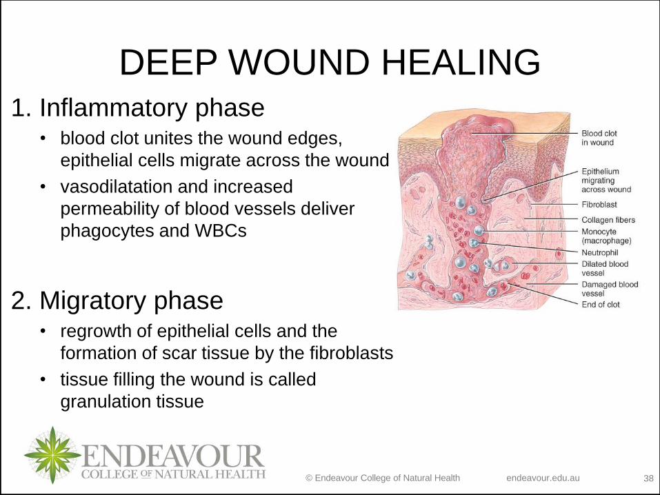
DEEP WOUND HEALING1. Inflammatory phase
• blood clot unites the wound edges,
epithelial cells migrate across the wound
• vasodilatation and increased
permeability of blood vessels deliver
phagocytes and WBCs
2. Migratory phase• regrowth of epithelial cells and the
formation of scar tissue by the fibroblasts
• tissue filling the wound is called
granulation tissue
© Endeavour College of Natural Health endeavour.edu.au 39
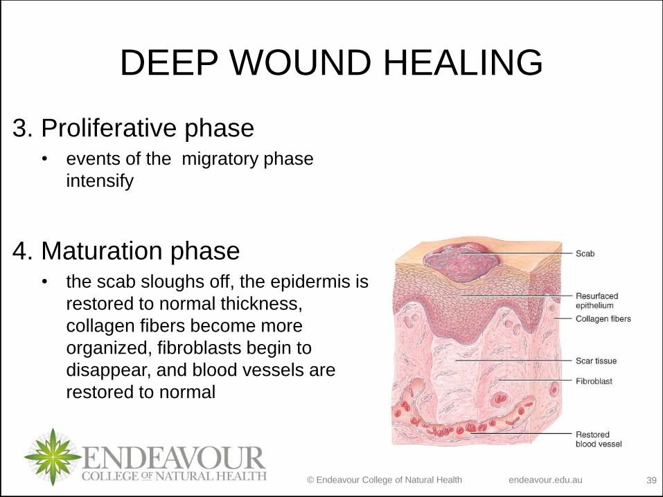
DEEP WOUND HEALING
3. Proliferative phase• events of the migratory phase
intensify
4. Maturation phase• the scab sloughs off, the epidermis is
restored to normal thickness,
collagen fibers become more
organized, fibroblasts begin to
disappear, and blood vessels are
restored to normal
© Endeavour College of Natural Health endeavour.edu.au 40
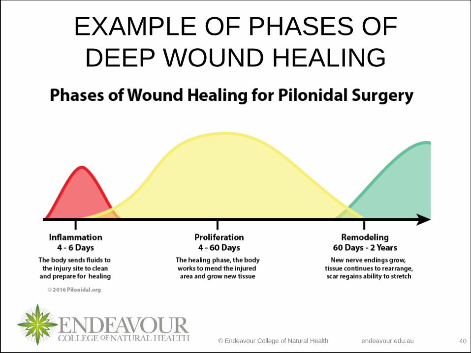
EXAMPLE OF PHASES OF
DEEP WOUND HEALING
© Endeavour College of Natural Health endeavour.edu.au 41
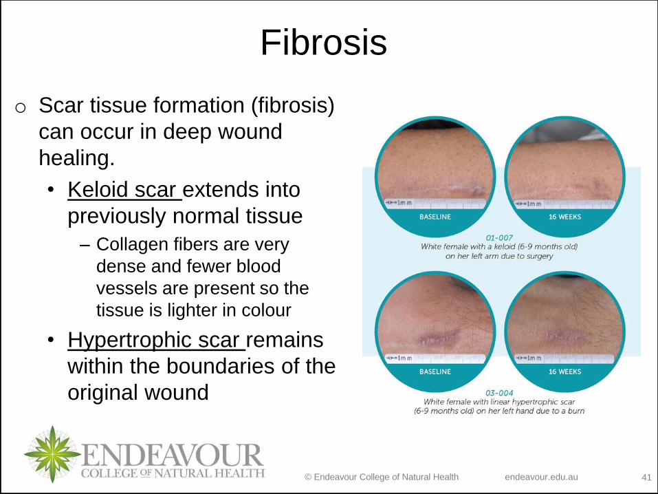
Fibrosis
o Scar tissue formation (fibrosis)
can occur in deep wound
healing.
• Keloid scar extends into
previously normal tissue
– Collagen fibers are very
dense and fewer blood
vessels are present so the
tissue is lighter in colour
• Hypertrophic scar remains
within the boundaries of the
original wound
© Endeavour College of Natural Health endeavour.edu.au 42
BURNS
o Definition: tissue damage from excessive heat, electricity,
radioactivity, or corrosive chemicals.
o Effects:
• Denaturing of proteins in the exposed cells
• Shock due to water, plasma and plasma protein loss
• Circulatory kidney problems from loss of plasma
• Bacterial infection
o Types of burns:
1. First-degree
2. Second-degree burn
3. Third-degree or full-thickness
© Endeavour College of Natural Health endeavour.edu.au 43
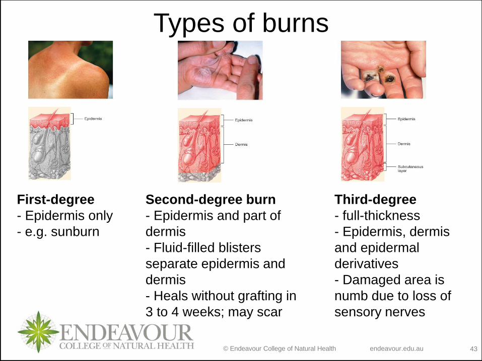
Types of burns
Third-degree
- full-thickness
- Epidermis, dermis
and epidermal
derivatives
- Damaged area is
numb due to loss of
sensory nerves
First-degree
- Epidermis only
- e.g. sunburn
Second-degree burn
- Epidermis and part of
dermis
- Fluid-filled blisters
separate epidermis and
dermis
- Heals without grafting in
3 to 4 weeks; may scar
© Endeavour College of Natural Health endeavour.edu.au 44
BURNS
o Determination of how serious is a burn is done by
considering:
• depth, extent, and area involved
• person’s age and general health
o E.g. when the burn area exceeds 70%, over half of the
victims die
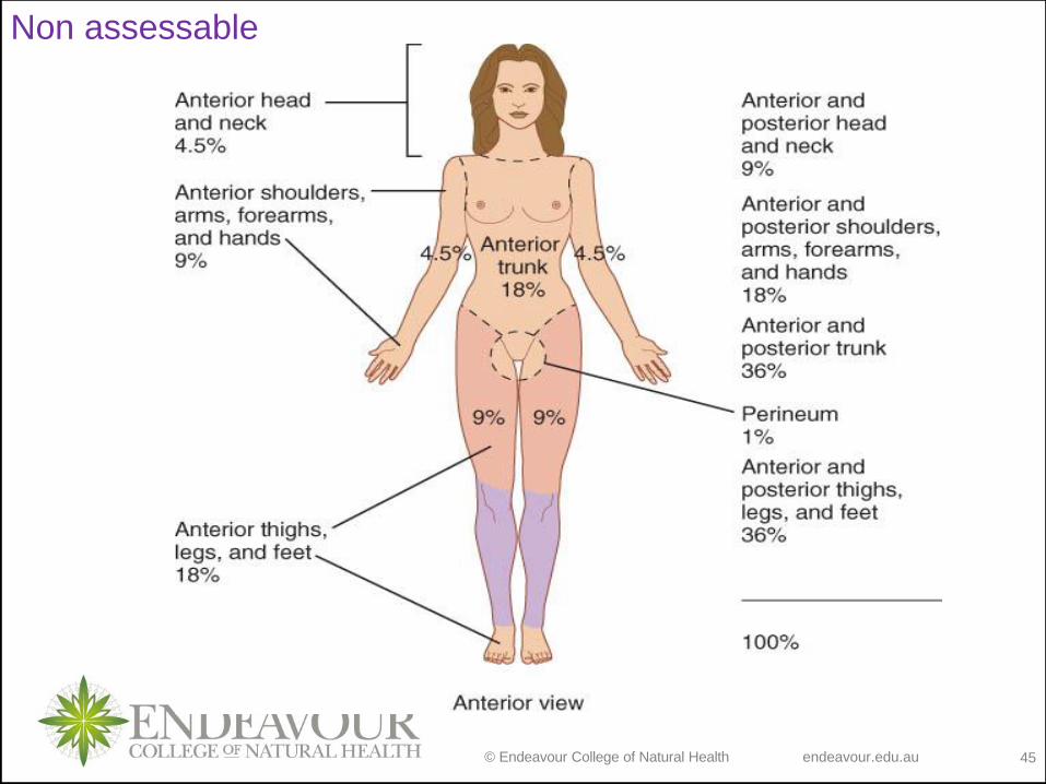
o Two methods for measuring burn extent:
1. rule of nines
2. Lund-Bowder method
© Endeavour College of Natural Health endeavour.edu.au 45
Non assessable
© Endeavour College of Natural Health endeavour.edu.au 46
Recap of Session 6
o Thermoregulation
o Blood reservoir
o Protection
o Cutaneous sensations
o Synthesis of Vitamin D
o Excretion
© Endeavour College of Natural Health endeavour.edu.au 47
Integumentary system is composed of cutaneous membrane (+subcutaneous
layer) and accessory structures
Epidermis contains specialised cells which allow several functions of the
integumentary system (e.g. melanocytes mediate UV protection)
Dermis contains several structures which allow specific functions of the
integumentary system (e.g. free nerve endings allow for cutaneous sensation
of pain)
Subcutaneous layer is composed of specific connective tissue specialised for
energy storage
Accessory structures contribute to the protection and sensation functions of
integumentary system
Recap of Session 6
© Endeavour College of Natural Health endeavour.edu.au 48
Preparation for next session
o Complete any missing concepts and linking words from
Session 6
o NOTE: your Map 1 missing concepts and linking
words should now be complete or near complete.
Please submit the answers for feedback to your lecturer
via a link you will find in Week 5 tab on LMS. This
document is to be submitted in .pdf format.
o Review cytoplasm and extracellular matrix structure and
functions
o Write down any medical terminology you already know