Embed Size (px)
Citation preview
SC I ENCE ADVANCES | R E S EARCH ART I C L E
PHOTON ICS
1Department of Polymer Science, The University of Akron, Akron, OH 44325, USA.2Department of Chemistry, Northwestern University, Evanston, IL 60208, USA.3Materials Science and Engineering Program, University of California, San Diego,La Jolla, CA 92093, USA. 4Department of Chemistry and Biochemistry, Universityof California, San Diego, La Jolla, CA 92093, USA. 5College of Polymer Scienceand Engineering, State Key Laboratory of Polymer Materials Engineering, SichuanUniversity, Chengdu 610065, P. R. China. 6Photonics Research Group, Department ofInformation Technology, Ghent University–imec, Center for Nano- and Biophotonics(NB-Photonics), 9000 Ghent, Belgium. 7Institute of Engineering Thermophysics,Shanghai Jiao Tong University, Shanghai 200240, China. 8Evolution and Optics ofNanostructures Group, Department of Biology, The University of Ghent, 9000 Ghent,Belgium. 9Department of Biology and Integrated Bioscience Program, University ofAkron, Akron, OH 44325, USA.*These authors contributed equally to this work.†Corresponding author. Email: [email protected] (A.D.); [email protected](M.D.S.); [email protected] (N.C.G.)
Xiao et al., Sci. Adv. 2017;3 : e1701151 15 September 2017
Copyright © 2017
The Authors, some
rights reserved;
exclusive licensee
American Association
for the Advancement
of Science. No claim to
original U.S. Government
Works. Distributed
under a Creative
Commons Attribution
NonCommercial
License 4.0 (CC BY-NC).
Bioinspired bright noniridescent photonicmelanin supraballsMing Xiao,1* Ziying Hu,2,3* Zhao Wang,4 Yiwen Li,5 Alejandro Diaz Tormo,6 Nicolas Le Thomas,6
Boxiang Wang,7 Nathan C. Gianneschi,2,3,4† Matthew D. Shawkey,8,9† Ali Dhinojwala1†
Structural colors enable the creation of a spectrumof nonfading colorswithout pigments, potentially replacing toxicmetal oxides and conjugated organic pigments. However, significant challenges remain to achieve the contrastneeded for a complete gamut of colors and a scalable process for industrial application. We demonstrate a feasiblesolution for producing structural colors inspired by bird feathers. We have designed core-shell nanoparticles usinghigh–refractive index (RI) (~1.74)melanin cores and low-RI (~1.45) silica shells. The design of these nanoparticleswasguided by finite-difference time-domain simulations. These nanoparticles were self-assembled using a one-potreverse emulsion process, which resulted in bright and noniridescent supraballs. With the combination of onlytwo ingredients, synthetic melanin and silica, we can generate a full spectrum of colors. These supraballs couldbe directly added to paints, plastics, and coatings and also used as ultraviolet-resistant inks or cosmetics.
Do
on June 4, 2018http://advances.sciencem
ag.org/w
nloaded from
INTRODUCTIONIn the colorful world in which we live, colors are significant not onlyfor aesthetics and pleasure but also for communication, signaling, andsecurity. Colors are produced through either absorption of light bymolecules (pigmentary colors) or scattering of light by nanostructures(structural colors) (1). Structural colors are superior to pigmentarycolors in many ways, because of their tunability, resistance to (photoor chemical) bleaching, and reduced dependence on toxic materials.Many recent studies have demonstrated the use of self-assembly toproduce photonic crystals that generate colors across the visiblespectrum (2). However, we still face significant challenges. Many tradi-tional structural colors are iridescent and thus are not useful for wide-angle displays. Recent examples of noniridescent structural colors lacksufficient color saturation in the absence of absorbingmaterials (carbonblack, gold nanoparticles, or black polypyrrole) to reduce incoherentscattering (3–7). Core-shell nanoparticles with a shell refractive index(RI) similar to water have been used to tune the spacing between coresto achieve optimal scattering for noniridescent colors, but only in solu-tion (8, 9). Although both bottom-up and top-downmethods have beenwidely used (10–12), there is a demand for a scalable process for massproduction of structural colors.
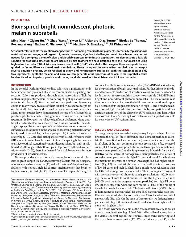
Nature provides many spectacular examples of structural colors,such as green-winged teal (Anas crecca) wing feathers that use hexagonalnonclose-packedmelanosomes (13) andwild turkey (Meleagris gallopavo)feathers with hollow, high-RI contrast melanosomes that brightenfeather colors (Fig. 1A) (14, 15). These examples inspire the design of
core-shell syntheticmelanin nanoparticles (CS-SMNPs) described here,for the production of bright structural colors. Further driven by the de-mand for scalable production of structural colors, we have developed afacile one-pot reverse emulsion process to assemble CS-SMNPs intobright and noniridescent photonic supraballs. The use of melanin asthe core material can increase the brightness and saturation of supra-balls because of its unique combination of high RI and broadband ab-sorption of light. In addition, melanin is biocompatible and candissipate almost 90% of the ultraviolet (UV) radiation into heat withina nanosecond (16, 17), making those melanin-based supraballs suitablefor cosmetics or UV-resistant inks.
RESULTS AND DISCUSSIONTo design an optimal core-shell morphology for producing colors, wefirst used the FDTD (finite-difference time-domain) method to calcu-late the theoretical reflectance spectra (normal incidence) from the(111) plane of themost common photonic crystal with a face-centeredcubic (FCC) packing composed of core-shell nanoparticles and homo-geneous nanoparticles (see the Supplementary Materials for details).Relative to the lattice of homogeneous nanoparticles, the lattice ofcore-shell nanoparticles with high-RI cores and low-RI shells showsthe maximum intensity at a similar wavelength but has higher reflec-tance (Fig. 1B). In contrast, the reverse core-shell structure consistingof low-RI cores and high-RI shells has a much lower reflectance thanthe lattice of homogeneous nanoparticles. These findings are consistentwith previously reported photonic bandgap calculations (18). By vary-ing the ratio of core-to-total radius, we find the highest reflectance(~130% relative to homogeneous nanoparticles) for high-RI core/low-RI shell structure when the core radius is ~60% of the radius ofthe whole core-shell nanoparticle. The lowest reflectance (~52% relativeto homogeneous nanoparticles) for core-shell nanoparticles with low-RI cores is obtained when the core radius is ~80% of the whole core-shellnanoparticle (Fig. 1C). On the basis of these results, we designed nano-particles with high-RI cores and low-RI shells to obtain higher reflec-tance and brighter colors.
We chose synthetic melanin as the core material because it has anunusual combination of high RI (~1.74) and broadband absorption inthe visible spectral region that reduces incoherent scattering andthereby enhances color purity (19). We used silica (RI, ~1.45) as the
1 of 7
SC I ENCE ADVANCES | R E S EARCH ART I C L E
on June 4, 2018http://advances.sciencem
ag.org/D
ownloaded from
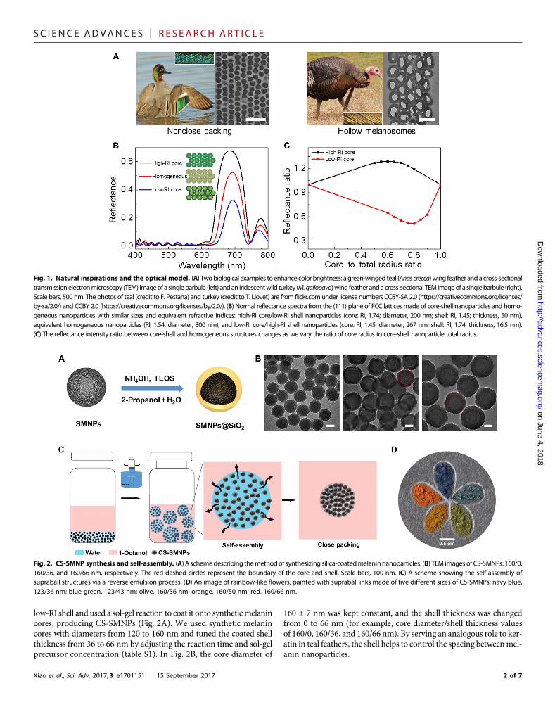
low-RI shell and used a sol-gel reaction to coat it onto syntheticmelanincores, producing CS-SMNPs (Fig. 2A). We used synthetic melanincores with diameters from 120 to 160 nm and tuned the coated shellthickness from 36 to 66 nm by adjusting the reaction time and sol-gelprecursor concentration (table S1). In Fig. 2B, the core diameter of
Xiao et al., Sci. Adv. 2017;3 : e1701151 15 September 2017
160 ± 7 nm was kept constant, and the shell thickness was changedfrom 0 to 66 nm (for example, core diameter/shell thickness valuesof 160/0, 160/36, and 160/66 nm). By serving an analogous role to ker-atin in teal feathers, the shell helps to control the spacing betweenmel-anin nanoparticles.
Fig. 1. Natural inspirations and the optical model. (A) Twobiological examples to enhance color brightness: a green-winged teal (Anas crecca)wing feather andacross-sectionaltransmissionelectronmicroscopy (TEM) imageofa singlebarbule (left) andan iridescentwild turkey (M.gallopavo)wing featherandacross-sectional TEM imageof a singlebarbule (right).Scale bars, 500 nm. The photos of teal (credit to F. Pestana) and turkey (credit to T. Llovet) are from flickr.comunder license numbers CCBY-SA 2.0 (https://creativecommons.org/licenses/by-sa/2.0/) and CCBY 2.0 (https://creativecommons.org/licenses/by/2.0/). (B) Normal reflectance spectra from the (111) plane of FCC latticesmade of core-shell nanoparticles and homo-geneous nanoparticles with similar sizes and equivalent refractive indices: high-RI core/low-RI shell nanoparticles (core: RI, 1.74; diameter, 200 nm; shell: RI, 1.45; thickness, 50 nm),equivalent homogeneous nanoparticles (RI, 1.54; diameter, 300 nm), and low-RI core/high-RI shell nanoparticles (core: RI, 1.45; diameter, 267 nm; shell: RI, 1.74; thickness, 16.5 nm).(C) The reflectance intensity ratio between core-shell and homogeneous structures changes as we vary the ratio of core radius to core-shell nanoparticle total radius.
Fig. 2. CS-SMNP synthesis and self-assembly. (A) A schemedescribing themethodof synthesizing silica-coatedmelaninnanoparticles. (B) TEM images of CS-SMNPs: 160/0,160/36, and 160/66 nm, respectively. The red dashed circles represent the boundary of the core and shell. Scale bars, 100 nm. (C) A scheme showing the self-assembly ofsupraball structures via a reverse emulsion process. (D) An image of rainbow-like flowers, painted with supraball inks made of five different sizes of CS-SMNPs: navy blue,123/36 nm; blue-green, 123/43 nm; olive, 160/36 nm; orange, 160/50 nm; red, 160/66 nm.
2 of 7
SC I ENCE ADVANCES | R E S EARCH ART I C L E
http://advancD
ownloaded from
We used a simple water-in-oil reverse emulsion template method toassemble CS-SMNPs into micrometer-sized supraballs (51 ± 14 mm)(Fig. 2C) (20, 21). No surfactant molecules were used to stabilize theemulsion, and the transient stable emulsion droplets were formedupon shear mixing. The oil phase 1-octanol absorbed small amountsof water (20) and helped to reduce the amount of water in the aqueousphase containing CS-SMNPs. This process slowly removed the waterand helped packing of CS-SMNPs into well-ordered supraballs. Thesesupraballs produce a full spectrum of colors depending on the sizes ofCS-SMNPs (Fig. 2D). This one-pot process is carried out at room tem-perature without additional posttreatment to remove water, and thesupraballs can be easily separated by centrifugation. This process hasa clear advantage over other emulsion-like processes used to producecolorful supraballs that require microwaves or heat to remove water(22–24). In contrast to microfluidic approaches, the reverse emulsionmethod is also easily scalable to produce larger quantities of supraballparticles (5, 9, 25).
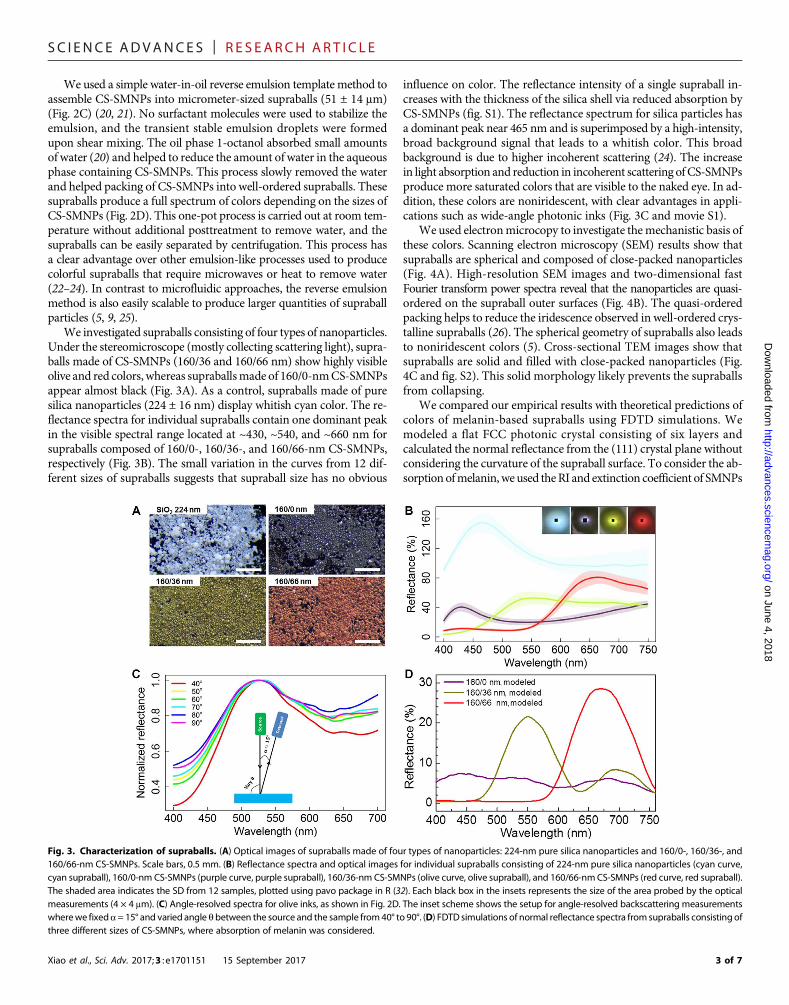
We investigated supraballs consisting of four types of nanoparticles.Under the stereomicroscope (mostly collecting scattering light), supra-balls made of CS-SMNPs (160/36 and 160/66 nm) show highly visibleolive and red colors, whereas supraballsmade of 160/0-nmCS-SMNPsappear almost black (Fig. 3A). As a control, supraballs made of puresilica nanoparticles (224 ± 16 nm) display whitish cyan color. The re-flectance spectra for individual supraballs contain one dominant peakin the visible spectral range located at ~430, ~540, and ~660 nm forsupraballs composed of 160/0-, 160/36-, and 160/66-nm CS-SMNPs,respectively (Fig. 3B). The small variation in the curves from 12 dif-ferent sizes of supraballs suggests that supraball size has no obvious
Xiao et al., Sci. Adv. 2017;3 : e1701151 15 September 2017
influence on color. The reflectance intensity of a single supraball in-creases with the thickness of the silica shell via reduced absorption byCS-SMNPs (fig. S1). The reflectance spectrum for silica particles hasa dominant peak near 465 nm and is superimposed by a high-intensity,broad background signal that leads to a whitish color. This broadbackground is due to higher incoherent scattering (24). The increasein light absorption and reduction in incoherent scattering ofCS-SMNPsproduce more saturated colors that are visible to the naked eye. In ad-dition, these colors are noniridescent, with clear advantages in appli-cations such as wide-angle photonic inks (Fig. 3C and movie S1).
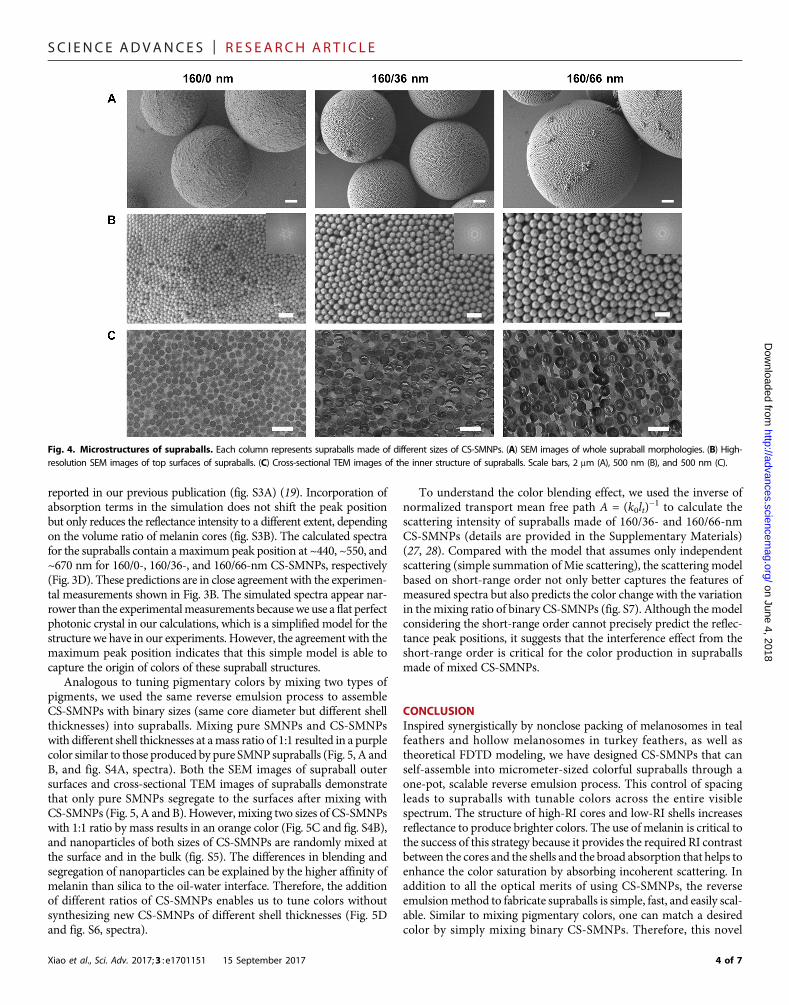
We used electronmicrocopy to investigate themechanistic basis ofthese colors. Scanning electron microscopy (SEM) results show thatsupraballs are spherical and composed of close-packed nanoparticles(Fig. 4A). High-resolution SEM images and two-dimensional fastFourier transform power spectra reveal that the nanoparticles are quasi-ordered on the supraball outer surfaces (Fig. 4B). The quasi-orderedpacking helps to reduce the iridescence observed in well-ordered crys-talline supraballs (26). The spherical geometry of supraballs also leadsto noniridescent colors (5). Cross-sectional TEM images show thatsupraballs are solid and filled with close-packed nanoparticles (Fig.4C and fig. S2). This solid morphology likely prevents the supraballsfrom collapsing.
We compared our empirical results with theoretical predictions ofcolors of melanin-based supraballs using FDTD simulations. Wemodeled a flat FCC photonic crystal consisting of six layers andcalculated the normal reflectance from the (111) crystal plane withoutconsidering the curvature of the supraball surface. To consider the ab-sorptionofmelanin, we used theRI and extinction coefficient of SMNPs
on June 4, 2018es.sciencem
ag.org/
Fig. 3. Characterization of supraballs. (A) Optical images of supraballs made of four types of nanoparticles: 224-nm pure silica nanoparticles and 160/0-, 160/36-, and160/66-nm CS-SMNPs. Scale bars, 0.5 mm. (B) Reflectance spectra and optical images for individual supraballs consisting of 224-nm pure silica nanoparticles (cyan curve,cyan supraball), 160/0-nm CS-SMNPs (purple curve, purple supraball), 160/36-nm CS-SMNPs (olive curve, olive supraball), and 160/66-nmCS-SMNPs (red curve, red supraball).The shaded area indicates the SD from 12 samples, plotted using pavo package in R (32). Each black box in the insets represents the size of the area probed by the opticalmeasurements (4 × 4 mm). (C) Angle-resolved spectra for olive inks, as shown in Fig. 2D. The inset scheme shows the setup for angle-resolved backscattering measurementswherewe fixeda= 15° and varied angle q between the source and the sample from40° to 90°. (D) FDTD simulations of normal reflectance spectra fromsupraballs consisting ofthree different sizes of CS-SMNPs, where absorption of melanin was considered.
3 of 7
SC I ENCE ADVANCES | R E S EARCH ART I C L E
on June 4, 2018http://advances.sciencem
ag.org/D
ownloaded from
reported in our previous publication (fig. S3A) (19). Incorporation ofabsorption terms in the simulation does not shift the peak positionbut only reduces the reflectance intensity to a different extent, dependingon the volume ratio of melanin cores (fig. S3B). The calculated spectrafor the supraballs contain amaximumpeak position at ~440, ~550, and~670 nm for 160/0-, 160/36-, and 160/66-nm CS-SMNPs, respectively(Fig. 3D). These predictions are in close agreement with the experimen-tal measurements shown in Fig. 3B. The simulated spectra appear nar-rower than the experimentalmeasurements becausewe use a flat perfectphotonic crystal in our calculations, which is a simplified model for thestructure we have in our experiments. However, the agreement with themaximum peak position indicates that this simple model is able tocapture the origin of colors of these supraball structures.
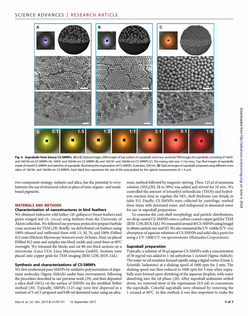
Analogous to tuning pigmentary colors by mixing two types ofpigments, we used the same reverse emulsion process to assembleCS-SMNPs with binary sizes (same core diameter but different shellthicknesses) into supraballs. Mixing pure SMNPs and CS-SMNPswith different shell thicknesses at amass ratio of 1:1 resulted in a purplecolor similar to those produced by pure SMNPsupraballs (Fig. 5, A andB, and fig. S4A, spectra). Both the SEM images of supraball outersurfaces and cross-sectional TEM images of supraballs demonstratethat only pure SMNPs segregate to the surfaces after mixing withCS-SMNPs (Fig. 5, A and B). However, mixing two sizes of CS-SMNPswith 1:1 ratio by mass results in an orange color (Fig. 5C and fig. S4B),and nanoparticles of both sizes of CS-SMNPs are randomly mixed atthe surface and in the bulk (fig. S5). The differences in blending andsegregation of nanoparticles can be explained by the higher affinity ofmelanin than silica to the oil-water interface. Therefore, the additionof different ratios of CS-SMNPs enables us to tune colors withoutsynthesizing new CS-SMNPs of different shell thicknesses (Fig. 5Dand fig. S6, spectra).
Xiao et al., Sci. Adv. 2017;3 : e1701151 15 September 2017
To understand the color blending effect, we used the inverse ofnormalized transport mean free path A = (k0lt)
−1 to calculate thescattering intensity of supraballs made of 160/36- and 160/66-nmCS-SMNPs (details are provided in the Supplementary Materials)(27, 28). Compared with the model that assumes only independentscattering (simple summation ofMie scattering), the scattering modelbased on short-range order not only better captures the features ofmeasured spectra but also predicts the color change with the variationin the mixing ratio of binary CS-SMNPs (fig. S7). Although the modelconsidering the short-range order cannot precisely predict the reflec-tance peak positions, it suggests that the interference effect from theshort-range order is critical for the color production in supraballsmade of mixed CS-SMNPs.
CONCLUSIONInspired synergistically by nonclose packing of melanosomes in tealfeathers and hollow melanosomes in turkey feathers, as well astheoretical FDTD modeling, we have designed CS-SMNPs that canself-assemble into micrometer-sized colorful supraballs through aone-pot, scalable reverse emulsion process. This control of spacingleads to supraballs with tunable colors across the entire visiblespectrum. The structure of high-RI cores and low-RI shells increasesreflectance to produce brighter colors. The use of melanin is critical tothe success of this strategy because it provides the required RI contrastbetween the cores and the shells and the broad absorption that helps toenhance the color saturation by absorbing incoherent scattering. Inaddition to all the optical merits of using CS-SMNPs, the reverseemulsionmethod to fabricate supraballs is simple, fast, and easily scal-able. Similar to mixing pigmentary colors, one can match a desiredcolor by simply mixing binary CS-SMNPs. Therefore, this novel
Fig. 4. Microstructures of supraballs. Each column represents supraballs made of different sizes of CS-SMNPs. (A) SEM images of whole supraball morphologies. (B) High-resolution SEM images of top surfaces of supraballs. (C) Cross-sectional TEM images of the inner structure of supraballs. Scale bars, 2 mm (A), 500 nm (B), and 500 nm (C).
4 of 7
SC I ENCE ADVANCES | R E S EARCH ART I C L E
on Junehttp://advances.sciencem
ag.org/D
ownloaded from
two-component strategy, melanin and silica, has the potential to revo-lutionize the use of structural colors in place of toxic organic- andmetal-based pigments.
4, 2018
MATERIALS AND METHODSCharacterization of nanostructures in bird feathersWe obtained iridescent wild turkey (M. gallopavo) breast feathers andgreen-winged teal (A. crecca) wing feathers from the University ofAkron collection.We followed our previous protocol to prepare barbulecross sections for TEM (29). Briefly, we dehydrated cut feathers using100% ethanol and infiltrated them with 15, 50, 70, and 100% EMbed812 resin (ElectronMicroscopy Sciences) every 24 hours. Next, we placedEMbed 812 resin and samples into block molds and cured them at 60°Covernight. We trimmed the blocks and cut 80-nm-thick sections on amicrotome (Leica UC6, Leica Microsystems GmbH). Sections wereplaced onto copper grids for TEM imaging (JEM-1230, JEOL Ltd.).Synthesis and characterizations of CS-SMNPsWe first synthesized pure SMNPs by oxidative polymerization of dopa-mine molecules (Sigma-Aldrich) under base environment, followingthe procedure described in our previous work (19), and then depositeda silica shell (SiO2) on the surface of SMNPs via the modified Stöbermethod (30). Typically, SMNPs (3.25 mg) were first dispersed in amixture of 5-ml 2-propanol and 0.88-ml deionized water using an ultra-
Xiao et al., Sci. Adv. 2017;3 : e1701151 15 September 2017
sonic method followed bymagnetic stirring. Then, 125 ml of ammoniasolution (NH4OH; 28 to 30%) was added and stirred for 10 min. Wecontrolled the amount of tetraethyl orthosilicate (TEOS) and hydrol-ysis reaction time to regulate the SiO2 shell thickness (see details intable S1). Finally, CS-SMNPs were collected by centrifuge, washedthree times with deionized water, and redispersed in deionized waterfor use in supraball preparation.
To examine the core-shell morphology and particle distributions,we drop-castedCS-SMNPsonto a carbon-coated copper grid forTEM(JEM-1230,JEOLLtd.).Wemeasuredaround40CS-SMNPsusingImageJto obtainparticle size andSD.Wealsomeasured theUV-visible (UV-vis)absorption of aqueous solutions of CS-SMNPs and solid silica particlesusing a UV-1800 UV-vis spectrometer (Shimadzu Corporation).
Supraball preparationTypically, a solution of 30-ml aqueous CS-SMNPs with a concentrationof 30 mg/ml was added to 1-ml anhydrous 1-octanol (Sigma-Aldrich).Thewater-in-oil emulsion formed rapidly using a digital vortex (Genie 2,Scientific Industries) at a shaking speed of 1600 rpm for 2 min. Theshaking speed was then reduced to 1000 rpm for 3 min when supra-balls were formed upon shrinking of the aqueous droplets, with waterdissolving into the oil phase (20). After supraball sediments settleddown, we removed most of the supernatant (0.9 ml) to concentratethe supraballs. Colorful supraballs were obtained by removing the1-octanol at 60°C. In this method, it was also important to make the
Fig. 5. Supraballs from binary CS-SMNPs. (A toC) Optical images, SEM images of top surface of supraballs, and cross-sectional TEM images for supraballs consisting of 160/0-and 160/36-nm CS-SMNPs (A), 160/0- and 160/66-nm CS-SMNPs (B), and 160/36- and 160/66-nm CS-SMNPs (C). Themixing ratio was 1:1 bymass. Top: Real images of supraballsmadeofmixed CS-SMNPs and sketches of supraballs, illustrating the organization of CS-SMNPs. Scale bars, 500nm. (D) Optical images of supraballs preparedusingdifferentmassratios of 160/36- and 160/66-nm CS-SMNPs. Each black box represents the size of the area probed by the optical measurements (4 × 4 mm).
5 of 7
SC I ENCE ADVANCES | R E S EARCH ART I C L E
onhttp://advances.sciencem
ag.org/D
ownloaded from
glass vials hydrophobic so that aqueous droplets did not adhere tothemand break upon contact.We coated an octadecyltrimethoxysilane(OTS) self-assembled monolayer (SAM) onto glass vials following amodified protocol (31). We added 2–volume % OTS toluene solutioninto dry and clean glass vials and degassed for 15 min before tightlyclosing the cap. After 16 hours at room temperature, we rinsed the vialsthree times with toluene and ethanol. Finally, the vials were annealed at120°C under vacuum for 2 hours. To quantify whether OTS SAM wassuccessfully grown onto glass vial, we put a clean glass slide inside thevial during the OTS growth and measured the contact angle of theglass slide (water contact angle, 112° ± 0.6°; fig. S8).
Supraball characterizationThe dried supraballs were imaged under a Leica M80 stereo micro-scope (Leica Microsystems), and we used high-density Teflon tape(TaegaTech) as a white balance. The microscope contained light-emitting diode lights as the source and was connected to a LeicaDMC 4500 camera. The reflectance spectrum of individual supraballswas measured using a CRAIC AX10 UV-vis–near-infrared micro-spectrophotometer (CRAIC Technologies Inc.), with a 75-W xenonshort-arc lamp (Ushio UXL75XE) as a light source. We averagedspectra from 12 supraballs and calculated the SD using pavo packagein R programming software (32). To investigate whether the colors ofsupraballs are angle-independent, we deposited thick films of supra-balls and measured the scattering spectra from different angles usingan AvaSpec spectrometer, with a xenon light source (Avantes Inc.)attached to a custom-built goniometer (Fig. 3C).
The nanostructure of supraball surfaces was characterized using afield-emission SEM (JEOL-7401, JEOL Ltd.). To investigate the innerstructure of supraballs, we dispersed powders of supraballs intoEMbed 812 resin in block molds and cured them at 60°C for 16 hour.The hard blocks were trimmed to a sharp trapezoidal tip using a LeicaS6 EM-Trim 2 (Leica Microsystems), and we then cut 80-nm-thicksections using a diamond knife (Diatome Ltd.) on a Leica UC7 ultra-microtome for TEM.
June 4, 2018
SUPPLEMENTARY MATERIALSSupplementary material for this article is available at http://advances.sciencemag.org/cgi/content/full/3/9/e1701151/DC1Optical Modeltable S1. Conditions used for synthesizing different sizes of CS-SMNPs.fig. S1. UV-vis absorption spectra of pure SMNPs, CS-SMNPs, and pure silica nanoparticles inaqueous solution (20 mg/liter).fig. S2. Representative TEM image of a small supraball made of 160/0-nm CS-SMNPs.fig. S3. FDTD simulations of reflectance spectra at normal incidence using the dimensions ofthe core-shell particles in supraballs.fig. S4. Comparisons of reflectance spectra collected for single supraballs made of melanin,core-shell nanoparticles, and mixtures of melanin and core-shell nanoparticles.fig. S5. TEM images of the inner structure of supraballs of binary CS-SMNPs.fig. S6. Normal reflectance spectra of supraballs made from 160/66- and 160/36-nm CS-SMNPs,and supraballs made by mixing different ratios of 160/66- and 160/36-nm CS-SMNPs.fig. S7. Calculations of inverse of normalized transport mean free path as a function ofdifferent mixing ratios of 160/36- and 160/66-nm CS-SMNPs using the scattering theoryoutlined in SI.fig. S8. Image of a drop of water on an OTS-coated glass.movie S1. Supraball-painted flowers do not change colors when the sample is rotating atdifferent angles.References (33–44)
REFERENCES AND NOTES1. A. G. Dumanli, T. Savin, Recent advances in the biomimicry of structural colours.
Chem. Soc. Rev. 45, 6698–6724 (2016).
Xiao et al., Sci. Adv. 2017;3 : e1701151 15 September 2017
2. J. F. Galisteo‐López, M. Ibisate, R. Sapienza, L. S. Froufe-Pérez, Á. Blanco, C. López,Self-assembled photonic structures. Adv. Mater. 23, 30–69 (2011).
3. Y. Takeoka, S. Yoshioka, A. Takano, S. Arai, K. Nueangnoraj, H. Nishihara, M. Teshima,Y. Ohtsuka, T. Seki, Production of colored pigments with amorphous arrays of black andwhite colloidal particles. Angew. Chem. Int. Ed. 52, 7261–7265 (2013).
4. K. Katagiri, Y. Tanaka, K. Uemura, K. Inumaru, T. Seki, Y. Takeoka, Structural color coatingfilms composed of an amorphous array of colloidal particles via electrophoreticdeposition. NPG Asia Mater. 9, e355 (2017).
5. N. Vogel, S. Utech, G. T. England, T. Shirman, K. R. Phillips, N. Koay, I. B. Burgess, M. Kolle,D. A. Weitz, J. Aizenberg, Color from hierarchy: Diverse optical properties of micron-sizedspherical colloidal assemblies. Proc. Natl. Acad. Sci. U.S.A. 112, 10845–10850 (2015).
6. X. Yang, D. Ge, G. Wu, Z. Liao, S. Yang, Production of structural colors with highcontrast and wide viewing angles from assemblies of polypyrrole black coated polystyrenenanoparticles. ACS Appl. Mater. Interfaces 8, 16289–16295 (2016).
7. Y. Zhang, B. Dong, A. Chen, X. Liu, L. Shi, J. Zi, Using cuttlefish ink as an additive toproduce non-iridescent structural colors of high color visibility. Adv. Mater. 27,4719–4724 (2015).
8. S. Magkiriadou, J.-G. Park, Y.-S. Kim, V. N. Manoharan, Disordered packings of core-shellparticles with angle-independent structural colors. Opt. Mater. Express 2, 1343–1352(2012).
9. J.-G. Park, S.-H. Kim, S. Magkiriadou, T. M. Choi, Y.-S. Kim, V. N. Manoharan, Full-spectrumphotonic pigments with non-iridescent structural colors through colloidal assembly.Angew. Chem. Int. Ed. 53, 2899–2903 (2014).
10. J. D. Forster, H. Noh, S. F. Liew, V. Saranathan, C. F. Schreck, L. Yang, J. G. Park,R. O. Prum, S. G. J. Mochrie, C. S. O’Hern, H. Cao, E. R. Dufresne, Biomimetic isotropicnanostructures for structural coloration. Adv. Mater. 22, 2939–2944 (2010).
11. J. Cui, W. Zhu, N. Gao, J. Li, H. Yang, Y. Jiang, P. Seidel, B. J. Ravoo, G. Li, Inverse opalspheres based on polyionic liquids as functional microspheres with tunable opticalproperties and molecular recognition capabilities. Angew. Chem. Int. Ed. 53,3844–3848 (2014).
12. Z. Gan, M. D. Turner, M. Gu, Biomimetic gyroid nanostructures exceeding their naturalorigins. Sci. Adv. 2, e1600084 (2016).
13. C. M. Eliason, M. D. Shawkey, A photonic heterostructure produces diverse iridescentcolours in duck wing patches. J. R. Soc. Interface 9, 2279–2289 (2012).
14. C. M. Eliason, P.-P. Bitton, M. D. Shawkey, How hollow melanosomes affect iridescentcolour production in birds. Proc. Biol. Sci. 280, 20131505 (2013).
15. M. D. Shawkey, L. D’Alba, M. Xiao, M. Schutte, R. Buchholz, Ontogeny of an iridescentnanostructure composed of hollow melanosomes. J. Morphol. 276, 378–384 (2015).
16. J. B. Nofsinger, S. E. Forest, J. D. Simon, Explanation for the disparity among absorptionand action spectra of eumelanin. J. Phys. Chem. B 103, 11428–11432 (1999).
17. A. Corani, A. Huijser, T. Gustavsson, D. Markovitsi, P.-Å. Malmqvist, A. Pezzella,M. d’Ischia, V. Sundström, Superior photoprotective motifs and mechanisms ineumelanins uncovered. J. Am. Chem. Soc. 136, 11626–11635 (2014).
18. K. P. Velikov, A. Moroz, A. van Blaaderen, Photonic crystals of core-shell colloidal particles.Appl. Phys. Lett. 80, 49–51 (2002).
19. M. Xiao, Y. Li, M. C. Allen, D. D. Deheyn, X. Yue, J. Zhao, N. C. Gianneschi, M. D. Shawkey,A. Dhinojwala, Bio-inspired structural colors produced via self-assembly of syntheticmelanin nanoparticles. ACS Nano 9, 5454–5460 (2015).
20. O. Velev, K. Nagayama, Assembly of latex particles by using emulsion droplets. 3.Reverse (water in oil) system. Langmuir 13, 1856–1859 (1997).
21. D. Liu, F. Zhou, C. Li, T. Zhang, H. Zhang, W. Cai, Y. Li, Black gold: Plasmonic colloidosomeswith broadband absorption self-assembled from monodispersed gold nanospheres byusing a reverse emulsion system. Angew. Chem. Int. Ed. 54, 9596–9600 (2015).
22. S.-H. Kim, S. Y. Lee, G.-R. Yi, D. J. Pine, S.-M. Yang, Microwave-assisted self-organizationof colloidal particles in confining aqueous droplets. J. Am. Chem. Soc. 128,10897–10904 (2006).
23. H. Li, H. Wang, A. Chen, B. Meng, X. Li, Ordered macroporous titania photonic balls bymicrometer-scale spherical assembly templating. J. Mater. Chem. 15, 2551–2556 (2005).
24. Y. Takeoka, S. Yoshioka, M. Teshima, A. Takano, M. Harun-Ur-Rashid, T. Seki, Structurallycoloured secondary particles composed of black and white colloidal particles.Sci. Rep. 3, 2371 (2013).
25. S.-H. Kim, J.-G. Park, T. M. Choi, V. N. Manoharan, D. A. Weitz, Osmotic-pressure-controlledconcentration of colloidal particles in thin-shelled capsules. Nat. Commun. 5, 3068 (2014).
26. O. D. Velev, A. M. Lenhoff, E. W. Kaler, A class of microstructured particles throughcolloidal crystallization. Science 287, 2240–2243 (2000).
27. M. C. W. van Rossum, T. M. Nieuwenhuizen, Multiple scattering of classical waves:Microscopy, mesoscopy, and diffusion. Rev. Mod. Phys. 71, 313–371 (1999).
28. P. Sheng, Introduction to Wave Scattering, Localization and Mesoscopic Phenomena, vol. 88of Springer Series in Materials Science (Springer, 2006).
29. M. Xiao, A. Dhinojwala, M. Shawkey, Nanostructural basis of rainbow-like iridescencein common bronzewing Phaps chalcoptera feathers. Opt. Express 22, 14625–14636(2014).
6 of 7
SC I ENCE ADVANCES | R E S EARCH ART I C L E
Dow
nloaded from
30. W. Stöber, A. Fink, E. Bohn, Controlled growth of monodisperse silica spheres in themicron size range. J. Colloid Interface Sci. 26, 62–69 (1968).
31. Y. Zhang, E. Anim-Danso, S. Bekele, A. Dhinojwala, Effect of surface energy on freezingtemperature of water. ACS Appl. Mater. Interfaces 8, 17583–17590 (2016).
32. R. Maia, C. M. Eliason, P.-P. Bitton, S. M. Doucet, M. D. Shawkey, pavo: an R package forthe analysis, visualization and organization of spectral data. Methods Ecol. Evol. 4,906–913 (2013).
33. E. Akkermans, P. E. Wolf, R. Maynard, G. Maret, Theoretical study of the coherentbackscattering of light by disordered media. J. Geophys. Res. 49, 77–98 (1988).
34. D. S. Wiersma, P. Bartolini, A. Lagendijk, R. Righini, Localization of light in a disorderedmedium. Nature 390, 671–673 (1997).
35. E. Akkermans, G. Montambaux, Mesoscopic Physics of Electrons and Photons(Cambridge Univ. Press, 2007).
36. M. Reufer, L. F. Rojas-Ochoa, S. Eiden, J. J. Sáenz, F. Scheffold, Transport of light inamorphous photonic materials. Appl. Phys. Lett. 91, 171904 (2007).
37. L. Shi, Y. Zhang, B. Dong, T. Zhan, X. Liu, J. Zi, Amorphous photonic crystals with onlyshort-range order. Adv. Mater. 25, 5314–5320 (2013).
38. L. S. Froufe-Pérez, M. Engel, P. F. Damasceno, N. Muller, J. Haberko, S. C. Glotzer,F. Scheffold, Role of short-range order and hyperuniformity in the formation of bandgaps in disordered photonic materials. Phys. Rev. Lett. 117, 053902 (2016).
39. S. F. Liew, J. Forster, H. Noh, C. F. Schreck, V. Saranathan, X. Lu, L. Yang, R. O. Prum,C. S. O’Hern, E. R. Dufresne, H. Cao, Short-range order and near-field effects on opticalscattering and structural coloration. Opt. Express 19, 8208–8217 (2011).
40. L. Tsang, J. A. Kong, Scattering of Electromagnetic Waves: Advanced Topics, vol. 26 of WileySeries in Remote Sensing and Image Processing (John Wiley & Sons, 2004).
41. M. S. Wertheim, Exact solution of the Percus-Yevick integral equation for hard spheres.Phys. Rev. Lett. 10, 321–323 (1963).
42. C. F. Bohren, D. R. Huffman, Absorption and Scattering of Light by Small Particles(John Wiley & Sons, 2008).
Xiao et al., Sci. Adv. 2017;3 : e1701151 15 September 2017
43. C. Mätzler, MATLAB functions for Mie scattering and absorption: Version 2 (ResearchReport No. 2002-11, Institut für Angewandte Physik, 2002); www.atmo.arizona.edu/students/courselinks/spring09/atmo656b/maetzler_mie_v2.pdf.
44. A. Lagendijk, B. Nienhuis, B. A. van Tiggelen, P. de Vries, Microscopic approach to thelorentz cavity in dielectrics. Phys. Rev. Lett. 79, 657 (1997).
Acknowledgments: We thank C. M. Eliason for providing the TEM images for duck and turkeyfeathers. We also thank A. S. Carlini for drawing the three-dimensional schemes of supraballs.Funding: This work was supported by the Air Force Office of Scientific Research (PECASEFA9550-11-1-0105 and FA9550-16-1-0331), NSF (EAR-1251895 and DMR-1105370), and HumanFrontier Science Program (RGY-0083). Author contributions: M.X., Z.H., A.D., M.D.S., and N.C.G.designed the study. M.X., Z.H., Z.W., Y.L., and M.D.S. conducted the experiments. M.X., B.W.,A.D.T., and N.L.T. ran the optical models. All authors contributed to the data interpretation andmanuscript writing. Competing interests: M.X., Z.H., A.D., M.D.S., and N.C.G. are authors on apending patent related to this work filed by the University of Akron (U.S. serial number62/425285; filed on 22 November 2016). The other authors declare that they have no competinginterests. Data and materials availability: All data needed to evaluate the conclusions inthe paper are present in the paper and/or the Supplementary Materials. Additional datarelated to this paper may be requested from the authors.
Submitted 5 April 2017Accepted 17 August 2017Published 15 September 201710.1126/sciadv.1701151
Citation: M. Xiao, Z. Hu, Z. Wang, Y. Li, A. D. Tormo, N. Le Thomas, B. Wang, N. C. Gianneschi,M. D. Shawkey, A. Dhinojwala, Bioinspired bright noniridescent photonic melanin supraballs.Sci. Adv. 3, e1701151 (2017).
htt
7 of 7
on June 4, 2018p://advances.sciencem
ag.org/

Bioinspired bright noniridescent photonic melanin supraballs
Gianneschi, Matthew D. Shawkey and Ali DhinojwalaMing Xiao, Ziying Hu, Zhao Wang, Yiwen Li, Alejandro Diaz Tormo, Nicolas Le Thomas, Boxiang Wang, Nathan C.
DOI: 10.1126/sciadv.1701151 (9), e1701151.3Sci Adv
ARTICLE TOOLS http://advances.sciencemag.org/content/3/9/e1701151
MATERIALSSUPPLEMENTARY http://advances.sciencemag.org/content/suppl/2017/09/11/3.9.e1701151.DC1
REFERENCES
http://advances.sciencemag.org/content/3/9/e1701151#BIBLThis article cites 39 articles, 5 of which you can access for free
PERMISSIONS http://www.sciencemag.org/help/reprints-and-permissions
Terms of ServiceUse of this article is subject to the
registered trademark of AAAS.is aScience Advances Association for the Advancement of Science. No claim to original U.S. Government Works. The title
York Avenue NW, Washington, DC 20005. 2017 © The Authors, some rights reserved; exclusive licensee American (ISSN 2375-2548) is published by the American Association for the Advancement of Science, 1200 NewScience Advances
on June 4, 2018http://advances.sciencem
ag.org/D
ownloaded from