Embed Size (px)
Citation preview
BioOne sees sustainable scholarly publishing as an inherently collaborative enterprise connecting authors, nonprofit publishers, academic institutions, researchlibraries, and research funders in the common goal of maximizing access to critical research.
Biological Responses in Known Bystander Cells Relative to Known Microbeam-Irradiated CellsAuthor(s): Brian Ponnaiya, Gloria Jenkins-Baker, David J. Brenner, Eric J. Hall, Gerhard Randers-Pehrson, and Charles R. GeardSource: Radiation Research, 162(4):426-432. 2004.Published By: Radiation Research SocietyDOI: http://dx.doi.org/10.1667/RR3236URL: http://www.bioone.org/doi/full/10.1667/RR3236
BioOne (www.bioone.org) is a nonprofit, online aggregation of core research in the biological, ecological, andenvironmental sciences. BioOne provides a sustainable online platform for over 170 journals and books publishedby nonprofit societies, associations, museums, institutions, and presses.
Your use of this PDF, the BioOne Web site, and all posted and associated content indicates your acceptance ofBioOne’s Terms of Use, available at www.bioone.org/page/terms_of_use.
Usage of BioOne content is strictly limited to personal, educational, and non-commercial use. Commercial inquiriesor rights and permissions requests should be directed to the individual publisher as copyright holder.

426
RADIATION RESEARCH 162, 426–432 (2004)0033-7587/04 $15.00q 2004 by Radiation Research Society.All rights of reproduction in any form reserved.
Biological Responses in Known Bystander Cells Relative toKnown Microbeam-Irradiated Cells
Brian Ponnaiya,1 Gloria Jenkins-Baker, David J. Brenner, Eric J. Hall, Gerhard Randers-Pehrson and Charles R. Geard
Center for Radiological Research, Columbia University, New York, New York 10032
Ponnaiya, B., Jenkins-Baker, G., Brenner, D. J., Hall, E. J.,Randers-Pehrson, G. and Geard, C. R. Biological Responsesin Known Bystander Cells Relative to Known Microbeam-Irradiated Cells. Radiat. Res. 162, 426–432 (2004).
Normal human fibroblasts in plateau phase (ù95% G1
phase) were stained with the vital nuclear dye Hoechst 33342(blue fluorescence) or the vital cytoplasmic dye Cell TrackerOrange (orange fluorescence) and plated at a ratio of 1:1.Only the blue-fluorescing nuclei were microbeam-irradiatedwith a defined number of 90 keV/mm a particles. The orange-fluorescing cells were then ‘‘bystanders’’, i.e. not themselveshit but adjacent to cells that were. Hit cells showed a fluence-dependent induction of micronuclei as well as delays in pro-gression from G1 to S phase. Known bystander cells alsoshowed enhanced frequencies of micronuclei (intermediate be-tween those seen in irradiated and control cells) and transientcell cycle delays. However, the induction of micronuclei inbystander cells did not appear to be dependent on the fluenceof the particles delivered to the neighboring hit cells. Theseare the first studies in which the bystander effect has beenvisualized directly rather than inferred. They indicate that thephenomenon has a quantitative basis and imply that the targetfor radiation effects cannot be considered to be the individualcell. q 2004 by Radiation Research Society
INTRODUCTION
A basic paradigm in considering the potential risk asso-ciated with exposure to ionizing radiation is that cell nu-clear DNA is the target. In recent years this basic conceptand consequent extrapolations of risk from high to low dos-es have been brought into question. An increasing amountof data from radiation studies has led to the proposal thatthe overall frequency at which a particular end point isobserved in an irradiated population has a component fromcells that are not directly hit by the initial irradiation. Thenonirradiated cells that respond have been termed bystand-ers, a term borrowed from a similar phenomenon reportedin viral transfection experiments (1– 4).
1 Address for correspondence: Radiological Research Facility, Centerfor Radiological Research, Columbia University, 136 S. Broadway, P.O.Box 21, Irvington, NY 10533; e-mail: [email protected].
It has been reported that exposure to very low doses ofa particles initiated sister chromatid exchanges in morecells than it was calculated could have been hit by an aparticle (5, 6). These non-hit, responding cells were ‘‘by-standers’’ either of directly hit cells or of energy deposi-tions in extracellular medium. Similar types of experimentshave also demonstrated the induction of specific genes inmore cells than were estimated to have been hit by a par-ticles (7, 8). Some subsequent studies have confirmed theseresults and pointed to extracellular factors as being respon-sible for these effects, with reactive oxygen species stronglyimplicated (9–12).
Other investigators have transferred medium from cul-tures of irradiated cells into cultures of unirradiated cellsand have observed enhanced cell death and specific geneinduction in the nonirradiated populations (13, 14). Theseresults have been interpreted to indicate that the irradiatedcells release factors into the medium that result in the ob-served changes in the unirradiated recipient cells.
More recent studies of the bystander effect have used acharged-particle microbeam. A microbeam has the abilityto deliver a defined number of a particles (including a sin-gle particle) at very precise locations in a cell population.Thus it is possible to target the nuclei of individual cellswith a defined number of particles. This is in contrast towhole-population ‘‘broad-beam’’ irradiations, where only afraction of the particles actually traverse cell nuclei. Fur-thermore, the number of particles delivered to a populationtypically follows a Poisson distribution, and therefore anyanalysis of the population is by definition an average ofthat population. By contrast, the microbeam allows the pre-cise analysis of each cell in a population, in which thenumber of a-particle traversals received by each cell isknown.
In previous studies, a microbeam was used to irradiate afew cells in a population, with levels of micronuclei and ofapoptosis being much higher than expected, i.e. evidenceof a bystander effect (15, 16). Further, both mutation in-duction (18) and oncogenic transformation (19) have beenshown to be enhanced in bystander cells after microbeamirradiation of known proportions of cells in a population.
Here we compare the cell cycle progression as well asinduction of micronuclei in known human fibroblast by-
427DEFINED BYSTANDER EFFECTS
TABLE 1Cell Numbers and BrdU Labeling Index of Cells Stained with Hoechst 33342 and CTO
Dye 2 h Postirradiation
Number of aparticles
Number of cells counted(number of dishes)
Hoechst-stained CTO-stainedRatio of Hoechst:CTO-stained cells
Percentage cells labeled
Hoechst-stained CTO-stained
0125
25
2114 (9)2138 (9)765 (3)
1338 (6)1607 (7)
2120 (9)2366 (9)668 (3)
1574 (6)1884 (7)
0.9970.9041.1450.8500.853
2.8 6 0.45.5 6 2.63.4 6 2.53.5 6 1.77.0 6 2.1
3.2 6 3.14.8 6 1.94.4 6 2.81.8 6 0.84.1 6 1.8
stander cells to that of microbeam-irradiated cells using amodified staining technique that allows the direct identifi-cation of hit and bystander cells. This approach takes fulladvantage of the ability of the microbeam to target specificcells in a population, and it is the first study in whichknown hit and bystander cells in the same population arevisualized directly and the biological responses in both setsof cells are compared.
MATERIALS AND METHODS
Cell Culture and Microbeam Irradiation
Normal human fibroblasts (Clonetics) were grown to plateau phase inT-25 flasks for 3–7 days prior to experiments. Cells were then split 1:2and reseeded onto T-25 flasks. Once the cells had attached, one flask wasstained with 100 nM Cell Tracker Orange (CTO, Molecular Probes, Eu-gene, OR) for 30 min. At this concentration the dye does not cause anymeasurable cell perturbations, and it gives a clear cytoplasmic signalthrough three to four cell divisions. The other flask was stained with 50nM Hoechst 33342 for 30 min, as done routinely in microbeam studies(18, 19). Flasks were then rinsed and incubated with fresh medium for30 min. Both sets of cells were then trypsinized, counted, mixed in a1:1 ratio, and seeded in a 2-ml drop onto microbeam dishes (coated withCell-Tak to enhance attachment of cells) at a density of 500 cells per dish(250 cells of each type). This resulted in a mean distance of 300 mmbetween cells. Construction of the microbeam dishes and the microbeamirradiation protocol used have been described elsewhere (21). Briefly,these dishes are 60-mm non-tissue-culture dishes that have a 0.25-inchhole drilled in the center to which a 3.8-mm-thick polypropylene film isattached to create a miniwell in which the cells are cultivated. Only theHoechst-stained cells were irradiated with the indicated number of a par-ticles, while the CTO-stained cells were the bystander cells. Cells wereirradiated within 5 h of plating to minimize progression through the cellcycle and movement relative to one another.
Micronucleus Analyses
After irradiation (;7 min per dish) cells were fixed in situ with 100%ice-cold methanol 8, 16, 24, 32 and 48 h after irradiation. Cells werecounterstained with DAPI and were scored for presence of micronuclei(blue fluorescence) in irradiated and bystander cells (orange fluores-cence). Given the limited numbers of cells in a single microbeam exper-iment, data from nine different experiments were pooled. Differences inmicronucleus frequencies between control and irradiated or bystandercells were evaluated using the x2 test.
Analysis of Cell Cycle Progression
After irradiation, BrdU was added (final concentration 1 mM in me-dium) and cells were fixed in situ with 100% ice-cold methanol 2, 24 or
48 h postirradiation. Dividing cells, as indicated by BrdU uptake, wereidentified using a FITC-labeled anti-BrdU antibody (Becton Dickinson)following the manufacturer’s recommendations. Cells were counterstainedwith DAPI and were scored for presence of BrdU uptake (green fluores-cence) in irradiated and bystander cells (orange fluorescence).
RESULTS
Status of Cells at the Time of Irradiation
At each fluence, dishes were fixed in situ at 2 h postir-radiation, and the numbers of each cell type were recorded.In addition, the cell cycle distributions of both populationswere determined using BrdU uptake. These data are pre-sented in Table 1. The number of each cell type rangedfrom 223 to 262 per dish, with a mean of 244 cells. As canbe seen, the ratios of Hoechst-stained to CTO-stained cellsranged from 0.85 to 1.14, in line with our experimentaldesign goal of equal numbers of irradiated and bystandercells. There were no significant differences in the BrdUlabeling index between Hoechst-stained and CTO-stainedcells. In both populations, the percentage of labeled cellswas around 5%, indicating that most of the cells were inG0/G1 at the time of the irradiation. After we had deter-mined conditions at the time of irradiation, irradiated andbystander cells were examined for the presence of micro-nuclei as a function of time postirradiation.
Induction of Micronuclei in Irradiated and BystanderCells
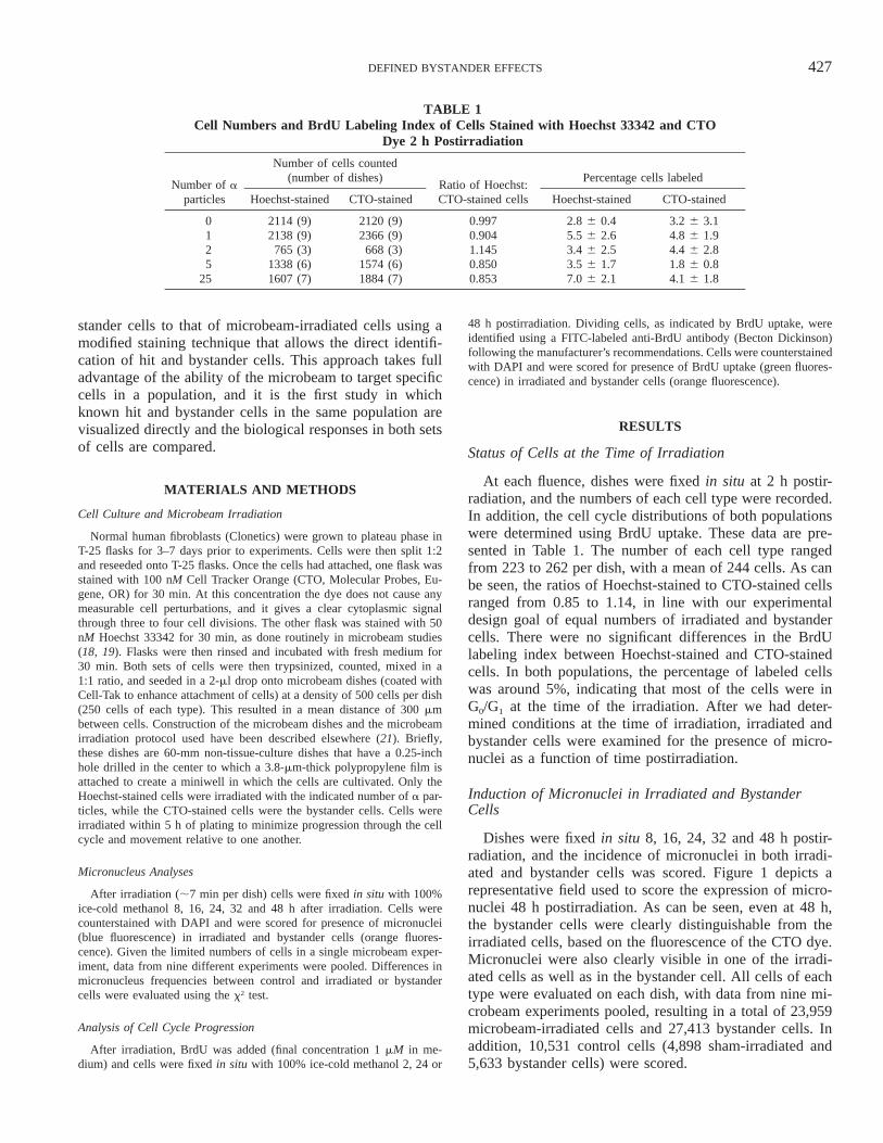
Dishes were fixed in situ 8, 16, 24, 32 and 48 h postir-radiation, and the incidence of micronuclei in both irradi-ated and bystander cells was scored. Figure 1 depicts arepresentative field used to score the expression of micro-nuclei 48 h postirradiation. As can be seen, even at 48 h,the bystander cells were clearly distinguishable from theirradiated cells, based on the fluorescence of the CTO dye.Micronuclei were also clearly visible in one of the irradi-ated cells as well as in the bystander cell. All cells of eachtype were evaluated on each dish, with data from nine mi-crobeam experiments pooled, resulting in a total of 23,959microbeam-irradiated cells and 27,413 bystander cells. Inaddition, 10,531 control cells (4,898 sham-irradiated and5,633 bystander cells) were scored.
428 PONNAIYA ET AL.
FIG. 1. Detection of micronuclei in irradiated and bystander cells at 48 h postirradiation. Bright blue nuclei, onewith a micronucleus, stained with DAPI were originally stained with Hoechst 33342 dye and microbeam-irradiated.Orange fluorescent cells, one with a micronucleus, are bystander cells originally stained with CTO dye. Each celltype can be readily discriminated and categorized. As can be seen, at 48 h, the increase in cell numbers has reducedthe distances between hit and bystander cells.
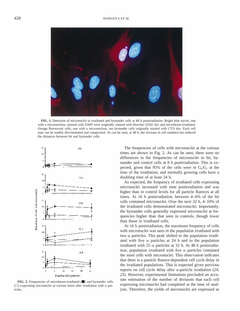
FIG. 2. Frequencies of microbeam-irradiated (m) and bystander cells(V) expressing micronuclei at various times after irradiation with a par-ticles.
The frequencies of cells with micronuclei at the varioustimes are shown in Fig. 2. As can be seen, there were nodifferences in the frequencies of micronuclei in hit, by-stander and control cells at 8 h postirradiation. This is ex-pected, given that 95% of the cells were in G0/G1 at thetime of the irradiation, and normally growing cells have adoubling time of at least 24 h.
As expected, the frequency of irradiated cells expressingmicronuclei increased with time postirradiation and washigher than in control levels for all particle fluences at alltimes. At 16 h postirradiation, between 4–6% of the hitcells contained micronuclei. Over the next 32 h, 4–10% ofthe irradiated cells demonstrated micronuclei. Importantly,the bystander cells generally expressed micronuclei at fre-quencies higher than that seen in controls, though lowerthan those in irradiated cells.
At 16 h postirradiation, the maximum frequency of cellswith micronuclei was seen in the population irradiated withtwo a particles. This peak shifted to the population irradi-ated with five a particles at 24 h and to the populationirradiated with 25 a particles at 32 h. At 48 h postirradia-tion, population irradiated with five a particles containedthe most cells with micronuclei. This observation indicatesthat there is a particle fluence-dependent cell cycle delay inthe irradiated populations. This is expected given previousreports on cell cycle delay after a-particle irradiation (24,25). However, experimental limitations precluded an accu-rate estimation of the number of divisions that each cellexpressing micronuclei had completed at the time of anal-ysis. Therefore, the yields of micronuclei are expressed as
429DEFINED BYSTANDER EFFECTS
TABLE 2Frequencies of Micronuclei Observed in Hit and Bystander Cells after
Microbeam Irradiation
Treatment conditionsNumber of cells
countedExpected numberof micronucleia
Number ofmicronuclei scored
Control
Hoechst-stainedCTO-stainedTotal
48985633
10,531
— 81113194
1 a particle
Hoechst-stainedCTO-stained
82219205 184
436256b
2 a particles
Hoechst-stainedCTO-stained
23061916 38
11439
5 a particles
Hoechst-stainedCTO-stained
68838309 166
547276b
25 a particles
Hoechst-stainedCTO-stained
47606745 135
313180b
a Expected frequencies of micronuclei for bystander cells based on corresponding control values in the absenceof a bystander effect.
b Observed frequencies are significantly different from expected values as determined by the x2 test (P , 0.001).
the totals of those observed at 16, 24, 48 and 72 h postir-radiation (Table 2).
There were no significant differences in the yields ofmicronuclei in control populations stained with eitherHoechst 33342 or CTO dye. Further, these frequencies aresimilar to those reported for normal human fibroblasts (26),indicating that, at the concentrations used, neither Hoechst33342 nor CTO dye induced micronuclei above back-ground levels.
The incidence of micronuclei in the irradiated cells wassomewhat higher than that determined previously in broad-beam experiments performed in this laboratory but was inagreement with frequencies observed in microbeam irradi-ation experiments. This may be due to the inherent hetero-geneity of population irradiations compared to the morehomogeneous nature of microbeam irradiations. Nonethe-less, there was a fluence-dependent increase in the inci-dence of micronuclei. One a particle through the nucleuswas sufficient to increase the incidence to more than threetimes that observed in control cells. The highest frequenciesof micronuclei were observed in cells irradiated with fivea particles (4.8-fold higher than background) that droppedto three times that of controls in cells that received 25 aparticles. This decline is most likely due to the lack of cellsentering mitosis, which is required for the expression ofmicronuclei.
The induction of micronucleus in the bystander cells wassomewhat different. While all bystander populations con-sistently showed increased micronucleus yields comparedto controls, this increase did not appear to be dependent onfluence. Micronucleus incidences in bystander cells were
between 1.3- and 1.65-fold higher than controls, with noclear increase with increasing numbers of a particles deliv-ered to the hit cells. With the exception of bystanders tocells that received two a particles, all bystander populationshad significantly higher incidences of micronuclei (P ,0.001) than expected based on yields of control cells. Im-portantly, at the times of analysis, only a few random by-stander cells were in contact with irradiated cells, and therewas no correlation between these cells and bystander cellsthat expressed micronuclei.
Cell Cycle Delay in Irradiated and Bystander Cells
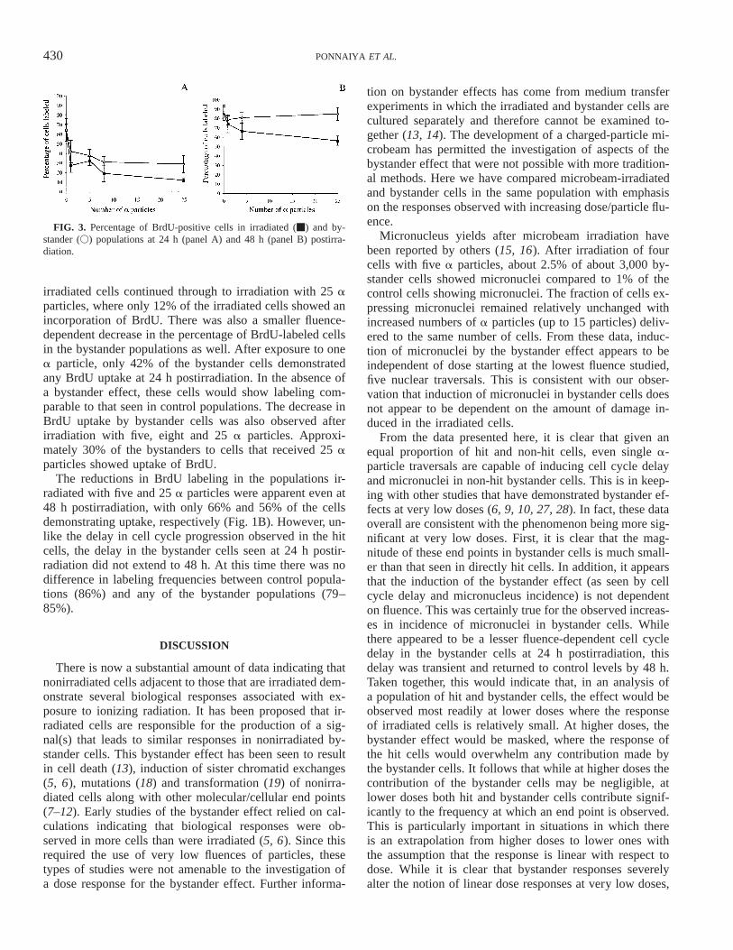
The data on micronucleus incidence discussed abovesuggested that there may be significant fluence-dependentdelay in cell cycle progression in irradiated cells, and thatbystander cells may exhibit similar responses. To determinewhether this was indeed the case, entry of cells into S phaseafter irradiation was monitored using BrdU uptake as anindicator of DNA synthesis. The patterns of BrdU labelingafter irradiation are shown in Fig. 3. At 24 h postirradiation(Fig. 1A), sham-irradiated and bystander control popula-tions contained about 65% labeled cells. This is expected,given that more that 95% of the cells were in G0/G1 at thetime of irradiation, and the labeling indices observed at 2h postirradiation. As expected, there was a fluence-depen-dent decrease in BrdU-positive cells in the irradiated pop-ulations at this time.
Irradiation with one a particle was sufficient to induce asignificant delay, with less than 28% of the irradiated cellsshowing the incorporation of BrdU. This trend in delay of
430 PONNAIYA ET AL.
FIG. 3. Percentage of BrdU-positive cells in irradiated (m) and by-stander (V) populations at 24 h (panel A) and 48 h (panel B) postirra-diation.
irradiated cells continued through to irradiation with 25 aparticles, where only 12% of the irradiated cells showed anincorporation of BrdU. There was also a smaller fluence-dependent decrease in the percentage of BrdU-labeled cellsin the bystander populations as well. After exposure to onea particle, only 42% of the bystander cells demonstratedany BrdU uptake at 24 h postirradiation. In the absence ofa bystander effect, these cells would show labeling com-parable to that seen in control populations. The decrease inBrdU uptake by bystander cells was also observed afterirradiation with five, eight and 25 a particles. Approxi-mately 30% of the bystanders to cells that received 25 aparticles showed uptake of BrdU.
The reductions in BrdU labeling in the populations ir-radiated with five and 25 a particles were apparent even at48 h postirradiation, with only 66% and 56% of the cellsdemonstrating uptake, respectively (Fig. 1B). However, un-like the delay in cell cycle progression observed in the hitcells, the delay in the bystander cells seen at 24 h postir-radiation did not extend to 48 h. At this time there was nodifference in labeling frequencies between control popula-tions (86%) and any of the bystander populations (79–85%).
DISCUSSION
There is now a substantial amount of data indicating thatnonirradiated cells adjacent to those that are irradiated dem-onstrate several biological responses associated with ex-posure to ionizing radiation. It has been proposed that ir-radiated cells are responsible for the production of a sig-nal(s) that leads to similar responses in nonirradiated by-stander cells. This bystander effect has been seen to resultin cell death (13), induction of sister chromatid exchanges(5, 6), mutations (18) and transformation (19) of nonirra-diated cells along with other molecular/cellular end points(7–12). Early studies of the bystander effect relied on cal-culations indicating that biological responses were ob-served in more cells than were irradiated (5, 6). Since thisrequired the use of very low fluences of particles, thesetypes of studies were not amenable to the investigation ofa dose response for the bystander effect. Further informa-
tion on bystander effects has come from medium transferexperiments in which the irradiated and bystander cells arecultured separately and therefore cannot be examined to-gether (13, 14). The development of a charged-particle mi-crobeam has permitted the investigation of aspects of thebystander effect that were not possible with more tradition-al methods. Here we have compared microbeam-irradiatedand bystander cells in the same population with emphasison the responses observed with increasing dose/particle flu-ence.
Micronucleus yields after microbeam irradiation havebeen reported by others (15, 16). After irradiation of fourcells with five a particles, about 2.5% of about 3,000 by-stander cells showed micronuclei compared to 1% of thecontrol cells showing micronuclei. The fraction of cells ex-pressing micronuclei remained relatively unchanged withincreased numbers of a particles (up to 15 particles) deliv-ered to the same number of cells. From these data, induc-tion of micronuclei by the bystander effect appears to beindependent of dose starting at the lowest fluence studied,five nuclear traversals. This is consistent with our obser-vation that induction of micronuclei in bystander cells doesnot appear to be dependent on the amount of damage in-duced in the irradiated cells.
From the data presented here, it is clear that given anequal proportion of hit and non-hit cells, even single a-particle traversals are capable of inducing cell cycle delayand micronuclei in non-hit bystander cells. This is in keep-ing with other studies that have demonstrated bystander ef-fects at very low doses (6, 9, 10, 27, 28). In fact, these dataoverall are consistent with the phenomenon being more sig-nificant at very low doses. First, it is clear that the mag-nitude of these end points in bystander cells is much small-er than that seen in directly hit cells. In addition, it appearsthat the induction of the bystander effect (as seen by cellcycle delay and micronucleus incidence) is not dependenton fluence. This was certainly true for the observed increas-es in incidence of micronuclei in bystander cells. Whilethere appeared to be a lesser fluence-dependent cell cycledelay in the bystander cells at 24 h postirradiation, thisdelay was transient and returned to control levels by 48 h.Taken together, this would indicate that, in an analysis ofa population of hit and bystander cells, the effect would beobserved most readily at lower doses where the responseof irradiated cells is relatively small. At higher doses, thebystander effect would be masked, where the response ofthe hit cells would overwhelm any contribution made bythe bystander cells. It follows that while at higher doses thecontribution of the bystander cells may be negligible, atlower doses both hit and bystander cells contribute signif-icantly to the frequency at which an end point is observed.This is particularly important in situations in which thereis an extrapolation from higher doses to lower ones withthe assumption that the response is linear with respect todose. While it is clear that bystander responses severelyalter the notion of linear dose responses at very low doses,
431DEFINED BYSTANDER EFFECTS
what impact this would have on risk assessment at lowdoses remains in question. The data presented here on DNAdamage as well as that on enhanced mutation and transfor-mation frequencies in bystander cells (18, 19) would sug-gest a supra-linear response. That is, at low doses, signifi-cantly more cells are damaged, mutated and transformedthan would be predicted by linear extrapolation from higherdoses, indicating a greater than expected risk at these doses.However, other data suggest a more protective role for thephenomenon. For example, the detection of apoptosis inunirradiated cells (29) would suggest that the presence ofbystander responses serves to eliminate altered cells andwould argue that alterations to risk estimates are unneces-sary.
It is important to stress that all the experiments per-formed here used densely ionizing (high-LET) radiations inwhich a clear delineation between hit and non-hit cells canbe achieved. How far these conclusions apply to sparselyionizing (low-LET) radiation such as X or g rays is stillunclear.
There appear to be at least two different mechanisms bywhich bystander effects are propagated. Several investiga-tors have suggested that in confluent cultures, gap junctioncommunications may be responsible for responses in by-stander cells (7, 8, 14, 18). In these situations the preven-tion of gap junction formation by the use of either mutantcell lines or inhibitors resulted in the suppression of theeffect. While gap junction communication may be involvedin the transfer of signals when irradiated and bystander cellsare in direct contact, it is unlikely in the present experi-ments. Here cells were seeded at low numbers to allowsufficient space to progress through the cell cycle, to moveand to express micronuclei after cellular division. As a re-sult, only a small fraction of cells were in contact with eachother. The bystander effects observed in this study are morelikely due to the release of factors from irradiated cells thatare responsible for the responses in unirradiated neighbor-ing cells. This mechanism has previously been postulatedto explain bystander effects observed in medium transferstudies in which the two sets of cells are never in contactwith each other (9, 14, 28). It is thought that deposition ofenergy in the medium or in cells generates factors in themedium that can then influence nonirradiated cells thatshare the same environment to produce responses similarto those observed in the irradiated cells. While little isknown about the precise mechanisms whereby this takesplace, several candidate molecules have been proposed, in-cluding IL8, TGFB and TNFA.
Others have reported data that suggest reactive oxygenspecies may be the signal between hit and non-hit cells (9–12). Given the nature of the experiments conducted here, itis not possible to either support or discount the possibilitythat reactive oxygen species play a role in this observedbystander effect.
In conclusion, this study is the first to take full advantageof microbeam irradiation techniques that allow the irradi-
ation of specific cells in a population. Using a unique stain-ing protocol, it was possible to follow the fate of individualirradiated and bystander cells in the same population, thishas allowed a direct contrast of the biological responsesobserved in irradiated and bystander cells.
ACKNOWLEDGMENTS
This work was supported by grants CA-75061 and CA-49062 from theNational Institutes of Health and by grant DE-FG03-00-ER62909 fromthe U.S. Department of Energy. All irradiations were conducted at theColumbia University Radiological Research Accelerator Facility (RAR-AF) an NIH-supported Research Center (RR-11623).
Received: February 24, 2004; accepted: June 1, 2004
REFERENCES
1. F. L. Moolten and J. M. Wells, Curability of tumors bearing herpesthymidine kinase genes transferred by retroviral vectors. J. Natl. Can-cer Inst. 82, 297–300 (1990).
2. K. W. Culver, Z. Ram, S. Wallbridge, H. Ishii, E. H. Oldfield andR. M. Blaese, In vivo gene transfer with retroviral vector-producercells for treatment of experimental brain tumors. Science 256, 1550–1552 (1992).
3. S. M. Freeman, C. N. Aboud, K. A. Whartenby, C. H. Packman,D. S. Koeplin, F. L. Moolten and G. N. Abraham, The ‘‘bystandereffect’’: tumor regression when a fraction of the tumor mass is ge-netically modified. Cancer Res. 53, 5274–5283 (1993).
4. W. Hamel, L. Magnelli, V. P. Chiarugi and M. A. Israel, Herpessimplex virus thymidine kinase/gancyclovir-mediated apoptotic deathof bystander cells. Cancer Res. 56, 2697–2702 (1996).
5. H. Nagasawa and J. B. Little, Induction of sister chromatid exchangesby extremely low doses of alpha particles. Cancer Res. 52, 6394–6396 (1992).
6. A. Deshpande, E. H. Goodwin, S. M. Bailey, B. Marrone and B.Lehnert, Alpha-particle-induced sister chromatid exchange in normalhuman lung fibroblasts: Evidence for an extranuclear target. Radiat.Res. 145, 260–267 (1996).
7. E. I. Azzam, S. M. de Toledo, T. Gooding and J. B. Little, Intercel-lular communication is involved in the bystander regulation of geneexpression in human cells exposed to very low fluences of alphaparticles. Radiat. Res. 150, 497–504 (1998).
8. E. I. Azzam, S. M. de Toledo and J. B. Little, Direct evidence forthe participation of gap junction-mediated intercellular communica-tion in the transmission of damage signals from a particle irradiatedto nonirradiated cells. Proc. Natl. Acad. Sci. USA 98, 473–478(2001).
9. B. E. Lehnert and E. H. Goodwin, A new mechanism for DNA al-terations induced by alpha particles like those emitted by radon andradon progeny. Cancer Res. 57, 2164–2171 (1997).
10. P. K. Narayanan, E. H. Goodwin and B. E. Lehnert, Alpha particlesinitiate biological production of superoxide anions and hydrogen per-oxide in human cells. Cancer Res. 57, 3963–3971 (1997).
11. P. K. Narayanan, K. E. A. LaRue, E. H. Goodwin and B. E. Lehnert,Alpha particles initiate biological production of interleukin-8 in hu-man cells. Radiat. Res. 152, 57–63 (1999).
12. A. W. Hickman, R. J. Jaramillo, J. F. Lechner and N. F. Johnson,Alpha-particle-induced p53 protein expression in a rat lung epithelialcell strain. Cancer Res. 54, 5797–5800 (1994).
13. C. Seymour and C. Mothersill, Delayed expression of lethal muta-tions and genomic instability in the progeny of human epithelial cellsthat survived in a bystander-killing environment. Radiat. Oncol. In-vest. 5, 106–110 (1997).
14. C. Mothersill and C. Seymour, Cell-cell contact during gamma irra-diation is not required to induce a bystander effect in normal human
432 PONNAIYA ET AL.
keratinocytes: Evidence for release during irradiation of a signal con-trolling survival into medium. Radiat. Res. 149, 256–262 (1998).
15. K. M. Prise, O. V. Belyakov, M. Folkard and B. D. Michael, Studiesof bystander effects in human fibroblasts using a charged particlemicrobeam. Int. J. Radiat. Biol. 74, 793–798 (1998).
16. O. V. Belyakov, A. M. Malcolmson, M. Folkard, K. M. Prise andB. D. Michael, Direct evidence for a bystander effect of ionizingradiation in primary human fibroblasts. Br. J. Cancer 84, 674–679(2001).
17. S. Lorimore, M. A. Kadhim, D. A. Pocock, D. Papworth, D. L. Ste-vens, D. T. Goodhead and E. G. Wright, Chromosomal instability inthe descendants of unirradiated surviving cells after a-particle irra-diation. Proc. Natl. Acad. Sci. USA 95, 5730–5733 (1998).
18. H. Zhou, G. Randers-Pehrson, C. A. Waldren, D. Vannais, E. J. Halland T. K. Hei, Induction of a bystander mutagenic effect of alphaparticles in mammalian cells. Proc. Natl. Acad. Sci. USA 97, 2099–2104 (2000).
19. S. Sawant, G. Randers-Pehrson, C. R. Geard, D. J. Brenner andE. J. Hall, The bystander effect in radiation oncogenesis: I. Trans-formation in C3H 10T½ cells in vitro can be initiated in the unirra-diated neighbors of irradiated cells. Radiat. Res. 155, 397–401(2001).
20. L. J. Wu, G. Randers-Pehrson, A. Xu, C. A. Waldren, C. R. Geard,Z. Yu and T. K. Hei, Targeted cytoplasmic irradiation with alphaparticles induces mutations in mammalian cells. Proc. Natl. Acad.Sci. USA 96, 4959–4964 (1999).
21. G. Randers-Pehrson, C. R. Geard, G. Johnson, C. D. Elliston andD. J. Brenner, The Columbia University single-ion microbeam. Ra-diat. Res. 156, 210–214 (2001).
22. N. Barboule, V. Baldin, S. Jozan, S. Vidal and A. Valette, Increasedlevel of p21 in human ovarian tumors is associated with increasedexpression of cdk2, cyclin A and PCNA. Int. J. Cancer 76, 891–896(1998).
23. S. Clasen, W. A. Schulz, C-D. Gerharz, M-O. Grimm, F. Christophand B. J. Schmitz-Drager, Frequent and heterogenous expression ofcyclin-dependent kinase inhibitor WAF1/p21 protein and mRNA inurothelial carcinoma. Br. J. Cancer 77, 515–521 (1998).
24. D. M. Gadbois, H. A. Crissman, A. Nastasi, R. Habbersett, S. K.Wang, D. Chen and B. E. Lenhert, Alterations in the progression ofcells through the cell cycle after exposure to alpha particles or gammarays. Radiat. Res. 146, 414–424 (1996).
25. H. Sasaki, Cell killing and division delay in asynchronous and syn-chronized HeLa cells irradiated with alpha particles or X rays. Radiat.Res. 99, 311–323 (1984).
26. M. N. Cornforth and E. H. Goodwin, Transmission of radiation-in-duced acentric chromosomal fragments to micronuclei in normal hu-man fibroblasts. Radiat. Res. 126, 210–217 (1991).
27. D. J. Brenner, J. B. Little and R. K. Sachs, The bystander effect inradiation oncogenesis: II. A quantitative model. Radiat. Res. 155,402–408 (2001).
28. R. Iyer and B. E. Lehnert, Factors underlying the cell growth-relatedbystander responses to alpha particles. Cancer Res. 60, 1290–1298(2000).


![[PPT]PowerPoint · Web viewWhite blood cells, broadly known as leukocytes, are actually several different cells. Furthermore, ... PowerPoint Presentation Last modified by: GLEICHER,](https://img.pdfslide.net/doc/110x75/5aa8a40d7f8b9a9a188bd96a/pptpowerpoint-viewwhite-blood-cells-broadly-known-as-leukocytes-are-actually.jpg)