Embed Size (px)
Citation preview
1
BIOLOGY OF ZIKA VIRUS INFECTION IN HUMAN SKIN CELLS 1 2 3
Running Title: Cellular tropism and entry receptors of Zika virus 4
5
Rodolphe Hamel1, Ophélie Dejarnac2$, Sineewanlaya Wichit1$, Peeraya Ekchariyawat1, 6
Aymeric Neyret3, Luplertlop Natthanej4, Manuel Perera-Lecoin1, Pornapat 7
Surasombatpattana,5 Loïc Talignani1, Frédéric Thomas1, Van-Mai Cao-Lormeau6, Valérie 8
Choumet7, Laurence Briant3, Philippe Desprès8, Ali Amara2, Hans Yssel9 and Dorothée 9
Missé1# 10
11
1Laboratoire MIVEGEC, UMR 224 IRD/CNRS/UM1, Montpellier, France 12
2Inserm, U944, Laboratoire de Pathologie et Virologie Moléculaire, Paris, France 13
3 Centre d’étude d’agents Pathogènes et Biotechnologies pour la Santé, CNRS-UMR 14
5236/UM1/UM2, Montpellier, France 15
4Department of Microbiology and Immunology, Faculty of Tropical Medicine, Mahidol 16
University, Bangkok, Thailand 17
5Pathology Department, Prince of Songkla University, Songkla, Thailand 18
6Institut Louis Malardé, Papeete, Tahiti, French Polynesia 19
7Unit Environment and Infectious Risks, Institut Pasteur, Paris, France 20 8Département infections et Epidémiologie, Institut Pasteur, 75724 Paris et UMR PIMIT (I2T 21
team), Université de La Réunion, Inserm U1187, CNRS 9192, IRD 249, GIP-CYROI, 97491 22
Saint Clotilde, La Réunion, France. 23 9 Centre d’Immunologie et des Maladies Infectieuses, Inserm, U1135, Sorbonne Universités, 24
UPMC, APHP Hôpital Pitié-Salpêtrière, Paris, France 25
26
Word count for the abstract: 249 27
Word count for the text: 7039 28
JVI Accepted Manuscript Posted Online 17 June 2015J. Virol. doi:10.1128/JVI.00354-15Copyright © 2015, American Society for Microbiology. All Rights Reserved.
2
29
Abbreviations: ZIKV, Zika virus; DENV, Dengue Virus; WNV, West Nile virus; PRRs, 30
pattern recognition receptors; TLR, Toll-like receptor; RIG-I, retinoic acid-inducible gene 1; 31
MDA-5, melanoma differentiation-associated gene 5; ISG, interferon-stimulated gene; DC-32
SIGN, dendritic cell-specific intracellular adhesion molecule 3-grabbing non-integrin; Ae., 33
Aedes; CLEC5, C-type lectin domain family 5; IU, international Unit; LC3, cytosolic 34
microtubule-associated light chain 3; OAS, oligoadenylate synthetase. 35
36
Keywords: Zika, arbovirus, Dengue, skin, fibroblast, keratinocyte, dendritic cells, AXL, 37
Phosphatidylserine receptor, antiviral response, autophagy, flavivirus. 38
39 $ contributed equally to this work 40
# Corresponding author: Address correspondence to Dorothée Missé, Email: 41 [email protected] 42 43
44
3
ABSTRACT 45
Zika virus (ZIKV) is an emerging arbovirus of the Flaviviridae family that includes Dengue, 46
West Nile, Yellow Fever and Japanese encephalitis viruses, causing a mosquito-borne disease 47
transmitted by the Aedes genus, with recent outbreaks in the South Pacific. Here, we 48
determine the importance of the human skin in the entry of ZIKV and its contribution to the 49
induction of anti-viral immune responses. We show that human dermal fibroblasts, epidermal 50
keratinocytes and immature dendritic cells are permissive to the most recent ZIKV isolate, 51
responsible for the epidemic in French Polynesia. Several entry and/or adhesion factors, 52
among which DC-SIGN, AXL, TYRO3, and to a lesser extent, TIM-1, permitted ZIKV entry 53
with a major role for the TAM receptor AXL. ZIKV permissiveness of human skin fibroblasts 54
was confirmed by the use of a neutralizing Ab and specific RNA silencing. ZIKV induced the 55
transcription of TLR-3, RIG-I and MDA5, as well as several interferon-stimulated genes, 56
including OAS2, ISG15 and MX1, characterized by a strongly enhanced interferon-β gene 57
expression. ZIKV was found to be sensitive to the antiviral effect of both type I and type II 58
interferons. Finally, infection of skin fibroblasts resulted in the formation of autophagosomes 59
whose presence was associated with enhanced viral replication, as shown by the use of Torin 60
1, a chemical inducer of autophagy or the specific autophagy inhibitor 3-Methyladenine. The 61
results presented herein permit to gain better insight in the biology of ZIKV and to devise 62
strategies aiming to interfere with the pathology caused by this emerging Flavivirus. 63
64
65
4
IMPORTANCE 66
Zika virus (ZIKV) is an arbovirus belonging to Flaviviridae family. Vector-mediated 67
transmission of ZIKV is initiated when a blood-feeding female Aedes mosquito injects the 68
virus into the skin of its mammalian host, followed by infection of permissive cells via 69
specific receptors. Indeed, skin immune cells, including dermal fibroblasts, epidermal 70
keratinocytes and immature dendritic cells, were all found to be permissive to ZIKV infection. 71
The results also show a major role for the phosphatidylserine receptor AXL as a ZIKV entry 72
receptor, and cellular autophagy in enhancing ZIKV replication in permissive cells. ZIKV 73
replication leads to activation of an antiviral innate immune response and the production of 74
type I interferons in infected cells. Taken together, these results provide for the first time a 75
general insight into the interaction between ZIKV and its mammalian host. 76
77
5
INTRODUCTION 78
Zika virus (ZIKV) is a little known emerging mosquito-borne Flavivirus, belonging to the 79
Flaviviridae family that is closely related to the Spondweni serocomplex. Like other members 80
of the Flavivirus genus, ZIKV contains a positive single-stranded genomic RNA, encoding a 81
polyprotein that is processed into three structural proteins, the capsid (C), the precursor of 82
membrane (prM) and the envelope (E), and seven nonstructural proteins NS1 to NS5 (1). 83
Virus replication occurs in the cellular cytoplasm. 84
Epidemiological studies point out to a wide-spread distribution of ZIKV in the northern half 85
of the African continent, as well as in many countries in south-east Asia, including Malaysia, 86
India, the Philippines, Thailand, Vietnam, Indonesia, and Pakistan (2-9). Many different 87
mosquito Aedes species can account for the transmission of ZIKV, including Ae. aegypti (10, 88
11) which is at present considered to be the main vector of the virus in South and South-East 89
Asia, (11, 12). The first human ZIKV infection has been reported in Uganda in 1964 (2, 3, 5, 90
13) and the virus was later isolated from humans in South East Asia (8, 14-16). Despite this 91
broad geographical distribution, human ZIKV infections have remained sporadic and limited 92
to small-scale epidemics for decades, until 2007 when a large epidemic was reported on Yap 93
Island, a territory of the Federated States of Micronesia, with nearly 75% of the population 94
being infected with the virus (17). Moreover, an outbreak of a syndrome due to Zika fever has 95
been reported in French Polynesia, in addition to several cases of ZIKV infection in New 96
Caledonia, Easter Island and the Cook Islands, indicating a rapid spreading of the virus in the 97
Pacific (18). Also, two imported cases of ZIKV infection of travellers from Indonesia and the 98
Cook Islands, respectively, to Australia and two from Thailand to Europe and Canada, 99
respectively have been described recently (19-22), emphasizing the capacity of ZIKV to 100
spread to non-endemic areas where the proper mosquito vector might be present. The largest 101
outbreak of ZIKV ever reported was characterized by fever, rash, arthralgia and conjunctivitis 102
6
in infected individuals. Moreover, during the recent outbreak in French Polynesia, ZIKV 103
infection-related neurological disorders were also described and the incidence of Guillain 104
Barré syndrome unexpectedly increased by 20 fold (23). In absence of monkeys in French 105
Polynesia, it is likely that humans served as primary amplification hosts for ZIKV. 106
Because ZIKV has received far less attention than other emerging arboviruses such as Yellow 107
fever, dengue (DENV), West Nile (WNV), Japanese encephalitis and Chikungunya viruses, 108
pathogenesis of ZIKV infection remains still poorly understood. Mosquito-mediated 109
transmission of ZIKV is initiated when a blood-feeding female Aedes mosquito injects the 110
virus into the human skin followed by infection of permissive cells (reviewed in (24)). 111
However, no information is available, neither on the nature of the skin cells that are 112
permissive to infection with ZIKV, nor on the entry receptor used by this Flavivirus. 113
Moreover, the mechanisms of ZIKV infection, the signaling pathways and the anti-viral 114
immune response of the host, elicited by this virus, remain to be determined. In the present 115
study, we describe the entry receptors and cellular targets of the most recent ZIKV isolate, 116
responsible for the recent epidemic in French Polynesia, to provide a general insight into the 117
interaction between this virus and its human host. 118
119
MATERIELS AND METHODS 120
Ethics Statement 121
The study was approved by the IRD ethics committee (registration number: AC-2013-1754) 122
and all donors gave written informed consent. 123
124
Cells, virus and reagents 125
Ae. albopictus C6/36 cells, used for propagation of the ZIKV strain were grown in DMEM 126
(Invitrogen, Cergy Pontoise, France), supplemented with 10% fetal calf serum (FCS, Lonza, 127
7
Basel, Switzerland) at 28°C, as previously described (25). Primary human dermal fibroblasts 128
were obtained from neonatal foreskins and cultured at 37°C, 5% CO2, in Fibroblast Basal 129
Medium-2, supplemented with FGM-2 supplements and growth factors (all purchased at 130
Lonza). The HFF-1 skin fibroblast cell line, HEK293T, A549 and Vero cells were maintained 131
in DMEM supplemented with 10% FCS. HEK293T cells stably expressing DC-SIGN, TIM-1, 132
TIM-4, AXL or Tyro3 have been reported previously (26). Primary human epidermal 133
keratinocytes were obtained from neonatal foreskins (Lonza, Basel, Switzerland) and cultured 134
at 37 °C, 5% CO2, in Keratinocyte Basal Medium-2 (Lonza, Basel, Switzerland) 135
supplemented with KGM-2 supplements and growth factors (Lonza, Basel, Switzerland). 136
Low-passaged PF-25013-18 strain of ZIKV (a generous gift of VM. Cao-Lormeau and D. 137
Musso ; ILM, Tahiti island, French Polynesia) isolated from a viremic patient in French 138
Polynesia in 2013, was used in the current study and grown in mosquito Ae. albopictus C6/36 139
cells. The DENV-2 Jamaica/N.1409 strain (GenBank: M20558.1) was propagated in AP61 140
cell monolayers after having undergone limited cell passages. Recombinant human IFN-α, 141
IFN-β, IFN-γ and Torin 1 autophagy inducer were purchased from R&D Systems (Lille, 142
France). The autophagy inhibitor 3-MA was purchased from Sigma. 143
144
Dendritic Cell Generation 145
Human peripheral blood mononuclear cells were isolated from healthy donors by density 146
centrifugation over Ficoll-Paque Plus (Amersham Biosciences, France). Monocytes were 147
negatively selected with magnetic beads coated with a mixture of antibodies (Miltenyi Biotec, 148
France) and seeded at 106 cells/ml in RPMI 1640 supplemented with 10% FCS, 1% 149
penicillin/streptomycin, 50 ng/ml recombinant human interleukin-4 (PeproTech, France), and 150
100 ng/ml recombinant human granulocyte macrophage colony-stimulating factor 151
(PeproTech, France) for 7 days. 152

8
ZIKV infection of cells 153
For infection, cells were seeded in culture plates at 4.104 cells per cm2. Then, cells were 154
rinsed once with PBS and the ZIKV diluted to the desired MOI was added to the cells. The 155
cells were incubated for 2 h at 37°C with gentle agitation every 30 min. Next, the inoculum 156
was removed and the cells were washed twice with PBS. Culture medium was added to each 157
well and the cells were incubated at 37 C, 5% CO2 for the duration of the experiment. As a 158
control, fibroblasts were incubated with culture supernatant from uninfected C6/36 cells, 159
referred to in the present study as mock-infected cells. 160
161
ZIKV infection of human skin biopsies 162
Fresh, sterile human skin biopsies with a size of 88 mm were obtained from three adult 163
healthy donors (age range: 38-43 years) following abdominoplastic surgery. The skin explants 164
were cultured, as described by Limon-Flores et al (27). For infection, 106 PFU in a final 165
volume of 50 μl DMEM were injected per biopsy. As a control, skin biopsies were injected 166
with culture supernatant from uninfected C6/36 cells in a final volume of 50 μl DMEM. At 167
different time points, the biopsies were harvested and soaked with enzymes which degrade the 168
extracellular matrix by using the whole skin dissociation kit (Miltenyi Biotec, Paris, France). 169
In a second step, single-cells were freed from the extracellular matrix by using the 170
gentleMACS™ Dissociator (Miltenyi Biotec, Paris, France) and cells were lysed with 171
TriREAGENT (Sigma, Saint Quentin Fallavier, France) for RT-PCR analysis. For 172
histological analysis biopsies were fixed in neutral-buffered formalin and embedded in 173
paraffin. Tissue sections (3-5 µm) were stained with hematoxilin-eosin, put on slides and 174
digitalized with Nanozoomer scanner (Hamamatsu, Massy, France) with a 20x objective. 175
176
177
9
ZIKV real-time RT-PCR 178
Total RNA was extracted from human fibroblasts using TriREAGENT (Sigma, Saint Quentin 179
Fallavier, France) according to manufacturer’s protocol. The RNA pellet was resuspended in 180
30 µL of RNAse free distilled water and stored at 80°C. 1µg was used for reverse 181
transcription using M-MLV Reverse Transcriptase (Promega, Charbonnieres, France) 182
according to manufacturer’s instruction. The Maxima™ Probe/ROX qPCR Master Mix 183
(Fermentas, Saint Remy les Chevreuses, France) was used in all qPCRs. Each reaction of 25 184
µL contained 500 nM of forward primer, 500 nM of reverse primer 250 nM of specific probe 185
and 1x Maxima™Probe/ROX qPCR Master Mix as final concentration. Primers and probe 186
sequence targeted ZIKV were already described (28). Amplification in an Applied Biosystem 187
7300 real-time PCR system involved activation at 95 C for 10 min followed by 40 188
amplification cycles of 95° C for 15 s, 60 C for 15 s and 72 C for 30 s. Real-time data were 189
analyzed using SDS software from Applied. Viral RNA was quantified by comparing the 190
sample’s threshold cycle (Ct) values with a ZIKV virus RNA standard curve which was 191
obtained as follows: firstly, total viral RNA from the cell culture was purified using QIAamp 192
Viral RNA kit (Qiagen, Courtaboeuf, France) following the manufacturer’s protocol. Then, 193
standard RT-PCR was carried out by using a primer containing the T7 promoter sequence 194
(T7-ZIKV_F-TAATACGACTCACTATAGGGTTGGTCATGATACTGCTGATTGC, 195
ZIKV_R- CCTTCCACAAAGTCCCTATTGC). The PCR product was used to generate 196
ZIKV RNA fragments by in vitro transcription using the MAXIscript kit (Ambion, Austin 197
Texas, USA). Then, RNA was purified by ethanol precipitation. RNA strands generated were 198
determined by spectrophotometry and converted to molecular copies using the following 199
formula: 200
231002.6340)(
×××
=bplenghttranscript
µLRNAXgµLYmolecules 201
10
RNA standards containing RNA copies were used to construct a standard curve. 202
203
Real-time PCR analysis 204
cDNA was synthesized using 2 µg RNA and MMLV reverse transcription kit (Promega, 205
Charbonière, France), following the manufacturer’s protocol. Gene expression was quantified 206
using real-time PCR with the Applied Biosystem 7300 real-time PCR system. Real-time PCR 207
was performed using 2 µl of cDNA with specific primers targeting the genes of interest 208
(Table S1) and 10 µl of Maxima™ Sybr/ROX qPCR Master Mix (Fermentas, Saint Remy les 209
Chevreuses, France) in a final reaction volume of 20 µl. The cycling conditions were 45 210
cycles of 95° C for 15 s, 60 °C for 15 s, and 72 °C for 30 s. mRNA expression (fold 211
induction) was quantified by calculating 2-ΔΔCT with GAPDH mRNA as an endogenous 212
control. 213
214
RT2 Profiler PCR Array 215
Total RNA was extracted from primary human skin fibroblasts, using TriReagent (Sigma, 216
Saint Quentin Fallavier, France), according to the manufacturer’s instructions. The 217
concentrations of all RNA samples were assessed using the NanoDrop spectrophotometer 218
(NanoDrop Technologies, Wilmington, DE). The same amount of total RNA (400 ng) from 219
each sample was subjected to a cDNA synthesis reaction using the RT2 First Strand Kit 220
(Qiagen, Valencia, CA). The resulting cDNA reaction (20 µL per sample) was diluted in 91 221
µl of nuclease-free H2O (Qiagen). The diluted cDNA (102 µL) was mixed with 1248 µl of 222
H2O plus 1350 µL of 2x RT2 SYBR green RT2 Master Mix. The cocktail was dispensed at 10 223
µl per well into the 384-well RT2 Profiler PCR Array plate for profiling a total of 84 genes, as 224
described in the manufacturer’s handbook (PAHS-122Z, SABiosciences, Frederick, MD). 225
Five Housekeeping genes (ACTB, B2M, GAPDH, HPRT1 and RPL13A) were used an an 226

11
internal control. DNA amplification was carried out with the Roche LightCycler 480 real-time 227
cycler using a two-step cycling program: 95°C for 10 min, followed by 40 cycles of 95° C for 228
15 sec and 60° C for 1 min followed by a melting curve acquisition step. The resulting 229
threshold cycle values for the plate were exported to a blank Excel work sheet. An automatic 230
datasheet for analysis was downloaded from the SABiosciences Web portal 231
(www.SABiosciences.com/pcrarraydataanalysis.php). The fold changes of gene expression 232
were calculated in comparison to the values of controls: 233
234
( )2 exp CtCtchangefold controleriment ΔΔ= −− 235
236
in which DCt =Ct (the gene of interest) - (the housekeeping gene), where Ct is the cycle 237
threshold. The average of reverse-transcription controls and positive PCR control cycle 238
threshold (Ct) values was used to normalize gene expression and determine fold change 239
between groups. To evaluate gene expression, we selected the fold change on the basis of the 240
criteria of at least a 2-fold up- or downregulation, as compared with the mock-infected cells. 241
Gene regulation was considered statistically significant at a 95% confidence level (p value 242
0.05). The confidence level was determined from the data obtained from each sample in 243
triplicate. Statistical analysis was performed using the RT2 profiler RT-PCR Array Data 244
Analysis version 3.5. 245
246
Immunolabeling 247
Twenty-four and 48 h following infection of fibroblasts, ZIKV infected and mock-infected 248
cells were fixed with 3.7% paraformaldehyde in PBS for 1h at room temperature. Slides were 249
blocked with an incubation in 10% FCS and 0.3% Triton X-100 for 30 min and incubated for 250
2 h at 37° C with the monoclonal antibody (mAb) 4G2 which is directed against the flavivirus 251
12
envelope protein. Cells were washed with PBS and incubated for 60 min at room temperature 252
with a FITC-conjugated anti-mouse IgG. Hoechst 33258 dye was used to stain the nucleus. 253
Preparations were examined with a Zeiss Apotome/Axioimager device. Autophagy was 254
monitored after fixation of the cells in a 3.7% paraformaldehyde-PBS solution for 10 min at 255
room temperature, permeabilization and labelling with anti-LC3 mAbs (Sigma). Coverslips 256
were and analyzed by epifluorescence using a Leica microscope. 257
258
Electron Microscopy 259
Primary human fibroblasts were exposed to ZIKV at a MOI 10 cultured at 5% CO2 for 72 h, 260
collected, washed twice with PBS and fixed for 1h at 4 °C in a solution containing 2.5 % 261
glutaraldehyde in 0.1 M cacodylate buffer pH 7.4. Cells were then rinsed three times in 262
cacodylate buffer and post-fixed for 1 h with 1% OsO4 (Electron Microscopy Sciences Inc.). 263
After an additional washing, the cells were incubated for 30 min in 0.5% tannic acid (Merck). 264
Dehydratation was obtained with a graded series of ethanol solutions (from 25 to 100%) 265
before embedding in Epok resin at 60 °C for 48h (Electron Microscopy Sciences Inc.). 266
Ultrathin sections were cut with a Reichert Ultracut microtome (Leica) and then examined 267
under a Hitachi H7100 transmission electron Microscope at 75kV. 268
269
ZIKV Plaque Assay 270
Four different 10-fold dilutions of purified virus were spread onto monolayers of VERO cells 271
at 37° C for 2 h to initiate binding to cells. Then, a mix of nutriment solution with agar 272
(Lonza) was added. The cells were maintained at 37°C for 6 days before the plaque assay. For 273
plaque counting, the cells were incubated with 3.7 % formaldehyde and 0.1% Crystal violet in 274
20% ethanol. This experiment was repeated three times. 275
276
13
Western blotting analysis 277
Cells were lysed on ice in RIPA buffer (150 mM NaCl, 5 mM β-mercaptoethanol, 1% NP-40, 278
0.1% sodium dodecyl sulfate, 50 mM Tris- HCl pH 8), supplemented with complete protease 279
inhibitor cocktail solution (Sigma). Protein concentration was determined by BCA assay 280
(ThermoScientific, Saint Herblain, France). Equal amounts of proteins were mixed with 281
Laemmli sample buffer, subjected to SDS-PAGE and electrotransferred onto a nitrocellulose 282
membrane. The membrane was blocked with PBS 0.05% Tween-20, containing 5% skimmed 283
milk, incubated overnight at 4°C with anti-MX1 as a primary antibody, washed three times 284
with PBS-Tween and subsequently incubated for 1h at RT with horseradish peroxidase-285
coupled secondary antibodies in PBS-Tween, containing 1% skimmed milk. The membrane 286
was washed three times and proteins were detected by chemiluminiscence, using the 287
SuperSignal West Pico Chemiluminescent Substrate kit (ThermoScientific). The immunoblot 288
was then stripped and reblotted with an anti-α-tubulin Ab to ensure that equivalent levels of 289
protein were loaded in each lane. 290
291
Flow Cytometry Analysis 292
Flow cytometry analysis was performed as previously described (26). ZIKV infection was 293
detected using the anti-4G2 mAb. 294
295
Inhibition of Infection Assay 296
Cells were incubated for 30 min prior to infection with media containing the indicated 297
quantities of goat anti-TIM and/or anti-AXL polyclonal antibodies. Identical concentrations of 298
purified normal goat IgG were used as control. Cells were then infected with ZIKV or DENV 299
for 3 hr incubation in the presence of inhibitors, washed and incubated with culture medium. 300
Infection was quantified by flow cytometry. 301
14
RNA Interference 302
Cells were transiently transfected using the Lipofectamine RNAiMax protocol (Life 303
Technologies) with 10 nM final siRNAs (26). After 48 hr, cells were infected at the indicated 304
MOI, and infected cell percentages were quantified 24 hr postinfection by flow cytometry. 305
For PRR signaling pathways, 50 nM final siRNAs were used. Pools of siRNAs (ON-306
TARGETplus SMARTpool) used in this study were from Dharmacon: TIM-1 (L-019856-00), 307
AXL (L-003104-00), TLR3 (L-007745-00), TLR7 (L004714-00), RIG-I (L-012511-00), 308
MDA5 (L-013041-00). A non-targeting pool (NT) was used as a negative control. 309
310
RESULTS 311
Human skin cells are permissive for ZIKV infection and replication 312
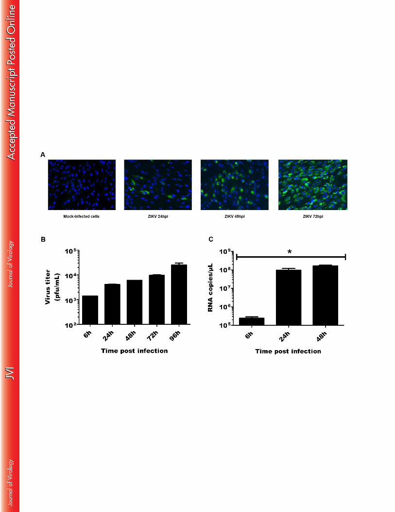
Given the capacity of mosquitoes to inoculate ZIKV into the human skin during the blood 313
feeding process, the potential target cells for infection with this virus are likely to be localized 314
in the epidermis and dermis which also constitute the first line of defense. We first determined 315
ZIKV susceptibility of skin fibroblasts, that have been recognized as a permissive target for 316
various arboviruses. Cells were infected in vitro with ZIKV and the presence of viral envelope 317
antigens was evaluated by immunofluorescence at different hours post infection (hpi). No 318
staining was observed in mock-infected cells or cells stained with an isotype control antibody 319
(Figure 1A). In contrast, as soon as 24h post-infection (hpi), the viral envelope protein was 320
detected in several cells, whereas at 72 hpi 100% of the infected cells expressed ZIKV (Figure 321
1A). Next, we evaluated the ability of these cells to produce viral progeny in vitro by 322
determining viral titers in the supernatants of ZIKV-infected primary human skin fibroblasts 323
using a standard plaque assay. The results show a gradual increase in the production or viral 324
particles over time indicating active viral replication in the infected cells (Figure 1B). 325
15
Intracellular viral RNA was also quantified by real time PCR at different time points post-326
infection. ZIKV RNA was detected in fibroblasts challenged with the virus, but not in mock-327
infected cells, as shown in Figure 1C. Viral RNA copy numbers were detected as soon as 6 328
hpi and increased during the course of infection. The amount of viral transcripts was markedly 329
high and could reach 108 RNA copies per microliter in cells infected with ZIKV and 330
maintained in culture for 24-48 h. 331
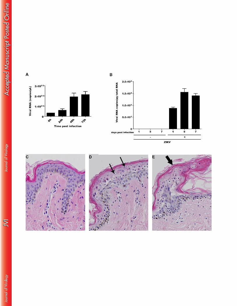
Then, given the observation that the epidermis layer is comprised mainly of keratinocytes, we 332
hypothesized that the latter cells also could be a target for ZIKV. Primary human epidermal 333
keratinocytes obtained from neonatal foreskin were infected with ZIKV and intracellular viral 334
RNA was quantified by quantitative PCR at different time points post-infection. As shown in 335
Figure 2A, ZIKV mRNA was detected in keratinocytes challenged with ZIKV, but not in 336
mock-infected cells. Viral RNA was found to increase over time and could be detected as 337
soon as 6 hpi, with a maximal amount of 105 viral copies per ml at both 48 and 72 hpi. The 338
capacity of ZIKV to replicate ex vivo in human skin cells was also studied. Infection of human 339
skin explants with ZIKV resulted in a gradual increase in viral copy numbers, with maximal 340
levels at 5 days post infection (dpi), pointing to a process of active viral replication (Fig. 2B). 341
Histological analysis of mock-infected human skin explants showed all aspects of a normal 342
healthy skin with a stratified epidermal layer, containing basal keratinocytes and 343
differentiated layers consisting of stratum granulosum and stratum corneum, respectively 344
(Figure 2C). In contrast, ZIKV-infected keratinocytes in human skin explants 5 dpi showed 345
the appearance of a cytoplasmic vacuolation, as well as the presence of pyknotic nuclei which 346
was however not generalized throughout the epidermis, but limited to the stratum granulosum 347
(Figure 2D). Moreover, ZIKV infection induced the sporadic formation of edema which was 348
also limited to this subcorneal layer (Figure 2E). 349
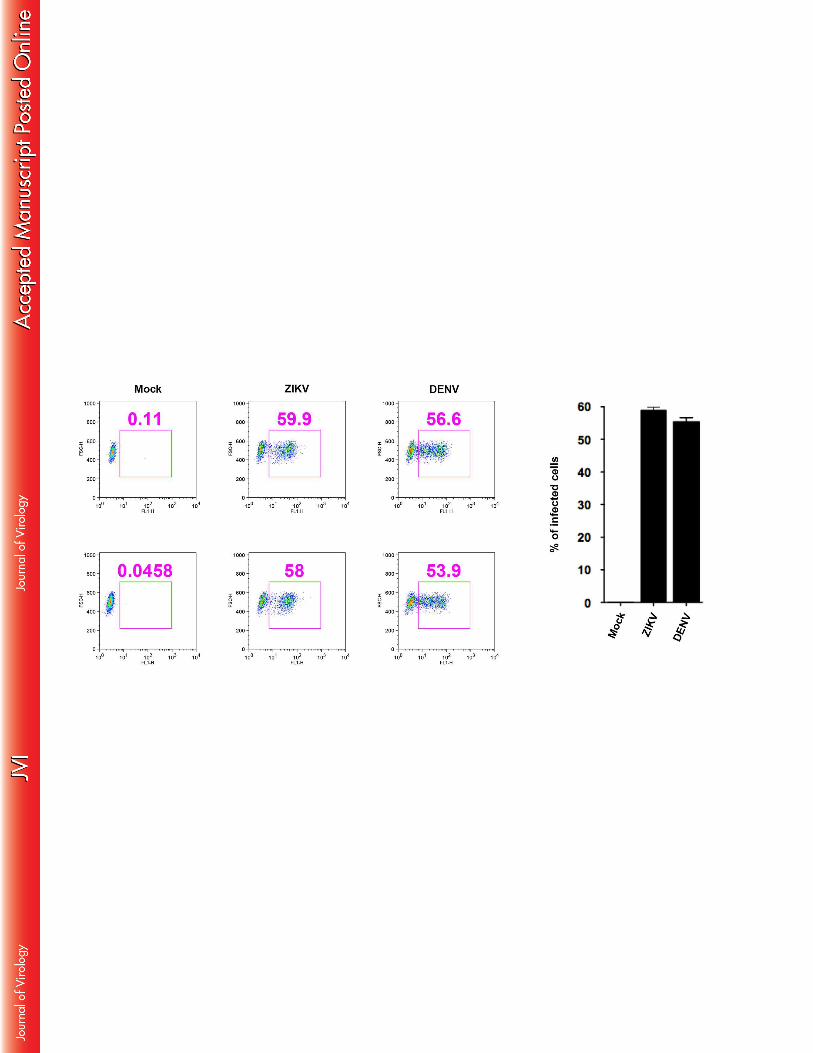
16
Immature dendritic cells have been reported to be permissive for DENV infection and as such 350
are recognized as an important target for propagation of this virus in the human skin. ZIKV 351
infection was therefore also investigated on this cell type, using DENV as a control, by 352
analyzing the intracellular presence of the viral envelope protein by flow cytometry. Our 353
results show that about 50% of human in vitro generated immature dendritic cells challenged 354
with ZIKV at MOI 0.5 for 24 hpi expressed the viral envelope (Figure 3). This percentage 355
was identical, as compared to that of the cells infected with DENV under the same 356
experimental conditions. These results indicate that immature dendritic cells are also 357
permissive to infection by this member of the Flavivirus family. 358
359
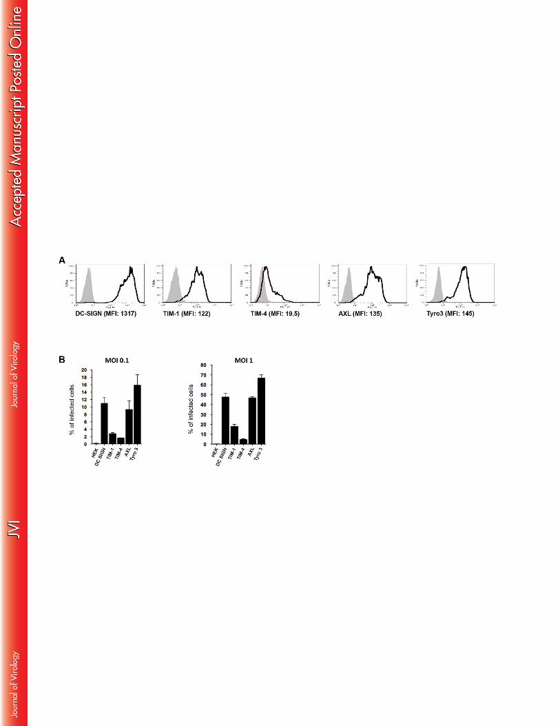
DC-SIGN, TIM and TAM receptors are involved in ZIVK infection 360
Several receptors, among which DC-SIGN, as well as certain TIM and TAM proteins, two 361
members of the phosphatidylserine receptor family, have been reported to facilitate viral entry 362
of DENV (review in (29)). To determine whether these receptors are also involved in ZIKV 363
entry, a series of HEK293T cell transfectants, expressing DC-SIGN, TIM1, TIM4, or the 364
TAM family members AXL and TYRO3, in a stable manner, were exposed to the virus. The 365
expression levels of each of these receptors are shown in Figure 4A. The parental, non-366
transfected HEK-293T cells were not susceptible to ZIKV-infection, as shown by the absence 367
of ZIKV antigen detection (Figure 4B). The expression of either DC-SIGN or AXL strongly 368
enhanced viral infection, already at MOI 0.1, resulting in about 50% of ZIKV-infected cells. 369
TYRO3-expressing HEK-293T cells were also highly permissive for ZIKV with nearly 70% 370
of the cells infected with the virus at 24 hpi. In contrast, the expression of TIM-1 or TIM-4 371
had only modest or marginal effects on ZIKV entry (Figure 4B). To further determine the 372
relative contribution of TIM and TAM receptors on ZIKV infection, A549 cells that 373
17
endogenously express TIM-1 and AXL, but not DC-SIGN (Figure 5A) were infected with the 374
virus. In keeping with the potent ZIKV infection-inducing activity of AXL, a neutralizing Ab, 375
specific for this receptor, strongly inhibited viral infection of A549 cells (Figure 5B). In 376
contrast, the presence of a neutralizing anti-TIM1 Ab did not have an impact on the 377
percentage of ZIKV infected cells at 24 hpi, as compared to cells infected with ZIKV alone. 378
However, the combination of the anti-TIM-1 and anti-AXL Abs completely abrogated ZIKV 379
infection (Figure 5B). We also used the RNA silencing technique to downregulate TIM-1 380
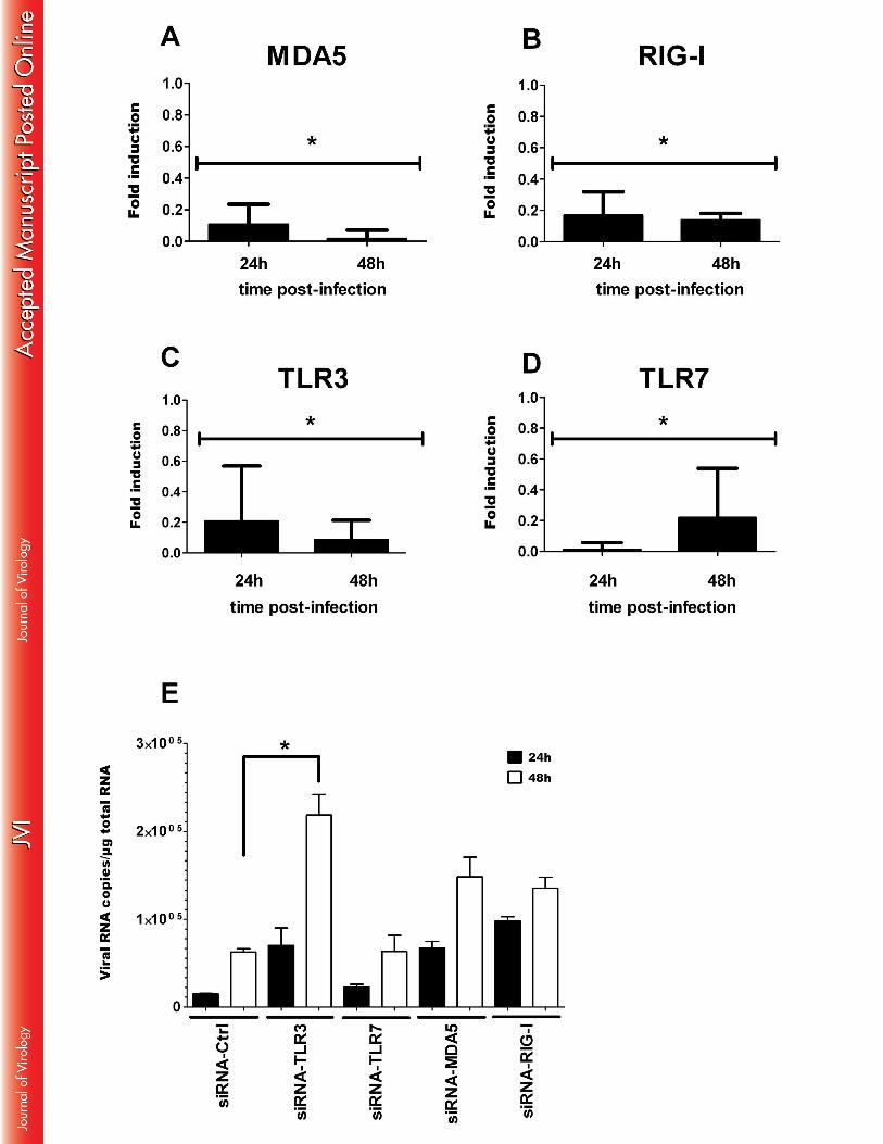
and/or AXL expression in A549 cells (Figure 5C). The results mirrored those obtained with 381
the neutralizing Ab, in that ZIKV infection was only slightly reduced in TIM-1-silenced cells, 382
strongly inhibited in AXL-silenced cells and totally abrogated when both genes were silenced 383
(Figure 5D). Finally, to determine the importance of the AXL in ZIKV infection of human 384
skin fibroblasts that express AXL but not TIM-1 (Figure 6A) were infected with the virus in 385
the absence or presence of a neutralizing Ab or specific siRNA. Exposure of the human skin 386
fibroblast cell line HFF1 to ZIKV, or DENV as a positive control, resulted in a comparable 387
number of infected cells that was inhibited by 70% and 50%, respectively, in the presence of a 388
neutralizing anti-AXL Ab (Figure 6B). Strikingly, the presence of specific AXL siRNA 389
totally inhibited the AXL expression (Figure 6C) and effectively abrogated the infection with 390
either virus, thus demonstrating the importance of AXL in the permissiveness of human skin 391
fibroblasts to infection and replication of ZIKV. Taken together, the data indicate an essential 392
and cooperative role for both TIM and TAM family members in ZIKV infection by 393
permissive cells. 394
395
ZIKV induces an innate anti-viral response in primary human skin fibroblasts 396
In order to determine whether ZIKV induces an innate anti-viral immune response in 397
permissive cells, the anti-viral gene expression profile in infected primary human fibroblasts at 398
18
early time points following ZIKV infection was determined using a human qPCR array 399
covering 84 human antiviral genes. This comparative analysis with mock-infected cells 400
showed the specific induction of pattern recognition receptors (PRRs), able to detect the 401
presence of pathogen-associated molecular patterns (PAMPs) in response to ZIKV infection. 402
This is particularly illustrated by the upregulation of the Toll-like receptor 3 (TLR-3) mRNA 403
expression, as well as by enhanced transcription of the DDX58 (RIG-I) and MDA5 (IFIH1) 404
genes that reportedly are involved in the detection of other Flavivirus members (Table 1). 405
Increased PRR expression levels and kinetics of expression, during an extended time course of 406
infection, were confirmed by individual qRT-PCR analysis. As shown in Figure 7A, RIG-I, 407
MDA5 and TLR3 expression was upregulated in ZIKV-infected fibroblasts as soon as 6 hpi 408
with maximal mRNA levels detected at 48 hpi. In contrast, no activation of the TLR7 gene 409
was observed in these cells following infection with ZIKV. The detection of viral PAMPs by 410
TLR3 and other PRRs initiates downstream signaling pathways that account for the 411
enhancement of transcription factors known to mobilize the antiviral machinery. The results 412
shown in Table 1 and Figure 7A are consistent with this general notion, as IRF7 mRNA levels 413
were increased in ZIKV-infected cells. IRF7 is a transcription factor that binds to the 414
interferon-stimulated response element, located on the promoters of type I IFN genes (30). 415
This result not only corroborates the enhanced IFN-α and IFN-β gene expression detected 416
following infection with ZIKV, but also the upregulation of the expression of several 417
interferon-stimulated genes (ISGs), including OAS2, ISG15 and MX1 (Table 1 and Figure 418
7B). The expression of the CXCR3 ligand CXCL10, as well as the inflammatory antiviral 419
chemokine CCL5, was also induced by ZIKV. Finally, ZIKV infection of skin fibroblasts was 420
also found to activate certain inflammasome components, as evidenced by a strong increase in 421
the expression of AIM2 and IL-1β transcripts (Figure 7A). In order to determine the 422
involvement of each of the upregulated PRRs in the anti-viral response against ZIKV, the 423
19
effect of specific siRNAs on viral replication was studied. Expression levels of MDA-5, RIG-424
I, TLR3 and TLR-7 in HFF1 cells were decreased by 80%, 24h following the transfection of 425
these cells with specific siRNA, and were completely inhibited after 48h (Figure 8A-D), thus 426
validating the efficacy of this approach. Inhibition of TLR3 expression, unlike that of the other 427
PRRs, resulted in a strong increase in the viral RNA copy numbers 48h following viral 428
infection of the cells (Figure 8E). However, inhibition of TLR3 expression did not modulate 429
type I IFN mRNA expression in the infected cells (Results not shown). Taken together, these 430
results underscore the importance of TLR3 in the induction of an antiviral response against 431
ZIKV. 432
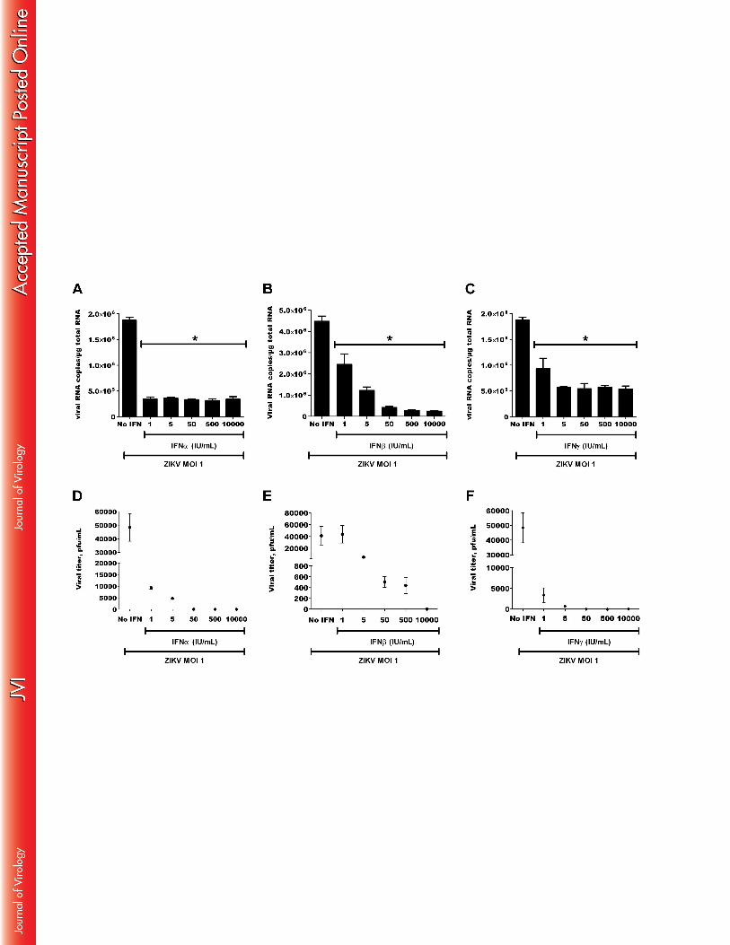
Type I and type II IFNs inhibit ZIKV replication 433
Because of the observed induction of type I IFNs by ZIKV-infected skin fibroblasts, their 434
effects on viral replication in the latter cells was investigated. Primary skin fibroblasts were 435
pretreated for 6h with increasing doses of recombinant human IFN-α, IFN-β or IFN-γ, 436
infected with ZIKV at an MOI of 1, and viral RNA copy numbers were determined by real-437
time PCR. At this viral titer, both type I and type II IFNs strongly, and dose-dependently, 438
inhibited viral replication with similar efficacy (Figure 9A-C). The effect of IFNs was 439
corroborated by a decrease in the release of viral particles as measured by plaque assay in the 440
culture supernatants of the infected cells (Fig. 9D-F). These results show that ZIKV is highly 441
sensitive to the antiviral effect of both type I and type II IFNs. 442
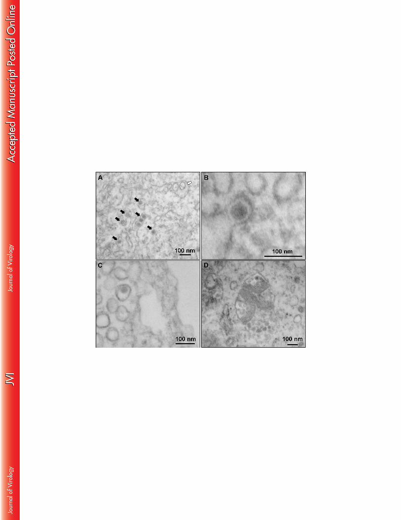
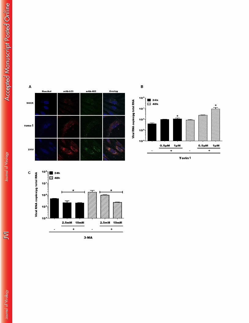
Autophagosome formation in infected skin fibroblasts increase ZIKV replication 443
Autophagy is a multi-step process responsible for degradation and recycling of cytoplasmic 444
components that augments the replication and dissemination of several arboviruses. We 445
therefore analyzed whether infection of skin fibroblasts with ZIKV resulted in the formation 446
of autophagosomes. First, an electron microscopy study was carried out to demonstrate the 447
20
presence of ZIKV particles in cytoplasmic compartments, as a result of exposure of these cells 448
to the virus. At 72 hpi, intravacuolar structures in ZIKV-infected fibroblasts were found to 449
contain capsids, in combination with enveloped and electron dense spherical viral particles 450
that were 70 to 100 nm in diameter which is a general feature of Flavivirus particles (Figure 451
10A and 10B). Moreover, ZIKV infection was associated with the formation of numerous 452
double-membrane intracytoplasmic vacuoles characteristic of autophagosomes (Figure 10C 453
and 10D) that were not observed in mock-infected cells (results not shown). To further 454
determine whether autophagy was induced following ZIKV infection, the skin fibroblast cell 455
line HFF1 was infected with the virus and the co-expression of the viral envelope protein and 456
the cytosolic microtubule-associated light chain 3 (LC3), an autophagosome-specific marker, 457
was determined by confocal microscopy. Torin 1, a chemical inducer of autophagy was used 458
as a positive control. As shown in Figure 11A, ZIKV infection induced the formation of LC3 459
punctae in infected fibroblasts while LC3 labelling was more diffuse in mock-infected cells. 460
Interestingly, LC3 signal in infected cells completely co-localized with that of the viral 461
envelope protein detected with specific antibodies. Moreover, the simultaneous addition of 462
ZIKV and Torin 1 to primary fibroblasts enhanced viral replication as shown by an increase in 463
the viral RNA copy number (Figure 11B). Conversely, addition of the 3-Methyladenine (3-464
MA) autophagy inhibitor decreased the number of viral copies in ZIKV-infected cells without 465
any cytotoxic effect on the cells (results not shown), thus formally confirming the association 466
between enhanced autophagosome formation and increased viral replication. Taken together, 467
these results show that ZIKV is able to increase its replication via induction of the autophagy 468
in the host cell. 469
470
471
21
472
DISCUSSION 473
ZIKV is a Flavivirus, related to Yellow fever, Dengue, West-Nile and Japanese encephalitis 474
viruses, that causes an arthropod-borne disease in human known as ZIKA fever. Originally 475
detected in a sentinel Rhesus monkey in Uganda in 1947 (31) and twenty years later isolated 476
from humans in Nigeria, the virus has since spread to other regions of the world. Importantly, 477
following recent outbreaks in Micronesia, French Polynesia, Cook Island and Easter Island, 478
ZIKV has become an emerging arbovirus (18). However, other than its phylogenetic 479
relationship to other members of the Flavivirus family, no information is available on the 480
cellular tropism of ZIKV and the nature of the cellular receptors that mediate its entry. In the 481
present study, we have identified the initial target cells of the ZIKV in the skin compartment, 482
as well as its entry receptors, and have furthermore characterized the anti-viral response 483
elicited following infection of permissive cells with the PF-13 ZIKV strain isolated during the 484
recent outbreak in French Polynesia (18). This strain is closely related to those isolated from 485
patients infected during the ZIVK outbreaks in Cambodia in 2010 and Yap State in 2007 and 486
its thus relevant for the results reported here. 487
ZIKV is transmitted by the Aedes mosquito that deposits the virus in the epidermis and dermis 488
of the bitten host during a blood meal. Indeed, both skin fibroblasts and epidermal 489
keratinocytes were found to be highly permissive to infection with ZIKV. Infection of skin 490
fibroblasts rapidly resulted in the presence of high levels of RNA copy numbers and a gradual 491
increase in the production or ZIKV particles over time, indicating active viral replication in 492
the infected cells. 493
ZIKV infection of epidermal keratinocytes resulted in the appearance of cytoplasmic 494
vacuolation, as well as the presence of pyknotic nuclei in the stratum granulosum, indicative 495
for cells that undergo apoptosis. This bears similarity to observations made with DENV that 496
22
induces the appearance of apoptotic cells in the epidermis of infected human skin explants 497
(27). It can be speculated that the induction of apoptotic cell death is a mechanism by which 498
ZIKV, like DENV, is able to divert anti-viral immune responses by increasing their 499
dissemination from dying cells. These results also corroborate previous reports in the 500
literature showing the importance of keratinocytes in infection with other flaviviruses, such as 501
WNV (32) and DENV (25). In addition to dermal fibroblasts and epidermal keratinocytes, we 502
report that dendritic cells are permissive to infection with ZIKV. This comes as no surprise 503
given the involvement of skin antigen presenting cells in the replication of other flavivirus 504
members, in particular DENV that efficiently infects Langerhans cells (33). The selective 505
susceptibility of permissive cells in the dermis and epidermis, including Langerhans cells, 506
dermal dendritic cells, macrophages, as well as fibroblast and keratinocytes, to infection with 507
ZIKV needs however to be determined. 508
The first step of Flavivirus entry into a host cell is mediated by the viral envelope protein that 509
interacts with several cell surface receptors and attachment factors, the differential expression 510
of which determines the cellular tropism of the virus. At present, more than a dozen putative 511
entry receptors and factors, in particular for DENV, have been described. Several of them, 512
such as heat-shock proteins, laminin receptor, integrin αvβ3, prohibitin, claudin-1, scavenger 513
receptor class B and natural killer cells receptor NKp44, can interact with viral particles in 514
mammalian and/or mosquito cells, but their exact role in the flavivirus entry program, as well 515
as their physiologic relevance, is not well understood (reviewed in (29). Heparan sulfate, a 516
sulfated polysaccharide associated to proteins from the extracellular matrix, has been 517
described as a non-specific attachment factor of flaviviruses, concentrating viral particles on 518
the cell surface and facilitating their interaction with primary receptors (34-38). Among them, 519
C-type lectin receptors such as the dendritic cell-specific intracellular adhesion molecule 3-520
grabbing non-integrin (DC-SIGN, CD209), the mannose receptor and the C-type lectin 521
23
domain family 5, member A (CLEC5A, MDL-1), play an important role in flavivirus binding 522
and infection of myeloid cells (39-41). Recently, TIM and TAM proteins, two distinct 523
families of transmembrane receptors that participate in the phosphatidylserine (PtdSer)-524
dependent phagocytic engulfment and removal of apoptotic cells, have also been shown to act 525
as DENV entry factors, promoting viral infection by attaching and possibly internalizing viral 526
particles in human cell cultures and primary cells targeted by flaviviruses (26, 29). 527
We show here that ZIKV entry is mediated by DC-SIGN, AXL, Tyro3 and, although to a 528
lesser extent, by TIM-1. Although TIM-1 by itself contributed little to ZIKV infection, its 529
expression nevertheless had an additive effect on the efficacy of AXL-mediated viral entry. 530
This raises the interesting possibility of a cooperation between both receptors, with TIM-1 531
acting as an attachment factor that binds viral particles and transfers them to AXL which 532
could in turn participate in viral internalization. In that sense, TIM-1 might not be 533
indispensable for ZIKV endocytosis and infection, but would rather concentrate virions on the 534
cell surface to facilitate their interaction with AXL, as well as the subsequent infection, which 535
might explain the additive inhibitory effect observed when both receptors are blocked with 536
neutralizing antibodies. However, additional experiments are required to assess the exact role 537
played by TIM and TAM receptors in ZIKV infection. 538
As has been reported for DENV, there seems to be a large number of receptors and/or 539
attachment factors that are able to mediate entry of ZIKV in permissive cells. It is of note 540
however that the permissiveness of skin cells to ZIKV is also determined by the profile of 541
receptor expression by these target cells. In this respect, unlike immature dendritic cells that 542
also are a primary target cell type for ZIKV infection, neither cutaneous fibroblasts, nor 543
epidermal keratinocytes express DC-SIGN. In contrast, the latter cells, as well as 544
macrophages, vascular endothelium cells and astrocytes (reviewed in ref (42), express AXL 545
that, as shown in the present study, is of major importance for ZIKV entry. The availability of 546
24
different entry receptors is likely to provide an evolutionary advantage for the virus that, as a 547
result, is able to infect a wide range of target cells and invade the human host. Nevertheless, 548
the contribution of each of these receptors and/or attachment factors to ZIKV infection and 549
pathogenesis is currently unknown and remain to be established. It is also important to 550
consider that other, as yet to be identified cell surface molecules exist that might account for 551
the tropism of ZIKV. 552
The outcome of viral infection is determined by a competition between viral replication and 553
the host immune response. The latter is programmed to rapidly control viral replication and to 554
limit virus spread by recognizing non-self nucleic acid as pathogen-associated molecular 555
patterns and triggering an antiviral response. Indeed, infection of fibroblasts in vitro with 556
ZIKV strongly induced the expression of several antiviral gene clusters, in particular PRRs, 557
such as RIG-I, MDA-5 and TLR3 that are able to detect the presence of PAMPs. These results 558
corroborate previous reports in the literature showing that these gene products play a sensory 559
role in the detection of other flaviviruses, such as DENV and WNV (25, 43). The induction of 560
TLR3 expression is rapid and already detectable at 6 hpi, whereas that of RIG-I and MDA-5 561
is delayed. It can therefore be hypothesized that these molecules trigger a coordinated 562
induction of the antiviral immune reaction against ZIKV with TLR3 priming an early 563
response that is amplified by RIG-I and MDA-5 at a later stage. This sequence of events has 564
also been suggested previously with respect to the immune response of fibroblasts following 565
infection with DENV (44). However, in the latter study, the involvement of only TLR3 and 566
RIG-I was considered, because, contrary to ZIKV, DENV infection did not enhance the 567
expression of MDA5 in skin fibroblasts. 568
Both TLR3 and TLR7 are implicated in the induction of an immune response against 569
flavivirus and triggering of these PRRs has been shown to initiate signaling pathways, leading 570
to the production of type I IFNs, as well as other inflammatory cytokines and chemokines by 571
25
hepatocytes and macrophages (review in (45). Indeed, ZIKV infection strongly enhanced 572
TLR3 expression, associated with the production of IFN-α and IFN-β in infected cells. 573
However, whereas inhibition of TLR3 expression by siRNA indeed resulted in a strong 574
enhancement of viral replication, no effect on type I IFN mRNA expression was detected. 575
Although TLR3 seem to play an important in role in the antiviral response to ZIKV, the 576
mechanism by which this receptor contributes to the control of viral replication remains to be 577
determined. In contrast, no modulation of TLR7 expression was observed, which is 578
reminiscent to results obtained with DENV-infected skin fibroblasts (44). The absence of 579
TLR7 induction was also reported in a separate study in which expression of PRRs in virally-580
infected fibroblasts of different origin was analyzed (46). Taken together, these findings 581
confirm the notion that the involvement of various TLR members seems to be dependent on 582
virus and cell type. 583
The detection of ZIKV-expressed PAMPs also resulted in an increase in transcriptional levels 584
of IRF7, a transcription factor that binds to the interferon-stimulated response element, 585
located on the promoters of type I IFN genes (30). This result corroborates the enhanced IFN-586
α and IFN-β gene expression, detected following infection with ZIKV, as well as the 587
upregulation of the expression of several interferon-stimulated genes, including OAS2, ISG15 588
and MX1. The expression of the two CXCR3 ligands, CXCL10 and CXCL11 was also 589
induced by ZIKV. The latter chemokines not only play a role in innate and adaptive immunity 590
by attracting T cells and other leukocytes to sites of inflammation, but also display direct, 591
receptor-independent, defensin-like antimicrobial activity when present at elevated 592
concentrations in dermal fibroblasts (47). In addition, infection of skin fibroblasts by ZIKV 593
resulted in upregulation of CCL5, another inflammatory chemokine known for it antiviral 594
activity. 595
26
Whereas TLR3 transcription was significantly enhanced, IRF3 gene expression, in contrast, 596
remained unchanged during the course of ZIVK infection of fibroblasts. A similar observation 597
was made in DENV-infected epidermal keratinocytes in which also no enhanced IRF3 598
expression could be detected. This is somewhat surprising in that IFR3 is known to play an 599
important role in the induction of IFN-β production in cells exposed to PAMPS from various 600
viruses (48). Moreover, dsRNA-mediated triggering of RIG-I and MDA5, both molecules 601
whose expression is upregulated following infection with ZIKV and other flaviviruses, seems 602
to be crucial for IRF3 activation (25). It has been reported that IFN-β production, which is 603
essential for the early antiviral immune response, was observed in both wild-type and IRF3-/- 604
mice following WNV infection (48). These results corroborate the present and previously 605
published data (25), indicating that the production of the type I IFN in response to DENV and 606
ZIKV infection is apparently independent of the IRF3 pathway, both in flavivirus-infected 607
epidermal keratinocytes and skin fibroblasts. It is of note that the replication of ZIKV was 608
significantly inhibited by both type I and type II IFNs, in keeping with the general antiviral 609
activity of these cytokines with critical functions in host defense mechanisms. 610
Electron microscopy analysis of ZIKV-infected primary skin fibroblasts showed the presence 611
of membrane vesicles with a size between 70 and 100 nm that were located in intimate 612
association with the endoplasmic reticulum, indicating that ZIKV replication occurs in close 613
association with host cell membranes. These results are in line with an earlier report in the 614
literature underscoring the importance of fibroblasts as a primary cell type of replication for 615
flaviruses, like DENV, that through the release of viral particles may contribute to subsequent 616
viral dissemination (44). ZIKV infection also induced an autophagy program, as demonstrated 617
by the presence of characteristic autophagosome-like vesicles in the infected fibroblasts. 618
Autophagy is a process characterized by the presence of double-membrane vesicles, known as 619
autophagosomes, that recruit cytoplasmic material and subsequently fuse with lysosomes for 620
27
protein degradation. Autophagy not only participates in the degradation of proteins and 621
damaged organelles in the cytoplasm to maintain homeostasis (49), but is also involved in 622
host immunity against pathogen infection. This is particularly illustrated by Vesicular 623
stomatitis virus (50), Sendai virus (51), and Herpes simplex virus-1 (52) infected cells in 624
which, autophagy-mediated degradation of viral proteins limits viral replication and promotes 625
cell survival. In contrast, the autophagy process can be subverted by viruses. This is true for 626
several arboviruses, including DENV (53, 54), Chikungunya virus (55) and Japanese 627
encephalitis (56) virus that use components of the autophagy pathway to promote their 628
replication and dissemination by clearing cells through multiple mechanisms. In this regard, 629
autophagy may thus have both pro- and antiviral effects. 630
Autophagy in ZIKV-infected fibroblasts was furthermore confirmed by the demonstration of 631
co-localization of the viral envelope protein and the cytosolic microtubule-associated 632
molecule LC3. The results also show that stimulation of autophagosome formation by Torin 1 633
further enhances replication of ZIKV in permissive cells, whereas the presence of 3 M-A, an 634
inhibitor of autophagosome formation, strongly reduced viral copy numbers in the infected 635
fibroblasts, indicating that autophagy promotes replication of ZIKV in permissive cells. In 636
this respect, ZIKV behaves like most other flavivirus members, with the exception of WNV 637
(57), by its capacity to interact with the conventional autophagy pathway in mammalian cells. 638
The precise mechanism by which ZIKV induces autophagy still needs to be determined. 639
Nevertheless, similar to DENV (58), the results from our study demonstrating the co-640
localization of ZIKV with LC3 strongly suggests that autophagocytic vacuoles are the site of 641
viral replication. It can furthermore speculated that autophagy may promote replication of 642
ZIKV infection through restriction of the antiviral innate immune response (59), enhancement 643
of translation of the viral genome that has entered the mammalian cells (60) or by providing 644
additional energy and relevant membrane structures for viral replication (61). However, the 645
28
exact molecular mechanism(s) by which ZIKV highjacks components of the autophagome 646
pathways remain to be determined. 647
At present, ZIKV has received far less attention in the literature than the other mosquito-borne 648
flavivirus members. Nevertheless, it is considered to be an emerging virus because of its 649
global spreading during the last decades and its pathogenic potential reminiscent to that of 650
DENV. Importantly, ZIKV has recently been isolated in Gabon from the Asian tiger mosquito 651
Ae. albopictus (62), a rapidly expanding Aedes species that lives in close contact with human 652
urban populations (63, 64) and that typically feeds not only at dusk and dawn, but also in the 653
daytime. This underscores its menacing character, as this vector is known for its capacity to 654
colonize new environments, either by progressive extension from already occupied zones, or 655
by jumping to new areas, in particular to those in heavily populated urban areas. In this 656
respect, a better understanding of the role of mosquito saliva in ZIKV infection is an 657
important point that must be addressed in the future as well. 658
Taken together, the results presented in this study pertaining to the identification of the 659
cellular tropism, molecular mechanisms of infection and replication, as well as signaling 660
pathways involved the anti-viral immune response of ZIKV, permit to gain better insight in its 661
mode of action and to devise strategies aiming to interfere with the pathology caused by this 662
emerging flavivirus. 663
664
ACKNOWLEDGMENTS 665
The authors thank Dr François Renaud for critical discussions, Chantal Cazevieille for expert 666
help with electron microscopy and Eric Bernard for technical assistance. This work was 667
supported by grants from the French Research Agency “Agence Nationale de la Recherche” 668
(ANR-12-BSV3-0004-01; ANR-14-CE14-0029). Sineewanlaya Wichit was supported by a 669
29
fellowship of the Infectiopôle Sud foundation. The funders had no role in study design, data 670
collection and analysis, decision to publish, or preparation of the manuscript. 671
672
673
674
675
676
REFERENCES 677
1. Kuno G, Chang GJ, Tsuchiya KR, Karabatsos N, Cropp CB. 1998. Phylogeny of the genus 678 Flavivirus. J Virol 72:73-83. 679
2. Moore DL, Causey OR, Carey DE, Reddy S, Cooke AR, Akinkugbe FM, David-West TS, Kemp 680 GE. 1975. Arthropod-borne viral infections of man in Nigeria, 1964-1970. Ann Trop Med 681 Parasitol 69:49-64. 682
3. Simpson DI. 1964. Zika Virus Infection in Man. Transactions of the Royal Society of Tropical 683 Medicine and Hygiene 58:335-338. 684
4. Smithburn KC. 1954. Neutralizing antibodies against arthropod-borne viruses in the sera of 685 long-time residents of Malaya and Borneo. American journal of hygiene 59:157-163. 686
5. Fagbami AH. 1979. Zika virus infections in Nigeria: virological and seroepidemiological 687 investigations in Oyo State. The Journal of hygiene 83:213-219. 688
6. Hammon WM, Schrack WD, Sather GE. 1958. Serological survey for a arthropod-borne virus 689 infections in the Philippines. Am J Trop Med Hyg 7:323-328. 690
7. Pond WL. 1963. Arthropod-Borne Virus Antibodies in Sera from Residents of South-East Asia. 691 Transactions of the Royal Society of Tropical Medicine and Hygiene 57:364-371. 692
8. Olson JG, Ksiazek TG, Suhandiman, Triwibowo. 1981. Zika virus, a cause of fever in Central 693 Java, Indonesia. Transactions of the Royal Society of Tropical Medicine and Hygiene 75:389-694 393. 695
9. Darwish MA, Hoogstraal H, Roberts TJ, Ghazi R, Amer T. 1983. A sero-epidemiological 696 survey for Bunyaviridae and certain other arboviruses in Pakistan. Trans R Soc Trop Med Hyg 697 77:446-450. 698
10. Marchette NJ, Garcia R, Rudnick A. 1969. Isolation of Zika virus from Aedes aegypti 699 mosquitoes in Malaysia. Am J Trop Med Hyg 18:411-415. 700
11. Li MI, Wong PS, Ng LC, Tan CH. 2012. Oral susceptibility of Singapore Aedes (Stegomyia) 701 aegypti (Linnaeus) to Zika virus. PLoS Negl Trop Dis 6:e1792. 702
12. Boorman JP, Porterfield JS. 1956. A simple technique for infection of mosquitoes with 703 viruses; transmission of Zika virus. Transactions of the Royal Society of Tropical Medicine and 704 Hygiene 50:238-242. 705
13. Monlun E, Zeller H, Le Guenno B, Traoré-Lamizana M, Hervy JP, Adam F, Ferrara L, 706 Fontenille D, Sylla R, Mondo M. 1993. [Surveillance of the circulation of arbovirus of medical 707 interest in the region of eastern Senegal]. Bull Soc Pathol Exot 86:21-28. 708
14. Weinbren MP, Williams MC. 1958. Zika virus: further isolations in the Zika area, and some 709 studies on the strains isolated. Transactions of the Royal Society of Tropical Medicine and 710 Hygiene 52:263-268. 711
30
15. Haddow AJ, Williams MC, Woodall JP, Simpson DI, Goma LK. 1964. Twelve Isolations of Zika 712 Virus from Aedes (Stegomyia) Africanus (Theobald) Taken in and above a Uganda Forest. 713 Bulletin of the World Health Organization 31:57-69. 714
16. Haddow AD, Schuh AJ, Yasuda CY, Kasper MR, Heang V, Huy R, Guzman H, Tesh RB, Weaver 715 SC. 2012. Genetic characterization of Zika virus strains: geographic expansion of the Asian 716 lineage. PLoS Negl Trop Dis 6:e1477. 717
17. Duffy MR, Chen TH, Hancock WT, Powers AM, Kool JL, Lanciotti RS, Pretrick M, Marfel M, 718 Holzbauer S, Dubray C, Guillaumot L, Griggs A, Bel M, Lambert AJ, Laven J, Kosoy O, Panella 719 A, Biggerstaff BJ, Fischer M, Hayes EB. 2009. Zika virus outbreak on Yap Island, Federated 720 States of Micronesia. The New England journal of medicine 360:2536-2543. 721
18. Musso D, Nilles EJ, Cao-Lormeau VM. 2014. Rapid spread of emerging Zika virus in the 722 Pacific area. Clin Microbiol Infect 20:O595-596. 723
19. Kwong JC, Druce JD, Leder K. 2013. Zika virus infection acquired during brief travel to 724 Indonesia. Am J Trop Med Hyg 89:516-517. 725
20. Tappe D, Rissland J, Gabriel M, Emmerich P, Gunther S, Held G, Smola S, Schmidt-Chanasit 726 J. 2014. First case of laboratory-confirmed Zika virus infection imported into Europe, 727 November 2013. Euro surveillance : bulletin Europeen sur les maladies transmissibles = 728 European communicable disease bulletin 19. 729
21. Pyke AT, Daly MT, Cameron JN, Moore PR, Taylor CT, Hewitson GR, Humphreys JL, Gair R. 730 2014. Imported zika virus infection from the cook islands into australia, 2014. PLoS currents 731 6. 732
22. Fonseca K, Meatherall B, Zarra D, Drebot M, MacDonald J, Pabbaraju K, Wong S, Webster 733 P, Lindsay R, Tellier R. 2014. First case of zika virus infection in a returning canadian traveler. 734 Am J Trop Med Hyg 91:1035-1038. 735
23. Oehler E, Watrin L, Larre P, Leparc-Goffart I, Lastere S, Valour F, Baudouin L, Mallet H, 736 Musso D, Ghawche F. 2014. Zika virus infection complicated by Guillain-Barre syndrome--737 case report, French Polynesia, December 2013. Euro surveillance : bulletin Europeen sur les 738 maladies transmissibles = European communicable disease bulletin 19. 739
24. Briant L, Desprès P, Choumet V, Missé D. 2014. Role of skin immune cells on the host 740 susceptibility to mosquito-borne viruses. Virology 464-465:26-32. 741
25. Surasombatpattana P, Hamel R, Patramool S, Luplertlop N, Thomas F, Despres P, Briant L, 742 Yssel H, Misse D. 2011. Dengue virus replication in infected human keratinocytes leads to 743 activation of antiviral innate immune responses. Infection, genetics and evolution : journal of 744 molecular epidemiology and evolutionary genetics in infectious diseases 11:1664-1673. 745
26. Meertens L, Carnec X, Lecoin MP, Ramdasi R, Guivel-Benhassine F, Lew E, Lemke G, 746 Schwartz O, Amara A. 2012. The TIM and TAM families of phosphatidylserine receptors 747 mediate dengue virus entry. Cell Host Microbe 12:544-557. 748
27. Limon-Flores AY, Perez-Tapia M, Estrada-Garcia I, Vaughan G, Escobar-Gutierrez A, 749 Calderon-Amador J, Herrera-Rodriguez SE, Brizuela-Garcia A, Heras-Chavarria M, Flores-750 Langarica A, Cedillo-Barron L, Flores-Romo L. 2005. Dengue virus inoculation to human skin 751 explants: an effective approach to assess in situ the early infection and the effects on 752 cutaneous dendritic cells. Int J Exp Pathol 86:323-334. 753
28. Lanciotti RS, Kosoy OL, Laven JJ, Velez JO, Lambert AJ, Johnson AJ, Stanfield SM, Duffy MR. 754 2008. Genetic and serologic properties of Zika virus associated with an epidemic, Yap State, 755 Micronesia, 2007. Emerging infectious diseases 14:1232-1239. 756
29. Perera-Lecoin M, Meertens L, Carnec X, Amara A. 2014. Flavivirus entry receptors: an 757 update. Viruses 6:69-88. 758
30. Honda K, Yanai H, Negishi H, Asagiri M, Sato M, Mizutani T, Shimada N, Ohba Y, Takaoka A, 759 Yoshida N, Taniguchi T. 2005. IRF-7 is the master regulator of type-I interferon-dependent 760 immune responses. Nature 434:772-777. 761
31
31. Dick GW, Kitchen SF, Haddow AJ. 1952. Zika virus. I. Isolations and serological specificity. 762 Transactions of the Royal Society of Tropical Medicine and Hygiene 46:509-520. 763
32. Lim PY, Behr MJ, Chadwick CM, Shi PY, Bernard KA. 2011. Keratinocytes are cell targets of 764 West Nile virus in vivo. J Virol 85:5197-5201. 765
33. Cerny D, Haniffa M, Shin A, Bigliardi P, Tan BK, Lee B, Poidinger M, Tan EY, Ginhoux F, Fink 766 K. 2014. Selective susceptibility of human skin antigen presenting cells to productive dengue 767 virus infection. PLoS Pathog 10:e1004548. 768
34. Chen Y, Maguire T, Hileman RE, Fromm JR, Esko JD, Linhardt RJ, Marks RM. 1997. Dengue 769 virus infectivity depends on envelope protein binding to target cell heparan sulfate. Nat Med 770 3:866-871. 771
35. Germi R, Crance JM, Garin D, Guimet J, Lortat-Jacob H, Ruigrok RW, Zarski JP, Drouet E. 772 2002. Heparan sulfate-mediated binding of infectious dengue virus type 2 and yellow fever 773 virus. Virology 292:162-168. 774
36. Hilgard P, Stockert R. 2000. Heparan sulfate proteoglycans initiate dengue virus infection of 775 hepatocytes. Hepatology 32:1069-1077. 776
37. Kroschewski H, Allison SL, Heinz FX, Mandl CW. 2003. Role of heparan sulfate for 777 attachment and entry of tick-borne encephalitis virus. Virology 308:92-100. 778
38. Lee E, Pavy M, Young N, Freeman C, Lobigs M. 2006. Antiviral effect of the heparan sulfate 779 mimetic, PI-88, against dengue and encephalitic flaviviruses. Antiviral Res 69:31-38. 780
39. Navarro-Sanchez E, Altmeyer R, Amara A, Schwartz O, Fieschi F, Virelizier JL, Arenzana-781 Seisdedos F, Despres P. 2003. Dendritic-cell-specific ICAM3-grabbing non-integrin is essential 782 for the productive infection of human dendritic cells by mosquito-cell-derived dengue 783 viruses. EMBO Rep 4:723-728. 784
40. Tassaneetrithep B, Burgess TH, Granelli-Piperno A, Trumpfheller C, Finke J, Sun W, Eller 785 MA, Pattanapanyasat K, Sarasombath S, Birx DL, Steinman RM, Schlesinger S, Marovich 786 MA. 2003. DC-SIGN (CD209) mediates dengue virus infection of human dendritic cells. J Exp 787 Med 197:823-829. 788
41. Chen ST, Lin YL, Huang MT, Wu MF, Cheng SC, Lei HY, Lee CK, Chiou TW, Wong CH, Hsieh SL. 789 2008. CLEC5A is critical for dengue-virus-induced lethal disease. Nature 453:672-676. 790
42. Lemke G, Rothlin CV. 2008. Immunobiology of the TAM receptors. Nat Rev Immunol 8:327-791 336. 792
43. Fredericksen BL, Keller BC, Fornek J, Katze MG, Gale M. 2008. Establishment and 793 maintenance of the innate antiviral response to West Nile Virus involves both RIG-I and 794 MDA5 signaling through IPS-1. J Virol 82:609-616. 795
44. Bustos-Arriaga J, Garcia-Machorro J, Leon-Juarez M, Garcia-Cordero J, Santos-Argumedo L, 796 Flores-Romo L, Mendez-Cruz AR, Juarez-Delgado FJ, Cedillo-Barron L. 2011. Activation of 797 the innate immune response against DENV in normal non-transformed human fibroblasts. 798 PLoS Negl Trop Dis 5:e1420. 799
45. Nazmi A, Dutta K, Hazra B, Basu A. 2014. Role of pattern recognition receptors in flavivirus 800 infections. Virus Res 185:32-40. 801
46. Paladino P, Cummings DT, Noyce RS, Mossman KL. 2006. The IFN-independent response to 802 virus particle entry provides a first line of antiviral defense that is independent of TLRs and 803 retinoic acid-inducible gene I. J Immunol 177:8008-8016. 804
47. Proost P, Vynckier AK, Mahieu F, Put W, Grillet B, Struyf S, Wuyts A, Opdenakker G, Van 805 Damme J. 2003. Microbial Toll-like receptor ligands differentially regulate CXCL10/IP-10 806 expression in fibroblasts and mononuclear leukocytes in synergy with IFN-gamma and 807 provide a mechanism for enhanced synovial chemokine levels in septic arthritis. Eur J 808 Immunol 33:3146-3153. 809
48. Bourne N, Scholle F, Silva MC, Rossi SL, Dewsbury N, Judy B, De Aguiar JB, Leon MA, Estes 810 DM, Fayzulin R, Mason PW. 2007. Early production of type I interferon during West Nile 811
32
virus infection: role for lymphoid tissues in IRF3-independent interferon production. J Virol 812 81:9100-9108. 813
49. Xie Z, Klionsky DJ. 2007. Autophagosome formation: core machinery and adaptations. Nat 814 Cell Biol 9:1102-1109. 815
50. Shelly S, Lukinova N, Bambina S, Berman A, Cherry S. 2009. Autophagy is an essential 816 component of Drosophila immunity against vesicular stomatitis virus. Immunity 30:588-598. 817
51. Lee HK, Lund JM, Ramanathan B, Mizushima N, Iwasaki A. 2007. Autophagy-dependent viral 818 recognition by plasmacytoid dendritic cells. Science 315:1398-1401. 819
52. Tallóczy Z, Virgin HW, Levine B. 2006. PKR-dependent autophagic degradation of herpes 820 simplex virus type 1. Autophagy 2:24-29. 821
53. Lee YR, Lei HY, Liu MT, Wang JR, Chen SH, Jiang-Shieh YF, Lin YS, Yeh TM, Liu CC, Liu HS. 822 2008. Autophagic machinery activated by dengue virus enhances virus replication. Virology 823 374:240-248. 824
54. Heaton NS, Randall G. 2011. Dengue virus and autophagy. Viruses 3:1332-1341. 825 55. Krejbich-Trotot P, Gay B, Li-Pat-Yuen G, Hoarau JJ, Jaffar-Bandjee MC, Briant L, Gasque P, 826
Denizot M. 2011. Chikungunya triggers an autophagic process which promotes viral 827 replication. Virol J 8:432. 828
56. Li JK, Liang JJ, Liao CL, Lin YL. 2012. Autophagy is involved in the early step of Japanese 829 encephalitis virus infection. Microbes Infect 14:159-168. 830
57. Vandergaast R, Fredericksen BL. 2012. West Nile virus (WNV) replication is independent of 831 autophagy in mammalian cells. PLoS One 7:e45800. 832
58. Panyasrivanit M, Greenwood MP, Murphy D, Isidoro C, Auewarakul P, Smith DR. 2011. 833 Induced autophagy reduces virus output in dengue infected monocytic cells. Virology 418:74-834 84. 835
59. Ke PY, Chen SS. 2011. Activation of the unfolded protein response and autophagy after 836 hepatitis C virus infection suppresses innate antiviral immunity in vitro. J Clin Invest 121:37-837 56. 838
60. Dreux M, Gastaminza P, Wieland SF, Chisari FV. 2009. The autophagy machinery is required 839 to initiate hepatitis C virus replication. Proc Natl Acad Sci U S A 106:14046-14051. 840
61. Heaton NS, Randall G. 2010. Dengue virus-induced autophagy regulates lipid metabolism. 841 Cell Host Microbe 8:422-432. 842
62. Grard G, Caron M, Mombo IM, Nkoghe D, Mboui Ondo S, Jiolle D, Fontenille D, Paupy C, 843 Leroy EM. 2014. Zika virus in Gabon (Central Africa)--2007: a new threat from Aedes 844 albopictus? PLoS Negl Trop Dis 8:e2681. 845
63. Benedict MQ, Levine RS, Hawley WA, Lounibos LP. 2007. Spread of the tiger: global risk of 846 invasion by the mosquito Aedes albopictus. Vector Borne Zoonotic Dis 7:76-85. 847
64. Medlock JM, Hansford KM, Schaffner F, Versteirt V, Hendrickx G, Zeller H, Van Bortel W. 848 2012. A review of the invasive mosquitoes in Europe: ecology, public health risks, and control 849 options. Vector Borne Zoonotic Dis 12:435-447. 850
851
852
853
854
855
33
856
FIGURE LEGENDS 857
Figure 1: Primary human fibroblasts are susceptible to ZIKV. (A) Primary fibroblasts 858
infected with ZIKV (MOI1) and mock-infected cells were analyzed at different times post-859
infection for the presence of the viral envelope protein by immunofluoresence with the 4G2 860
mAb and an FITC-conjugated anti-mouse IgG. (B) Viral replication was determined by 861
plaque assay analysis of culture supernatants of ZIKV-infected cells. (C) Expression of viral 862
RNA was determined by real-time RT-PCR. Data are representative of three independent 863
experiments each performed in duplicate (error bars represent standard error of the mean). 864
Wilcox–Mann–Whitney test was employed to analyze the difference between sets of data. 865
*indicates p values < 0.05. 866
867
Figure 2: ZIKV infects human keratinocytes and induces morphological changes in 868
human skin biopsies 869
(A) Primary human keratinocytes or (B) human skin biopsies were infected with ZIKV (MOI 870
1 and 106 PFU, respectively) and expression of viral RNA was determined at different time 871
points by real-time RT-PCR. Data are representative of three independent experiments each 872
performed in duplicate (error bars represent standard error of the mean). Wilcox–Mann–873
Whitney test was employed to analyze the difference between sets of data. *indicates p values 874
< 0.05. Microscopic observation of (C) Mock- or (D and E) ZIKV-infected human skin 875
biopsies. Small arrows indicate keratinocyte cytoplasmic vacuolation. Large arrow indicates a 876
superficial sub-corneous edema with also cytoplasmic vacuolation. Magnification 20x. Data 877
are representative of two independent experiments. 878
879
880
34
881
Figure 3: Dendritic cells are permissive to ZIKV and DENV. 882
Human immature dendritic cells were infected with ZIKV or DENV (MOI 1) for 24 hpi and 883
the intracellular presence of the viral envelope protein was detected using the pan-flavivirus 884
Ab 4G2 by flow cytometry. Mean fluorescence intensity was determined and the percentage 885
of infected cells was calculated, as compared to non-infected cells. Data are representative of 886
three independent experiments. 887
888
Figure 4: Entry receptors involved in ZIKV infection 889
(A) Expression profile of different cell surface receptors by HEK293T cells stably expressing 890
DC-SIGN, TIM-1, TIM-4, AXL or Tyro3 (white histograms) or parental, non-transfected 891
cells (grey histograms). (B) HEK293T cells expressing the indicated receptors were incubated 892
with ZIKV (MOI 0.1 and 1) and the percentage of infected cells was determined by 893
measuring the expression of viral envelope protein by flow cytometry at 24 hpi. Data are 894
representative of three independent experiments. 895
896
Figure 5: Involvement of AXL and TIM-1 in ZIKV infection of A549 cells 897
(A) Cell surface expression levels of AXL, TIM-1 and DC-SIGN on A549 cells, as 898
determined by flow cytometry. Immunofluorescence staining of cells with specific mAb 899
(white histogram) is superimposed on those with isotype control mAb (grey histograms). (B) 900
A549 cells were incubated with ZIKV (MOI 1) for 1 hr at 4°C in the presence of neutralizing 901
anti-TIM-1 (5µg/mL) and/or anti-AXL (10µg/mL), respectively, or with different 902
concentration of a goat IgG as control. The percentage of infected cells was measured by flow 903
cytometry and normalized to that in presence of control IgG. Data are shown as representative 904
flow cytometry analysis (upper panel) and are represented as mean +/-SEM of at least three 905
independent experiments (lower panel). (C) A549 cells were transfected by the indicated 906
35
siRNA, and TIM-1 and AXL expression was assessed by flow cytometry after 24hpi, at the 907
time of infection. (D) Cells were infected with ZIKV (MOI 1). Infection was normalized to 908
infection in nontargeting (siNT) siRNA-transfected cells. To test the significance of the 909
differences, analysis of the variance (ANOVA) was performed with GraphPad Prism 910
software. Statistically significant differences between each condition and control cells are 911
denoted by an asterisk (*) and are indicated p values < 0.05. Data are representative of three 912
independent experiments. 913
914
Figure 6: Expression of AXL permits ZIKV infection of skin fibroblasts 915
(A) Cell surface expression levels of AXL and TIM-1 on HFF1 cells was monitored by flow 916
cytometry. Immunofluorescence staining of cells with specific mAb (white histogram) is 917
superimposed on those with isotype control mAb (grey histograms). (B) HFF1 cells were 918
incubated with ZIKV (MOI 3) or DENV (MOI 5) for 1 hr at 4°C in the presence of 919
neutralizing anti-AXL, or normal goat IgG as control. The percentage of infected cells was 920
measured by flow cytometry and normalized to that in presence of control IgG. Data are 921
shown as representative flow cytometry analysis (upper panel) and are represented as mean 922
+/-SEM of at least three independent experiments (lower panel). (C) HFF1 cells were 923
transfected by the indicated siRNA for 24h then cells were infected with ZIKV (MOI 3) or 924
DENV (MOI 5). Infection was normalized to infection in non-targeting (siNT) siRNA-925
transfected cells. To test the significance of the differences, analysis of the variance 926
(ANOVA) was performed with GraphPad Prism software. Statistically significant differences 927
between each condition and control cells are denoted by an asterisk (*) and are indicated p 928
values < 0.05. Data are representative of three independent experiments. 929
930
Figure 7: ZIKV induces an innate anti-viral response in primary human skin fibroblasts 931
36
(A) Primary human fibroblasts were exposed to ZIKV (MOI 1) and mRNA levels were 932
quantified over time by real-time RT-PCR. Results are expressed as fold induction of 933
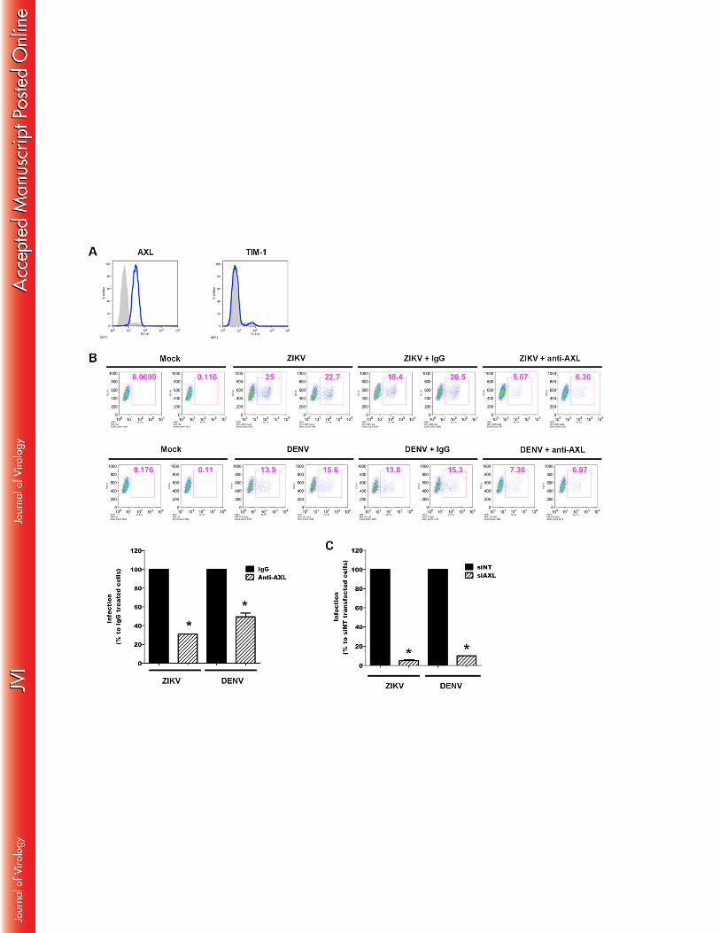
transcripts in ZIKV-infected cells relative to those in mock-infected cells. Data are 934
representative of three independent experiments each performed in duplicate (errors bars 935
represent standard error of the mean). Wilcoxon–Mann–Whitney test was employed to 936
analyze the difference between sets of data. A value of p < 0.05 was considered significant. * 937
indicates p values < 0.05. (B) Cells were exposed to ZIKV (MOI 1) at the indicated time and 938
MX1 protein levels were detected by Western blotting using a specific antibody. The 939
immunoblot was stripped and reblotted with an anti-α-tubulin Ab as a control for protein 940
loading. Data are representative of three independent experiments. 941
942
Figure 8: Effect of PRR silencing on ZIKV replication and IFN expression 943
(A-D) siRNAs specific for MDA5 (siRNA-MDA5), RIG-I (siRNA-RIG-I), TLR3 (siRNA-944
TLR3), TLR7 (siRNA-TLR7), as well as a non-specific siRNA (siRNA-Ctrl), were 945
transfected into HFF1 cells 24 h before infection with ZIKV (MOI 0.1). Reduction of mRNA 946
levels by siRNA was confirmed by real-time RT-PCR at 24 and 48 hpi. Results are expressed 947
as fold induction of expression of transcripts in specific siRNA-transfected cells, relative to 948
that in siRNA-Ctrl-transfected cells. The latter value corresponds to 1 on the ordinate of each 949
histogram. Data are representative of two independent experiments, each performed in 950
triplicate, and are normalized according to 18S mRNA levels in the samples (errors bars 951
represent standard error of the mean). The Wilcoxon–Mann–Whitney test was used to analyze 952
the difference between sets of data. A value of p < 0.05 was considered significant and 953
denoted by an asterisk (*). (E) Viral copy numbers in siRNA-transfected cells were measured 954
by real-time RT-PCR at 24 and 48 hpi. Statistically significant differences (p values < 0.05) 955
between specific siRNA- and siRNA-Ctrl-transfected cells were determined using the analysis 956
37
of the variance (ANOVA) with GraphPad Prism software and denoted by an asterisk (*). Data 957
are representative of two independent experiments each performed in triplicate. 958
959
Figure 9: IFNs inhibit ZIKV infection 960
Primary skin fibroblasts were pretreated 6 h before infection with different concentrations 961
IFN-α, IFN-β and IFN-γ and were then exposed to ZIKV at MOI 1. (A-C) inhibition of viral 962
replication was measured, at 24 hpi, by real-time RT-PCR and (D-F) the release of viral 963
particle quantified by plaque assay in the culture supernatants. Statistical significance of the 964
data, were determined using analysis of the variance (ANOVA) and GraphPad Prism software 965
and are denoted by an asterisk (*) and p values < 0.05. Data are representative of three 966
independent experiments each performed in duplicate (error bars represent standard error of 967
the mean). 968
969
Figure 10: Electron microscopic imaging of ZIKV-infected primary fibroblasts. (A) 970
Membrane vesicles with size between 70 and 100 nm observed in intimate association with 971
endoplasmic reticulum are indicated by white arrow. Black arrows indicate the presence of 972
spherical capsids detected in intracellular vacuoles or docked to intracellular membranes. (B) 973
Enlargement of a ZIKV particle. Intracellular electron dense spherical capsid is 40 nm in size. 974
(C) Assembled capsids are transported to the cell surface in intracellular vacuoles. (D) 975
Autophagosomes are frequently detected in infected fibroblasts and assembled capsids are 976
observed inside this compartment. Data are representative of two independent experiments. 977
978
Figure 11: ZIKV induces autophagy in infected skin fibroblasts. (A) Visualization of 979
autophagosome formation by LC3 aggregation in Mock- or ZIKV-infected cells and cells 980
treated with Torin 1. Cells were fixed 24 hpi and the colocalization of autophagosomes and 981
38
ZIKV was determined by immunofluorescence using mAbs specific for LC3 or the viral 982
envelope protein (4G2). Data are representative of three independent experiments. (B) 983
Primary human skin fibroblasts were exposed to ZIKV (MOI 2) in the absence (cells were 984
treated with the vehicle 0.05% DMSO) or presence of (B) Torin 1 or (C) 3-MA, at the 985
indicated concentrations, and viral replication was quantified by real-time RT-PCR at 24 and 986
48 hpi. Data are representative of three independent experiments. To test the significance of 987
the differences, analysis of the variance (ANOVA) was performed with GraphPad Prism 988
software. Statistically significant differences between each condition and control cells are 989
denoted by an asterisk (*) and are indicated by p values < 0.05. 990
991
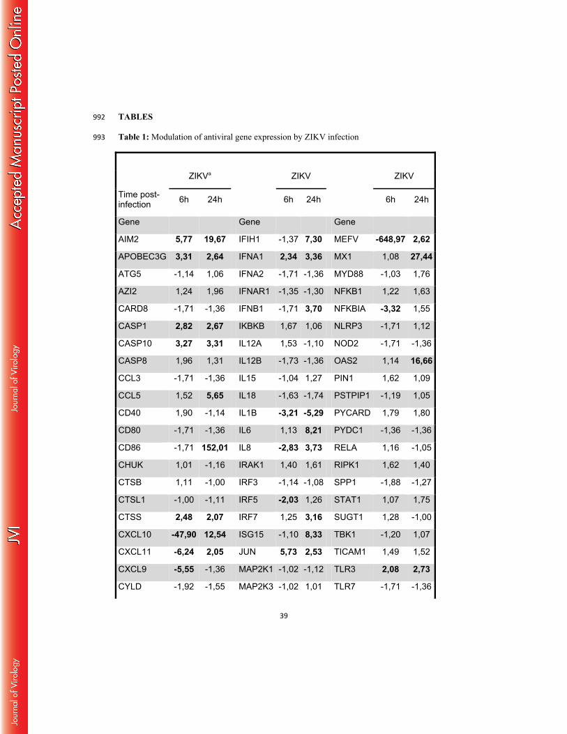
39
TABLES 992
Table 1: Modulation of antiviral gene expression by ZIKV infection 993
ZIKVa ZIKV ZIKV
Time post-infection 6h 24h 6h 24h 6h 24h
Gene Gene Gene
AIM2 5,77 19,67 IFIH1 -1,37 7,30 MEFV -648,97 2,62
APOBEC3G 3,31 2,64 IFNA1 2,34 3,36 MX1 1,08 27,44
ATG5 -1,14 1,06 IFNA2 -1,71 -1,36 MYD88 -1,03 1,76
AZI2 1,24 1,96 IFNAR1 -1,35 -1,30 NFKB1 1,22 1,63
CARD8 -1,71 -1,36 IFNB1 -1,71 3,70 NFKBIA -3,32 1,55
CASP1 2,82 2,67 IKBKB 1,67 1,06 NLRP3 -1,71 1,12
CASP10 3,27 3,31 IL12A 1,53 -1,10 NOD2 -1,71 -1,36
CASP8 1,96 1,31 IL12B -1,73 -1,36 OAS2 1,14 16,66
CCL3 -1,71 -1,36 IL15 -1,04 1,27 PIN1 1,62 1,09
CCL5 1,52 5,65 IL18 -1,63 -1,74 PSTPIP1 -1,19 1,05
CD40 1,90 -1,14 IL1B -3,21 -5,29 PYCARD 1,79 1,80
CD80 -1,71 -1,36 IL6 1,13 8,21 PYDC1 -1,36 -1,36
CD86 -1,71 152,01 IL8 -2,83 3,73 RELA 1,16 -1,05
CHUK 1,01 -1,16 IRAK1 1,40 1,61 RIPK1 1,62 1,40
CTSB 1,11 -1,00 IRF3 -1,14 -1,08 SPP1 -1,88 -1,27
CTSL1 -1,00 -1,11 IRF5 -2,03 1,26 STAT1 1,07 1,75
CTSS 2,48 2,07 IRF7 1,25 3,16 SUGT1 1,28 -1,00
CXCL10 -47,90 12,54 ISG15 -1,10 8,33 TBK1 -1,20 1,07
CXCL11 -6,24 2,05 JUN 5,73 2,53 TICAM1 1,49 1,52
CXCL9 -5,55 -1,36 MAP2K1 -1,02 -1,12 TLR3 2,08 2,73
CYLD -1,92 -1,55 MAP2K3 -1,02 1,01 TLR7 -1,71 -1,36
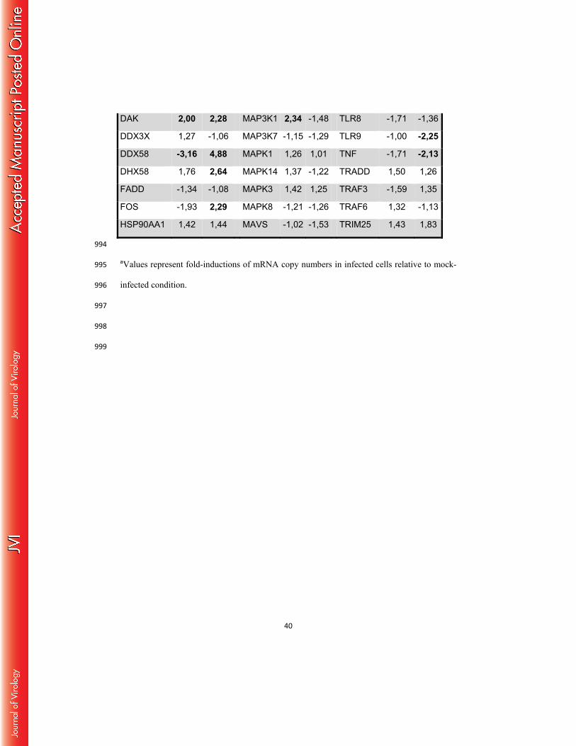
40
DAK 2,00 2,28 MAP3K1 2,34 -1,48 TLR8 -1,71 -1,36
DDX3X 1,27 -1,06 MAP3K7 -1,15 -1,29 TLR9 -1,00 -2,25
DDX58 -3,16 4,88 MAPK1 1,26 1,01 TNF -1,71 -2,13
DHX58 1,76 2,64 MAPK14 1,37 -1,22 TRADD 1,50 1,26
FADD -1,34 -1,08 MAPK3 1,42 1,25 TRAF3 -1,59 1,35
FOS -1,93 2,29 MAPK8 -1,21 -1,26 TRAF6 1,32 -1,13
HSP90AA1 1,42 1,44 MAVS -1,02 -1,53 TRIM25 1,43 1,83
994
aValues represent fold-inductions of mRNA copy numbers in infected cells relative to mock-995
infected condition. 996
997
998
999