Embed Size (px)
Citation preview
From Department of Microbiology, Tumor and Cell Biology
Karolinska Institutet, Stockholm, Sweden
BIOMARKERS IN NASOPHARYNGEAL CARCINOMA
Zi-Ming Du
Stockholm, 2012

All previously published papers were reproduced with permission from the publisher.
Published by Karolinska Institutet. Printed by Larserics Digital Print AB, Sweden.
© Zi-Ming Du, 2012
ISBN 978-91-7457-666-5

To my family

ABSTRACT
Nasopharyngeal carcinoma (NPC) is one of the most common malignancies in certain areas of Southern China, Southeastern Asia and Northern Africa. Currently, evaluation of NPC prognosis is mainly based on the tumor-node-metastasis (TNM) staging system. However, NPC patients with the same clinical stage often present different clinical courses, suggesting that the TNM staging is insufficient to predict prognosis of this disease. Therefore, it is important to find molecular biomarkers, which can help clinicians to identify NPC patients with worse prognosis and develop therapeutic interventions in NPC patients.
This thesis presents the identification and investigation of mechanism of several novel markers in NPC. In the first paper, Caveolin-1 (Cav-1), a major structural component of caveolae, and CD147 (also known as extracellular matrix metalloproteinase inducer, EMMPRIN), a glycoprotein, were found to be overexpressed in NPC. Both Cav-1 and CD147 expression levels correlated significantly with metastasis and poor prognosis of NPC patients. Further studies revealed that Cav-1 and CD147 enhance NPC cell migration, which is associated with MMP-3 and MMP-11 (active) secretion.
The role of microRNA-155 (miR-155) is associated with oncogenesis of several human tumors. In the second paper, miR-155 was found to be upregulated in NPC cell lines and clinical samples. EBV encoded LMP1 and LMP2A could further enhance the expression of miR-155 in NPC CNE1 and TW03 cells. JMJD1A was identified as a direct target of miR-155 in NPC. Downregulation of JMJD1A was significantly correlated with N stage of the TNM classification, a lower five-year survival rate, and a lower five-year disease-free survival rate of NPC patients.
Spleen tyrosine kinase (Syk) is a nonreceptor tyrosine kinase and often aberrantly expressed in human cancers. In the third paper, high expression of Syk was detected in 24% of NPC cases. High expression of Syk, resulted partly from LMP2A expression in NPC, is associated with tumor recurrence and poor prognosis of NPC patients.
Human chromosome 3 (Chr. 3) contains clusters of tumor suppressor genes (TSGs) involved in many cancer types. In the fourth paper, using Not I Chr. 3 microarray, ten candidate TSGs were found in NPC. Among them, the CpG island in the promoter region of Wingless-type Mouse mammary tumor virus integration site family, member 7A (WNT7a) and the intron 1 region of Integrin α9 (ITGA9) were confirmed to be hypermethylated in NPC by bisulfite sequencing and methylation specific PCR. Demethylating agent 5-aza-2′-deoxycytidine (5-aza-CdR) treatment could restore the expression of WNT7a and ITGA9 in NPC cell lines. Furthermore, WNT7a and ITGA9 were downregulated in NPC clinical samples. As both proteins execute significant functions related to the tumor cell biology, the potential of WNT7a and ITGA9 as diagnosis or therapeutic targets for NPC should be considered.
LIST OF PUBLICATIONS Articles included in this thesis: 1. Zi-Ming Du, Chun-Fang Hu, Qiong Shao, Ma-Yan Huang, Chang-Wei Kou, Xiao-Feng Zhu, Yi-Xin Zeng, and Jian-Yong Shao*. Upregulation of Caveolin-1 and CD147 Expression in Nasopharyngeal Carcinoma Enhanced Tumor Cell Migration and Correlated with Poor Prognosis of the Patients. Int. J. Cancer: 125, 1832-1841 (2009). PMID: 19582878 2. Zi-Ming Du, Li-Fu Hu, Hai-Yun Wang, Li-Xu Yan, Yi-Xin Zeng, Jian-Yong Shao*, Ingemar Ernberg*. Upregulation of MiR-155 in Nasopharyngeal Carcinoma is partly driven by LMP1 and LMP2A and downregulates a Negative Prognostic Marker JMJD1A. PLoS ONE. 2011 Apr 26;6(4):e19137. PMID: 21541331 3. Zi-Ming Du#, Chang-Wei Kou#, Chun-Fang Hu, Jing Chen, Hai-Yun Wang, Li-Xu Yan, Li-Fu Hu, Ingemar Ernberg, Yi-Xin Zeng and Jian-Yong Shao*. Clinical Significance of Elevated Spleen Tyrosine Kinase Expression in Nasopharyngeal Carcinoma. Head Neck. 2012 Jan 27. PMID: 22287277
4. Imran Nawaz#, Zi-Ming Du#, Ilya Ignatyev#, Tatiana V Pavlova, Vladimir Kashuba, Eugene R Zabarovsky, Ingemar Ernberg, Li-Fu Hu*. WNT7a and ITGA9 Gene are Hypermethylated and Downregulated in Nasopharyngeal Carcinoma. (manuscript)
Related publications: 1. Yan Zhang#, Li-Xu Yan#, Qi-Nian Wu#, Zi-Ming Du#, Jing Chen, Ding-Zhun Liao, Ma-Yan Huang, Jing-Hui Hou, Qiu-Liang Wu, Mu-Sheng Zeng, Wen-Lin Huang, Yi-Xin Zeng and Jian-Yong Shao*. miR-125b is Methylated and Functions as A Tumor Suppressor by Regulating the ETS1 proto-oncogene in Human Invasive Breast Cancer. Cancer Res. 2011 May 15;71(10):3552-62. PMID: 21444677 2. Hai-Yun Wang, Bing-Yu Sun, Zhi-Hua Zhu, Ellen T Chang, Ka-Fai To, Jacqueline SG Hwang, Hao Jiang, Michael Koon Ming Kam, Gang Chen, Shie-Lee Cheah, Ming Lee, Zhi-Wei Liu, Jing Chen, Jia-Xing Zhang, Hui-Zhong Zhang, Jie-Hua He, Fa-Long Chen, Xiao-Dong Zhu, Ma-Yan Huang, Ding-Zhun Liao, Jia Fu, Qiong Shao, Man-Bo Cai, Zi-Ming Du, Li-Xu Yan, Chun-Fang Hu, Ho Keung Ng, Joseph TS Wee, Weimin Ye, Ingemar Ernberg, Hans-Olov Adami, Anthony T Chan, Yi-Xin Zeng, and Jian-Yong Shao*. An Eight-signature Classifier for Prediction of Nasopharnyngeal Carcinoma Survival. J Clinl Oncol. 2011 Dec 1;29(34):4516-25. PMID: 22025164
# These authors contributed equally * Corresponding author
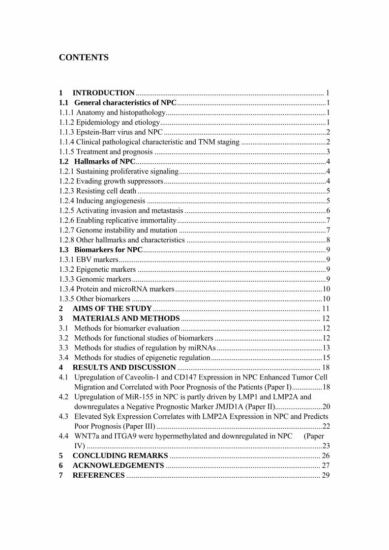
CONTENTS
1 INTRODUCTION .................................................................................................... 1 1.1 General characteristics of NPC...............................................................................1 1.1.1 Anatomy and histopathology.....................................................................................1 1.1.2 Epidemiology and etiology........................................................................................1 1.1.3 Epstein-Barr virus and NPC ......................................................................................2 1.1.4 Clinical pathological characteristic and TNM staging .............................................2 1.1.5 Treatment and prognosis ...........................................................................................3 1.2 Hallmarks of NPC.....................................................................................................4 1.2.1 Sustaining proliferative signaling..............................................................................4 1.2.2 Evading growth suppressors......................................................................................4 1.2.3 Resisting cell death ....................................................................................................5 1.2.4 Inducing angiogenesis ...............................................................................................5 1.2.5 Activating invasion and metastasis ...........................................................................6 1.2.6 Enabling replicative immortality...............................................................................7 1.2.7 Genome instability and mutation ..............................................................................7 1.2.8 Other hallmarks and characteristics ..........................................................................8 1.3 Biomarkers for NPC.................................................................................................9 1.3.1 EBV markers..............................................................................................................9 1.3.2 Epigenetic markers ....................................................................................................9 1.3.3 Genomic markers.......................................................................................................9 1.3.4 Protein and microRNA markers ..............................................................................10 1.3.5 Other biomarkers .....................................................................................................10 2 AIMS OF THE STUDY ......................................................................................... 11 3 MATERIALS AND METHODS .......................................................................... 12 3.1 Methods for biomarker evaluation ...........................................................................12 3.2 Methods for functional studies of biomarkers .........................................................12 3.3 Methods for studies of regulation by miRNAs........................................................13 3.4 Methods for studies of epigenetic regulation...........................................................15 4 RESULTS AND DISCUSSION ............................................................................ 18 4.1 Upregulation of Caveolin-1 and CD147 Expression in NPC Enhanced Tumor Cell
Migration and Correlated with Poor Prognosis of the Patients (Paper I)................18 4.2 Upregulation of MiR-155 in NPC is partly driven by LMP1 and LMP2A and
downregulates a Negative Prognostic Marker JMJD1A (Paper II).........................20 4.3 Elevated Syk Expression Correlates with LMP2A Expression in NPC and Predicts
Poor Prognosis (Paper III) ........................................................................................22 4.4 WNT7a and ITGA9 were hypermethylated and downregulated in NPC (Paper
IV) .............................................................................................................................23 5 CONCLUDING REMARKS ................................................................................ 26 6 ACKNOWLEDGEMENTS .................................................................................. 27 7 REFERENCES ....................................................................................................... 29
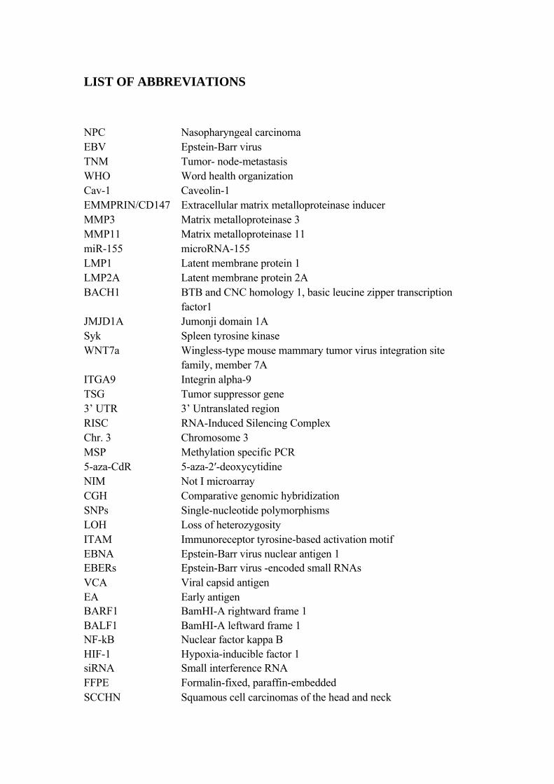
LIST OF ABBREVIATIONS
NPC Nasopharyngeal carcinoma EBV Epstein-Barr virus TNM Tumor- node-metastasis WHO Word health organization Cav-1 Caveolin-1 EMMPRIN/CD147 Extracellular matrix metalloproteinase inducer MMP3 Matrix metalloproteinase 3 MMP11 Matrix metalloproteinase 11 miR-155 microRNA-155 LMP1 Latent membrane protein 1 LMP2A Latent membrane protein 2A BACH1 BTB and CNC homology 1, basic leucine zipper transcription
factor1 JMJD1A Jumonji domain 1A Syk Spleen tyrosine kinase WNT7a Wingless-type mouse mammary tumor virus integration site
family, member 7A ITGA9 Integrin alpha-9 TSG Tumor suppressor gene 3’ UTR 3’ Untranslated region RISC RNA-Induced Silencing Complex Chr. 3 Chromosome 3 MSP Methylation specific PCR 5-aza-CdR 5-aza-2′-deoxycytidine NIM Not I microarray CGH Comparative genomic hybridization SNPs Single-nucleotide polymorphisms LOH Loss of heterozygosity ITAM Immunoreceptor tyrosine-based activation motif EBNA Epstein-Barr virus nuclear antigen 1 EBERs Epstein-Barr virus -encoded small RNAs VCA Viral capsid antigen EA Early antigen BARF1 BamHI-A rightward frame 1 BALF1 BamHI-A leftward frame 1 NF-kB Nuclear factor kappa B HIF-1 Hypoxia-inducible factor 1 siRNA Small interference RNA FFPE Formalin-fixed, paraffin-embedded SCCHN Squamous cell carcinomas of the head and neck
Biomarkers in Nasopharyngeal Carcinoma
1 INTRODUCTION
1.1 GENERNAL CHARACTERISTICS OF NPC 1.1.1 Anatomy and histopathology Nasopharyngeal carcinoma (NPC) is a tumor arising from the mucosal epithelium covering the nasopharyngeal surface, most often within the lateral nasopharyngeal recess or fossa of Rosenmüller (the space behind the nose). The histological classification system of the World Health Organization (WHO), which was most recently revised in 1991, defines NPC as either keratinizing squamous cell carcinoma (Type 1) or non-keratinizing carcinoma (Type 2) ; the latter is further subdivided into non-keratinizing differentiated carcinoma (Type 2a) and non-keratinizing undifferentiated carcinoma (Type 2b) (Table 1) 1. The most common form, non-keratinizing undifferentiated carcinoma, shows a close to 100% prevalence of Epstein-Barr virus (EBV) in the tumor cells2.
Table 1 Histological subtypes of NPC classified by World Health Organization Histological subtypes Characteristics WHO
Types
Keratinizing squamous cell carcinoma Highly differentiated, epithelial growth patterns, keratin filaments
Type 1 (I)
Non-keratinizing differentiated carcinoma
Retaining epithelial cell shape and growth pattern
Type 2a (II)Non-keratinizing carcinoma Non-keratinizing
undifferentiated carcinoma Lack of distinctive epithelial growth pattern
Type 2b (III)
1.1.2 Epidemiology and etiology NPC is an uncommon disease in most countries of the world, and its age-adjusted incidence for male and female is less than one per 100,000 persons 3. However NPC is one of the most common malignancies in certain areas of Southern China, Southeastern Asia and Northern Africa 4-5. The highest incidence of 25-40 per 100,000 persons was reported in the Sihui area of Guangdong province, Southern China6. The incidence rate among Chinese people born in North America is lower than in those born in Southern China, but still remains considerable higher than the local7-8. These findings suggest that genetic, ethnic and environmental factors play a role in the etiology of NPC. Familial NPC has been linked to susceptibility loci on chromosome 3p 219, 4p15.1-q12 10and 5p 1311. Epstein-Barr virus (EBV) infection has been verified to be strongly associated with NPC 12-13. Moreover, consumption of salted fish and other preserved foods containing volatile nitrosamines, especially during childhood, is important carcinogenic factor for NPC 14-15.
1
Biomarkers in Nasopharyngeal Carcinoma
1.1.3 Epstein-Barr virus and NPC EBV is a prototype gamma herpes virus which infects more than 90% of the world’s adult population16. Humans are the only natural host for EBV. Primary infection with EBV normally occurs in early childhood and is usually asymptomatic in most countries. However, in some individuals, EBV is implicated in the pathogenesis of several human malignancies including Burkitt’s lymphoma 17, Hodgkin’s lymphoma 18, gastric carcinoma 19 and NPC20. EBV infection in these tumors is characterized by the expression of viral genes expressed during latent infection, in particular, Epstein-Barr virus nuclear antigen 1 (EBNA1), Latent membrane protein 1 (LMP1), Latent membrane protein 2 (LMP2), BamHI-A rightward frame 1 (BARF1), BamHI-A leftward frame 1 (BALF1) and Epstein-Barr virus -encoded small RNAs (EBERs) 21-24 (Table 2).
Table 2 Expression of EBV latent genes in human diseases Latency types EBV genes expressed Disease associated Type I EBNA1, EBERs Burkitt’s lymphoma Type II EBNA1, LMP1, LMP2,
EBERs, BARF1 NPC, Hodgkin’s disease, periphpheral T cell lymphoma
Type III EBNA1, EBNA2, EBNA3, LMP1, LMP2, EBERs
lymhoproliferative disease, X-linked lymphoproliferative disease, infectious mononucleosis
LMP1 and LMP2A are not expressed at a significant level in all NPCs, and the level of expression varies in different studies depending on material and methods used for detection. LMP1, which was reported to be expressed in as much as approximately 65% of NPC patients25, is the first EBV latent gene found to be able to transform cell lines and alter the phenotype of cells due to its oncogenic potential 26. In human epithelial cells, LMP1 alters many functional properties that are involved in tumor progression and invasion 27. LMP2A, which is expressed in around 45% of NPC patients28, was reported to induce primary epithelial cell migration and invasion 29. More importantly, Guasparri et al 30 supported that EBV LMP2A protein could affect LMP1-mediated Nuclear factor kappa B (NF-kB) signaling and survival of lymphoma cells. LMP1 and LMP2A may partly affect the same pathway, also including transcriptional regulation of miRNAs. Moreover, BARF131-33 and BALF134 have been found to be expressed in NPC biopsies, suggesting that they play a role in the pathogenesis of NPC. EBERs are the most abundant EBV viral transcripts and are used to detect EBV-infected cells in tissues by in situ hybridization. EBERs could contribute to oncogenesis by modulating innate immunity in patients with NPC32 1.1.4 Clinical pathological characteristic and TNM staging NPC has a dominant clinico-pathological behavior of loco-regional recurrence and metastasis, which differs from that of other types of head and neck cancers35. Currently,
2
Biomarkers in Nasopharyngeal Carcinoma
tumor-node-metastasis (TNM) staging of NPC is based on clinical and radiologic examination. There are various ways of classifying NPC. At present, the American Joint Committee on Cancer Staging and End Result Reporting / International Union Against Cancer (AJC/UICC) system is preferred in Europe and America36. The Chinese NPC staging system of 1992 (Sidebar 1) has incorporated prognostic significance, and according to this staging criteria, the 5-year survival rates of NPC patients in China for stages I, II, III and IV are 89.7%, 75.9%, 51.3%, and 22.2%, respectively37. The Chinese 1992 NPC staging system is considered satisfactory and has been widely used in China 37-39.
Sidebar 1 The characteristics of the 1992 NPC staging system The staging system is characterized according to the following model: T, primary tumor: T1, limited to the nasopharynx; T2, involvement of the nasal cavity, oropharynx, soft palatine, anterior cervical vertebrae soft tissue, and parapharyngeal space extension before the SO line (the SO line is between the styloid process and the midpoint on the posterior edge of the great occipital foramen);T3, extension over the SO line, involvement of the anterior or posterior cranial nerves alone, the base of the skull, the pterygoprocess zone, and the pterygopalatine fossa; T4, involvement of both anterior and posterior cranial nerves, parabasal sinus, cavernous sinus, orbit, infratemporal fossa, and direct invasion of the first or second cervical vertebrae; N, regional lymph node involvement: N0, no enlarged lymph nodes; N1, greatest dimension of upper neck lymph node <4 cm, movable; N2, lower neck lymph node or greatest lymph node dimension between 4 and 7 cm; N3, supraclavicular lymph node, lymph node greatest dimension >7 cm, fixed, or skin infiltration (the border between the upper neck and the lower neck is the inferior margin of the cricoid cartilage); M, distant metastasis: M0, absence of distant metastasis; M1, presence of distant metastasis; staging: Stage I, T1N0M0; Stage II, T2N0-N1M0, T0-T2N1M0; Stage III, T3N0-N2M0, T0-T3N2M0; Stage IVa, T4N0-N3M0, T0-T4N3M0; Stage IVb, M1.
1.1.5 Treatment and prognosis NPC is a relatively radiosensitive tumor and therefore radiotherapy remains the standard treatment for almost all NPC patients. The fields for targeting of radiotherapy to the primary tumor and the surrounding anatomical structures at risk of tumor invasion were defined clinically by radiographic imaging. Although more than 70% overall survival rates can be expected for stages I and II, treatment outcomes are unsatisfactory for advanced-stage NPC when radiotherapy is offered alone 40-43. Utilization of radiochemotherapy, which is combined modality therapy using chemotherapy and radiotherapy, is an important strategy for improving tumor control of advanced NPC to achieve improvements in survival 44-50. Although NPC is radiosensitive, the average five-year survival rate remains between 50-60%. Regional lymph node and distant metastasis and loco-regional recurrence are the two major reasons resulting in failure of treatment for this cancer39, 51.
3
Biomarkers in Nasopharyngeal Carcinoma
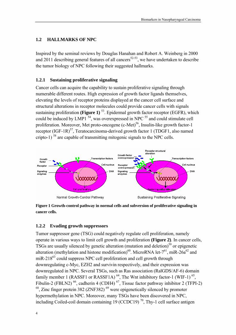
1.2 HALLMARKS OF NPC Inspired by the seminal reviews by Douglas Hanahan and Robert A. Weinberg in 2000 and 2011 describing general features of all cancers52-53, we have undertaken to describe the tumor biology of NPC following their suggested hallmarks. 1.2.1 Sustaining proliferative signaling Cancer cells can acquire the capability to sustain proliferative signaling through numerable different routes. High expression of growth factor ligands themselves, elevating the levels of receptor proteins displayed at the cancer cell surface and structural alterations in receptor molecules could provide cancer cells with signals sustaining proliferation (Figure 1) 53. Epidermal growth factor receptor (EGFR), which could be induced by LMP1 54, was overexpressed in NPC 55 and could stimulate cell proliferation. Moreover, Met proto-oncogene (c-Met)56, Insulin-like growth factor-1 receptor (IGF-1R)57, Teratocarcinoma-derived growth factor 1 (TDGF1, also named cripto-1) 58 are capable of transmitting mitogenic signals to the NPC cells.
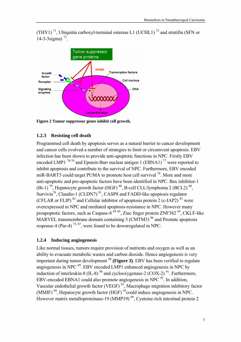
Figure 1 Growth-control pathway in normal cells and subversion of proliferative signaling in cancer cells. 1.2.2 Evading growth suppressors Tumor suppressor gene (TSG) could negatively regulate cell proliferation, namely operate in various ways to limit cell growth and proliferation (Figure 2). In cancer cells, TSGs are usually silenced by genetic alteration (mutation and deletion)59 or epigenetic alteration (methylation and histone modification)60. MicroRNA let-761, miR-26a62 and miR-21863 could suppress NPC cell proliferation and cell growth through downregulating c-Myc, EZH2 and survivin respectively, and their expression was downregulated in NPC. Several TSGs, such as Ras association (RalGDS/AF-6) domain family member 1 (RASSF1 or RASSF1A) 64, The Wnt inhibitory factor-1 (WIF-1) 65, Fibulin-2 (FBLN2) 66, cadherin 4 (CDH4) 67, Tissue factor pathway inhibitor 2 (TFPI-2) 68, Zinc finger protein 382 (ZNF382) 69 were epigenetically silenced by promoter hypermethylation in NPC. Moreover, many TSGs have been discovered in NPC, including Coiled-coil domain containing 19 (CCDC19) 70, Thy-1 cell surface antigen
4
Biomarkers in Nasopharyngeal Carcinoma
(THY1) 71, Ubiquitin carboxyl-terminal esterase L1 (UCHL1) 72 and stratifin (SFN or 14-3-3sigma) 73.
Figure 2 Tumor suppressor genes inhibit cell growth. 1.2.3 Resisting cell death Programmed cell death by apoptosis serves as a natural barrier to cancer development and cancer cells evolved a number of strategies to limit or circumvent apoptosis. EBV infection has been shown to provide anti-apoptotic functions in NPC. Firstly EBV encoded LMP1 74-76 and Epstein-Barr nuclear antigen 1 (EBNA1) 77 were reported to inhibit apoptosis and contribute to the survival of NPC. Furthermore, EBV encoded miR-BART5 could target PUMA to promote host cell survival 78. More and more anti-apoptotic and pro-apoptotic factors have been identified in NPC. Bax inhibitor-1 (Bi-1) 79, Hepatocyte growth factor (HGF) 80, B-cell CLL/lymphoma 2 (BCL2) 80, Survivin76, Claudin-1 (CLDN7) 81, CASP8 and FADD-like apoptosis regulator (CFLAR or FLIP) 82 and Cellular inhibitor of apoptosis protein 2 (c-IAP2) 83 were overexpressed in NPC and mediated apoptosis-resistance in NPC. However many proapoptotic factors, such as Caspase-8 84 85, Zinc finger protein ZNF382 69, CKLF-like MARVEL transmembrane domain containing 3 (CMTM3) 86 and Prostate apoptosis response-4 (Par-4) 75, 87, were found to be downregulated in NPC. 1.2.4 Inducing angiogenesis Like normal tissues, tumors require provision of nutrients and oxygen as well as an ability to evacuate metabolic wastes and carbon dioxide. Hence angiogenesis is very important during tumor development 88 (Figure 3). EBV has been verified to regulate angiogenesis in NPC 89. EBV encoded LMP1 enhanced angiogenesis in NPC by induction of interleukin-8 (IL-8) 90 and cyclooxygenase-2 (COX-2) 91. Furthermore, EBV-encoded EBNA1 could also promote angiogenesis in NPC 92. In addition, Vascular endothelial growth factor (VEGF) 93, Macrophage migration inhibitory factor (MMIF) 94, Hepatocyte growth factor (HGF) 95could induce angiogenesis in NPC. However matrix metalloproteinase-19 (MMP19) 96, Cysteine-rich intestinal protein 2
5
Biomarkers in Nasopharyngeal Carcinoma
(CRIP2) 97 and Fibulin-2 (FBLN2) 66 had anti-angiogenic role and were downregulated in NPC.
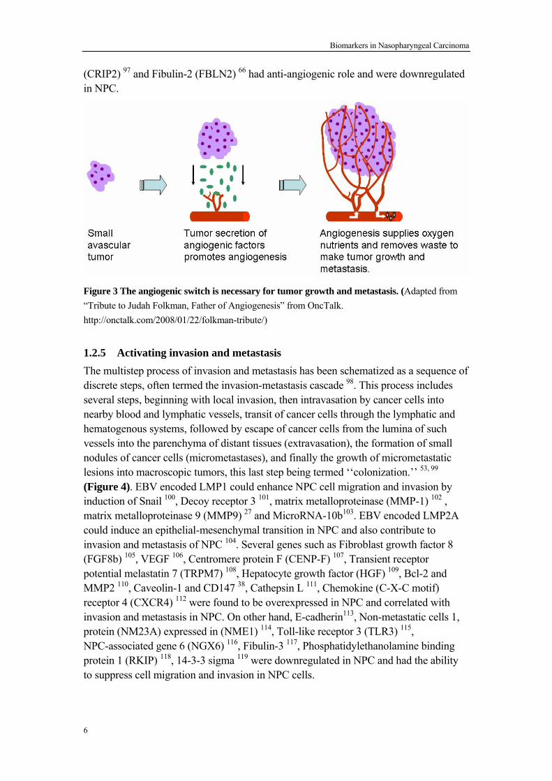
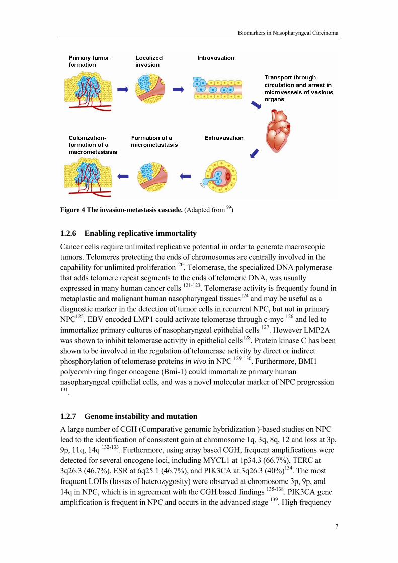
Figure 3 The angiogenic switch is necessary for tumor growth and metastasis. (Adapted from “Tribute to Judah Folkman, Father of Angiogenesis” from OncTalk. http://onctalk.com/2008/01/22/folkman-tribute/) 1.2.5 Activating invasion and metastasis The multistep process of invasion and metastasis has been schematized as a sequence of discrete steps, often termed the invasion-metastasis cascade 98. This process includes several steps, beginning with local invasion, then intravasation by cancer cells into nearby blood and lymphatic vessels, transit of cancer cells through the lymphatic and hematogenous systems, followed by escape of cancer cells from the lumina of such vessels into the parenchyma of distant tissues (extravasation), the formation of small nodules of cancer cells (micrometastases), and finally the growth of micrometastatic lesions into macroscopic tumors, this last step being termed ‘‘colonization.’’ 53, 99 (Figure 4). EBV encoded LMP1 could enhance NPC cell migration and invasion by induction of Snail 100, Decoy receptor 3 101, matrix metalloproteinase (MMP-1) 102 , matrix metalloproteinase 9 (MMP9) 27 and MicroRNA-10b103. EBV encoded LMP2A could induce an epithelial-mesenchymal transition in NPC and also contribute to invasion and metastasis of NPC 104. Several genes such as Fibroblast growth factor 8 (FGF8b) 105, VEGF 106, Centromere protein F (CENP-F) 107, Transient receptor potential melastatin 7 (TRPM7) 108, Hepatocyte growth factor (HGF) 109, Bcl-2 and MMP2 110, Caveolin-1 and CD147 38, Cathepsin L 111, Chemokine (C-X-C motif) receptor 4 (CXCR4) 112 were found to be overexpressed in NPC and correlated with invasion and metastasis in NPC. On other hand, E-cadherin113, Non-metastatic cells 1, protein (NM23A) expressed in (NME1) 114, Toll-like receptor 3 (TLR3) 115, NPC-associated gene 6 (NGX6) 116, Fibulin-3 117, Phosphatidylethanolamine binding protein 1 (RKIP) 118, 14-3-3 sigma 119 were downregulated in NPC and had the ability to suppress cell migration and invasion in NPC cells.
6
Biomarkers in Nasopharyngeal Carcinoma
Figure 4 The invasion-metastasis cascade. (Adapted from 99) 1.2.6 Enabling replicative immortality Cancer cells require unlimited replicative potential in order to generate macroscopic tumors. Telomeres protecting the ends of chromosomes are centrally involved in the capability for unlimited proliferation120. Telomerase, the specialized DNA polymerase that adds telomere repeat segments to the ends of telomeric DNA, was usually expressed in many human cancer cells 121-123. Telomerase activity is frequently found in metaplastic and malignant human nasopharyngeal tissues124 and may be useful as a diagnostic marker in the detection of tumor cells in recurrent NPC, but not in primary NPC125. EBV encoded LMP1 could activate telomerase through c-myc 126 and led to immortalize primary cultures of nasopharyngeal epithelial cells 127. However LMP2A was shown to inhibit telomerase activity in epithelial cells128. Protein kinase C has been shown to be involved in the regulation of telomerase activity by direct or indirect phosphorylation of telomerase proteins in vivo in NPC 129 130. Furthermore, BMI1 polycomb ring finger oncogene (Bmi-1) could immortalize primary human nasopharyngeal epithelial cells, and was a novel molecular marker of NPC progression 131.
1.2.7 Genome instability and mutation A large number of CGH (Comparative genomic hybridization )-based studies on NPC lead to the identification of consistent gain at chromosome 1q, 3q, 8q, 12 and loss at 3p, 9p, 11q, 14q 132-133. Furthermore, using array based CGH, frequent amplifications were detected for several oncogene loci, including MYCL1 at 1p34.3 (66.7%), TERC at 3q26.3 (46.7%), ESR at 6q25.1 (46.7%), and PIK3CA at 3q26.3 (40%)134. The most frequent LOHs (losses of heterozygosity) were observed at chromosome 3p, 9p, and 14q in NPC, which is in agreement with the CGH based findings 135-138. PIK3CA gene amplification is frequent in NPC and occurs in the advanced stage 139. High frequency
7
Biomarkers in Nasopharyngeal Carcinoma
somatic mutations in RASSF1A is seen 140. Moreover, mitochondrial DNA somatic mutations are frequent in NPC 141. 1.2.8 Other hallmarks and characteristics The role of defective immunological monitoring of tumors seem to be validated by the striking increases of certain cancers in immunocompromised individuals 142. EBV encoded EBERs induced the expression of insulin-like growth factor 1 (IGF-I) in NPC cells and modulated the innate immunity system143. Inhibition of NF-kB by EBV encoded EBNA1 is employed by viruses as an immune evasion strategy which is also closely linked to oncogenesis during persistent viral infection 144. The imbalances of T(reg) and effector T cell phenotypes and down-regulation of signal-transducing molecules in TILs, play important roles in suppression of immune response and immune evasion of NPC145.
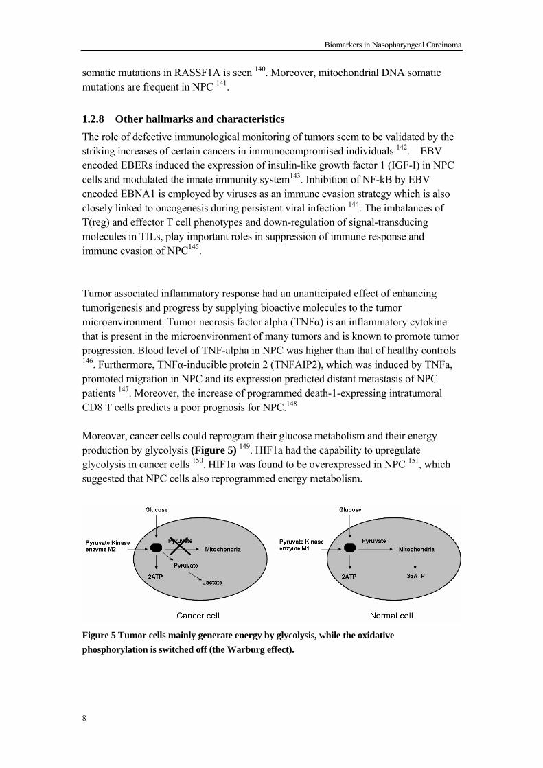
Tumor associated inflammatory response had an unanticipated effect of enhancing tumorigenesis and progress by supplying bioactive molecules to the tumor microenvironment. Tumor necrosis factor alpha (TNFα) is an inflammatory cytokine that is present in the microenvironment of many tumors and is known to promote tumor progression. Blood level of TNF-alpha in NPC was higher than that of healthy controls 146. Furthermore, TNFα-inducible protein 2 (TNFAIP2), which was induced by TNFa, promoted migration in NPC and its expression predicted distant metastasis of NPC patients 147. Moreover, the increase of programmed death-1-expressing intratumoral CD8 T cells predicts a poor prognosis for NPC.148 Moreover, cancer cells could reprogram their glucose metabolism and their energy production by glycolysis (Figure 5) 149. HIF1a had the capability to upregulate glycolysis in cancer cells 150. HIF1a was found to be overexpressed in NPC 151, which suggested that NPC cells also reprogrammed energy metabolism.
Figure 5 Tumor cells mainly generate energy by glycolysis, while the oxidative phosphorylation is switched off (the Warburg effect).
8
Biomarkers in Nasopharyngeal Carcinoma
1.3 BIOMARKERS FOR NPC 1.3.1 EBV markers EBV has been verified to correlate with the etiology of NPC, and EBV-specific antibody-based assays such as serum titers of IgA antibodies to viral capsid antigen (VCA), early antigen (EA), nuclear antigen (EBNA) and DNase assay have been commonly used for at least twenty years as standard markers for screening and monitoring the disease152. Serological levels of VCA/IgA and EA/IgA were significantly associated with increased risks for NPC, and VCA/IgA had better predictive performance for NPC incidence than EA/IgA153. Some studies suggested that combination of EBV EA and EBNA recombinant antigens has high sensitivity and acceptable specificity and accuracy in early diagnosis and mass screening of NPC patients154-155. Antibodies targeting the BARF1 protein could be used as a new diagnostic tool to identify NPC patients33. Recently, one paper reported that pretreatment serologic antienzyme rate (AER) of EBV DNase-specific neutralizing antibody could serve as an independent prognostic marker complementing TNM stage in NPC 156. EBV viral load measurement in serum or plasma has been shown to be useful for diagnosis and monitoring in NPC patients. It can reflect tumor stage, is predictive for overall survival, correlated with the residual tumor load after treatment and accurately predict recurrence and prognosis 157-161. Hence cell-free EBV DNA could become one of the potential diagnostic and prognostic molecular markers for NPC.
1.3.2 Epigenetic markers DNA methylation of TSGs is widely known to be an early event in carcinogenesis and methylation analysis in the promoter of TSGs may serve as a complementary marker for identifying early cases. Epigenetic changes are frequently found in NPC and the high detection rate in body fluids suggests its potential application in non-invasive screening of NPC or detection of residual carcinoma after treatment 162. Hutajulu et al 163 found that combined analysis of five methylation markers (RASSF1A, p16, WIF1, CHFR and RIZ1) in brushings shows good discrimination between NPC and non-NPC with a detection rate of 98% in a high risk population. Moreover, hypermethylated promoter DNA of at least one of the three genes (CDH1, DAPK1, and p16) was detectable in post-treatment plasma of 5 of 13 (38%) recurrent NPC patients and none of the patients in remission, which suggested that cell-free circulating methylated gene promoter DNA is a potential useful serological marker in assisting in screening of potentially local or regional recurrent NPC 164. 1.3.3 Genomic markers Several genomic markers in NPC have been identified as possible candidates for risk prediction. Familial NPC has been linked to genetic predispositions such as leukocyte
9
Biomarkers in Nasopharyngeal Carcinoma
antigen (HLA) genotypes 165-166 and susceptibility loci on chromosome 3p 219, 4p15.1-q12 10and 5p 1311. Recently, a genome-wide association study was performed using 464,328 autosomal single-nucleotide polymorphisms (SNPs) in 1,583 NPCs and 1,894 controls, and three new susceptibility loci, including TNFRSF19 on 13q12, MDS1-EVI1 on 3q26 and the CDKN2A-CDKN2B gene cluster on 9p21 were identified166. Moreover, the allele of Cav-1 T29107A was found to be protective against the development of NPC and may be a novel useful genomic marker for early screening and prediction of NPC 167. The endothelin A receptor (EDNRA) / H323H polymorphism was a novel and independent prognostic marker for patients with locoregionally advanced NPC 168. MDM2 SNP309 T/G polymorphism 169 and Tumor necrosis factor alpha 308G/A polymorphism 170 could be considered a risk marker for the development of NPC. Moreover, the FAS-670 G allele 171 and MCP-1 SNP-2518 172 have been suggested as a genetic marker for risk of distant metastasis of NPC patients.
1.3.4 Protein and microRNA markers Many aberrantly expressed cellular genes have been found in NPC cancer cells and patients’ serum, and many studies indicated that the expression pattern of some particular genes could be the biomarkers for NPC diagnosis and prognosis prediction. Wang et al 173 developed a new NPC-SVM classifier, which integrated patient sex and the protein expression level of seven genes, including EBV encoded LMP1, CD147, caveolin-1, phospho-P70S6 kinase, matrix metalloproteinase 11 (MMP11), survivin, and secreted protein acidic and rich in cysteine (SPARC). The NPC-SVM classifier distinguished patients with NPC into low- and high-risk groups with significant differences in 5-year DSS in the evaluated patients (87% v 37.7%; P < .001) in the validation cohort. Moreover, the patients with NPC with higher levels of interleukin-8 (IL-8), vascular endothelial growth factor (VEGF), macrophage inflammatory protein (MIP)-3α, and EBV DNA in plasma had worse prognosis for overall survival 174. Until now, useful miRNA markers have not yet been identified in either NPC tumor tissue or the NPC patient serum. 1.3.5 Other biomarkers Metabolic profiling revealed that kynurenine, N-acetylglucosaminylamine, N-acetylglucosamine and hydroxyphenylpyruvate are increased in NPC patient sera. After radiotherapy, these four metabolites decreased gradually, tended to normalize, and were associated with rate of tumor reduction. The results revealed that kynurenine, N-acetylglucosaminylamine, N-acetylglucosamine and hydroxyphenylpyruvate are potential markers for NPC diagnosis 175.
10
Biomarkers in Nasopharyngeal Carcinoma
2 AIMS OF THE STUDY Currently, NPC prognosis evaluation and the choice of treatment are mainly based on the tumor-node-metastasis (TNM) staging system. However, NPC patients with the same clinical stage often present with different clinical courses, suggesting that the TNM stage is insufficient to provide satisfactory prediction of prognosis. Therefore, it is important to find molecular biomarkers, which can help clinicians to identify high risk NPC patient, thus improving prognosis prediction, and therapeutic interventions in NPC patients. To identify novel prognostic markers for NPC, we have studied the following aspects: 1. Investigating the expression pattern and clinical significance of Caveolin-1 and CD147 in NPC, and their possible functional mechanism. 2 Exploring the expression pattern of miR-155 and its possible mechanism of action in NPC, identifying targets and evaluating prognostic value in NPC
3 Investigating the expression of Syk and evaluating its prognostic value in NPC
4 Identifying putative TSGs in Chr.3 in NPC, and their possible regulatory mechanism in NPC
11
Biomarkers in Nasopharyngeal Carcinoma
3 MATERIALS AND METHODS 3.1 METHODS FOR BIOMARKER EVALUATION To evaluate the clinical value of certain biomarkers, immunohistochemistry was performed in NPC clinical samples, and then the prognostic value was analyzed by Kaplan-Meier analysis and log-rank tests. The detailed information and protocol of these methods are described in Paper I, II and III. Clinical Samples For this retrospective study, archival formalin-fixed, paraffin-embedded (FFPE) tissue specimens from primary NPC patients with clinical information were obtained from Sun Yat-Sen University Cancer Center (Guangzhou, China). All NPC samples in our study were obtained before treatment with standard curative radiotherapy, with or without chemotherapy. The study was approved by the Research Ethics Committee of Sun Yat-Sen University Cancer Center, Guangzhou, China (Reference number: YP-2009175) and Karolinska Institutet, Stockholm, Sweden (Reference number: 00-302). Immunohistochemistry Immunohistochemistry is applicable for the detection of protein expression in tissue sections with specific antibodies, which can provide both location and intensity of positive signal. The immunohistochemistry results were evaluated and scored by senior pathologists without knowledge of the clinicopathological outcomes of the patients. Then the NPC patients were divided into high expression group and low expression group according to the composite pathology score which each patient was given. Statistical analysis SPSS12.0 software was used to statistically evaluate the clinical biomarker data. The association between biomarker expression and clinicopathological parameters were assessed using a Chi-Square test. Kaplan-Meier analysis and log-rank tests were used to assess the survival rate and to compare the difference in survival curves. Differences were considered significant when p < 0.05. 3.2 METHODS FOR FUNCTIONAL STUDIES OF BIOMARKERS To study the function of the certain biomarkers, gene transfection technology was used in NPC cell lines to knockdown or overexpress protein. Transwell migration assay was performed to evaluate the migration ability of the cells. All the detailed methods used can be found in section “Materials and methods” of Paper I. Cell lines
12
Biomarkers in Nasopharyngeal Carcinoma
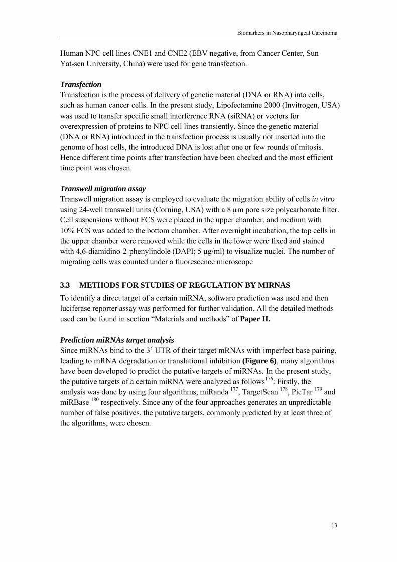
Human NPC cell lines CNE1 and CNE2 (EBV negative, from Cancer Center, Sun Yat-sen University, China) were used for gene transfection. Transfection Transfection is the process of delivery of genetic material (DNA or RNA) into cells, such as human cancer cells. In the present study, Lipofectamine 2000 (Invitrogen, USA) was used to transfer specific small interference RNA (siRNA) or vectors for overexpression of proteins to NPC cell lines transiently. Since the genetic material (DNA or RNA) introduced in the transfection process is usually not inserted into the genome of host cells, the introduced DNA is lost after one or few rounds of mitosis. Hence different time points after transfection have been checked and the most efficient time point was chosen. Transwell migration assay Transwell migration assay is employed to evaluate the migration ability of cells in vitro using 24-well transwell units (Corning, USA) with a 8 μm pore size polycarbonate filter. Cell suspensions without FCS were placed in the upper chamber, and medium with 10% FCS was added to the bottom chamber. After overnight incubation, the top cells in the upper chamber were removed while the cells in the lower were fixed and stained with 4,6-diamidino-2-phenylindole (DAPI; 5 μg/ml) to visualize nuclei. The number of migrating cells was counted under a fluorescence microscope 3.3 METHODS FOR STUDIES OF REGULATION BY MIRNAS To identify a direct target of a certain miRNA, software prediction was used and then luciferase reporter assay was performed for further validation. All the detailed methods used can be found in section “Materials and methods” of Paper II. Prediction miRNAs target analysis Since miRNAs bind to the 3’ UTR of their target mRNAs with imperfect base pairing, leading to mRNA degradation or translational inhibition (Figure 6), many algorithms have been developed to predict the putative targets of miRNAs. In the present study, the putative targets of a certain miRNA were analyzed as follows176: Firstly, the analysis was done by using four algorithms, miRanda 177, TargetScan 178, PicTar 179 and miRBase 180 respectively. Since any of the four approaches generates an unpredictable number of false positives, the putative targets, commonly predicted by at least three of the algorithms, were chosen.
13
Biomarkers in Nasopharyngeal Carcinoma
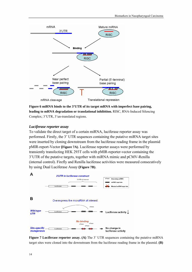
Figure 6 miRNA binds to the 3’UTR of its target mRNA with imperfect base pairing, leading to mRNA degradation or translational inhibition. RISC, RNA-Induced Silencing Complex; 3’UTR, 3’un-translated regions. Luciferase reporter assay To validate the direct target of a certain miRNA, luciferase reporter assay was performed. Firstly, the 3’ UTR sequences containing the putative miRNA target sites were inserted by cloning downstream from the luciferase reading frame in the plasmid pMIR-report-Vector (Figure 7A). Luciferase reporter assays were performed by transiently transfecting HEK 293T cells with pMIR-reporter-vector containing the 3’UTR of the putative targets, together with miRNA mimic and pCMV-Renilla (internal control). Firefly and Renilla luciferase activities were measured consecutively by using Dual Luciferase Assay (Figure 7B).
Figure 7 Luciferase reporter assay. (A) The 3’ UTR sequences containing the putative miRNA target sites were cloned into the downstream from the luciferase reading frame in the plasmid. (B)
14
Biomarkers in Nasopharyngeal Carcinoma
A result was positive when the miRNA could repress the luciferase activity in the wild type 3’UTR construct, but could not in the site-specific mutagenesis construct. 3.4 METHODS FOR STUDIES OF EPIGENETIC REGULATION
To investigate whether epigenetic modification might be involved in regulation of putative biomarkers, i.e. to identify epigenetically silenced biomarkers (tumor suppressor genes), Not I microarrays were performed to screen methylated or deleted genes in chromosome 3 in NPC and then Bisulfite sequencing and methylation specific PCR (MSP) were performed for further validation. All the detailed methods used can be found in section “Materials and methods” of Paper IV.
Chr. 3 specific NotI microarrays
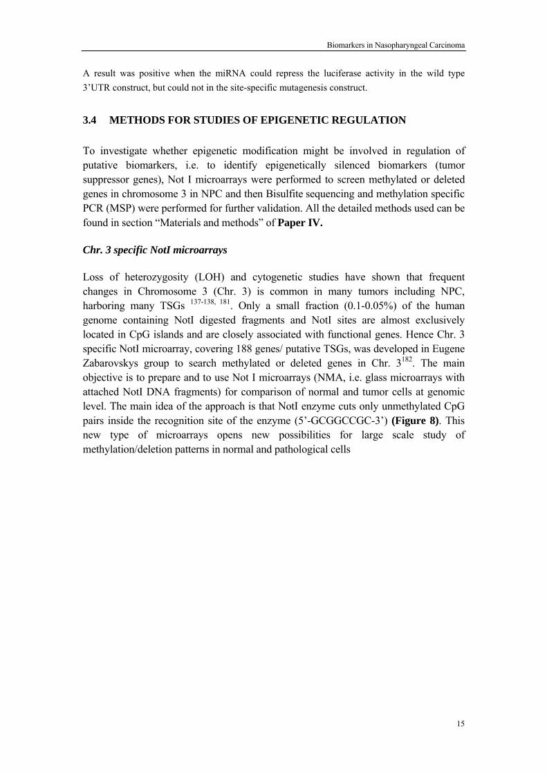
Loss of heterozygosity (LOH) and cytogenetic studies have shown that frequent changes in Chromosome 3 (Chr. 3) is common in many tumors including NPC, harboring many TSGs 137-138, 181. Only a small fraction (0.1-0.05%) of the human genome containing NotI digested fragments and NotI sites are almost exclusively located in CpG islands and are closely associated with functional genes. Hence Chr. 3 specific NotI microarray, covering 188 genes/ putative TSGs, was developed in Eugene Zabarovskys group to search methylated or deleted genes in Chr. 3182. The main objective is to prepare and to use Not I microarrays (NMA, i.e. glass microarrays with attached NotI DNA fragments) for comparison of normal and tumor cells at genomic level. The main idea of the approach is that NotI enzyme cuts only unmethylated CpG pairs inside the recognition site of the enzyme (5’-GCGGCCGC-3’) (Figure 8). This new type of microarrays opens new possibilities for large scale study of methylation/deletion patterns in normal and pathological cells
15
Biomarkers in Nasopharyngeal Carcinoma
Figure 8 Schematic representation how NotI microarrays are employed to identify methylated or deleted sequences (The main idea of using NR for NotI microaarays). N and B represent Not I and BamH I / Bgl II sites, respectively. Methylated NotI sites are indicated by an asterisk. NR: NotI- representation. (Adapted from182)
Bisulfite sequencing
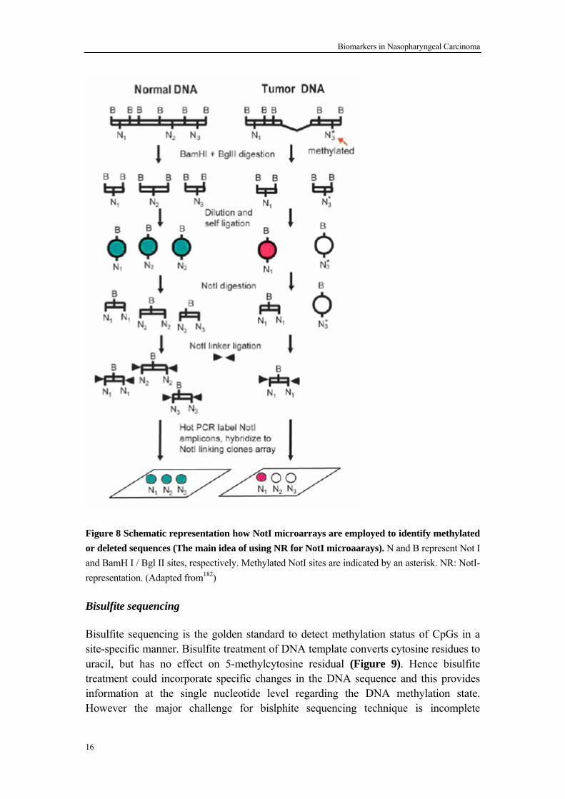
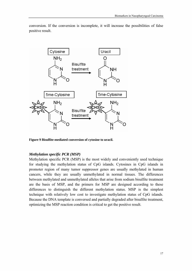
Bisulfite sequencing is the golden standard to detect methylation status of CpGs in a site-specific manner. Bisulfite treatment of DNA template converts cytosine residues to uracil, but has no effect on 5-methylcytosine residual (Figure 9). Hence bisulfite treatment could incorporate specific changes in the DNA sequence and this provides information at the single nucleotide level regarding the DNA methylation state. However the major challenge for bislphite sequencing technique is incomplete
16
Biomarkers in Nasopharyngeal Carcinoma
conversion. If the conversion is incomplete, it will increase the possibilities of false positive result.
Figure 9 Bisulfite-mediated conversion of cytosine to uracil.
Methylation specific PCR (MSP) Methylation specific PCR (MSP) is the most widely and conveniently used technique for studying the methylation status of CpG islands. Cytosines in CpG islands in promoter region of many tumor suppressor genes are usually methylated in human cancers, while they are usually unmethylated in normal tissues. The differences between methylated and unmethylated alleles that arise from sodium bisulfite treatment are the basis of MSP, and the primers for MSP are designed according to these differences to distinguish the different methylation status. MSP is the simplest technique with relatively low cost to investigate methylation status of CpG islands. Because the DNA template is conversed and partially degraded after bisulfite treatment, optimizing the MSP reaction condition is critical to get the positive result.
17
Biomarkers in Nasopharyngeal Carcinoma
4 RESULTS AND DISCUSSION 4.1 UPREGULATION OF CAVEOLIN-1 AND CD147 EXPRESSION IN NPC
ENHANCED TUMOR CELL MIGRATION AND CORRELATED WITH
POOR PROGNOSIS OF THE PATIENTS (PAPER I)
Expression of Cav-1 and CD147 and their associations with clinico- pathological parameters in NPC Cav-1 is a major structural component of caveolae, which are involved in several cellular functions, including vesicle trafficking, cholesterol homeostasis and signal transduction 183-184. Reduced Cav-1 expression has been reported in ovarian cancer185, breast cancer186 and lung cancer187. On the other hand, Cav-1 overexpression has been observed in bladder cancer188, prostate cancer189 and esophageal squamous cell
carcinoma (ESCC) 190. Extracellular matrix metalloproteinase inducer (EMMPRIN), also named CD147, is a glycoprotein that belongs to the immunoglobulin superfamily 191. Due to serendipitous observation, we thought the expression patterns of Cav-1 and CD147 in NPC was of interest. In the present study, Cav-1 and CD147 were found to be upregulated in NPC cell lines as well as in NPC clinical samples. We found that Cav-1 was expressed at a high level in 96 cases (49.5%) of NPC, which associated significantly with local recurrence (p = 0.038) and metastasis (p = 0.025) of NPC. No significant association between Cav-1 expression and age, gender and TNM stage of the patients was observed in this study. Overexpression of CD147 was observed in 117 cases (59.4%) of NPC, which was associated significantly with metastasis of the disease (p = 0.017). No significant association between CD147 expression and age, gender, clinical staging and recurrence of the disease was observed in this study. Cav-1 overexpression was reported previously to be correlated with the metastasis of several malignancies, including prostate cancer 189, ESCC 190, breast cancer 192 and lung cancer 193. In the present study, we revealed that Cav-1 overexpression in NPC tumor cells was significantly correlated with recurrence (p = 0.038) and metastasis (p = 0.025) of the disease. Overexpression of CD147 in the tumor cells has been reported to be correlated with metastasis of breast cancer 194, and oral squamous cell carcinoma 195; and with poor prognosis of patients with ESCC 196, breast cancer 194, serous ovarian cancer 197 and gastric cancer 198. We found that CD147 overexpression in NPC tumor cells was significantly correlated with metastasis (p = 0.017) of the disease. Combined Cav-1 and CD147 overexpression predicts poor survival of the NPC patients
18
Biomarkers in Nasopharyngeal Carcinoma
Cav-1 overexpression in tumor cells also has been reported to be correlated with poor prognosis in patients with ESCC 190, renal clear cell carcinoma 199, prostate cancer 200, lung cancer 201, and pancreatic ductal adenocarcinoma 202. In the present study, we revealed that Cav-1 overexpression in NPC tumor cells was significantly correlated with poor prognosis in NPC patients. Several recent studies have demonstrated that overexpression of CD147 is correlated with poor prognosis in human cancers, including ESCC 196, breast carcinoma 194, serous ovarian carcinoma 197 and gastric carcinoma 198. We found that CD147 overexpression in NPC tumor cells was significantly correlated with poor prognosis in NPC patients. In addition, we found that NPC patients with overexpression of both Cav-1 and CD147 in tumor cells had a significantly worse prognosis and a significantly lower five-year overall survival rate relative to NPC patients with low expression of Cav-1 and / or CD147 (p = 0.001). Cav-1 and CD147 increase the migration ability of CNE1 and CNE2 cells through increasing the secretion of MMP-3 and MMP-11 (active) Recent evidence suggests a central role for Cav-1 in the regulation of cellular invasion and metastasis 203-204. CD147 is enriched on the plasma membrane of tumor cells and triggers the production or release of MMPs in surrounding mesenchymal cells and tumor cells 205-206. In this study, Cav-1 and CD147 overexpression were found to be associated with metastasis in NPC patients. Transwell migration assays further revealed that loss of Cav-1 and CD147 expression inhibited CNE1 and CNE2 cell migration ability, whereas Cav-1 and CD147 overexpression promoted cell migration ability. Taken together, our results are consistent with previous studies of Cav-1 and CD147 expression in other malignancies, and indicate that these two genes may play a key role in the invasion and metastasis of NPC. How do Cav-1 and CD147 regulate the invasion and metastasis of NPC? Growing evidence suggests a close association between Cav-1, CD147 and the expression or secretion of MMPs. Overexpression of Cav-1 in HEK293 cells decreased MMP-1 secretion in a co-culture assay with primary human fibroblasts 207, and overexpression of Cav-1 in metastatic mammary tumor cells could inhibit MMP-2 and MMP-9 secretion, although the expression of MMP-2 and MMP-9 in whole cell lysates was not altered 186. In contrast, Cav-1 can induce MMP-11 secretion and the invasive potential of a murine hepatocarcinoma cell line 208. CD147 is a tumor cell-derived MMP inducer that is expressed on the tumor cell surface and triggers the production or release of MMP-1, MMP-2, MMP-3, MMP-9, MT1-MMP and MT2-MMP in the surrounding mesenchymal cells and tumor cells 205-206, 209-211. In the present study, we found that the suppression of Cav-1 and CD147 expression led to decrease MMP-3 and MMP-11 (active) secretion in CNE1 and CNE2 cells, whereas overexpression of Cav-1 in CNE1 and CNE2 cells promoted MMP-3 and MMP-11 (active) secretion. Furthermore, suppression of CD147 expression in Cav-1 overexpressing CNE1 cells could also lead to decrease of MMP-3 and MMP-11 (active) secretion. Taken together, these results indicate that Cav-1 and CD147 overexpression in NPC tumors can promote tumor cell migration by stimulating MMP-3 and MMP-11 (active) secretion in NPC tumor cells,
19
Biomarkers in Nasopharyngeal Carcinoma
and Cav-1 regulates MMP-3 and MMP-11 (active) secretion partially, maybe directly, through CD147 in NPC tumor cells.
4.2 UPREGULATION OF MIR-155 IN NPC IS PARTLY DRIVEN BY LMP1
AND LMP2A AND DOWNREGULATES A NEGATIVE PROGNOSTIC
MARKER JMJD1A (PAPER II)
EBV LMP1 and LMP2A further enhance miR-155 expression in NPC
MiR-155 was upregulated in several human tumors, such as lymphoma 212-213, thyroid carcinoma214 , breast cancer215-216, colon cancer215, cervical cancer217, pancreatic cancer218, and lung cancer219. Furthermore, elevated expression of miR-155 was associated with poor prognosis of pancreatic cancer218 and lung cancer 219. Recently, Chen et al used a stem-loop real-time-PCR method to quantify the expression levels of 270 human miRNAs in 13 NPC samples and 9 adjacent normal tissues. They identified 35 miRNAs whose expression levels were significantly altered in NPC samples, including upregulation of miR-155 220. In the present study, we reported that miR-155 expression was enhanced in NPC. Several studies have demonstrated that EBV could induce miR-155 expression in B cells and cell lines which in turn modulates EBV-regulated pathways 221-223. LMP1222-225 and EBNA2222 were responsible for the upregulation of miR-155 after EBV infection of B-lymphocytes, while LMP2A did not influence miR-155 expression223, 225. Furthermore LMP1 was demonstrated to trans-activate miR-155 transcription through the NF-kappaB and AP1 pathways 223, 225. In contrast, EBV did not induce the expression of miR155 in HEK 293 and Hela cells222. We found that both LMP1 and LMP2A could induce the miR-155 expression in NPC CNE1 and TW03 cells. To our knowledge, this is the first report that LMP2A could induce miR-155 expression in NPC. Guasparri et al reported that EBV LMP2A protein could affect LMP1-mediated NF-κB signaling and survival of lymphoma cells, hence LMP2A might increase miR-155 expression through NF- kappaB pathway 30. In addition, LMP1 was reported to induce the expression of miR-146a in B-lymphocytes 224. However, in our study, we found that neither LMP1 nor LMP2A could induce the miR-146a expression in NPC cells. These differences might be due to different tumor cell type backgrounds. LMP1 and LMP2A were expressed in approximately 65% and 45% NPC patients, repectively 25, 28, and miR-155 was also found to be upregulated in many human tumors, which were not related to EBV 214-219. Furthermore, in our study, both CNE1 and TW03 were EBV negative NPC derived cell lines, the expression of miR-155 in these two cell lines was higher than that in the immortalized nasopharyngeal epithelial cell line NP69. Hence there are also other factors than EBV genes which might upregulate miR-155
20
Biomarkers in Nasopharyngeal Carcinoma
expression in NPC. TGF-beta (Transforming Growth Factor – beta) was verified to induce miR-155 expression by increased promoter activity due to SMAD4 (SMAD family member 5)226. Elevated serum levels of TGF-beta1 was also found in NPC patients227. Hence TGF-beta and SMAD4 pathways might also contributed to miR-155 overexpression in NPC.
Identification of JMJD1A as a direct target of miR-155 in NPC
At present, many direct targets of miR-155 have been identified and show oncogenic features of miR-155. MiR-155 promoted the proliferation of breast cancer cells through down-regulation of SOCS1 (Suppressor of cytokine signaling 1) 228 and FOXO3a (forkhead box O3) 229. MiR-155 has also been reported to be involved in the development of lymphoma by targeting SMAD5 (SMAD family member 5) 230 and SHIP1 (inositol polyphosphate-5-phosphatase) 231. MiR-155 could promote pancreatic tumor development through downregulation of TP53INP1 (Tumor protein p53 induced nuclear protein 1) 232. Moreover, some other genes implicated in differentiation, inflammation and transcriptional regulation, were direct targets of miR-155, including HIF-1 (Hypoxia-inducible factor 1)233-234, BACH1 (BTB and CNC homology 1, basic leucine zipper transcription factor1)223, 233, HIVEP2 (Human immunodeficiency virus type I enhancer binding protein 2)223, IKKɛ (Inhibitor of kappa light polypeptide gene enhancer in B cells, kinase ɛ)235, and so on. In our study, we found that miR-155 could repress endogenous JMJD1A and BACH1 protein expression in NP69 cells. Luciferase reporter assay was performed to identify both JMJD1A and BACH1 as direct targets of miR-155 in NPC cells. This is the first report that JMJD1A is a direct target of miR-155. Furthermore, JMJD1A and BACH1 are downregulated in NPC cell lines and NPC tumor tissues.
Downregulation of JMJD1A predicts poor survival in NPC The expression of JMJD1A and BACH1 was detected by immunostaining in 185 NPC cases. Low expression of JMJD1A was observed in 113 (61%), and was associated significantly with N-stage (p = 0.023). No significant association was seen between JMJD1A expression and age, gender, T stage, TNM stage, recurrence or metastasis. In addition, no significant association was seen between BACH1 expression and age, gender, T stage, N stage, TNM stage, recurrence or metastasis. The five-year overall survival rate was 61.3% for patients with low JMJD1A expression (n = 113), and 77.2% for patients with high JMJD1A expression (n = 72), which was a significant difference (p = 0.021). The five-year overall survival rate was 66.8% for patients with low BACH1 expression (n = 94), and 68.2% for patients with high BACH1 expression (n = 91), which was not significant (p = 0.759). Furthermore, the five-year disease-free survival rate was 57.0% for NPC patients with low levels of JMJD1A expression (n = 113), and 68.7% for those with high levels of JMJD1A expression (n = 72), and this difference in the disease-free survival rate was significant
21
Biomarkers in Nasopharyngeal Carcinoma
(p = 0.049). No significant difference was seen in the disease-free survival rate of NPC patients, with or without BACH1 overexpression (p = 0.895). Adam et al. 236 demonstrated that loss of JMJD1A is sufficient to reduce tumor growth of renal cell carcinoma and colon carcinoma in vivo, suggesting that function of JMJD1A in different cells and tissues depend on cell microenvironment. Hence, the function of JMJD1A and BACH1 in NPC deserve for further attention. 4.3 ELEVATED SYK EXPRESSION CORRELATES WITH LMP2A
EXPRESSION IN NPC AND PREDICTS POOR PROGNOSIS (PAPER
III)
High expression of Syk correlates with clinicopathological parameters and predicts poor survival in NPC
Spleen tyrosine kinase (Syk) is a nonreceptor tyrosine kinase that contains tandem NH2 terminal Src homology 2 domains, multiple tyrosine phosphorylation sites, and a COOH-terminal tyrosine kinase domain237. Moreover, Syk promoter hypermethylation leads to gene silencing and reduced Syk expression correlates with tumor metastasis and poor prognosis in breast cancer238, pancreatic adenocarcinoma239, hepatocellular carcinoma240, bladder carcinoma241, and gastric carcinoma242. In contrast, overexpression of Syk is associated with recurrence and poor prognosis of squamous cell carcinomas of the head and neck (SCCHN) patients 243. In addition, overexpression of Syk has also been observed in peripheral T-cell lymphomas 244-245, chronic lymphocytic leukemia246, and B-lineage acute lymphoblastic leukemia247. In this study, we showed that high expression of Syk was significantly associated with T-stage and local recurrence of NPC. Our data also indicated that NPC patients with high expression levels of Syk had shorter survival time, shorter disease free time, and that Syk was an independent prognosis factor in NPC. These results are consistent with the study of another head and neck cancer, SCCHN, in which, Sutima et al. 243 reported that overexpression of Syk was associated with recurrence and poor prognosis of the patients. Syk has been reported to be involved in tumor metastasis in many human cancers, either suppressing or enhancing metastasis. Reduced expression of Syk could promote cell migration in breast cancer 238, while overexpression of Syk could enhance cell migration in SCCHN cells 243 . However, in our study, no significant correlation was found between Syk expression and tumor metastasis.It would have been highly interesting to compare Syk expression in paired samples of primary tumors and metastasis, but such clinical material with sufficient number of samples has not been
22
Biomarkers in Nasopharyngeal Carcinoma
made available as yet (biopsies are rarely removed by surgery). Taken together, our results indicate that Syk may play a role in local NPC recurrence, and correlates with poor prognosis of NPC patients, suggesting that Syk could be used clinically as one of several biomarkers for NPC prognosis.
LMP2A induces Syk expression in NPC
It has been demonstrated that Syk could bind to the ITAM motif in EBV encoded LMP2A in B cells and epithelial cells 248-249. Moreover, Syk interacts selectively in immune cells with proteins containing a phosphorylated immunoreceptor tyrosine-based activation motif (ITAM) through its distinct tandem Src homology 2 domain 250. LMP2A, which is detected in the majority of NPC, contains an ITAM 251. In our study, we could confirm the interaction and co-localization of LMP2A with Syk as demonstrated by co-IP and IF in NPC cell lines. Interestingly, our results of ISH and IF indicated that LMP2A could enhance Syk expression in two NPC-cell lines SUNE1 and CNE2 cells, and LMP2A high expression positively correlated with Syk high expression in NPC clinical samples. However, LMP2A has been reported to promote the degradation of Syk in B cells by ubiquitination 252-253. The increased expression of Syk in LMP2A positive NPC might be a compensation for a shorter half life of Syk when LMP2A binds and affects Syk degradation. LMP2A was reported to induce cell migration and invasion in epithelial cells. 29. Lu et al 249 further demonstrated that endogenous Syk was activated in epithelial cells in the presence of LMP2A, and that Syk was required for LMP2A-triggered cell migration in epithelial cells. Taken together, these data suggested that LMP2A expression partly contributes to Syk high expression in NPC, and that Syk and LMP2A may have co-operative roles in NPC development. The precise relationship between Syk and LMP2A and the mechanism by which LMP2A induces Syk in NPC deserve further experimental studies.
4.4 WNT7A AND ITGA9 WERE HYPERMETHYLATED AND
DOWNREGULATED IN NPC (PAPER IV) Screening for potentially methylated genes in NPC using NotI micrarrays
Loss of heterozygosity (LOH) and cytogenetic studies have shown that frequent changes in Chromosome 3 (Chr. 3) is common in many tumors including NPC, harboring many TSGs 181. To screen the potentially methylated or deleted genes in chr. 3 in NPC, Not I micorarrays were performed on genomic DNA from three NPC biopsies (T9, T10 and T18), two normal nasopharyngeal epithelia tissues (N1 and N2), three NPC cell lines (CNE1, TW03 and C666-1) and one normal nasopharyngeal epithelium derived cell line NP69. Ten genes (WNT7a, FGD5, ITGA9, RBSP3,
23
Biomarkers in Nasopharyngeal Carcinoma
ALDH1L1, NUDT16P, ZIC4, EPHB3, BCL6 and FGF12) were identified as showing reduced signal suggesting methylation or deletion. These genes were selected for further validation by MSP and Q-PCR. Among these, we selected two interesting genes WNT7a and ITGA9, for further study.
WNT7a and ITGA9 were hypermethylated in NPC
The methylation status of the CpG rich region in the WNT7a gene promoter was investigated by bisulfite sequencing in four NPC biopsies (T1, T2, T5 and T6) and one normal nasopharyngeal epithelia biopsy (N10). Twenty-three CpG sites were included in this region. The WNT7a promoter was partially methylated in the four NPC samples, whereas there was almost no of methylation in the control sample. Similarly, methylation status of CpG island in ITGA9 gene intron 1 region was investigated in the same samples. Fourteen CpG sites were included in this region. The CpG island in ITGA9 gene intron 1 region was partially methylated in four NPC samples, whereas it was totally free of methylation in the normal sample.
To investigate whether 5-aza-CdR could restore WNT7a and ITGA9 expression, NPC CNE1 and TW03 cell lines were treated with 10 μM of the demethylating agent 5-aza-2′-deoxycytidine (5-aza-CdR) for 96 h. QPCR was performed to detect WNT7a and ITGA9 expression in 5-aza-CdR treated and untreated NPC cell lines. Both WNT7a and ITGA9 expression were found to be restored after 5-aza-CdR treatment.
Downregulation of WNT7a and ITGA9 were found in NPC
QPCR was performed to detect WNT7a and ITGA9 expression in the three clinical NPC samples (T2, T3 and T4) as well as in four normal epithelial tissues (N2, N4, N5 and N7). The average expression levels of WNT7a and ITGA9 in NPC tumors (9.4±7.7 fold; 4.0±2.6 fold) were downregulated, compared with the expression in normal epithelial tissues (28.9±18.9 fold; 19.9±10.2 fold). Moreover, immunostaining was performed to evaluate the expression of WNT7a and ITGA9 in three cases of NPC tumor cells and adjacent normal nasopharyngeal epithelium. Strong expression of WNT7a and ITGA9 were observed in the membrane and cytoplasm of normal adjacent nasopharyngeal epithelium cells, while weak expression of WNT7a and ITGA9 were observed in NPC tumor cells.
In Non-Small Cell Lung Cancer (NSCLC) cells, the re-expression of WNT-7a and signaling through Fzd-9 are associated with increased differentiation and thus represent a tumor suppressor pathway 254. It has been demonstrated that WNT7a is a positive regulator of E-cadherin expression in lung cancer cells and E-cadherin is also downregulated in NPC . Pharmacologic reversal of WNT7a losses could be achieved with the use of inhibitors of GSK3β, HDACs, or in some cases only with combined treatment .
255
256
255
24
Biomarkers in Nasopharyngeal Carcinoma
ITGA9 is abundantly expressed in lung cancers in general and small cell lung cancers (SCLC) in particular. Its aberrant upregulation in the SCLC samples, both cell lines and primary tumors has been suggested to play a role in metastasis and in the progression to more malignant phenotypes 257. This is in contrast to the downregulation of ITGA9 in NPC and in human papilloma virus associated head and neck squamous cell carcinoma 258.
In conclusion, we found that WNT7a and ITGA9 are methylated and downregulated in Chinese NPC patients. As both proteins execute significant functions related to the tumor cell biology, in depth functional studies has to be pursed to understand the functional role of WNT7a and ITGA9 downregulation in the development of NPC. With such studies these two genes might be useful for improved diagnosis and treatment in the future.
25
Biomarkers in Nasopharyngeal Carcinoma
5 CONCLUDING REMARKS Nowadays, several biomarkers in serum, saliva and tissue have been identified for NPC screening, early diagnosis and prognosis prediction. Using serological biomarkers is a common and convenient way to screen and monitor NPC, such as EBV-specific antibodies in serum for early diagnosis and mass screening of NPC patients154-155, and EBV viral load in serum for diagnosis and monitoring recurrence in NPC patients 157-161 and Cell-free circulating methylated gene promoter DNA in assisting in screening of potentially local or regional recurrent NPC 164. Using epigenetic biomarkers in saliva and body fluids is a non-invasive way to screen and detect residual carcinoma after treatment of NPC 162. Genomic biomarkers could be used for screening the high risk population through NPC specific susceptibility loci9-11. Protein biomarkers in tissues could be used for diagnosis and prognosis prediction of NPC 173. In this study, we have tried to identify the new protein biomarkers and epigenetic biomarkers for NPC early diagnosis and prognosis prediction. Several approaches to identify biomarkers of significant clinical and functional interest have been explored. We have investigated some genes with considerable impact in other tumors (Paper I and III), genes regulated by miRNAs (Paper II) and epigenetically deregulated genes (Paper IV). In paper I, II and III, four protein biomarkers have been identified for prognosis prediction of NPC, and a larger scale of NPC samples are needed for further validation. In paper IV, two putative epigenetic biomarkers have been found, and further study is needed to evaluate their potentials for early diagnosis of NPC using either saliva or blood. These are my main conclusions from the work: 1. Cav-1 and CD147 overexpression predict poor NPC prognosis and enhanced tumor
cell migration, which is associated with MMP-3 and MMP-11 (active) secretion. 2. Upregulation of miR-155 in NPC is partly drived by LMP1 and LMP2A, and
results in downregaultion of JMJD1A, which is associated with N stage and poor prognosis of NPC patients.
3. High expression of Syk, which is resulted partly from LMP2A expression in NPC, is associated with tumor recurrence and poor prognosis of NPC patients
4. Downregulation of WNT7A and ITGA9 in NPC is partly due to promoter hypermethylation. The potential of WNT7A and ITGA9 as diagnosis or therapeutic targets for NPC should be further investigated
26
Biomarkers in Nasopharyngeal Carcinoma
6 ACKNOWLEDGEMENTS I would like to express my gratitude to everybody who helped me and supported me in last four years. In particular I would like to thank: Prof. Ingemar Ernberg, my principle supervisor. Thanks for giving me a fantastic opportunity to study in your group, for your great interest in my work, for valuable discussions and encouragement. Your enthusiasm to science, culture and life, your generousness, kindness and your talent in organization, all these have greatly affected me. With your full support, I have a wonderful memory in Karolinka Institutet, and in Sweden. Thank you! Prof. Jian-Yong Shao, my co-supervisor at Sun Yat-sen University Cancer Center, Guangzhou, China. Thank you for taking me as your student in China, for bringing me into the scientific research field, for giving me so many opportunities and freedom to do my research, for introducing me to Karolinska Institutet, for the support you ever gave me during the past eight years. I learnt a lot from you, not only about science, but also life. Prof. Lifu Hu, my co-supervisor. Thank you for your generous help especially at my first arrival to Sweden, for your invitation to your family dinner, for sharing me with your experience in Sweden and in your life, for the interesting discussion of my project, for organizing the EBV journal club. Prof. Maria G. Masucci, my mentor. Thank you for the interesting discussion and suggestions. I would like to thank Prof. Eugene Zabarovsky, Vladimir Kashuba, Ilya Ignatyev and Tatiana Pavlova at MTC, Karolinska Institutet for the cooperation on NPC epigenetic project. I would also like to thank Prof. Erik Aurell, Aymeric Fouquier d'Herouel and Ilqar Abdullayev at KTH for the collaborative project on the non-coding RNA of EBERs, although nothing has come out yet. I would like to thank Dr. Matin Corcoran at Cancer Center Karolinska Institutet for good discussion of miRNAs project and sharing plasmid with me. Present and previous members of Ingemar’s group: Fu Chen, it is a pity that you could not see my graduation today, I am so grateful to you from my heart. Jiezhi Zou, for the all your always nice help in the lab and in the daily life. Gosta Winberg, Liudmila Matskova, Li-Sophie Zhao Rathje, Imran Nawaz, Xiaoying Zhou, Anna Birgersdotter, Prof. Gunnar Klein, Anders Wennborg, Henrik Brändén, Elvira Grigorieva, Anna Aleman, Helena Lonnqvist, Joakim Coster and Prof. Anneka Ehrnst and her group: Rozina Cardha, Lotta Pramanik and Sven Grutzmeier.
27
Biomarkers in Nasopharyngeal Carcinoma
Thank you all for your support and the harmonious moments we being together during these years, for the Friday coffee breaks, Christmas dinners, etc. I would also like to give my thanks to Prof. Geogre Klein, Prof. Eva Klein, Barbro Ehlin-Henriksson, Liang Wu, Noemi Nagy, Elena Kashuba, Lorand Levente Kis, Daniel Salamon, Eahsan Rasul, Harsha Madapura and Monika Adori for the instructive group meeting together and journal club. Thanks for the knowledge you ever brought to me. I would like to thank Prof. Yi-Xin Zeng, Prof. Xiao-Feng Zhu, Prof. Wen-Lin Huang, Dr. Rong Deng, Dr. Chun-Fang Hu, Chang-Wei Kou, Li-Xu Yan, Hai-Yun Wang, Qiong Shao at Sun Yat-sen University Cancer Center, Guangzhou, China, for your support, your help and your cooperation. I would like to thank China Scholarship Council for the financial support and many thanks to Counselor Ning Zhang from education section of Chinese Embassy in Sweden, Wei Wang, Hong Zhang, Rui Fan and Xudong Li, for the kind help. Present and previous friends at MTC: Prof. Yihai Cao for your interesting discussion on basic tumor biology and good suggestion for my future career. Dr. Renhai Cao, Xiaoda Wang, Shengju Qiao, Xun Huang, Wenjie Bao, Liying Chen, Ying Zhao, Yao Shi, Hai Li, Xingqi Chen, Chengxi Shi, Qanzi Yan, Zongwei Wang, Yuan Xue, Zhaodong Zong, Ziquan Cao, Junwei Zhang, Yunlong Yang, Xiaojuan Yang, Hong Ji, Sharom Lim, Ying Ye and Xiangqun Ye. Thank you all for your generous help in different ways, for the happy time we spent together. Present and previous friends outside MTC: Prof. Annelie Brauner, Jinfeng Shen, Bin Zhao, Qing Chen, Jie Yan, Zongli Zheng, Wei Jiao, Roujun Peng, Miaoli Lin, Yingqing Wang, Zhe Hu, Zhuochun Peng, Yatao Du, Jiangqiang Xu, Xun Wang, Anquan Liu, Jikui Guan, Xin Wang, Xiaofeng Zheng, Zheng Chang, Si Cong, Jiaqi Huang, Xinming Wang, Heng Wang, Jingyi Ren, Bo Sun, Yougen Chen, Xiaoli Feng, Daohua Lou, Na Wang, Qiao Li, Hongya Han, Juan Du, Zhiwei Liu, Meng Chen, Meng Li, Jia Sun, and other friends not listed here, Thank you all for your help and I treasure the friendship very much. To my family, my parents, my elder sister, my elder brother-in-law and my little nephew, thanks for your endless love, your consistent encouragement, your never fading support, your patience and also for the confidence in me. Special thanks to my dearest Qin, my greatest achievement in Sweden, for the happy time we spent, for your love, for your consideration, for your support and for everything. I appreciate whatever you did to me, brought to me, no matter happiness or so called troubles, all are my sweetest memories in my life!
28
Biomarkers in Nasopharyngeal Carcinoma
7 REFERENCES 1. Shanmugaratnam K, Sobin LH. The World Health Organization histological classification of tumours of the upper respiratory tract and ear. A commentary on the second edition. Cancer. 1993 Apr 15;71(8):2689-97. 2. Lo KW, To KF, Huang DP. Focus on nasopharyngeal carcinoma. Cancer Cell. 2004 May;5(5):423-8. 3. Cancer incidence in five continents. Volume VII. IARC Sci Publ. 1997(143):i-xxxiv, 1-1240. 4. Licitra L, Bernier J, Cvitkovic E, Grandi C, Spinazze S, Bruzzi P, et al. Cancer of the nasopharynx. Crit Rev Oncol Hematol. 2003 Feb;45(2):199-213. 5. Titcomb CP, Jr. High incidence of nasopharyngeal carcinoma in Asia. J Insur Med. 2001;33(3):235-8. 6. Cao SM, Simons MJ, Qian CN. The prevalence and prevention of nasopharyngeal carcinoma in China. Chin J Cancer. Feb;30(2):114-9. 7. Buell P. The effect of migration on the risk of nasopharyngeal cancer among Chinese. Cancer Res. 1974 May;34(5):1189-91. 8. Dickson RI, Flores AD. Nasopharyngeal carcinoma: an evaluation of 134 patients treated between 1971-1980. Laryngoscope. 1985 Mar;95(3):276-83. 9. Xiong W, Zeng ZY, Xia JH, Xia K, Shen SR, Li XL, et al. A susceptibility locus at chromosome 3p21 linked to familial nasopharyngeal carcinoma. Cancer Res. 2004 Mar 15;64(6):1972-4. 10. Feng BJ, Huang W, Shugart YY, Lee MK, Zhang F, Xia JC, et al. Genome-wide scan for familial nasopharyngeal carcinoma reveals evidence of linkage to chromosome 4. Nat Genet. 2002 Aug;31(4):395-9. 11. Hu LF, Qiu QH, Fu SM, Sun D, Magnusson K, He B, et al. A genome-wide scan suggests a susceptibility locus on 5p 13 for nasopharyngeal carcinoma. Eur J Hum Genet. 2008 Mar;16(3):343-9. 12. Kumar S, Mahanta J. Aetiology of nasopharyngeal carcinoma. A review. Indian J Cancer. 1998 Jun;35(2):47-56. 13. Shao JY, Li YH, Gao HY, Wu QL, Cui NJ, Zhang L, et al. Comparison of plasma Epstein-Barr virus (EBV) DNA levels and serum EBV immunoglobulin A/virus capsid antigen antibody titers in patients with nasopharyngeal carcinoma. Cancer. 2004 Mar 15;100(6):1162-70. 14. Yu MC, Mo CC, Chong WX, Yeh FS, Henderson BE. Preserved foods and nasopharyngeal carcinoma: a case-control study in Guangxi, China. Cancer Res. 1988 Apr 1;48(7):1954-9. 15. Jia WH, Luo XY, Feng BJ, Ruan HL, Bei JX, Liu WS, et al. Traditional Cantonese diet and nasopharyngeal carcinoma risk: a large-scale case-control study in Guangdong, China. BMC Cancer.10:446. 16. Liebowitz D. Nasopharyngeal carcinoma: the Epstein-Barr virus association. Semin Oncol. 1994 Jun;21(3):376-81. 17. Brady G, MacArthur GJ, Farrell PJ. Epstein-Barr virus and Burkitt lymphoma. J Clin Pathol. 2007 Dec;60(12):1397-402. 18. Kapatai G, Murray P. Contribution of the Epstein Barr virus to the molecular pathogenesis of Hodgkin lymphoma. J Clin Pathol. 2007 Dec;60(12):1342-9. 19. Mladenova I, Pellicano R. Infectious agents and gastric tumours. An increasing role for Epstein-Barr virus. Panminerva Med. 2003 Sep;45(3):183-8. 20. Kutok JL, Wang F. Spectrum of Epstein-Barr virus-associated diseases. Annu Rev Pathol. 2006;1:375-404. 21. Izumi KM. Identification of EBV transforming genes by recombinant EBV technology. Semin Cancer Biol. 2001 Dec;11(6):407-14. 22. Young LS, Murray PG. Epstein-Barr virus and oncogenesis: from latent genes to tumours. Oncogene. 2003 Aug 11;22(33):5108-21.
29
Biomarkers in Nasopharyngeal Carcinoma
23. Tsao SW, Tramoutanis G, Dawson CW, Lo AK, Huang DP. The significance of LMP1 expression in nasopharyngeal carcinoma. Semin Cancer Biol. 2002 Dec;12(6):473-87. 24. Kiss C, Nishikawa J, Takada K, Trivedi P, Klein G, Szekely L. T cell leukemia I oncogene expression depends on the presence of Epstein-Barr virus in the virus-carrying Burkitt lymphoma lines. Proc Natl Acad Sci U S A. 2003 Apr 15;100(8):4813-8. 25. Fahraeus R, Fu HL, Ernberg I, Finke J, Rowe M, Klein G, et al. Expression of Epstein-Barr virus-encoded proteins in nasopharyngeal carcinoma. Int J Cancer. 1988 Sep 15;42(3):329-38. 26. Dawson CW, Rickinson AB, Young LS. Epstein-Barr virus latent membrane protein inhibits human epithelial cell differentiation. Nature. 1990 Apr 19;344(6268):777-80. 27. Yoshizaki T, Sato H, Furukawa M, Pagano JS. The expression of matrix metalloproteinase 9 is enhanced by Epstein-Barr virus latent membrane protein 1. Proc Natl Acad Sci U S A. 1998 Mar 31;95(7):3621-6. 28. Heussinger N, Buttner M, Ott G, Brachtel E, Pilch BZ, Kremmer E, et al. Expression of the Epstein-Barr virus (EBV)-encoded latent membrane protein 2A (LMP2A) in EBV-associated nasopharyngeal carcinoma. J Pathol. 2004 Jun;203(2):696-9. 29. Pegtel DM, Subramanian A, Sheen TS, Tsai CH, Golub TR, Thorley-Lawson DA. Epstein-Barr-virus-encoded LMP2A induces primary epithelial cell migration and invasion: possible role in nasopharyngeal carcinoma metastasis. J Virol. 2005 Dec;79(24):15430-42. 30. Guasparri I, Bubman D, Cesarman E. EBV LMP2A affects LMP1-mediated NF-kappaB signaling and survival of lymphoma cells by regulating TRAF2 expression. Blood. 2008 Apr 1;111(7):3813-20. 31. Seto E, Yang L, Middeldorp J, Sheen TS, Chen JY, Fukayama M, et al. Epstein-Barr virus (EBV)-encoded BARF1 gene is expressed in nasopharyngeal carcinoma and EBV-associated gastric carcinoma tissues in the absence of lytic gene expression. J Med Virol. 2005 May;76(1):82-8. 32. Takada K. Role of EBER and BARF1 in nasopharyngeal carcinoma (NPC) tumorigenesis. Semin Cancer Biol. 2011 Dec 24. 33. Hoebe EK, Hutajulu SH, van Beek J, Stevens SJ, Paramita DK, Greijer AE, et al. Purified hexameric Epstein-Barr virus-encoded BARF1 protein for measuring anti-BARF1 antibody responses in nasopharyngeal carcinoma patients. Clin Vaccine Immunol. 2011 Feb;18(2):298-304. 34. Cabras G, Decaussin G, Zeng Y, Djennaoui D, Melouli H, Broully P, et al. Epstein-Barr virus encoded BALF1 gene is transcribed in Burkitt's lymphoma cell lines and in nasopharyngeal carcinoma's biopsies. J Clin Virol. 2005 Sep;34(1):26-34. 35. Farias TP, Dias FL, Lima RA, Kligerman J, de Sa GM, Barbosa MM, et al. Prognostic factors and outcome for nasopharyngeal carcinoma. Arch Otolaryngol Head Neck Surg. 2003 Jul;129(7):794-9. 36. Sobin LH, Wittekind, eds. TNM classification of malignant tumours. New York: Wiley-Liss. 1997;5th edn:6. 37. Min H, Hong M, Ma J, Zhang E, Zheng Q, Zhang J, et al. A new staging system for nasopharyngeal carcinoma in China. Int J Radiat Oncol Biol Phys. 1994 Dec 1;30(5):1037-42. 38. Du ZM, Hu CF, Shao Q, Huang MY, Kou CW, Zhu XF, et al. Upregulation of caveolin-1 and CD147 expression in nasopharyngeal carcinoma enhanced tumor cell migration and correlated with poor prognosis of the patients. Int J Cancer. 2009 Oct 15;125(8):1832-41. 39. Hong MH, Mai HQ, Min HQ, Ma J, Zhang EP, Cui NJ. A comparison of the Chinese 1992 and fifth-edition International Union Against Cancer staging systems for staging nasopharyngeal carcinoma. Cancer. 2000 Jul 15;89(2):242-7. 40. Chua DT, Sham JS, Kwong DL, Au GK. Treatment outcome after radiotherapy alone for patients with Stage I-II nasopharyngeal carcinoma. Cancer. 2003 Jul 1;98(1):74-80.
30
Biomarkers in Nasopharyngeal Carcinoma
41. Lee AW, Poon YF, Foo W, Law SC, Cheung FK, Chan DK, et al. Retrospective analysis of 5037 patients with nasopharyngeal carcinoma treated during 1976-1985: overall survival and patterns of failure. Int J Radiat Oncol Biol Phys. 1992;23(2):261-70. 42. Pak MW, To KF, Leung SF, van Hasselt CA. Prognostic significance of argyrophilic nucleolar organizer regions in nasopharyngeal carcinoma. Eur Arch Otorhinolaryngol. 2000 Nov;257(9):517-20. 43. Teo P, Yu P, Lee WY, Leung SF, Kwan WH, Yu KH, et al. Significant prognosticators after primary radiotherapy in 903 nondisseminated nasopharyngeal carcinoma evaluated by computer tomography. Int J Radiat Oncol Biol Phys. 1996 Sep 1;36(2):291-304. 44. Al-Sarraf M, LeBlanc M, Giri PG, Fu KK, Cooper J, Vuong T, et al. Chemoradiotherapy versus radiotherapy in patients with advanced nasopharyngeal cancer: phase III randomized Intergroup study 0099. J Clin Oncol. 1998 Apr;16(4):1310-7. 45. Oh JL, Vokes EE, Kies MS, Mittal BB, Witt ME, Weichselbaum RR, et al. Induction chemotherapy followed by concomitant chemoradiotherapy in the treatment of locoregionally advanced nasopharyngeal cancer. Ann Oncol. 2003 Apr;14(4):564-9. 46. Wee J, Tan EH, Tai BC, Wong HB, Leong SS, Tan T, et al. Randomized trial of radiotherapy versus concurrent chemoradiotherapy followed by adjuvant chemotherapy in patients with American Joint Committee on Cancer/International Union against cancer stage III and IV nasopharyngeal cancer of the endemic variety. J Clin Oncol. 2005 Sep 20;23(27):6730-8. 47. Lee AW, Lau WH, Tung SY, Chua DT, Chappell R, Xu L, et al. Preliminary results of a randomized study on therapeutic gain by concurrent chemotherapy for regionally-advanced nasopharyngeal carcinoma: NPC-9901 Trial by the Hong Kong Nasopharyngeal Cancer Study Group. J Clin Oncol. 2005 Oct 1;23(28):6966-75. 48. Chan AT, Leung SF, Ngan RK, Teo PM, Lau WH, Kwan WH, et al. Overall survival after concurrent cisplatin-radiotherapy compared with radiotherapy alone in locoregionally advanced nasopharyngeal carcinoma. J Natl Cancer Inst. 2005 Apr 6;97(7):536-9. 49. Chua DT, Ma J, Sham JS, Mai HQ, Choy DT, Hong MH, et al. Long-term survival after cisplatin-based induction chemotherapy and radiotherapy for nasopharyngeal carcinoma: a pooled data analysis of two phase III trials. J Clin Oncol. 2005 Feb 20;23(6):1118-24. 50. Lee AW, Tung SY, Chua DT, Ngan RK, Chappell R, Tung R, et al. Randomized trial of radiotherapy plus concurrent-adjuvant chemotherapy vs radiotherapy alone for regionally advanced nasopharyngeal carcinoma. J Natl Cancer Inst. 2010 Aug 4;102(15):1188-98. 51. Heng DM, Wee J, Fong KW, Lian LG, Sethi VK, Chua ET, et al. Prognostic factors in 677 patients in Singapore with nondisseminated nasopharyngeal carcinoma. Cancer. 1999 Nov 15;86(10):1912-20. 52. Hanahan D, Weinberg RA. The hallmarks of cancer. Cell. 2000 Jan 7;100(1):57-70. 53. Hanahan D, Weinberg RA. Hallmarks of cancer: the next generation. Cell. Mar 4;144(5):646-74. 54. Tao YG, Tan YN, Liu YP, Song X, Zeng L, Gu HH, et al. Epstein-Barr virus latent membrane protein 1 modulates epidermal growth factor receptor promoter activity in a nuclear factor kappa B-dependent manner. Cell Signal. 2004 Jul;16(7):781-90. 55. Taheri-Kadkhoda Z, Magnusson B, Svensson M, Mercke C, Bjork-Eriksson T. Expression modes and clinical manifestations of latent membrane protein 1, Ki-67, cyclin-B1, and epidermal growth factor receptor in nonendemic nasopharyngeal carcinoma. Head Neck. 2009 Apr;31(4):482-92. 56. Li Y, Zhang S, Tang Z, Chen J, Kong W. Silencing of c-Met by RNA interference inhibits the survival, proliferation, and invasion of nasopharyngeal carcinoma cells. Tumour Biol. 2011 Dec;32(6):1217-24.
31
Biomarkers in Nasopharyngeal Carcinoma
57. Friedrich RE, Hagel C, Bartel-Friedrich S. Insulin-like growth factor-1 receptor (IGF-1R) in primary and metastatic undifferentiated carcinoma of the head and neck: a possible target of immunotherapy. Anticancer Res. May;30(5):1641-3. 58. Wu Z, Li G, Wu L, Weng D, Li X, Yao K. Cripto-1 overexpression is involved in the tumorigenesis of nasopharyngeal carcinoma. BMC Cancer. 2009;9:315. 59. Nault JC, Zucman-Rossi J. Genetics of hepatobiliary carcinogenesis. Semin Liver Dis. 2011 May;31(2):173-87. 60. Kanwal R, Gupta S. Epigenetic modifications in cancer. Clin Genet. 2011 Nov 14. 61. Wong TS, Man OY, Tsang CM, Tsao SW, Tsang RK, Chan JY, et al. MicroRNA let-7 suppresses nasopharyngeal carcinoma cells proliferation through downregulating c-Myc expression. J Cancer Res Clin Oncol. 2011 Mar;137(3):415-22. 62. Lu J, He ML, Wang L, Chen Y, Liu X, Dong Q, et al. MiR-26a inhibits cell growth and tumorigenesis of nasopharyngeal carcinoma through repression of EZH2. Cancer Res. 2011 Jan 1;71(1):225-33. 63. Alajez NM, Lenarduzzi M, Ito E, Hui AB, Shi W, Bruce J, et al. MiR-218 suppresses nasopharyngeal cancer progression through downregulation of survivin and the SLIT2-ROBO1 pathway. Cancer Res. 2011 Mar 15;71(6):2381-91. 64. Wang T, Liu H, Chen Y, Liu W, Yu J, Wu G. Methylation associated inactivation of RASSF1A and its synergistic effect with activated K-Ras in nasopharyngeal carcinoma. J Exp Clin Cancer Res. 2009;28:160. 65. Fendri A, Khabir A, Hadri-Guiga B, Sellami-Boudawara T, Daoud J, Frikha M, et al. Epigenetic alteration of the Wnt inhibitory factor-1 promoter is common and occurs in advanced stage of Tunisian nasopharyngeal carcinoma. Cancer Invest. 2010 Nov;28(9):896-903. 66. Law EW, Cheung AK, Kashuba VI, Pavlova TV, Zabarovsky ER, Lung HL, et al. Anti-angiogenic and tumor-suppressive roles of candidate tumor-suppressor gene, Fibulin-2, in nasopharyngeal carcinoma. Oncogene. 2011 Jul 11. 67. Du C, Huang T, Sun D, Mo Y, Feng H, Zhou X, et al. CDH4 as a novel putative tumor suppressor gene epigenetically silenced by promoter hypermethylation in nasopharyngeal carcinoma. Cancer Lett. 2011 Oct 1;309(1):54-61. 68. Wang S, Xiao X, Zhou X, Huang T, Du C, Yu N, et al. TFPI-2 is a putative tumor suppressor gene frequently inactivated by promoter hypermethylation in nasopharyngeal carcinoma. BMC Cancer. 2010;10:617. 69. Cheng Y, Geng H, Cheng SH, Liang P, Bai Y, Li J, et al. KRAB zinc finger protein ZNF382 is a proapoptotic tumor suppressor that represses multiple oncogenes and is commonly silenced in multiple carcinomas. Cancer Res. 2010 Aug 15;70(16):6516-26. 70. Liu Z, Li X, He X, Jiang Q, Xie S, Yu X, et al. Decreased expression of updated NESG1 in nasopharyngeal carcinoma: its potential role and preliminarily functional mechanism. Int J Cancer. 2011 Jun 1;128(11):2562-71. 71. Lung HL, Cheung AK, Cheng Y, Kwong FM, Lo PH, Law EW, et al. Functional characterization of THY1 as a tumor suppressor gene with antiinvasive activity in nasopharyngeal carcinoma. Int J Cancer. 2010 Jul 15;127(2):304-12. 72. Li L, Tao Q, Jin H, van Hasselt A, Poon FF, Wang X, et al. The tumor suppressor UCHL1 forms a complex with p53/MDM2/ARF to promote p53 signaling and is frequently silenced in nasopharyngeal carcinoma. Clin Cancer Res. 2010 Jun 1;16(11):2949-58. 73. Yang H, Zhao R, Lee MH. 14-3-3sigma, a p53 regulator, suppresses tumor growth of nasopharyngeal carcinoma. Mol Cancer Ther. 2006 Feb;5(2):253-60. 74. Li SS, Yang S, Wang S, Yang XM, Tang QL, Wang SH. Latent membrane protein 1 mediates the resistance of nasopharyngeal PI3K/Akt signaling pathway. Oncol Rep. 2011 Dec;26(6):1573-9. 75. Lee JW, Liu PF, Hsu LP, Chen PR, Chang CH, Shih WL. EBV LMP-1 negatively regulates expression and pro-apoptotic activity of Par-4 in nasopharyngeal carcinoma cells. Cancer Lett. 2009 Jul 8;279(2):193-201. 76. Faqing T, Zhi H, Liqun Y, Min T, Huanhua G, Xiyun D, et al. Epstein-Barr virus LMP1 initiates cell proliferation and apoptosis inhibition via regulating expression of Survivin in nasopharyngeal carcinoma. Exp Oncol. 2005 Jun;27(2):96-101.
32
Biomarkers in Nasopharyngeal Carcinoma
77. Sivachandran N, Sarkari F, Frappier L. Epstein-Barr nuclear antigen 1 contributes to nasopharyngeal carcinoma through disruption of PML nuclear bodies. PLoS Pathog. 2008;4(10):e1000170. 78. Choy EY, Siu KL, Kok KH, Lung RW, Tsang CM, To KF, et al. An Epstein-Barr virus-encoded microRNA targets PUMA to promote host cell survival. J Exp Med. 2008 Oct 27;205(11):2551-60. 79. Zhang M, Li X, Zhang Y, Zhou K. Bax inhibitor-1 mediates apoptosis-resistance in human nasopharyngeal carcinoma cells. Mol Cell Biochem. 2010 Jan;333(1-2):1-7. 80. Li-juan B, Bing L, Zhi L, Yang L, Ying-jie L. Hepatocyte growth factor suppresses tumor cell apoptosis in nasopharyngeal carcinoma by upregulating Bcl-2 protein expression. Pathol Res Pract. 2009;205(12):828-37. 81. Lee JW, Hsiao WT, Chen HY, Hsu LP, Chen PR, Lin MD, et al. Upregulated claudin-1 expression confers resistance to cell death of nasopharyngeal carcinoma cells. Int J Cancer. 2010 Mar 15;126(6):1353-66. 82. Chen LC, Chung IC, Hsueh C, Tsang NM, Chi LM, Liang Y, et al. The antiapoptotic protein, FLIP, is regulated by heterogeneous nuclear ribonucleoprotein K and correlates with poor overall survival of nasopharyngeal carcinoma patients. Cell Death Differ. 2010 Sep;17(9):1463-73. 83. Friboulet L, Pioche-Durieu C, Rodriguez S, Valent A, Souquere S, Ripoche H, et al. Recurrent overexpression of c-IAP2 in EBV-associated nasopharyngeal carcinomas: critical role in resistance to Toll-like receptor 3-mediated apoptosis. Neoplasia. 2008 Nov;10(11):1183-94. 84. Ji C, Ren F, Xu M. Caspase-8 and p38MAPK in DATS-induced apoptosis of human CNE2 cells. Braz J Med Biol Res. 2010 Sep;43(9):821-7. 85. Wong TS, Tang KC, Kwong DL, Sham JS, Wei WI, Kwong YL, et al. Differential gene methylation in undifferentiated nasopharyngeal carcinoma. Int J Oncol. 2003 Apr;22(4):869-74. 86. Wang Y, Li J, Cui Y, Li T, Ng KM, Geng H, et al. CMTM3, located at the critical tumor suppressor locus 16q22.1, is silenced by CpG methylation in carcinomas and inhibits tumor cell growth through inducing apoptosis. Cancer Res. 2009 Jun 15;69(12):5194-201. 87. Lee JW, Lee KF, Hsu HY, Hsu LP, Shih WL, Chu YC, et al. Protein expression and intracellular localization of prostate apoptosis response-4 (Par-4) are associated with apoptosis induction in nasopharyngeal carcinoma cell lines. Cancer Lett. 2007 Nov 18;257(2):252-62. 88. Bergers G, Benjamin LE. Tumorigenesis and the angiogenic switch. Nat Rev Cancer. 2003 Jun;3(6):401-10. 89. Sakakibara S, Tosato G. Regulation of angiogenesis in malignancies associated with Epstein-Barr virus and Kaposi's sarcoma-associated herpes virus. Future Microbiol. 2009 Sep;4(7):903-17. 90. Yoshizaki T, Horikawa T, Qing-Chun R, Wakisaka N, Takeshita H, Sheen TS, et al. Induction of interleukin-8 by Epstein-Barr virus latent membrane protein-1 and its correlation to angiogenesis in nasopharyngeal carcinoma. Clin Cancer Res. 2001 Jul;7(7):1946-51. 91. Murono S, Inoue H, Tanabe T, Joab I, Yoshizaki T, Furukawa M, et al. Induction of cyclooxygenase-2 by Epstein-Barr virus latent membrane protein 1 is involved in vascular endothelial growth factor production in nasopharyngeal carcinoma cells. Proc Natl Acad Sci U S A. 2001 Jun 5;98(12):6905-10. 92. O'Neil JD, Owen TJ, Wood VH, Date KL, Valentine R, Chukwuma MB, et al. Epstein-Barr virus-encoded EBNA1 modulates the AP-1 transcription factor pathway in nasopharyngeal carcinoma cells and enhances angiogenesis in vitro. J Gen Virol. 2008 Nov;89(Pt 11):2833-42. 93. Wakisaka N, Wen QH, Yoshizaki T, Nishimura T, Furukawa M, Kawahara E, et al. Association of vascular endothelial growth factor expression with angiogenesis and lymph node metastasis in nasopharyngeal carcinoma. Laryngoscope. 1999 May;109(5):810-4. 94. Liao B, Zhong BL, Li Z, Tian XY, Li Y, Li B. Macrophage migration inhibitory factor contributes angiogenesis by up-regulating IL-8 and correlates with poor
33
Biomarkers in Nasopharyngeal Carcinoma
prognosis of patients with primary nasopharyngeal carcinoma. J Surg Oncol. 2010 Dec 1;102(7):844-51. 95. Xie LQ, Bian LJ, Li Z, Li Y, Liang YJ. Co-elevated expression of hepatocyte growth factor and Interleukin-8 contributes to poor prognosis of patients with primary nasopharyngeal carcinoma. Oncol Rep. 2010 Jan;23(1):141-50. 96. Chan KC, Ko JM, Lung HL, Sedlacek R, Zhang ZF, Luo DZ, et al. Catalytic activity of matrix metalloproteinase-19 is essential for tumor suppressor and anti-angiogenic activities in nasopharyngeal carcinoma. Int J Cancer. 2010 Dec 16. 97. Cheung AK, Ko JM, Lung HL, Chan KW, Stanbridge EJ, Zabarovsky E, et al. Cysteine-rich intestinal protein 2 (CRIP2) acts as a repressor of NF-kappaB-mediated proangiogenic cytokine transcription to suppress tumorigenesis and angiogenesis. Proc Natl Acad Sci U S A. 2011 May 17;108(20):8390-5. 98. Talmadge JE, Fidler IJ. AACR centennial series: the biology of cancer metastasis: historical perspective. Cancer Res. Jul 15;70(14):5649-69. 99. Fidler IJ. The pathogenesis of cancer metastasis: the 'seed and soil' hypothesis revisited. Nat Rev Cancer. 2003 Jun;3(6):453-8. 100. Horikawa T, Yoshizaki T, Kondo S, Furukawa M, Kaizaki Y, Pagano JS. Epstein-Barr Virus latent membrane protein 1 induces Snail and epithelial-mesenchymal transition in metastatic nasopharyngeal carcinoma. Br J Cancer. 2011 Mar 29;104(7):1160-7. 101. Ho CH, Chen CL, Li WY, Chen CJ. Decoy receptor 3, upregulated by Epstein-Barr virus latent membrane protein 1, enhances nasopharyngeal carcinoma cell migration and invasion. Carcinogenesis. 2009 Aug;30(8):1443-51. 102. Ben Nasr H, Chahed K, Remadi S, Zakhama A, Chouchane L. Expression and clinical significance of latent membrane protein-1, matrix metalloproteinase-1 and Ets-1 transcription factor in tunisian nasopharyngeal carcinoma patients. Arch Med Res. 2009 Apr;40(3):196-203. 103. Li G, Wu Z, Peng Y, Liu X, Lu J, Wang L, et al. MicroRNA-10b induced by Epstein-Barr virus-encoded latent membrane protein-1 promotes the metastasis of human nasopharyngeal carcinoma cells. Cancer Lett. 2010 Dec 18;299(1):29-36. 104. Kong QL, Hu LJ, Cao JY, Huang YJ, Xu LH, Liang Y, et al. Epstein-Barr virus-encoded LMP2A induces an epithelial-mesenchymal transition and increases the number of side population stem-like cancer cells in nasopharyngeal carcinoma. PLoS Pathog. 2010;6(6):e1000940. 105. Lui VW, Yau DM, Cheung CS, Wong SC, Chan AK, Zhou Q, et al. FGF8b oncogene mediates proliferation and invasion of Epstein-Barr virus-associated nasopharyngeal carcinoma cells: implication for viral-mediated FGF8b upregulation. Oncogene. 2011 Mar 31;30(13):1518-30. 106. Lv X, Xiang YQ, Cao SM, Qian CN, Li NW, Guo L, et al. Prospective validation of the prognostic value of elevated serum vascular endothelial growth factor in patients with nasopharyngeal carcinoma: more distant metastases and shorter overall survival after treatment. Head Neck. 2011 Jun;33(6):780-5. 107. Cao JY, Liu L, Chen SP, Zhang X, Mi YJ, Liu ZG, et al. Prognostic significance and therapeutic implications of centromere protein F expression in human nasopharyngeal carcinoma. Mol Cancer. 2010;9:237. 108. Chen JP, Luan Y, You CX, Chen XH, Luo RC, Li R. TRPM7 regulates the migration of human nasopharyngeal carcinoma cell by mediating Ca(2+) influx. Cell Calcium. 2010 May;47(5):425-32. 109. Xie LQ, Bian LJ, Li Z, Li Y, Li ZX, Li B. Altered expression of E-cadherin by hepatocyte growth factor and effect on the prognosis of nasopharyngeal carcinoma. Ann Surg Oncol. 2010 Jul;17(7):1927-36. 110. Chen MK, Yang SF, Lai JC, Yeh KT, Yang JS, Chen LS, et al. Expression of bcl-2 correlates with poor prognosis and modulates migration of nasopharyngeal carcinoma cells. Clin Chim Acta. 2010 Mar;411(5-6):400-5. 111. Xu X, Yuan G, Liu W, Zhang Y, Chen W. Expression of cathepsin L in nasopharyngeal carcinoma and its clinical significance. Exp Oncol. 2009 Jun;31(2):102-5.
34
Biomarkers in Nasopharyngeal Carcinoma
112. Segawa Y, Oda Y, Yamamoto H, Shiratsuchi H, Hirakawa N, Komune S, et al. Close correlation between CXCR4 and VEGF expression and their prognostic implications in nasopharyngeal carcinoma. Oncol Rep. 2009 May;21(5):1197-202. 113. Zheng Z, Pan J, Chu B, Wong YC, Cheung AL, Tsao SW. Downregulation and abnormal expression of E-cadherin and beta-catenin in nasopharyngeal carcinoma: close association with advanced disease stage and lymph node metastasis. Hum Pathol. 1999 Apr;30(4):458-66. 114. Cao XJ, Hao JF, Yang XH, Xie P, Liu LP, Yao CP, et al. Prognostic value of expression of EGFR and nm23 for locoregionally advanced nasopharyngeal carcinoma. Med Oncol. 2011 Jan 8. 115. Zhang Y, Sun R, Liu B, Deng M, Zhang W, Li Y, et al. TLR3 activation inhibits nasopharyngeal carcinoma metastasis via downregulation of chemokine receptor CXCR4. Cancer Biol Ther. 2009 Oct;8(19):1826-30. 116. Wang L, Xiang B, Yi M, Zhang WL, Yang JB, Peng SP, et al. Identification of a new seven-span transmembrane protein: NGX6a is downregulated in nasopharyngeal carcinoma and is associated with tumor metastasis. J Histochem Cytochem. 2010 Jan;58(1):41-51. 117. Hwang CF, Chien CY, Huang SC, Yin YF, Huang CC, Fang FM, et al. Fibulin-3 is associated with tumour progression and a poor prognosis in nasopharyngeal carcinomas and inhibits cell migration and invasion via suppressed AKT activity. J Pathol. 2010 Dec;222(4):367-79. 118. Chen Y, Ouyang GL, Yi H, Li MY, Zhang PF, Li C, et al. Identification of RKIP as an invasion suppressor protein in nasopharyngeal carcinoma by proteomic analysis. J Proteome Res. 2008 Dec;7(12):5254-62. 119. Yi B, Tan SX, Tang CE, Huang WG, Cheng AL, Li C, et al. Inactivation of 14-3-3 sigma by promoter methylation correlates with metastasis in nasopharyngeal carcinoma. J Cell Biochem. 2009 Apr 1;106(5):858-66. 120. Blasco MA. Telomeres and human disease: ageing, cancer and beyond. Nat Rev Genet. 2005 Aug;6(8):611-22. 121. Liu C, Fang X, Ge Z, Jalink M, Kyo S, Bjorkholm M, et al. The telomerase reverse transcriptase (hTERT) gene is a direct target of the histone methyltransferase SMYD3. Cancer Res. 2007 Mar 15;67(6):2626-31. 122. Zhang A, Zheng C, Hou M, Lindvall C, Wallin KL, Angstrom T, et al. Amplification of the telomerase reverse transcriptase (hTERT) gene in cervical carcinomas. Genes Chromosomes Cancer. 2002 Jul;34(3):269-75. 123. Zhang A, Zheng C, Lindvall C, Hou M, Ekedahl J, Lewensohn R, et al. Frequent amplification of the telomerase reverse transcriptase gene in human tumors. Cancer Res. 2000 Nov 15;60(22):6230-5. 124. Chang JT, Liao CT, Jung SM, Wang TC, See LC, Cheng AJ. Telomerase activity is frequently found in metaplastic and malignant human nasopharyngeal tissues. Br J Cancer. 2000 Jun;82(12):1946-51. 125. Hwang CF, Su CY, Kou SC, Huang SC, Leung SW, Huang CM, et al. Diagnostic usefulness of telomerase activity in nasopharyngeal carcinoma. Jpn J Cancer Res. 2000 Jul;91(7):760-6. 126. Yang J, Deng X, Deng L, Gu H, Fan W, Cao Y. Telomerase activation by Epstein-Barr virus latent membrane protein 1 is associated with c-Myc expression in human nasopharyngeal epithelial cells. J Exp Clin Cancer Res. 2004 Sep;23(3):495-506. 127. Yip YL, Tsang CM, Deng W, Cheung PY, Jin Y, Cheung AL, et al. Expression of Epstein-Barr virus-encoded LMP1 and hTERT extends the life span and immortalizes primary cultures of nasopharyngeal epithelial cells. J Med Virol. 2010 Oct;82(10):1711-23. 128. Chen F, Liu C, Lindvall C, Xu D, Ernberg I. Epstein-Barr virus latent membrane 2A (LMP2A) down-regulates telomerase reverse transcriptase (hTERT) in epithelial cell lines. Int J Cancer. 2005 Jan 10;113(2):284-9. 129. Ku WC, Cheng AJ, Wang TC. Inhibition of telomerase activity by PKC inhibitors in human nasopharyngeal cancer cells in culture. Biochem Biophys Res Commun. 1997 Dec 29;241(3):730-6.
35
Biomarkers in Nasopharyngeal Carcinoma
130. Yu CC, Lo SC, Wang TC. Telomerase is regulated by protein kinase C-zeta in human nasopharyngeal cancer cells. Biochem J. 2001 Apr 15;355(Pt 2):459-64. 131. Song LB, Zeng MS, Liao WT, Zhang L, Mo HY, Liu WL, et al. Bmi-1 is a novel molecular marker of nasopharyngeal carcinoma progression and immortalizes primary human nasopharyngeal epithelial cells. Cancer Res. 2006 Jun 15;66(12):6225-32. 132. Hui AB, Lo KW, Leung SF, Teo P, Fung MK, To KF, et al. Detection of recurrent chromosomal gains and losses in primary nasopharyngeal carcinoma by comparative genomic hybridisation. Int J Cancer. 1999 Aug 12;82(4):498-503. 133. Chien G, Yuen PW, Kwong D, Kwong YL. Comparative genomic hybridization analysis of nasopharygeal carcinoma: consistent patterns of genetic aberrations and clinicopathological correlations. Cancer Genet Cytogenet. 2001 Apr 1;126(1):63-7. 134. Hui AB, Lo KW, Teo PM, To KF, Huang DP. Genome wide detection of oncogene amplifications in nasopharyngeal carcinoma by array based comparative genomic hybridization. Int J Oncol. 2002 Mar;20(3):467-73. 135. Mutirangura A, Tanunyutthawongese C, Pornthanakasem W, Kerekhanjanarong V, Sriuranpong V, Yenrudi S, et al. Genomic alterations in nasopharyngeal carcinoma: loss of heterozygosity and Epstein-Barr virus infection. Br J Cancer. 1997;76(6):770-6. 136. Shao JY, Wang HY, Huang XM, Feng QS, Huang P, Feng BJ, et al. Genome-wide allelotype analysis of sporadic primary nasopharyngeal carcinoma from southern China. Int J Oncol. 2000 Dec;17(6):1267-75. 137. Deng L, Jing N, Tan G, Zhou M, Zhan F, Xie Y, et al. A common region of allelic loss on chromosome region 3p25.3-26.3 in nasopharyngeal carcinoma. Genes Chromosomes Cancer. 1998 Sep;23(1):21-5. 138. Lo KW, Teo PM, Hui AB, To KF, Tsang YS, Chan SY, et al. High resolution allelotype of microdissected primary nasopharyngeal carcinoma. Cancer Res. 2000 Jul 1;60(13):3348-53. 139. Fendri A, Khabir A, Mnejja W, Sellami-Boudawara T, Daoud J, Frikha M, et al. PIK3CA amplification is predictive of poor prognosis in Tunisian patients with nasopharyngeal carcinoma. Cancer Sci. 2009 Nov;100(11):2034-9. 140. Pan ZG, Kashuba VI, Liu XQ, Shao JY, Zhang RH, Jiang JH, et al. High frequency somatic mutations in RASSF1A in nasopharyngeal carcinoma. Cancer Biol Ther. 2005 Oct;4(10):1116-22. 141. Pang LJ, Shao JY, Liang XM, Xia YF, Zeng YX. Mitochondrial DNA somatic mutations are frequent in nasopharyngeal carcinoma. Cancer Biol Ther. 2008 Feb;7(2):198-207. 142. Vajdic CM, van Leeuwen MT. Cancer incidence and risk factors after solid organ transplantation. Int J Cancer. 2009 Oct 15;125(8):1747-54. 143. Samanta M, Takada K. Modulation of innate immunity system by Epstein-Barr virus-encoded non-coding RNA and oncogenesis. Cancer Sci. 2010 Jan;101(1):29-35. 144. Valentine R, Dawson CW, Hu C, Shah KM, Owen TJ, Date KL, et al. Epstein-Barr virus-encoded EBNA1 inhibits the canonical NF-kappaB pathway in carcinoma cells by inhibiting IKK phosphorylation. Mol Cancer. 2010;9:1. 145. Yip WK, Abdullah MA, Yusoff SM, Seow HF. Increase in tumour-infiltrating lymphocytes with regulatory T cell immunophenotypes and reduced zeta-chain expression in nasopharyngeal carcinoma patients. Clin Exp Immunol. 2009 Mar;155(3):412-22. 146. Hsiao SH, Lee MS, Lin HY, Su YC, Ho HC, Hwang JH, et al. Clinical significance of measuring levels of tumor necrosis factor-alpha and soluble interleukin-2 receptor in nasopharyngeal carcinoma. Acta Otolaryngol. 2009 Dec;129(12):1519-23. 147. Chen LC, Chen CC, Liang Y, Tsang NM, Chang YS, Hsueh C. A novel role for TNFAIP2: its correlation with invasion and metastasis in nasopharyngeal carcinoma. Mod Pathol. 2011 Feb;24(2):175-84. 148. Hsu MC, Hsiao JR, Chang KC, Wu YH, Su IJ, Jin YT, et al. Increase of programmed death-1-expressing intratumoral CD8 T cells predicts a poor prognosis for nasopharyngeal carcinoma. Mod Pathol. 2010 Oct;23(10):1393-403. 149. Warburg O. On the origin of cancer cells. Science. 1956 Feb 24;123(3191):309-14. 150. Semenza GL. Defining the role of hypoxia-inducible factor 1 in cancer biology and therapeutics. Oncogene. Feb 4;29(5):625-34.
36
Biomarkers in Nasopharyngeal Carcinoma
151. Hui EP, Chan AT, Pezzella F, Turley H, To KF, Poon TC, et al. Coexpression of hypoxia-inducible factors 1alpha and 2alpha, carbonic anhydrase IX, and vascular endothelial growth factor in nasopharyngeal carcinoma and relationship to survival. Clin Cancer Res. 2002 Aug;8(8):2595-604. 152. Ng MH, Chan KH, Ng SP, Zong YS. Epstein-Barr virus serology in early detection and screening of nasopharyngeal carcinoma. Ai Zheng. 2006 Feb;25(2):250-6. 153. Cao SM, Liu Z, Jia WH, Huang QH, Liu Q, Guo X, et al. Fluctuations of epstein-barr virus serological antibodies and risk for nasopharyngeal carcinoma: a prospective screening study with a 20-year follow-up. PLoS One. 2011;6(4):e19100. 154. Hsu MM, Hsu WC, Sheen TS, Kao CL. Specific IgA antibodies to recombinant early and nuclear antigens of Epstein-Barr virus in nasopharyngeal carcinoma. Clin Otolaryngol Allied Sci. 2001 Aug;26(4):334-8. 155. Chang KP, Hsu CL, Chang YL, Tsang NM, Chen CK, Lee TJ, et al. Complementary serum test of antibodies to Epstein-Barr virus nuclear antigen-1 and early antigen: a possible alternative for primary screening of nasopharyngeal carcinoma. Oral Oncol. 2008 Aug;44(8):784-92. 156. Xu J, Wan XB, Huang XF, Chan KC, Hong MH, Wang LH, et al. Serologic antienzyme rate of Epstein-Barr virus DNase-specific neutralizing antibody segregates TNM classification in nasopharyngeal carcinoma. J Clin Oncol. Dec 10;28(35):5202-9. 157. Chan AT, Lo YM, Zee B, Chan LY, Ma BB, Leung SF, et al. Plasma Epstein-Barr virus DNA and residual disease after radiotherapy for undifferentiated nasopharyngeal carcinoma. J Natl Cancer Inst. 2002 Nov 6;94(21):1614-9. 158. Lo YM, Chan LY, Chan AT, Leung SF, Lo KW, Zhang J, et al. Quantitative and temporal correlation between circulating cell-free Epstein-Barr virus DNA and tumor recurrence in nasopharyngeal carcinoma. Cancer Res. 1999 Nov 1;59(21):5452-5. 159. Lo YM, Chan AT, Chan LY, Leung SF, Lam CW, Huang DP, et al. Molecular prognostication of nasopharyngeal carcinoma by quantitative analysis of circulating Epstein-Barr virus DNA. Cancer Res. 2000 Dec 15;60(24):6878-81. 160. An X, Wang FH, Ding PR, Deng L, Jiang WQ, Zhang L, et al. Plasma Epstein-Barr virus DNA level strongly predicts survival in metastatic/recurrent nasopharyngeal carcinoma treated with palliative chemotherapy. Cancer. 2011 Feb 11. 161. Shao JY, Zhang Y, Li YH, Gao HY, Feng HX, Wu QL, et al. Comparison of Epstein-Barr virus DNA level in plasma, peripheral blood cell and tumor tissue in nasopharyngeal carcinoma. Anticancer Res. 2004 Nov-Dec;24(6):4059-66. 162. Chang HW, Chan A, Kwong DL, Wei WI, Sham JS, Yuen AP. Evaluation of hypermethylated tumor suppressor genes as tumor markers in mouth and throat rinsing fluid, nasopharyngeal swab and peripheral blood of nasopharygeal carcinoma patient. Int J Cancer. 2003 Jul 20;105(6):851-5. 163. Hutajulu SH, Indrasari SR, Indrawati LP, Harijadi A, Duin S, Haryana SM, et al. Epigenetic markers for early detection of nasopharyngeal carcinoma in a high risk population. Mol Cancer.10:48. 164. Wong TS, Kwong DL, Sham JS, Wei WI, Kwong YL, Yuen AP. Quantitative plasma hypermethylated DNA markers of undifferentiated nasopharyngeal carcinoma. Clin Cancer Res. 2004 Apr 1;10(7):2401-6. 165. Lu SJ, Day NE, Degos L, Lepage V, Wang PC, Chan SH, et al. Linkage of a nasopharyngeal carcinoma susceptibility locus to the HLA region. Nature. 1990 Aug 2;346(6283):470-1. 166. Bei JX, Li Y, Jia WH, Feng BJ, Zhou G, Chen LZ, et al. A genome-wide association study of nasopharyngeal carcinoma identifies three new susceptibility loci. Nat Genet. Jul;42(7):599-603. 167. Tsou YA, Tsai CW, Tsai MH, Chang WS, Li FJ, Liu YF, et al. Association of caveolin-1 genotypes with nasopharyngeal carcinoma susceptibility in Taiwan. Anticancer Res. Oct;31(10):3629-32. 168. Wen YF, Qi B, Liu H, Mo HY, Chen QY, Li J, et al. Polymorphisms in the endothelin-1 and endothelin a receptor genes and survival in patients with locoregionally advanced nasopharyngeal carcinoma. Clin Cancer Res. Apr 15;17(8):2451-8.
37
Biomarkers in Nasopharyngeal Carcinoma
169. Sousa H, Pando M, Breda E, Catarino R, Medeiros R. Role of the MDM2 SNP309 polymorphism in the initiation and early age of onset of nasopharyngeal carcinoma. Mol Carcinog. Feb;50(2):73-9. 170. Sousa H, Breda E, Santos AM, Catarino R, Pinto D, Medeiros R. Genetic risk markers for nasopharyngeal carcinoma in Portugal: tumor necrosis factor alpha -308G >A polymorphism. DNA Cell Biol. Feb;30(2):99-103. 171. Zhu Q, Wang T, Ren J, Hu K, Liu W, Wu G. FAS-670A/G polymorphism: A biomarker for the metastasis of nasopharyngeal carcinoma in a Chinese population. Clin Chim Acta. Feb;411(3-4):179-83. 172. Tse KP, Tsang NM, Chen KD, Li HP, Liang Y, Hsueh C, et al. MCP-1 Promoter Polymorphism at 2518 is associated with metastasis of nasopharyngeal carcinoma after treatment. Clin Cancer Res. 2007 Nov 1;13(21):6320-6. 173. Wang HY, Sun BY, Zhu ZH, Chang ET, To KF, Hwang JS, et al. Eight-signature classifier for prediction of nasopharnyngeal carcinoma survival. J Clin Oncol. Dec 1;29(34):4516-25. 174. Chang KP, Chang YT, Wu CC, Liu YL, Chen MC, Tsang NM, et al. Multiplexed immunobead-based profiling of cytokine markers for detection of nasopharyngeal carcinoma and prognosis of patient survival. Head Neck. Jun;33(6):886-97. 175. Tang F, Xie C, Huang D, Wu Y, Zeng M, Yi L, et al. Novel potential markers of nasopharyngeal carcinoma for diagnosis and therapy. Clin Biochem. Jun;44(8-9):711-8. 176. Rajewsky N. microRNA target predictions in animals. Nat Genet. 2006 Jun;38 Suppl:S8-13. 177. John B, Enright AJ, Aravin A, Tuschl T, Sander C, Marks DS. Human MicroRNA targets. PLoS Biol. 2004 Nov;2(11):e363. 178. Lewis BP, Burge CB, Bartel DP. Conserved seed pairing, often flanked by adenosines, indicates that thousands of human genes are microRNA targets. Cell. 2005 Jan 14;120(1):15-20. 179. Krek A, Grun D, Poy MN, Wolf R, Rosenberg L, Epstein EJ, et al. Combinatorial microRNA target predictions. Nat Genet. 2005 May;37(5):495-500. 180. Enright AJ, John B, Gaul U, Tuschl T, Sander C, Marks DS. MicroRNA targets in Drosophila. Genome Biol. 2003;5(1):R1. 181. Zabarovsky ER, Lerman MI, Minna JD. Tumor suppressor genes on chromosome 3p involved in the pathogenesis of lung and other cancers. Oncogene. 2002 Oct 7;21(45):6915-35. 182. Li J, Protopopov A, Wang F, Senchenko V, Petushkov V, Vorontsova O, et al. NotI subtraction and NotI-specific microarrays to detect copy number and methylation changes in whole genomes. Proc Natl Acad Sci U S A. 2002 Aug 6;99(16):10724-9. 183. Okamoto T, Schlegel A, Scherer PE, Lisanti MP. Caveolins, a family of scaffolding proteins for organizing "preassembled signaling complexes" at the plasma membrane. J Biol Chem. 1998 Mar 6;273(10):5419-22. 184. Anderson RG. The caveolae membrane system. Annu Rev Biochem. 1998;67:199-225. 185. Wiechen K, Diatchenko L, Agoulnik A, Scharff KM, Schober H, Arlt K, et al. Caveolin-1 is down-regulated in human ovarian carcinoma and acts as a candidate tumor suppressor gene. Am J Pathol. 2001 Nov;159(5):1635-43. 186. Williams TM, Medina F, Badano I, Hazan RB, Hutchinson J, Muller WJ, et al. Caveolin-1 gene disruption promotes mammary tumorigenesis and dramatically enhances lung metastasis in vivo. Role of Cav-1 in cell invasiveness and matrix metalloproteinase (MMP-2/9) secretion. J Biol Chem. 2004 Dec 3;279(49):51630-46. 187. Sunaga N, Miyajima K, Suzuki M, Sato M, White MA, Ramirez RD, et al. Different roles for caveolin-1 in the development of non-small cell lung cancer versus small cell lung cancer. Cancer Res. 2004 Jun 15;64(12):4277-85. 188. Sanchez-Carbayo M, Socci ND, Charytonowicz E, Lu M, Prystowsky M, Childs G, et al. Molecular profiling of bladder cancer using cDNA microarrays: defining histogenesis and biological phenotypes. Cancer Res. 2002 Dec 1;62(23):6973-80.
38
Biomarkers in Nasopharyngeal Carcinoma
189. Li L, Yang G, Ebara S, Satoh T, Nasu Y, Timme TL, et al. Caveolin-1 mediates testosterone-stimulated survival/clonal growth and promotes metastatic activities in prostate cancer cells. Cancer Res. 2001 Jun 1;61(11):4386-92. 190. Kato K, Hida Y, Miyamoto M, Hashida H, Shinohara T, Itoh T, et al. Overexpression of caveolin-1 in esophageal squamous cell carcinoma correlates with lymph node metastasis and pathologic stage. Cancer. 2002 Feb 15;94(4):929-33. 191. Biswas C, Zhang Y, DeCastro R, Guo H, Nakamura T, Kataoka H, et al. The human tumor cell-derived collagenase stimulatory factor (renamed EMMPRIN) is a member of the immunoglobulin superfamily. Cancer Res. 1995 Jan 15;55(2):434-9. 192. Nestl A, Von Stein OD, Zatloukal K, Thies WG, Herrlich P, Hofmann M, et al. Gene expression patterns associated with the metastatic phenotype in rodent and human tumors. Cancer Res. 2001 Feb 15;61(4):1569-77. 193. Ho CC, Huang PH, Huang HY, Chen YH, Yang PC, Hsu SM. Up-regulated caveolin-1 accentuates the metastasis capability of lung adenocarcinoma by inducing filopodia formation. Am J Pathol. 2002 Nov;161(5):1647-56. 194. Reimers N, Zafrakas K, Assmann V, Egen C, Riethdorf L, Riethdorf S, et al. Expression of extracellular matrix metalloproteases inducer on micrometastatic and primary mammary carcinoma cells. Clin Cancer Res. 2004 May 15;10(10):3422-8. 195. Bordador LC, Li X, Toole B, Chen B, Regezi J, Zardi L, et al. Expression of emmprin by oral squamous cell carcinoma. Int J Cancer. 2000 Feb 1;85(3):347-52. 196. Ishibashi Y, Matsumoto T, Niwa M, Suzuki Y, Omura N, Hanyu N, et al. CD147 and matrix metalloproteinase-2 protein expression as significant prognostic factors in esophageal squamous cell carcinoma. Cancer. 2004 Nov 1;101(9):1994-2000. 197. Davidson B, Goldberg I, Berner A, Kristensen GB, Reich R. EMMPRIN (extracellular matrix metalloproteinase inducer) is a novel marker of poor outcome in serous ovarian carcinoma. Clin Exp Metastasis. 2003;20(2):161-9. 198. Zheng HC, Takahashi H, Murai Y, Cui ZG, Nomoto K, Miwa S, et al. Upregulated EMMPRIN/CD147 might contribute to growth and angiogenesis of gastric carcinoma: a good marker for local invasion and prognosis. Br J Cancer. 2006 Nov 20;95(10):1371-8. 199. Joo HJ, Oh DK, Kim YS, Lee KB, Kim SJ. Increased expression of caveolin-1 and microvessel density correlates with metastasis and poor prognosis in clear cell renal cell carcinoma. BJU Int. 2004 Feb;93(3):291-6. 200. Yang G, Truong LD, Wheeler TM, Thompson TC. Caveolin-1 expression in clinically confined human prostate cancer: a novel prognostic marker. Cancer Res. 1999 Nov 15;59(22):5719-23. 201. Yoo SH, Park YS, Kim HR, Sung SW, Kim JH, Shim YS, et al. Expression of caveolin-1 is associated with poor prognosis of patients with squamous cell carcinoma of the lung. Lung Cancer. 2003 Nov;42(2):195-202. 202. Suzuoki M, Miyamoto M, Kato K, Hiraoka K, Oshikiri T, Nakakubo Y, et al. Impact of caveolin-1 expression on prognosis of pancreatic ductal adenocarcinoma. Br J Cancer. 2002 Nov 4;87(10):1140-4. 203. Lu Z, Ghosh S, Wang Z, Hunter T. Downregulation of caveolin-1 function by EGF leads to the loss of E-cadherin, increased transcriptional activity of beta-catenin, and enhanced tumor cell invasion. Cancer Cell. 2003 Dec;4(6):499-515. 204. Zhang W, Razani B, Altschuler Y, Bouzahzah B, Mostov KE, Pestell RG, et al. Caveolin-1 inhibits epidermal growth factor-stimulated lamellipod extension and cell migration in metastatic mammary adenocarcinoma cells (MTLn3). Transformation suppressor effects of adenovirus-mediated gene delivery of caveolin-1. J Biol Chem. 2000 Jul 7;275(27):20717-25. 205. Guo H, Li R, Zucker S, Toole BP. EMMPRIN (CD147), an inducer of matrix metalloproteinase synthesis, also binds interstitial collagenase to the tumor cell surface. Cancer Res. 2000 Feb 15;60(4):888-91. 206. Kanekura T, Chen X, Kanzaki T. Basigin (CD147) is expressed on melanoma cells and induces tumor cell invasion by stimulating production of matrix metalloproteinases by fibroblasts. Int J Cancer. 2002 Jun 1;99(4):520-8.
39
Biomarkers in Nasopharyngeal Carcinoma
207. Tang W, Hemler ME. Caveolin-1 regulates matrix metalloproteinases-1 induction and CD147/EMMPRIN cell surface clustering. J Biol Chem. 2004 Mar 19;279(12):11112-8. 208. Jia L, Wang S, Zhou H, Cao J, Hu Y, Zhang J. Caveolin-1 up-regulates CD147 glycosylation and the invasive capability of murine hepatocarcinoma cell lines. Int J Biochem Cell Biol. 2006;38(9):1584-93. 209. Yang JM, Xu Z, Wu H, Zhu H, Wu X, Hait WN. Overexpression of extracellular matrix metalloproteinase inducer in multidrug resistant cancer cells. Mol Cancer Res. 2003 Apr;1(6):420-7. 210. Sameshima T, Nabeshima K, Toole BP, Yokogami K, Okada Y, Goya T, et al. Glioma cell extracellular matrix metalloproteinase inducer (EMMPRIN) (CD147) stimulates production of membrane-type matrix metalloproteinases and activated gelatinase A in co-cultures with brain-derived fibroblasts. Cancer Lett. 2000 Sep 1;157(2):177-84. 211. Tang Y, Kesavan P, Nakada MT, Yan L. Tumor-stroma interaction: positive feedback regulation of extracellular matrix metalloproteinase inducer (EMMPRIN) expression and matrix metalloproteinase-dependent generation of soluble EMMPRIN. Mol Cancer Res. 2004 Feb;2(2):73-80. 212. Kluiver J, Poppema S, de Jong D, Blokzijl T, Harms G, Jacobs S, et al. BIC and miR-155 are highly expressed in Hodgkin, primary mediastinal and diffuse large B cell lymphomas. J Pathol. 2005 Oct;207(2):243-9. 213. Eis PS, Tam W, Sun L, Chadburn A, Li Z, Gomez MF, et al. Accumulation of miR-155 and BIC RNA in human B cell lymphomas. Proc Natl Acad Sci U S A. 2005 Mar 8;102(10):3627-32. 214. Nikiforova MN, Tseng GC, Steward D, Diorio D, Nikiforov YE. MicroRNA expression profiling of thyroid tumors: biological significance and diagnostic utility. J Clin Endocrinol Metab. 2008 May;93(5):1600-8. 215. Volinia S, Calin GA, Liu CG, Ambs S, Cimmino A, Petrocca F, et al. A microRNA expression signature of human solid tumors defines cancer gene targets. Proc Natl Acad Sci U S A. 2006 Feb 14;103(7):2257-61. 216. Yan LX, Huang XF, Shao Q, Huang MY, Deng L, Wu QL, et al. MicroRNA miR-21 overexpression in human breast cancer is associated with advanced clinical stage, lymph node metastasis and patient poor prognosis. RNA. 2008 Nov;14(11):2348-60. 217. Wang X, Tang S, Le SY, Lu R, Rader JS, Meyers C, et al. Aberrant expression of oncogenic and tumor-suppressive microRNAs in cervical cancer is required for cancer cell growth. PLoS One. 2008;3(7):e2557. 218. Greither T, Grochola LF, Udelnow A, Lautenschlager C, Wurl P, Taubert H. Elevated expression of microRNAs 155, 203, 210 and 222 in pancreatic tumors is associated with poorer survival. Int J Cancer. 2010 Jan 1;126(1):73-80. 219. Yanaihara N, Caplen N, Bowman E, Seike M, Kumamoto K, Yi M, et al. Unique microRNA molecular profiles in lung cancer diagnosis and prognosis. Cancer Cell. 2006 Mar;9(3):189-98. 220. Chen HC, Chen GH, Chen YH, Liao WL, Liu CY, Chang KP, et al. MicroRNA deregulation and pathway alterations in nasopharyngeal carcinoma. Br J Cancer. 2009 Mar 24;100(6):1002-11. 221. Mrazek J, Kreutmayer SB, Grasser FA, Polacek N, Huttenhofer A. Subtractive hybridization identifies novel differentially expressed ncRNA species in EBV-infected human B cells. Nucleic Acids Res. 2007;35(10):e73. 222. Lu F, Weidmer A, Liu CG, Volinia S, Croce CM, Lieberman PM. Epstein-Barr virus-induced miR-155 attenuates NF-kappaB signaling and stabilizes latent virus persistence. J Virol. 2008 Nov;82(21):10436-43. 223. Yin Q, McBride J, Fewell C, Lacey M, Wang X, Lin Z, et al. MicroRNA-155 is an Epstein-Barr virus-induced gene that modulates Epstein-Barr virus-regulated gene expression pathways. J Virol. 2008 Jun;82(11):5295-306. 224. Motsch N, Pfuhl T, Mrazek J, Barth S, Grasser FA. Epstein-Barr virus-encoded latent membrane protein 1 (LMP1) induces the expression of the cellular microRNA miR-146a. RNA Biol. 2007 Nov;4(3):131-7.
40
Biomarkers in Nasopharyngeal Carcinoma
225. Gatto G, Rossi A, Rossi D, Kroening S, Bonatti S, Mallardo M. Epstein-Barr virus latent membrane protein 1 trans-activates miR-155 transcription through the NF-kappaB pathway. Nucleic Acids Res. 2008 Nov;36(20):6608-19. 226. Kong W, Yang H, He L, Zhao JJ, Coppola D, Dalton WS, et al. MicroRNA-155 is regulated by the transforming growth factor beta/Smad pathway and contributes to epithelial cell plasticity by targeting RhoA. Mol Cell Biol. 2008 Nov;28(22):6773-84. 227. Xu J, Menezes J, Prasad U, Ahmad A. Elevated serum levels of transforming growth factor beta1 in Epstein-Barr virus-associated nasopharyngeal carcinoma patients. Int J Cancer. 1999 Aug 20;84(4):396-9. 228. Jiang S, Zhang HW, Lu MH, He XH, Li Y, Gu H, et al. MicroRNA-155 functions as an OncomiR in breast cancer by targeting the suppressor of cytokine signaling 1 gene. Cancer Res. 2010 Apr 15;70(8):3119-27. 229. Kong W, He L, Coppola M, Guo J, Esposito NN, Coppola D, et al. MicroRNA-155 regulates cell survival, growth, and chemosensitivity by targeting FOXO3a in breast cancer. J Biol Chem. 2010 Jun 4;285(23):17869-79. 230. Rai D, Kim SW, McKeller MR, Dahia PL, Aguiar RC. Targeting of SMAD5 links microRNA-155 to the TGF-beta pathway and lymphomagenesis. Proc Natl Acad Sci U S A. 2010 Feb 16;107(7):3111-6. 231. Pedersen IM, Otero D, Kao E, Miletic AV, Hother C, Ralfkiaer E, et al. Onco-miR-155 targets SHIP1 to promote TNFalpha-dependent growth of B cell lymphomas. EMBO Mol Med. 2009 Aug;1(5):288-95. 232. Gironella M, Seux M, Xie MJ, Cano C, Tomasini R, Gommeaux J, et al. Tumor protein 53-induced nuclear protein 1 expression is repressed by miR-155, and its restoration inhibits pancreatic tumor development. Proc Natl Acad Sci U S A. 2007 Oct 9;104(41):16170-5. 233. O'Connell RM, Rao DS, Chaudhuri AA, Boldin MP, Taganov KD, Nicoll J, et al. Sustained expression of microRNA-155 in hematopoietic stem cells causes a myeloproliferative disorder. J Exp Med. 2008 Mar 17;205(3):585-94. 234. Yeligar S, Tsukamoto H, Kalra VK. Ethanol-induced expression of ET-1 and ET-BR in liver sinusoidal endothelial cells and human endothelial cells involves hypoxia-inducible factor-1alpha and microrNA-199. J Immunol. 2009 Oct 15;183(8):5232-43. 235. Tili E, Michaille JJ, Cimino A, Costinean S, Dumitru CD, Adair B, et al. Modulation of miR-155 and miR-125b levels following lipopolysaccharide/TNF-alpha stimulation and their possible roles in regulating the response to endotoxin shock. J Immunol. 2007 Oct 15;179(8):5082-9. 236. Krieg AJ, Rankin EB, Chan D, Razorenova O, Fernandez S, Giaccia AJ. Regulation of the histone demethylase JMJD1A by hypoxia-inducible factor 1 alpha enhances hypoxic gene expression and tumor growth. Mol Cell Biol. 2010 Jan;30(1):344-53. 237. Sada K, Takano T, Yanagi S, Yamamura H. Structure and function of Syk protein-tyrosine kinase. J Biochem. 2001 Aug;130(2):177-86. 238. Toyama T, Iwase H, Yamashita H, Hara Y, Omoto Y, Sugiura H, et al. Reduced expression of the Syk gene is correlated with poor prognosis in human breast cancer. Cancer Lett. 2003 Jan 10;189(1):97-102. 239. Layton T, Stalens C, Gunderson F, Goodison S, Silletti S. Syk tyrosine kinase acts as a pancreatic adenocarcinoma tumor suppressor by regulating cellular growth and invasion. Am J Pathol. 2009 Dec;175(6):2625-36. 240. Yuan Y, Wang J, Li J, Wang L, Li M, Yang Z, et al. Frequent epigenetic inactivation of spleen tyrosine kinase gene in human hepatocellular carcinoma. Clin Cancer Res. 2006 Nov 15;12(22):6687-95. 241. Kunze E, Wendt M, Schlott T. Promoter hypermethylation of the 14-3-3 sigma, SYK and CAGE-1 genes is related to the various phenotypes of urinary bladder carcinomas and associated with progression of transitional cell carcinomas. Int J Mol Med. 2006 Oct;18(4):547-57. 242. Nakashima H, Natsugoe S, Ishigami S, Okumura H, Matsumoto M, Hokita S, et al. Clinical significance of nuclear expression of spleen tyrosine kinase (Syk) in gastric cancer. Cancer Lett. 2006 May 8;236(1):89-94.
41
Biomarkers in Nasopharyngeal Carcinoma
243. Luangdilok S, Box C, Patterson L, Court W, Harrington K, Pitkin L, et al. Syk tyrosine kinase is linked to cell motility and progression in squamous cell carcinomas of the head and neck. Cancer Res. 2007 Aug 15;67(16):7907-16. 244. Feldman AL, Sun DX, Law ME, Novak AJ, Attygalle AD, Thorland EC, et al. Overexpression of Syk tyrosine kinase in peripheral T-cell lymphomas. Leukemia. 2008 Jun;22(6):1139-43. 245. Wilcox RA, Sun DX, Novak A, Dogan A, Ansell SM, Feldman AL. Inhibition of Syk protein tyrosine kinase induces apoptosis and blocks proliferation in T-cell non-Hodgkin's lymphoma cell lines. Leukemia. 2010 Jan;24(1):229-32. 246. Baudot AD, Jeandel PY, Mouska X, Maurer U, Tartare-Deckert S, Raynaud SD, et al. The tyrosine kinase Syk regulates the survival of chronic lymphocytic leukemia B cells through PKCdelta and proteasome-dependent regulation of Mcl-1 expression. Oncogene. 2009 Sep 17;28(37):3261-73. 247. Uckun FM, Ozer Z, Qazi S, Tuel-Ahlgren L, Mao C. Polo-like-kinase 1 (PLK1) as a molecular target to overcome SYK-mediated resistance of B-lineage acute lymphoblastic leukaemia cells to oxidative stress. Br J Haematol. 2009 Nov 12. 248. Fruehling S, Swart R, Dolwick KM, Kremmer E, Longnecker R. Tyrosine 112 of latent membrane protein 2A is essential for protein tyrosine kinase loading and regulation of Epstein-Barr virus latency. J Virol. 1998 Oct;72(10):7796-806. 249. Lu J, Lin WH, Chen SY, Longnecker R, Tsai SC, Chen CL, et al. Syk tyrosine kinase mediates Epstein-Barr virus latent membrane protein 2A-induced cell migration in epithelial cells. J Biol Chem. 2006 Mar 31;281(13):8806-14. 250. Futterer K, Wong J, Grucza RA, Chan AC, Waksman G. Structural basis for Syk tyrosine kinase ubiquity in signal transduction pathways revealed by the crystal structure of its regulatory SH2 domains bound to a dually phosphorylated ITAM peptide. J Mol Biol. 1998 Aug 21;281(3):523-37. 251. Brooks L, Yao QY, Rickinson AB, Young LS. Epstein-Barr virus latent gene transcription in nasopharyngeal carcinoma cells: coexpression of EBNA1, LMP1, and LMP2 transcripts. J Virol. 1992 May;66(5):2689-97. 252. Merchant M, Caldwell RG, Longnecker R. The LMP2A ITAM is essential for providing B cells with development and survival signals in vivo. J Virol. 2000 Oct;74(19):9115-24. 253. Ikeda M, Longnecker R. The c-Cbl proto-oncoprotein downregulates EBV LMP2A signaling. Virology. 2009 Mar 1;385(1):183-91. 254. Winn RA, Marek L, Han SY, Rodriguez K, Rodriguez N, Hammond M, et al. Restoration of Wnt-7a expression reverses non-small cell lung cancer cellular transformation through frizzled-9-mediated growth inhibition and promotion of cell differentiation. Journal of Biological Chemistry. 2005 May 20;280(20):19625-34. 255. Ohira T, Gemmill RM, Ferguson K, Kusy S, Roche J, Brambilla E, et al. WNT7a induces E-cadherin in lung cancer cells. Proc Natl Acad Sci U S A. 2003 Sep 2;100(18):10429-34. 256. Krishna SM, Kattoor J, Balaram P. Down regulation of adhesion protein E-cadherin in Epstein-Barr virus infected nasopharyngeal carcinomas. Cancer Biomark. 2005;1(6):271-7. 257. Hibi K, Yamakawa K, Ueda R, Horio Y, Murata Y, Tamari M, et al. Aberrant upregulation of a novel integrin alpha subunit gene at 3p21.3 in small cell lung cancer. Oncogene. 1994 Feb;9(2):611-9. 258. Ghosh A, Ghosh S, Maiti GP, Sabbir MG, Zabarovsky ER, Roy A, et al. Frequent alterations of the candidate genes hMLH1, ITGA9 and RBSP3 in early dysplastic lesions of head and neck: clinical and prognostic significance. Cancer science. 2010 Jun;101(6):1511-20.
42


![Nasopharyngeal Carcinoma [Ind] - Fix 19](https://img.pdfslide.net/doc/110x75/55cf9043550346703ba47221/nasopharyngeal-carcinoma-ind-fix-19.jpg)