Embed Size (px)
Citation preview
BIOMECHANICS
Barefoot running strikes backWilliam L. Jungers
Detailed analyses of foot kinematics and kinetics in barefoot and shod runners offer a refined understanding of bipedalism in human evolution. This research will also prompt fresh studies of running injuries.
A commitment to walking and running on two legs distinguishes humans from apes, and has long been the defining adaptation of the hominins — the lineages that include both humans and our extinct relatives. This form of locomotion (bipedalism) has been around for millions of years, and we have been unshod for more than 99% of that time1. The uniquely specialized anatomy of the human foot2 is thus a product of barefoot bipedalism, which is still the norm in parts of the world. Lieberman and colleagues’ biomechanical research on the subject (page 531 of this issue)3, therefore has implications for interpreting human evolu-tion. It also has some potentially useful and thought-provoking implications for sports medicine and running-shoe design. (Full dis-closure: I don’t run any more, either barefoot or in shoes.)
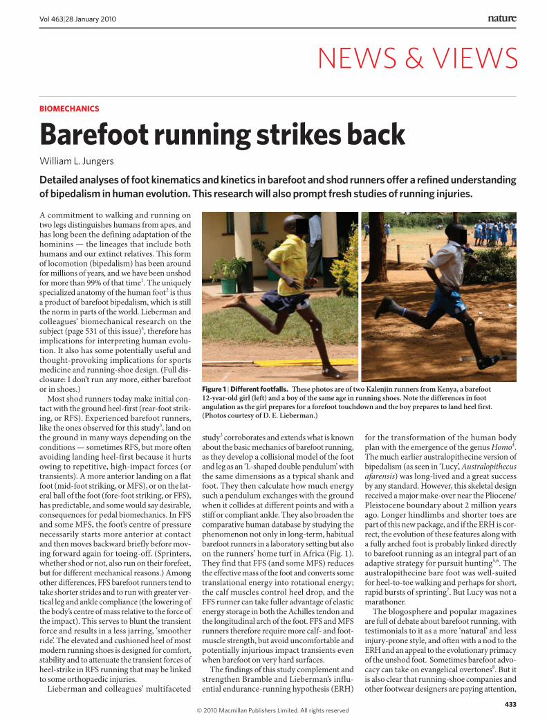
Most shod runners today make initial con-tact with the ground heel-first (rear-foot strik-ing, or RFS). Experienced barefoot runners, like the ones observed for this study3, land on the ground in many ways depending on the conditions — sometimes RFS, but more often avoiding landing heel-first because it hurts owing to repetitive, high-impact forces (or transients). A more anterior landing on a flat foot (mid-foot striking, or MFS), or on the lat-eral ball of the foot (fore-foot striking, or FFS), has predictable, and some would say desirable, consequences for pedal biomechanics. In FFS and some MFS, the foot’s centre of pressure necessarily starts more anterior at contact and then moves backward briefly before mov-ing forward again for toeing-off. (Sprinters, whether shod or not, also run on their forefeet, but for different mechanical reasons.) Among other differences, FFS barefoot runners tend to take shorter strides and to run with greater ver-tical leg and ankle compliance (the lowering of the body’s centre of mass relative to the force of the impact). This serves to blunt the transient force and results in a less jarring, ‘smoother ride’. The elevated and cushioned heel of most modern running shoes is designed for comfort, stability and to attenuate the transient forces of heel-strike in RFS running that may be linked to some orthopaedic injuries.
Lieberman and colleagues’ multifaceted
study3 corroborates and extends what is known about the basic mechanics of barefoot running, as they develop a collisional model of the foot and leg as an ‘L-shaped double pendulum’ with the same dimensions as a typical shank and foot. They then calculate how much energy such a pendulum exchanges with the ground when it collides at different points and with a stiff or compliant ankle. They also broaden the comparative human database by studying the phenomenon not only in long-term, habitual barefoot runners in a laboratory setting but also on the runners’ home turf in Africa (Fig. 1). They find that FFS (and some MFS) reduces the effective mass of the foot and converts some translational energy into rotational energy; the calf muscles control heel drop, and the FFS runner can take fuller advantage of elastic energy storage in both the Achilles tendon and the longitudinal arch of the foot. FFS and MFS runners therefore require more calf- and foot-muscle strength, but avoid uncomfortable and potentially injurious impact transients even when barefoot on very hard surfaces.
The findings of this study complement and strengthen Bramble and Lieberman’s influ-ential endurance-running hypothesis (ERH)
for the transformation of the human body plan with the emergence of the genus Homo4. The much earlier australopithecine version of bipedalism (as seen in ‘Lucy’, Australopithecus afarensis) was long-lived and a great success by any standard. However, this skeletal design received a major make-over near the Pliocene/Pleistocene boundary about 2 million years ago. Longer hindlimbs and shorter toes are part of this new package, and if the ERH is cor-rect, the evolution of these features along with a fully arched foot is probably linked directly to barefoot running as an integral part of an adaptive strategy for pursuit hunting5,6. The australopithecine bare foot was well-suited for heel-to-toe walking and perhaps for short, rapid bursts of sprinting7. But Lucy was not a marathoner.
The blogosphere and popular magazines are full of debate about barefoot running, with testimonials to it as a more ‘natural’ and less injury-prone style, and often with a nod to the ERH and an appeal to the evolutionary primacy of the unshod foot. Sometimes barefoot advo-cacy can take on evangelical overtones8. But it is also clear that running-shoe companies and other footwear designers are paying attention,
Figure 1 | Different footfalls. These photos are of two Kalenjin runners from Kenya, a barefoot 12-year-old girl (left) and a boy of the same age in running shoes. Note the differences in foot angulation as the girl prepares for a forefoot touchdown and the boy prepares to land heel first. (Photos courtesy of D. E. Lieberman.)
433
Vol 463|28 January 2010
NEWS & VIEWS
433-440 News and Views NEW MH IF.indd 433433-440 News and Views NEW MH IF.indd 433 25/1/10 09:23:3825/1/10 09:23:38
© 20 Macmillan Publishers Limited. All rights reserved10
and they should take note of this new study3. Barefoot-like designs for footwear are cur-rently the rage, even if they still constitute only a small slice of the enormous running-shoe industry. Many shod runners never develop injuries, but the available data indicate that at least some (19–79%) do9. Although there is no hard proof that running in shoes, especially hi-tech or PCECH (pronation control, elevated cushioned heel) versions, causes injuries, in my view there is no compelling evidence that it prevents them either10,11. However, there are data that implicate shoes more generally as a plausible source of some types of chronic foot problems12,13.
More studies like that of Lieberman et al.3 are required to provide data instead of opinion, and testable models and scientific explanation instead of anecdotes. It is also apparent that a carefully designed biomedical study with an evidence-based approach is badly needed to assess the competing claims as to what, if any-thing, is the best cover for a runner’s foot. It will
be interesting to see where the next foot falls, and how it is wrapped. ■ William L. Jungers is in the Department
of Anatomical Sciences, Stony Brook
University Medical Center, Stony Brook,
New York 11794-8081, USA.
e-mail: [email protected]
1. Trinkaus, E. J. Arch. Sci. 32, 1515–1526 (2005).
2. Klenerman, L. & Wood, B. A. The Human Foot: A Companion
to Clinical Studies (Springer, 2006).
3. Lieberman, D. E. et al. Nature 463, 531–535 (2010).
4. Bramble, D. M. & Lieberman, D. E. Nature 432, 345–352
(2004).
5. Rolian, C. et al. J. Exp. Biol. 212, 713–721 (2009).
6. Lieberman, D. E., Bramble, D. M., Raichlen, D. A. & Shea,
J. J. in The First Humans (eds Grine, F. E., Fleagle, J. G. &
Leakey, R. E.) 77–98 (Springer, 2009).
7. Lee, S. S. M. & Piazza, S. J. J. Exp. Biol. 212, 3700–3707 (2009).
8. McDougall, C. Born to Run: A Hidden Tribe, Superathletes, and
the Greatest Race the World Has Never Seen (Knopf, 2009).
9. van Gent, R. N. et al. Br. J. Sports Med. 41, 469–480 (2007).
10. Richards, C. E., Magin, P. J. & Callister, R. Br. J. Sports Med.
43, 159–162 (2009).
11. Kerrigan, D. C. et al. PM&R 1, 1058–1063 (2009).
12. Rao, U. B. & Joseph, B. J. Bone Joint Surg. 74B, 525–527 (1992).
13. Zipfel, B. & Berger, L. R. The Foot 17, 205–213 (2007).
because they lack the typical T-cell surface receptor and do not respond to antigens. They do, however, respond to IL-2, in line with their expression of the common γ-chain receptor, which functions as the signalling component of the IL-2 receptor.
Moro and colleagues4 name these cells ‘natural helper cells’ and, on the basis of similar activation properties5, draw analogies with the natural killer cell (NK cell) — a type of lym-phocyte that forms part of the innate immune defence against viruses and tumour cells, and that responds to cytokine stimulation and/or cell-surface components rather than specific antigens. However, natural helper cells lack NK-cell-lineage markers and, unlike NK cells, bear receptors commonly found on pro-genitor (rather than differentiated) cells, such as the IL-7 receptor and stem-cell-factor recep-tor (c-Kit). In view of these characteristics, it might be more accurate to call these cells ‘natural TH2 cells’.
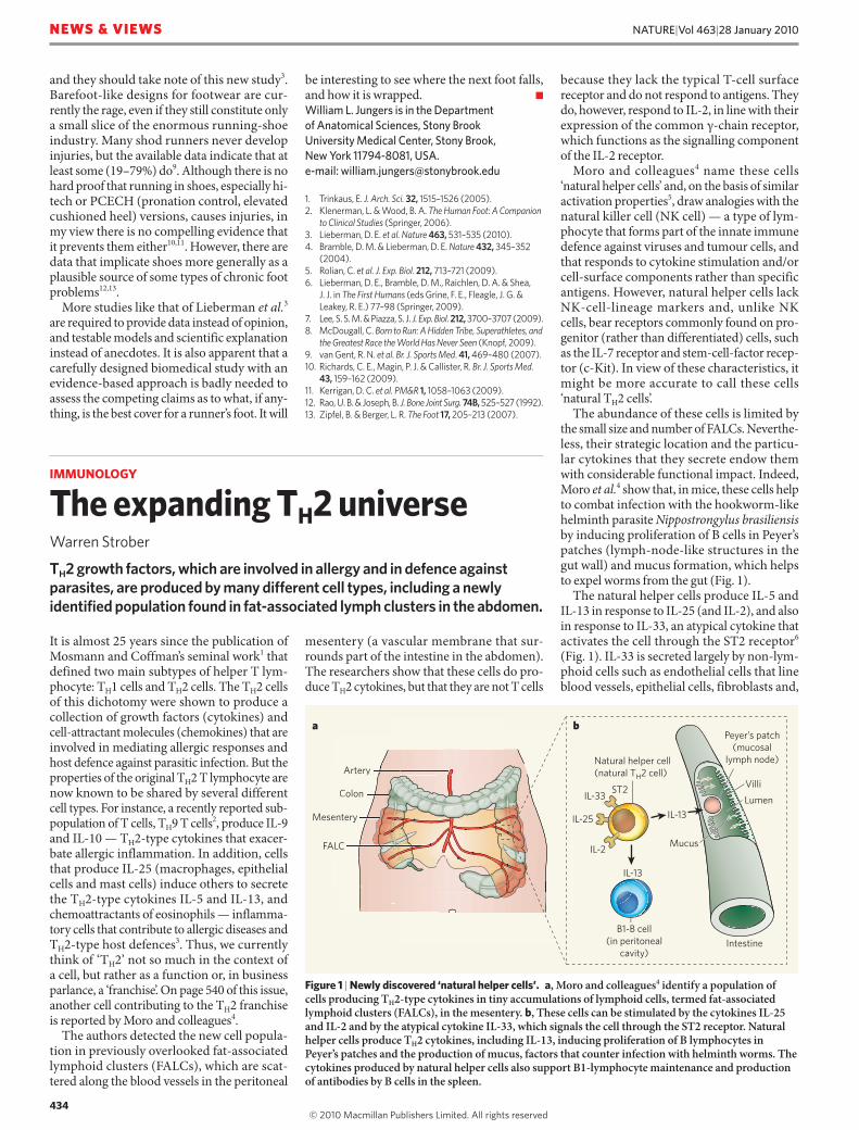
The abundance of these cells is limited by the small size and number of FALCs. Neverthe-less, their strategic location and the particu-lar cytokines that they secrete endow them with considerable functional impact. Indeed, Moro et al.4 show that, in mice, these cells help to combat infection with the hookworm-like helminth parasite Nippostrongylus brasiliensis by inducing proliferation of B cells in Peyer’s patches (lymph-node-like structures in the gut wall) and mucus formation, which helps to expel worms from the gut (Fig. 1).
The natural helper cells produce IL-5 and IL-13 in response to IL-25 (and IL-2), and also in response to IL-33, an atypical cytokine that activates the cell through the ST2 receptor6 (Fig. 1). IL-33 is secreted largely by non-lym-phoid cells such as endothelial cells that line blood vessels, epithelial cells, fibroblasts and,
IMMUNOLOGY
The expanding TH2 universeWarren Strober
TH2 growth factors, which are involved in allergy and in defence against parasites, are produced by many different cell types, including a newly identified population found in fat-associated lymph clusters in the abdomen.
It is almost 25 years since the publication of Mosmann and Coffman’s seminal work1 that defined two main subtypes of helper T lym-phocyte: TH1 cells and TH2 cells. The TH2 cells of this dichotomy were shown to produce a collection of growth factors (cytokines) and cell-attractant molecules (chemokines) that are involved in mediating allergic responses and host defence against parasitic infection. But the properties of the original TH2 T lymphocyte are now known to be shared by several different cell types. For instance, a recently reported sub-population of T cells, TH9 T cells2, produce IL-9 and IL-10 — TH2-type cytokines that exacer-bate allergic inflammation. In addition, cells that produce IL-25 (macrophages, epithelial cells and mast cells) induce others to secrete the TH2-type cytokines IL-5 and IL-13, and chemoattractants of eosinophils — inflamma-tory cells that contribute to allergic diseases and TH2-type host defences3. Thus, we currently think of ‘TH2’ not so much in the context of a cell, but rather as a function or, in business parlance, a ‘franchise’. On page 540 of this issue, another cell contributing to the TH2 franchise is reported by Moro and colleagues4.
The authors detected the new cell popula-tion in previously overlooked fat-associated lymphoid clusters (FALCs), which are scat-tered along the blood vessels in the peritoneal
mesentery (a vascular membrane that sur-rounds part of the intestine in the abdomen). The researchers show that these cells do pro-duce TH2 cytokines, but that they are not T cells
Figure 1 | Newly discovered ‘natural helper cells’. a, Moro and colleagues4 identify a population of cells producing TH2-type cytokines in tiny accumulations of lymphoid cells, termed fat-associated lymphoid clusters (FALCs), in the mesentery. b, These cells can be stimulated by the cytokines IL-25 and IL-2 and by the atypical cytokine IL-33, which signals the cell through the ST2 receptor. Natural helper cells produce TH2 cytokines, including IL-13, inducing proliferation of B lymphocytes in Peyer’s patches and the production of mucus, factors that counter infection with helminth worms. The cytokines produced by natural helper cells also support B1-lymphocyte maintenance and production of antibodies by B cells in the spleen.
a b
Artery
Colon
Mesentery
FALC
Natural helper cell
(natural TH2 cell)
B1-B cell
(in peritoneal
cavity)
Peyer’s patch
(mucosal
lymph node)
Villi
IL-33
IL-13
IL-2
IL-25
ST2
IL-13
A t
e
ALL
Artery
n
L
Colon
ntery
LC
Intestine
Lumen
Mucus
434
NATURE|Vol 463|28 January 2010NEWS & VIEWS
433-440 News and Views NEW MH IF.indd 434433-440 News and Views NEW MH IF.indd 434 25/1/10 09:23:4025/1/10 09:23:40
© 20 Macmillan Publishers Limited. All rights reserved10