Embed Size (px)
Citation preview
MICROBIOLOGICAL REVIEWS, Sept. 1986, p. 314-352 Vol. 50, No. 30146-0749/86/090314-39$02.00/0Copyright © 1986, American Society for Microbiology
Biosynthesis and Metabolism of Arginine in BacteriaRAYMOND CUNIN,' NICOLAS GLANSDORFF,l 3* ANDRE PIERARD,2'3 AND VICTOR STALON2
Erfelijkheidsleer en Microbiologie, Vrije Universiteit Brussel,' Laboratoire de Microbiologie, Universite Libre deBruxelles,2 and Institut de Recherches dii Centre d'Enseignement et de Recherches des Industries Alimentaires et
Chimiqiues, 3 B-1070 Brussels, Belgium
INTRODUCTION ...................................................... 315BIOSYNTHESIS OF ARGININE ...................................................... 315
Enzymatic Steps and Regulation of Metabolic Flow ...................................................... 315From glutamate to arginine ...................................................... 315
N-Acetylglutamate synthetase ...................................................... 316N-Acetylglutamate 5-phosphotransferase ...................................................... 317N-Acetylglutamate 5-semialdehyde reductase ...................................................... 317N-Acetylornithine aminotransferase ...................................................... 317N-Acetylornithinase ...................................................... 318Ornithine acetyltransferase ...................................................... 318OTCase ...................................................... 318Argininosuccinate synthetase ...................................................... 319Argininosuccinase ...................................................... 319
Biosynthesis of carbamoylphosphate ...................................................... 319Enterobacterial CPSases ...................................................... 319P. aeruginosa CPSase ...................................................... 320B. subtilis CPSase ...................................................... 321Other procaryotic CPSases ...................................................... 321
Control of Gene Expression in E. coli...................................................... 321Genetic organization ...................................................... 321Levels of control ...................................................... 321Formation of active repressor ...................................................... 322Structure of control regions and the repression response ...................................................... 323Bipolar argECBH operon and divergent transcription ...................................................... 324carAB operon and cumulative repression ...................................................... 325Arginyl-tRNA synthetase ...................................................... 325Concluding remarks ...................................................... 326
Control of Enzyme Synthesis in Other Procaryotes ...................................................... 326Bacteria lacking or showing limited repression control ...................................................... 326Pseudomonas spp.......................................................326Bacteria with extensive repression control ...................................................... 327S. typhimurium ............................................................... 327B. subtilis ...................................................... 327
Transport of Arginine and Related Metabolites ...................................................... 328CATABOLISM OF ARGININE ...................................................... 328
Arginase Pathway ...................................................... 328ADI Pathway ...................................................... 330
Control of energy provision ...................................................... 331Arginine Succinyltransferase Pathway ...................................................... 332Arginine Transaminase, Oxidase, and Oxygenase Pathways ...................................................... 333Arginine Decarboxylase Pathway ...................................................... 335
Lactic bacteria...................................................... 335E. coli...................................................... 335Klebsiella spp....................................................... 336Pseudomonas and Aeromonas spp....................................................... 336
Conversion of arginine into putrescine ...................................................... 336Enzymes of putrescine utilization ...................................................... 337
Other Pathways of Arginine, Citrulline, and Ornithine Utilization ................................................ 337Bacteria with Multiple Pathways ...................................................... 338
EVOLUTIONARY CONSIDERATIONS ...................................................... 339Biological Significance of Gene Organization ...................................................... 339Lack of Attenuation Control ...................................................... 339Chromosomal Rearrangements Involving Arginine Genes ...................................................... 340Cryptic argM Gene of E. coli .................................. .........................340Origin of CPSases...................... 340
314
on May 29, 2019 by guest
http://mm
br.asm.org/
Dow
nloaded from
ARGININE BIOSYNTHESIS AND METABOLISM IN BACTERIA
Genetic Control of Carbamoylation: Origin of Carbamoyltransferases ...........................................341Acquisition of the Ability to Use Anabolic or Catabolic OTCases in the Reverse
Direction of Their Normal Operation ............................................................................341Evolutionary Significance and Origin of Multiple Catabolic Pathways in Pseudomonas Species ...........342
ACKNOWLEDGMENTS ........................................................................... 342LITERATURE CITED ........................................................................... 342
INTRODUCTION
The reasons why the biosynthesis and metabolism ofarginine have been a focus of interest over the last 30 yearsreside for a large part in their higher degree of complexitythan other pathways chosen as paradigms for studies in
molecular physiology. This is evident in the extensive scat-tering of anabolic arginine genes displayed by most of theorganisms investigated, in the occurrence of a biosyntheticbranch point at the level of carbamoylphosphate (a precursorcommon to arginine and the pyrimidines), in the fact thatornithine or arginine is a potential precursor of polyamines,and in the impressive variety of degradative pathways thatwere found to occur, sometimes in the same organism. Itwas thus expected that the study of this system would lead tofindings of general interest in the fields of enzymology,genetic control mechanisms, and metabolic physiology.For historical reasons mainly, the present state of the art
is very different whether one considers arginine biosynthesisin Escherichia coli (in particular, genetic control mecha-nisms) or other topics such as the nature and regulation ofcatabolic pathways. This situation is reflected in the differentsections into which this review has been divided. Studies on
the regulation of arginine biosynthesis in E. coli have seen
the birth of the very concepts of repression (375) andregulons (202), as well as their extensive substantiation atthe molecular level; this matter is therefore treated as a
major section, after an account of the enzymological aspectsof arginine biosynthesis and before a survey of geneticregulatory mechanisms in other bacteria. Data regardingcatabolic pathways concern mainly the nature of the enzy-
matic steps involved and their physiological significance;despite the maze of information at hand, molecular geneticstudies are still scarce. Therefore, we have considered itappropriate to treat each degradative pathway as an inclu-sive and separate section. Last but not least, studies on
genes and enzymes involved in arginine metabolism haveprovided several observations of evolutionary interest,which are dealt with in the last section.Polyamine biosynthesis is not considered in this review as
a topic per se, but relevant information on this subject isconsidered in the subsection on the arginine decarboxylasepathway, which also deals with agmatine and putrescinecatabolism. Several reviews on polyamines are available(342, 343; N. Glansdorff, In F. C. Neidhardt, J. L. Ingraham,K. B. Low, B. Magasanik, M. Schaechter, and H. E.Umbarger, ed., Escherichia /oli anid Sal/moiella typhi-mlurium: Celll/lar and Molecular Biology, in press).
Various aspects of arginine biosynthesis and degradationhave been reviewed over the last 15 years (1, 27, 73, 75, 204,270, 335, 382; Glansdorff, in press).The role played by ornithine in the biosynthesis of iron-
chelating hydroxamate siderophores (98) and the resultinginterference exerted by arginine on some iron-requiringprocesses (176) are outside the scope of this review. This is
* Corresponding author.
also the case for the part taken by arginine, its precursors,and some polyamines in the biosynthesis of antibiotics (302).
BIOSYNTHESIS OF ARGININE
Enzymatic Steps and Regulation of Metabolic Flow
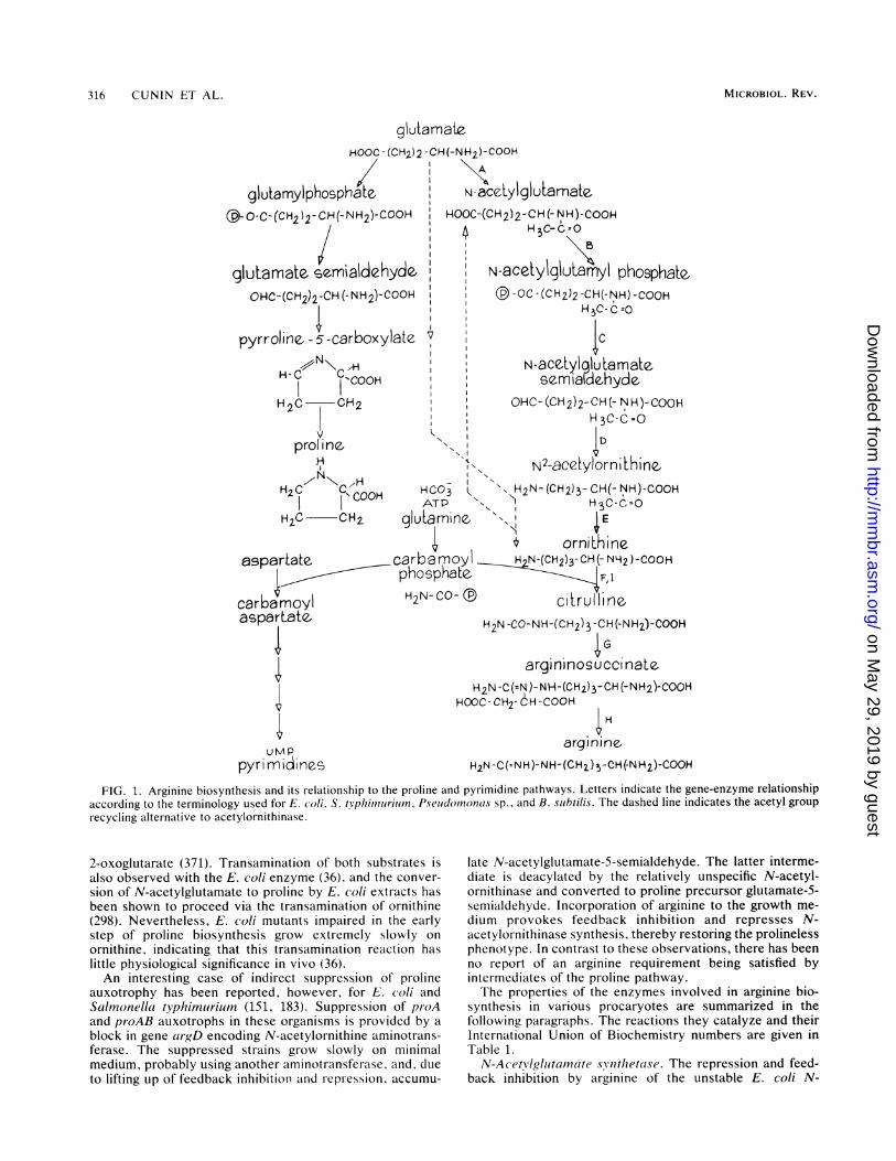
From glutamate to arginine. The biosynthesis of arginineproceeds from glutamate in eight enzymatic steps (Fig. 1;Table 1). Five steps involving N-acetylated intermediateslead to ornithine. The conversion of ornithine to argininerequires three additional steps, the first of which involvescarbamoylphosphate utilization. We deal in a separate sec-tion with the synthesis of this energy-rich metabolite, whichparticipates also in the biosynthesis of pyrimidines.The synthesis of ornithine, like that of proline, involves
the activation and reduction of the 5-carboxyl group ofglutamate. The product of this reaction is glutamate-5-semialdehyde, which in the nonacetylated form undergoesspontaneous cyclization, yielding the proline precursor 1-pyrroline-5-carboxylate. Acetylation of the 2-amino group ofglutamate prevents this cyclization, thus keeping the twopathways separate (374, 376). After transamination onto thesemialdehyde group to produce N'-acetylornithine, theacetyl group is removed in a subsequent step, yieldingornithine. In procaryotes the pathway of arginine biosynthe-sis follows two alternative patterns which differ by thestrategies used for the removal of the acetyl group and forthe control of the metabolite flow along the pathway. En-terobacteriaceae and Bacilla/caa use a linear pathway inwhich the formation of ornithine from N'-acetylornithine ismediated by the hydrolytic enzyme acetylornithinase (286,354, 378). In these organisms, N-acetylglutamate synthetase,the first enzyme of the pathway, is the target enzyme forfeedback inhibition by arginine (383). In contrast, Micro-(cocs)S glitaminicas and various other bacteria, includingpseudomonads, cyanobacteria, photosynthetic bacteria, andThermolis aquaticuis recycle the acetyl group by transacetyla-tion of N2-acetylornithine and glutamate (81, 140, 354, 356).In those organisms which utilize this energetically moreeconomical version of arginine biosynthesis, N-acetylgluta-mate synthetase fulfills an anaplerotic function, and it is thesecond enzyme of the pathway, N-acetylglutamate 5-phosphotransferase, which is feedback inhibited by arginine(354, 357). This "more evolved" form of arginine biosynthe-sis is also present in eucaryotic organisms such as the fungiSaccharoinvces (erevisi(iae and Nell}rospor-a spp. (79) and ingreen alga Ch/ainydomonas sp. (84). It has recently beenfound in several methanogenic bacteria (212).
Although the use of acetylated intermediates achieves aneffective separation of arginine and proline biosyntheses,exchanges of metabolites between these pathways may beobserved under particular conditions. Pseiudomonas aeru-,gi-nosa mutants with an early block in proline synthesis grow inthe presence of high concentrations of L-ornithine (20 mM).This agrees with the observation that N-acetylornithine5-aminotransferase of P. aeruiiginosal catalyzes the trans-amination of both N--acetylornithine and L-ornithine with
315VOL. 50, 1986
on May 29, 2019 by guest
http://mm
br.asm.org/
Dow
nloaded from
316 CUNIN ET AL.
glulamateHOOC - (CH2) 2
glutamylphospha e--O-C-(CIH2 )2- CH(-NH2)-COOH
glutamate- sermialdehydeOHC-(CH2)2-CH (-NH2)-COOH
pyrroline. -5-carboxylate.
H-C cCOfH2C CH2
prol neH
H2C cOOH
-CH(-NH2)-COOH%A
N-aceLylgluLamate.HOOC-(CH2)2-CH (-NH)-COOH
4 HHC-C=O
N-acetylgluIamy1 phosphatbqE-OC -(CH )2 -CH(-NH) -COOH
H?HC-C=Ov I
4cN-aCeLylgluWamatese.maldehyde
OHC-(CH2)2-CH(-NH)-COOHH 3C-C=O
N N22-acetylo rfLhiflQCO " H2N- (CH2)3- CH(-NH)-COOHATP H 2C-C<O
H2C CH2 glutamine N E
asparLatecrbamol orniLhinecarbatmoyl N-(CH2)3-CH(- NH2)-COOHLspartae. phosphate. 2 F,I
carbamoyl H2N-CO- ci1ruHlineasparLate. H1N -CO-NH-(CH )2 -CH(-NH?)-COO)H
umppyri mnidines
'/ - -- _- D _-*4.3 - % 1 --
7Gargininosuccinate
H2N -C(=N)-NH-(CH2)3- CH (-NH2)-COOHHOOC - CH2- CH -COOH
H
argin ne-H2N -C(=NH )-NH-(CH2) 3-CH (-NH2)-COOH
FIG. 1. Arginine biosynthesis and its relationship to the proline and pyrimidine pathways. Letters indicate the gene-enzyme relationshipaccording to the terminology used for E. co/i, S. tVphimurium, Pseudomnonas sp., and B. sibtilis. The dashed line indicates the acetyl grouprecycling alternative to acetylornithinase.
2-oxoglutarate (371). Transamination of both substrates isalso observed with the E. coli enzyme (36), and the conver-sion of N-acetylglutamate to proline by E. coli extracts hasbeen shown to proceed via the transamination of ornithine(298). Nevertheless, E. coli mutants impaired in the earlystep of proline biosynthesis grow extremely slowly onornithine, indicating that this transamination reaction haslittle physiological significance in vivo (36).An interesting case of indirect suppression of proline
auxotrophy has been reported, however, for E. (oli andSalmnonella typhimiurium (151, 183). Suppression of proAand proAB auxotrophs in these organisms is provided by ablock in gene argD encoding N-acetylornithine aminotrans-ferase. The suppressed strains grow slowly on minimalmedium, probably using another aminotransferase. and, dueto lifting up of feedback inhibition Cand repression, accumu-
late N-acetylglutamate-5-semialdehyde. The latter interme-diate is deacylated by the relatively unspecific N-acetyl-ornithinase and converted to proline precursor glutamate-5-semialdehyde. Incorporation of arginine to the growth me-dium provokes feedback inhibition and represses N-acetylornithinase synthesis, thereby restoring the prolinelessphenotype. In contrast to these observations, there has beenno report of an arginine requirement being satisfied byintermediates of the proline pathway.The properties of the enzymes involved in arginine bio-
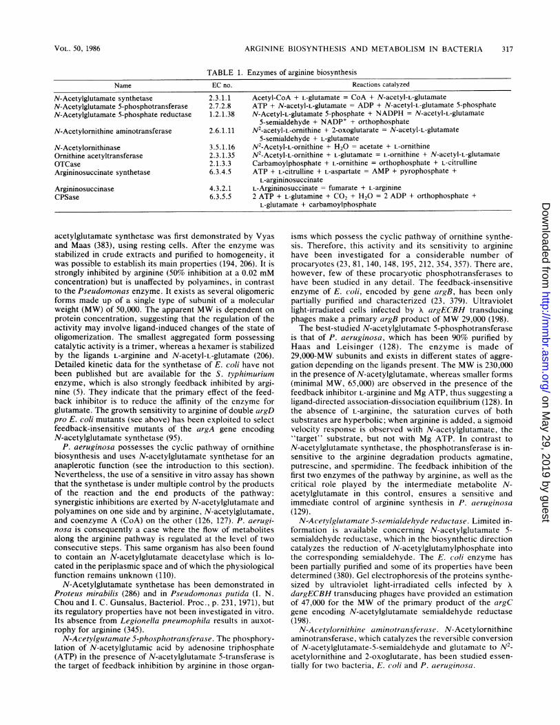
synthesis in various procaryotes are summarized in thefollowing paragraphs. The reactions they catalyze and theirInternational Union of Biochemistry numbers are given inTable 1.
N-Acet/lglutarnate svnthetase. The repression and feed-back inhibition by arginine of the unstable E. coli N-
MICROBIOL. REV.
on May 29, 2019 by guest
http://mm
br.asm.org/
Dow
nloaded from
ARGININE BIOSYNTHESIS AND METABOLISM IN BACTERIA
TABLE 1. Enzymes of arginine biosynthesisName EC no. Reactions catalyzed
N-Acetylglutamate synthetase 2.3.1.1 Acetyl-CoA + L-glutamate = CoA + N-acetyl-L-glutamateN-Acetylglutamate 5-phosphotransferase 2.7.2.8 ATP + N-acetyl-L-glutamate = ADP + N-acetyl-L-glutamate 5-phosphateN-Acetylglutamate 5-phosphate reductase 1.2.1.38 N-Acetyl-L-glutamate 5-phosphate + NADPH = N-acetyl-L-glutamate
5-semialdehyde + NADP+ + orthophosphateN-Acetylornithine aminotransferase 2.6.1.11 N2-acetyl-L-ornithine + 2-oxoglutarate = N-acetyl-L-glutamate
5-semialdehyde + L-glutamateN-Acetylornithinase 3.5.1.16 N2-Acetyl-L-ornithine + H20 = acetate + L-ornithineOrnithine acetyltransferase 2.3.1.35 N2-Acetyl-L-ornithine + L-glutamate = L-ornithine + N-acetyl-L-glutamateOTCase 2.1.3.3 Carbamoylphosphate + L-ornithine = orthophosphate + L-citrullineArgininosuccinate synthetase 6.3.4.5 ATP + L-citrulline + L-aspartate = AMP + pyrophosphate +
L-argininosuccinateArgininosuccinase 4.3.2.1 L-Argininosuccinate = fumarate + L-arginineCPSase 6.3.5.5 2 ATP + L-glutamine + CO2 + H20 = 2 ADP + orthophosphate +
L-glutamate + carbamoylphosphate
acetylglutamate synthetase was first demonstrated by Vyasand Maas (383), using resting cells. After the enzyme wasstabilized in crude extracts and purified to homogeneity, itwas possible to establish its main properties (194, 206). It isstrongly inhibited by arginine (50% inhibition at a 0.02 mMconcentration) but is unaffected by polyamines, in contrastto the Pseudomonas enzyme. It exists as several oligomericforms made up of a single type of subunit of a molecularweight (MW) of 50,000. The apparent MW is dependent onprotein concentration, suggesting that the regulation of theactivity may involve ligand-induced changes of the state ofoligomerization. The smallest aggregated form possessingcatalytic activity is a trimer, whereas a hexamer is stabilizedby the ligands L-arginine and N-acetyl-L-glutamate (206).Detailed kinetic data for the synthetase of E. coli have notbeen published but are available for the S. typhimuriumenzyme, which is also strongly feedback inhibited by argi-nine (5). They indicate that the primary effect of the feed-back inhibitor is to reduce the affinity of the enzyme forglutamate. The growth sensitivity to arginine of double argDpro E. coli mutants (see above) has been exploited to selectfeedback-insensitive mutants of the argA gene encodingN-acetylglutamate synthetase (95).
P. aeruginosa possesses the cyclic pathway of ornithinebiosynthesis and uses N-acetylglutamate synthetase for ananaplerotic function (see the introduction to this section).Nevertheless, the use of a sensitive in vitro assay has shownthat the synthetase is under multiple control by the productsof the reaction and the end products of the pathway:synergistic inhibitions are exerted by N-acetylglutamate andpolyamines on one side and by arginine, N-acetylglutamate,and coenzyme A (CoA) on the other (126, 127). P. aerugi-nosa is consequently a case where the flow of metabolitesalong the arginine pathway is regulated at the level of twoconsecutive steps. This same organism has also been foundto contain an N-acetylglutamate deacetylase which is lo-cated in the periplasmic space and of which the physiologicalfunction remains unknown (110).
N-Acetylglutamate synthetase has been demonstrated inProteus mirabilis (286) and in Pseudomonas putida (I. N.Chou and I. C. Gunsalus, Bacteriol. Proc., p. 231, 1971), butits regulatory properties have not been investigated in vitro.Its absence from Legionella pneumophila results in auxot-rophy for arginine (345).N-Acetylgutamate 5-phosphotransjerase. The phosphory-
lation of N-acetylglutamic acid by adenosine triphosphate(ATP) in the presence of N-acetylglutamate 5-transferase isthe target of feedback inhibition by arginine in those organ-
isms which possess the cyclic pathway of ornithine synthe-sis. Therefore, this activity and its sensitivity to argininehave been investigated for a considerable number ofprocaryotes (23, 81, 140, 148, 195, 212, 354, 357). There are,however, few of these procaryotic phosphotransferases tohave been studied in any detail. The feedback-insensitiveenzyme of E. coli, encoded by gene argB, has been onlypartially purified and characterized (23, 379). Ultravioletlight-irradiated cells infected by A argECBH transducingphages make a primary argB product of MW 29,000 (198).The best-studied N-acetylglutamate 5-phosphotransferase
is that of P. aeruginosa, which has been 90% purified byHaas and Leisinger (128). The enzyme is made of29,000-MW subunits and exists in different states of aggre-gation depending on the ligands present. The MW is 230,000in the presence of N-acetylglutamate, whereas smaller forms(minimal MW, 65,000) are observed in the presence of thefeedback inhibitor L-arginine and Mg ATP, thus suggesting aligand-directed association-dissociation equilibrium (128). Inthe absence of L-arginine, the saturation curves of bothsubstrates are hyperbolic; when arginine is added, a sigmoidvelocity response is observed with N-acetylglutamate, the"target" substrate, but not with Mg ATP. In contrast toN-acetylglutamate synthetase, the phosphotransferase is in-sensitive to the arginine degradation products agmatine,putrescine, and spermidine. The feedback inhibition of thefirst two enzymes of the pathway by arginine, as well as thecritical role played by the intermediate metabolite N-acetylglutamate in this control, ensures a sensitive andimmediate control of arginine synthesis in P. aeruginosa(129).N-Acetylglutamate 5-semialdehyde reductase. Limited in-
formation is available concerning N-acetylglutamate 5-semialdehyde reductase, which in the biosynthetic directioncatalyzes the reduction of N-acetylglutamylphosphate intothe corresponding semialdehyde. The E. coli enzyme hasbeen partially purified and some of its properties have beendetermined (380). Gel electrophoresis of the proteins synthe-sized by ultraviolet light-irradiated cells infected by XdargECBH transducing phages have provided an estimationof 47,000 for the MW of the primary product of the argCgene encoding N-acetylglutamate semialdehyde reductase(198).
N-Acetylornithine aminotransferase. N-Acetylornithineaminotransferase, which catalyzes the reversible conversionof N-acetylglutamate-5-semialdehyde and glutamate to N2-acetylornithine and 2-oxoglutarate, has been studied essen-tially for two bacteria, E. coli and P. aeruginosa.
VOL. 50, 1986 317
on May 29, 2019 by guest
http://mm
br.asm.org/
Dow
nloaded from
318 CUNIN ET AL.
E. coli W and K-12 have an amidotransaminase which isrepressible by arginine (12). The enzyme is also active withglutamate-5-semialdehyde, thus yielding ornithine, althoughthis activity probably has no physiological significance (36).By selecting for suppressors of argD mutations, mutantshave been isolated in both strains that exhibit an arginine-inducible transaminase (22, 377; T. Eckhardt, Ph.D. thesis,ETH-Ziurich, 1975). The inducible activity appears to resultfrom the activation of argM, a cryptic gene unlinked toargD. The induction of argM, like the repression of argD, ismediated by the argR gene product (22). Both the inducibleand the repressible N-acetylornithine aminotransferaseshave been purified to homogeneity (37, 105). The twoenzymes differ by their MWs (119,000 for the wild-typetransaminase, 61,000 for the inducible enzyme) and show noimmunochemical cross-reactivity. Yet, both are made of31,000-MW subunits and exhibit nearly identical trypticdigestion patterns, thus suggesting a common origin forthese two genes. The evolutionary implications of suchobservations are discussed in another section of this review.
In P. aeruiginosa, the catalytic properties of thetransaminase and the regulation of its synthesis indicate thatthis enzyme is involved in the biosynthesis as well as thecatabolism of L-arginine (371, 373). The enzyme, which hasbeen purified to electrophoretic homogeneity, has an ap-proximate MW of 110,000 and consists of two 55,000subunits. It catalyzes the transamination of N2-acetyl-ornithine as well as that of L-ornithine with 2-oxoglutarate,the Km for N2-acetylornithine and ornithine being 1.1 mMand 10.0 mM, respectively (371). Ihe transaminase is in-duced during growth on arginine as the only carbon andnitrogen souce and is repressed by various carbon sources(373). Recent results (D. Haas, personal communication; V.Stalon, unpublished observations) suggest that this enzymeis identical to succinylornithine aminotransferase, an en-zyme of the succinyltransferase pathway of arginine catab-olism (D. Vander Wauven, C. Legrain, and V. Stalon,manuscript in preparation). The properties of a mutant withan inactive acetylornithine aminotransferase indicate thatthis enzyme can be replaced by 4-aminobutyrate aminotrans-ferase, an enzyme of putrescine catabolism (372).
In contrast to the previous organisms, Klebsiella aCe-o-genes forms two separable acetylornithine aminotrans-ferases (108). One is repressed by arginine and participatesin its biosynthesis, whereas the second is induced by argi-nine and ornithine and functions in their catabolism. Theinducible enzyme, which has been purified to near homoge-neity, has an MW of 59,000 and exhibits activity withN2-acetylornithine as well as with ornithine (107). Thefinding that this inducible aminotransferase has a fourfoldlower K,,, for succinylornithine than for N2-acetylornithinesuggests that it is also identical to succinylornithine amino-transferase (Vander Wauven et al., in preparation, seesubsection, "Arginine Succinyltransferase Pathway").
N-AcetWolrnithinase. The hydrolytic enzyme N-acetyl-ornithinase catalyzes the ornithine-yielding step of the linearpathway of arginine biosynthesis. It requires the presence ofCo2+ ions and a thiol compound such as glutathion for itsactivity (378) and has been detected in a number ofenterobacteria and bacilli (286, 354, 378). The best-characterized acetylornithinase is that of E. coli. It has beenpurified to homogeneity and, based on molecular sievingexperiments, appears to be a monomer of MW 62,000 (J.Charlier, FEBS Meet. 1983, Brussels, Belgium, Abstr. SO-5;Charlier, personal communication). Acetylornithinasereadily deacylates N-acetylglutamate semialdehyde, N-
acetylarginine, N-acetylhistidine, N-acetylmethionine, andN-formylmethionine (26, 381). Advantage has been taken ofthis low substrate specificity of acetylornithinase and of itsrepression by arginine to select for fast-growing argR deriv-atives among Iiis auxotrophs growing slowly on acetyl-histidine (26), as well as (is-dominant mutations affectinga1rgECBH expression (43, 67).
Orniiitdliine acetvltransferause. Ornithine acetyltransferasecatalyzes the transfer of the acetyl group of N2-acetylornithine onto glutamate to yield ornithine and N-acetylglutamate; it is the key enzymatic step of the so-calledcyclic pathway of ornithine synthesis (see the introduction tothis section). It was first identified in M. glutamicus byUdaka and Kinoshita (356) and has since been demonstratedin a number of procaryotes including pseudomonads, photo-synthetic bacteria, the thermophilic bacterium T. aquaticus,cyanobacteria, and methanogenic bacteria (81, 140, 212,354). Yet none of these acetyltransferases appears to havebeen studied in any detail.Some organisms have been found to possess both activi-
ties that achieve the conversion of N -acetylornithine toornithine in vitro: ornithine acetyltransferase and acetyl-ornithinase. In yeasts, mutants impaired in ornithine acetyl-transferase grow slowly in the absence of arginine (220; F.Messenguy, personal communication), and the acetyl-ornithinase measured in vitro can be ascribed to a metal-activated carboxypeptidase with little biosynthetic function(80). Such a carboxypeptidase is probably also responsiblefor the acetylornithinase activity of T. aquaticus (81). In P.aerulginiosa, the two activities are separable by gel filtration,but their relative contributions to ornithine synthesis areunknown since no mutants lacking these activities have beenisolated to date (125).OTCase. Ornithine carbamoyltransferase (OTCase)
serves two functions in arginine metabolism. In argininebiosynthesis it catalyzes the transfer of the carbamoyl moi-ety of carbamoylphosphate to the 5-amino group ofornithine, forming citrulline. ln the catabolic argininedeiminase pathway (see the section on arginine catabolism),it mediates the thermodynamically less favored reversereaction, the phosphorolysis of citrulline, yielding ornithineand carbamoylphosphate. Organisms that use both of thesefunctions elaborate distinct anabolic and catabolic OTCases(186, 193, 338). The various anabolic OTCases exhibit sim-ilar structural, kinetic, and mechanistic properties. Most ofthem have MWs between 100,000 and 150,000; they aretrimers of identical subunits, with MWs from 35,000 to40,000 (193). This pattern of quaternary structure has beenfound for the anabolic OTCases of E. coli (189, 190), S.typhim,uriiim (11), P. plJtidai (339), P. aleruginosa (Stalon andMomin, unpublished data), and Saccharomyces cerevisiae(268). In Aeromonas. forimicans this basic trimeric structure(MW 125,000) exists in equilibrium with a heavier form (MW420,000); the two forms do not differ by their kinetic con-stants (Momin and Stalon, unpublished data). A notableexception to the trimeric rule for the anabolic OTCases is theenzyme of Bacillus subtilis (243), which exists as a mixtureof dimeric, tetrameric, and hexameric forms of a 44,000-MWsubunit. Interestingly, OTCase is repressed by arginine ingrowing B. siubtilis cells but is induced by arginine at the endof exponential growth. The physiological role of this induc-tion by arginine is not known, but the enzyme synthesizedunder all conditions exhibits the same response towardsantibodies against purified OTCase. This same enzyme issubject to inactivation followed by proteolytic degradation insporulating cells (244).
MICROBIOL. REV.
on May 29, 2019 by guest
http://mm
br.asm.org/
Dow
nloaded from
ARGININE BIOSYNTHESIS AND METABOLISM IN BACTERIA
In contrast to the anabolic OTCases, the catabolicOTCases usually show much more diverse structural fea-tures (100, 193). The evolutionary implications of suchfindings are discussed in a separate section.The kinetic behaviors of the anabolic OTCases are usually
consistent with ordered mechanisms in which carbamoyl-phosphate is the first substrate to bind and phosphate is thelast product to be released or with random addition of thereactants with a preferred binding of carbamoylphosphate as
the leading substrate (11, 190, 193, 243, 385). The patterns ofinhibition of the anabolic OTCases of E. coli and B. subtilisby the bisubstrate analog N-8-phosphono-acetyl-L-ornithine(PALO) are consistent with such mechanisms (243, 266). E.coli cells are impervious to PALO but are able to take up theoligopeptide gly-gly-PALO through the oligopeptidepermease; the toxicity of PALO towards OTCase is ex-
pressed following its liberation by an intracellular peptidase(267). Such "illicit uptake" may have therapeutical implica-tions as a means of driving into the cell a substance which isnormally unable to penetrate into it (265).
All anabolic OTCases, except that of Pseludomonas spp.,
are able to catalyze both directions of the reaction (193). Thefunctional irreversibility of the Pseudomonas enzyme resultsfrom the formation of a binary dead-end complex betweenthe enzyme and citrulline that reduces the apparent maxi-mum velocity (336). Such functional specialization may beimportant in organisms which use both directions of theOTCase reaction.The position of OTCase half-way through the arginine
pathway and the fact that the production of its secondsubstrate, carbamoylphosphate, is in general highly regu-
lated make a control of its activity unnecessary. A differentsituation is created, however, in organisms which possess an
inducible arginase: after the addition of arginine and beforethe dilution of repressible OTCase by growth, an energy-
wasteful urea cycle could operate that immediately degradesarginine formed in the biosynthetic pathway. In some yeasts
and in B. subtilis this potential urea cycle is avoided by an
arginine- and ornithine-dependent binding and inhibition ofOTCase by arginase, called epi-arginasic regulation (149,221). In Agrobacterium tumefaciens and several Rhizobiumspecies in which arginase is inducible and OTCase is consti-tutive, the disadvantage of the simultaneous presence ofboth enzymes at a high level is corrected by feedbackinhibition of OTCase by arginine (370; S. Vissers et al.,manuscript in preparation). OTCase inhibition in S.typhimurium and P. putida occurs at relatively high arginineconcentrations and has probably less physiological impor-tance (11, 336).The structure of E. coli OTCase has been studied in
particular detail. Interestingly, E. coli K-12 carries twogenes for OTCase, argF and argI, both repressible byarginine (118); their products interact to form a family of fourtrimeric isoenzymes which can be separated by ion-exchange chromatography (189). The F and I isoenzymeshave similar kinetic parameters but differ in theirthermostabilities (191). Only gene argI or its equivalent can
be found in E. coli B and W or in other Enterobacteriaceae(189, 191). The occurrence of hybrid F-I isoenzymes hassuggested that these genes originate from the duplication ofa common ancestral gene. In addition, the adjacency of argland pyrBl (152) encoding aspartate carbamoyltransferases as
well as the structural and catalytic similarities observedbetween the two carbamoyltransferases has led to a beliefthat these enzymes arose from an analogous genetic event(189). These hypotheses are supported by the recent deter-
mination of the primary structure of these genes and areconsidered in more detail under "Evolutionary Consider-ations."
Argininosuccinate synthetase. Argininosuccinate synthe-tase, which catalyzes the conversion of citrulline, aspartate,and ATP into argininosuccinate, has not been well studiedfor procaryotes but its yeast counterpart has been charac-terized in some detail; it is a tetramer of identical 49,000-MWsubunits (137). From denaturing gel electrophoresis of ex-tracts from minicells producing a plasmid-encoded arginin-osuccinate synthetase, the E. coli enzyme appears to consistof a basic polypeptide of similar MW, 48,000 (239).
Argininosluccinase. Little is known of the bacterialargininosuccinases, which hydrolyze argininosuccinate intoarginine and fumarate. Extracts of ultraviolet light-irradiatedE. coli cells infected with an argH transducing phage containa polypeptide of 55,000 MW (198), which may be similar tothe 50,000 subunit of the better-characterized tetramericmammalian argininosuccinase (295).
Biosynthesis of carbamoylphosphate. Three types of organ-ization may be distinguished among procaryotes with regardto the biosynthesis of carbamoylphosphate. The first typecorresponds to organisms which use a single enzyme toproduce carbamoylphosphate required for arginine and py-rimidine biosynthesis. It is widely distributed among gram-negative bacteria and, in particular, Enterobacteriaceae.This group of organisms is best illustrated by E. coli. In thisorganism, one-step mutants can be isolated which lack asingle glutamine-dependent carbamoylphosphate synthetasethat is regulated in a manner consistent with its dual meta-bolic function: cumulative repression by arginine and pyrim-idine and modulation of the activity by effectors belonging tothe two pathways which utilize carbamoylphosphate (18,269, 271, 273). A low carbamate kinase activity detectable inE. coli and in some other enteric bacteria is due to aconstitutive acetate kinase and probably has no biosyntheticsignificance (71, 165, 273, 346). Carbamate kinase itself,previously believed to play a role in carbamoylphosphatesynthesis (162), is now assigned an essentially catabolicfunction as an enzyme of the arginine deiminase pathway(see the section on arginine catabolism).A second type of organization is represented by B. subti-
lis, which elaborates two independently regulated carbamo-ylphosphate synthetases: one is repressed by arginine; thesecond is repressed and feedback inhibited by pyrimidines(262). No other gram-positive bacterium has been studied incomparable detail. Thus it is not known whether B. subtilisis unique among procaryotes in displaying this type oforganization or whether it is representative of a wider groupof organisms.
Still another way of forming carbamoylphosphate is usedby Lactobacillus leichmanii, and possibly other lactic bac-teria, which seems to lack any carbamoylphosphate synthe-tase activity but possesses the arginine deiminase pathwayof arginine catabolism. Tracer studies suggest that thisorganism derives the carbamoylphosphate required or py-rimidine biosynthesis from the degradation of arginine (146).
This section is devoted to a summary and discussion of theproperties and control of the activity of E. coli and otherwell-characterized bacterial carbamoylphosphate synthe-tases.
Enterobacterial CPSases. The single carbamoylphosphatesynthetases (CPSases) from E. coli (for review see reference214), S. typhimuriuim (1, 8), and Serrcatia marcescens (71) arewell characterized and appear very similar in structural,catalytic, and regulatory properties. All three are subject to
319VOL. 50, 1986
on May 29, 2019 by guest
http://mm
br.asm.org/
Dow
nloaded from
320 CUNIN ET AL.
cumulative repression by arginine and uracil (1, 71, 273) aswell as to activation by ornithine and feedback inhibition byuridine monophosphate (UMP) (1. 18. 71, 269, 271).
E. coli CPSase is the most thoroughly studied of theseenzymes. It catalyzes a reaction in which the amide group ofglutamine, a bicarbonate ion, and two ATPs are used for thesynthesis of carbamoylphosphate (16). Ammonia, a lower-affinity nitrogen donor for the reaction in vitro, probablyplays no physiological role (165, 166). Studies with highlypurified CPSase preparations have shown that the reactionproceeds in four steps: (i) ATP-dependent activation ofcarbon dioxide under the form of enzyme-bound carbonate-phosphate anhydride; (ii) reaction of this intermediate withglutamine; (iii) transfer of the amide nitrogen group ofglutamine to activated carbon dioxide to form enzyme-bound carbamate; (iv) use of a second ATP to phosphorylatecarbamate and liberation of carbamoylphosphate (17).The observation that a chloroketone analog of glutamine
inactivates the glutamine binding site of CPSase withoutaffecting the ammonia-dependent activity has provided clearevidence that these two nitrogen donors react with separatebinding sites on the enzyme (177). In addition, relativelymild conditions were shown to promote the reversible dis-sociation of the enzyme into two nonidentical subunits: asmall subunit (MW 42,000) displaying glutaminase activity invitro and a large subunit (MW 130,000) which catalyzes thesynthesis of carbamoylphosphate from ammonia, bicarbon-ate, and ATP (348). These two subunits are encoded by theadjacent genes carA and catB of E. coli (219) which areorganized in an operon oriented from A to B (70, 114). Thecomplete sequences of these genes, as determined recently,provide MWs of 41,270 and 117,710 for their respectiveprotein products (250, 277). The equivalent proteins in S.typhimruriurm are encoded by the locus pvrA (8). No infor-mation is available concerning their genetic determinants inSerraltiu marcescenis.The mechanisms of glutamine utilization for carbam-
oylphosphate synthesis is similar to that proposed for otherglutamine amidotransferases (133). It involves binding ofglutamine to the small subunit and transfer of its amidenitrogen group to an ammonia binding site on the largesubunit (280, 348, 349). Extensive interactions between thelight and heavy subunits of the enzyme appear to facilitate itscatalytic function. For example. the isolated light subunitexhibits a much lower affinity for glutamine than does thenative enzyme (349). In addition, the binding of thechloroketone analog of glutamine to the light subunit de-creases the apparent K,,, for ammonia (280).CPSase is a highly regulated enzyme which is inhibited by
UMP, and to a lesser extent by other uridylic nucleotides,and activated by ornithine; its activity is also enhanced byammonium ions, by inosine monophosphate, and by variousother purine nucleotides (18, 269, 271). In E. coli the heavysubunit of CPSase, which carries all of the catalytic func-tions of the enzyme except the hydrolysis of glutamine,clearly also bears the binding sites for the allosteric effectors(348, 349). In S. tNphirnuiimn, however, the purified heavysubunit is but weakly inhibited by UMP though addition ofthe light subunit restores normal sensitivity towardis thisnucleotide; it is not known whether this effect reflectsinteractions between the two subunits or the presence of anUMP binding site on the light subunit. The antagonisticeffects of UMP and ornithine provide an elegant manner toregulate the supply of carbamoylphosphate according to theneeds of the two pathways which utilize it (269). Indeed thecellular concentration of ornithine varies in inverse ratio to
arginine owing to feedback inhibition of N-acetylglutamatesynthetase by arginine (269). The activation of CPSase byinosine monophosphate and the other purine nucleotidesachieves a balance between the relative rates of purine andpyrimidine biosynthesis (18).CPSase, which displays a sigmoidal ATP saturation curve
(18), exists in at least three conformational states: a formwhich has no affinity tor the substrate ATP and is stabilizedby UMP; in equilibrium with this form, a second form, whichis stabilized by ornithine and inosine monophosphate andhas affinity for ATP; and a third form, which is catalyticallyactive and into which the second one is converted by ATPbinding (14, 15). 'IThe monomer undergoes a reversible self-association in the presence of the allosteric effectors (349)which is not related to the regulatory and catalytic propertiesof the enzyme (13).The ATP molecules which participate in two different
steps of the reaction catalyzed by CPSase bind at separatesites on the heavy subunit (39, 285). Nyunoya and Lusty(250). after determining the sequence of curB, have observedan homology between the two halves of the sequence, thussuggesting that this gene arose from the duplication of anancestral gene. They have also proposed that the allostericbehavior of the enzyme reflects interactions between distinctfolding domains corresponding to the two half sequences,each possibly carrying one of the ATP binding sites.Due to the complexity of its catalytic and regulatory
properties, it is not surpr-ising that a variety of non-biauxotrophic phenotypes were observed among mutantsaffected in E. coli CPSase (3, 219, 271). The first of thesemutants to be studied were uracil sensitive (271; G.Leclercq, Ph.D. thesis, Universit6 Libre de Bruxelles, Brus-sels, Belgium, 1971). The mutant enzyme of strain P678M1,for example, displays an increased K,,, for ATP in such amanner that in the presence of UMP, and even in thepresence of ornithine, the activity is too low to supportgrowth. The apparent affinity for ATP is so low in theabsence of ornithine that the mutant is also partially sensi-tive to ar-ginine.Such phenotypes have also been obtained in S. typhi-
mnuriiim. Uracil sensitivity in a cold-sensitive mutant resultsfrom the increased inhibition of the enzyme by UMP at lowtemperature (2). Other phenotypes are of particular interestsince they seem to impair complex enzyme interactions andprotein maturation mechanisms. They are discussed in thesection devoted to control of gene expression in S.tvPhim7Zl)illin (9, 10).
P. (erllginlosa CPSa.u . The single CPSase of P. aeriugi-niosa shows much similarity with its enteric counterpart (7).The enzyme uses either glutamine (K,,, 0.15 mM) or NH3 (K,17 mM) as the nitrogen donor. It has an MW of 165,000 andis composed of two nonidentical subunits (MW 44,000 and122,000) which are probably equivalent to the carA and curBproducts of E. coli (7). Mutations resulting in doubleauxotrophy for arginine and uracil have been obtained (125,200). They map in a single cur locus in which it has not beenpossible to distinguish the equivalent to genes carA and carB(125: C. Vander Wauven, personal communication). TheCPStase of P. uCrugitnosa( is subject to partial cumulativerepression by arginine and pyrimidines; it is feedback inhib-ited by UMP and activated by ornithine and N2-acetyl-ornithine (7; C. Vander Wauven, unpublished data). Theeffect of N-cacetylornithine can be considered an index ofarginine limitation under conditions in which ornithine israpidly catabolized (7).A particularity of Psculomionuas spp. is the simultaneous
MIC ROBIOL. REV.
on May 29, 2019 by guest
http://mm
br.asm.org/
Dow
nloaded from
ARGININE BIOSYNTHESIS AND METABOLISM IN BACTERIA
presence of CPSase and carbamate kinase under conditionsin which the arginine deiminase pathway is induced. Thiswould allow the operation of a wasteful cycle resulting in theloss of one molecule of ATP per molecule of carbam-oylphosphate processed. The energetic disadvantage of sucha situation is probably minimized owing to the inhibition byATP of the activity of carbamate kinase (6). The catabolicOTCase is also inhibited by ATP, thus avoiding an exagger-ate conversion of carbamoylphosphate into citrulline (C.Legrain and V. Stalon, unpublished observations).
B. siihtilis CPSase. B. siibtilis, in contrast to the gram-negative bacteria discussed in the previous paragraph, hastwo CPSases with different physical and catalytic propertiesand with different sensitivities to feedback inhibition and torepression (262). Already suggested by the isolation ofmutant strains which display arginine-sensitive or uracil-sensitive phenotypes (283), the existence of these two en-zymes has been confirmed by the characterization of theCPSases present in extracts of wild-type and mutant strains(262). CPSase A, absent from uracil-sensitive strains, has anMW of 200,000 and is repressible by arginine. It is insensi-tive to metabolites of the arginine or pyrimidine pathways.CPSase P (MW 90,000 to 100,000), which is repressible byuracil, is lacking in arginine-sensitive strains; it is severelyinhibited by uridine nucleotides and activated by PRPP andguanosine monophosphate. Both isoenzymes utilize gluta-mine and, less efficiently, ammonia and require K4 ions foractivity and stability (262). This previously unrecognizedfact may explain earlier difficulties encountered in charac-terizing CPSases of this organism (150, 283).
B. siubtilis is unique among procaryotes studied to date inpossessing two CPSases and in this respect displays astriking similarity to fungi and, in particular, yeasts (185). Itwill be interesting to study the regulation of carbamoyl-phosphate synthesis in other gram-positive bacteria.
Other procarvotic CPSases. Glutamine-dependent CPsasewas detected in various other procaryotes including Neisse-nia gonorrhloeae (240), T. aquaticus (81). and at least twocyanobacteria, Synechoccus sp. strain PCC 6301 andSynechocystis sp. strain PCC 6308 (A. Feller, D. VanderWauven, V. Stalon, and A. Pierard, unpublished data). Theuse of ammonia for carbamoylphosphate synthesis, possiblythrough carbamate kinase, has been reported in Ectothio-rhodospir(I shlaposhna iko vii and Rhlodospirill//m riibriii(178).
Evidence based on the isolation of double auxotrophs forarginine and pyrimidine suggests the existence of a singleCPSase in Proteuis mirabhilis (288) and N. gonotrhoeae (240).A nonrepressible glutamine-dependent CPSase was demon-strated in the latter organism.
Control of Gene Expression in E. coli
The genes coding for the arginine-biosynthetic enzymes ofE. coli are scattered around the chromosome (19). Theynevertheless constitute a physiological unit controlled by asingle repressor, i.e., a regulon (202). As the extent of therepression response varies considerably from enzyme toenzyme, it has been proposed very early that the repressor(the argR gene product) interacts with different thoughrelated operators (120, 201). The genes coding for CPSaseare particularly interesting in this respect since the synthesisof this enzyme is cumulatively repressed by arginine and thepyrimidines (273).From the outset, regulation of arginine biosynthesis in E.
(oli presented a paradox: while in strains K-12 and W theenzymes of the pathway are repressible by arginine, they areinduced by this amino acid in strain B. In a first unitaryaccount of regulation in the arginine system, Jacoby andGorini (154) showed by genetic studies that E. coli Bpossessed a regulatory gene (argRB) allelic to argRK42. In E.coli B excess arginine appeared to favor a form of therepressor displaying reduced affinity for the operator sites; ina lower concentration range arginine actually repressedenzyme synthesis. A single amino acid substitution causedthe B repressor to behave like its K-12 counterpart. Indiploids combining B and K-12 alleles (or wild-type andmutant ones) the allele conferring the lowest enzyme levelwas always dominant (164).
Establishing the level of control (transcriptional or post-transcriptional) required the construction of transducingphages and plasmid vectors to be used in deoxyribonucleicacid-ribonucleic acid (DNA-RNA) hybridization experi-ments and as in vitro templates. The relevant experimentalstrategies have been reviewed in detail by Cunin (73) andGlansdorff (in press), who also described the steps takenmore recently to clone and sequence individual airg genes.Only the final results of these investigations are discussedhere. From the information available emerges a simplemodel for transcriptional regulation; in essence, it bears outthe early proposal. Interestingly, no attenuation controlappears to be involved, a feature shared by lysine andmethionine biosynthesis (91, 308, 341). The possibility of asecond site control remains a pending question, discussed inthe subsection, '"Levels of control."
Genetic organization. Of all E. coli aCrg genes, argECBHconstitutes the only cluster. It is a divergent operon (97, 153)the two arms of which, argE and argCBH, are transcribedfrom two promoters facing each other over an internaloperator region (97 and below). argF is peculiar to E. coliK-12; this strain appears unique among the Enterobacteria-ceae in having this second gene for OTCase in addition toar-gl. argM, which codes for a cryptic, inducible acetylo-rnithine transaminase, lies in the i/vA-argECBH region (seeabove, N-acetylornithine aminotransferase) but is not local-ized accurately (301; Eckhardt, Ph.D. thesis). The genes forCPSase, carA, and carB (formerly pvrA) form an operoncontrolled from adjacent tandem promoters (40, 277) respec-tively controlled by arginine and the pyrimidines (277; seebelow).
It is not yet known whether argS, the structural gene forarginyl-transfer RNA (tRNA) synthetase, belongs to theregulon. The genes involved in arginine transport apparentlydo not (see section on transport). The possible significanceof the genetic layout of the ar-g regulon is discussed in'"Evolutionary Considerations." Mutants with mutations inar-g genes can be obtained by a variety of methods, includingforward selections for aig and car auxotrophs. Several waysof selecting for argR (derepressed) mutants have also beendescribed (Glansdorff, in press).
Levels of control. Estimates of pulse-labeled RNA hybrid-izing with the DNA of phages transducing the orgECBHgenes indicated that the major part of the control of thosegenes was transcriptional (74, 182). This conclusion wasextended in vitro and shown to apply as well to aIrgA, -F, and-/ and cur (199, 272, 303, 318, 319). However, only in thecase of argECBH and carAB operons were the measure-ments sufficiently precise to assess whether repression ofDNA transcription and repression of enzyme synthesisparalleled each other over the whole range of gene expres-sion. Using purified single-strands of X argECBH transduc-
VOL. A), 1986 321
on May 29, 2019 by guest
http://mm
br.asm.org/
Dow
nloaded from
322 CUNIN ET AL.
ing phages as hybridization probes, it could be shown thatthe rate of argE or argCBH messenger RNA (mRNA)synthesis varied three- to fourfold less than the cognateenzyme activities (74). Others (182) confirmed the generaltrend of these data by measuring the bulk of argECBHhybridizable mRNA and were able to reproduce the phe-nomenon in vitro (401). In contrast, estimates of carABmRNA and CPSase activities paralleled each other closely(272).The relative excess of argECBH RNA observed in repres-
sion revived the notion (209) that the control of arg genesmight be at least partly translational. Since restricting thearginine supply increased three- to fourfold the chemicalhalf-life of argECBH hybridizable RNA, it was proposedthat derepression stabilized arg mRNA (210). However,since bulk RNA proved to be affected in the same way (182),no specific argR-mediated effect on argECBH mRNA sta-bility could be regarded as an explanation for the mRNA-enzyme discrepancy. The possibility of such an effect wasalso critically investigated in the case of OTCase mRNA,with negative results (131).The discrepancy could have been explained in the frame of
Lavalle's kinetic analysis of enzyme repression (188). Asaddition of arginine to a growing culture provoked a tempo-rary stagnation of acetylornithinase and OTCase synthesisfollowed by slow recovery, the author proposed that animmediate effect of arginine addition, via the repressor andanother protein X of the regulon, was to freeze mRNA in aninactive state, until the quantity of this protein becamediluted by growth. If, as a result of this interaction, theefficiency of translation had remained relatively lower inrepression than in derepression, a discrepancy between therespective evolutions of enzyme-specific activities and ofmRNA levels would have been observed. However, laterexperiments indicated that the temporary stagnation of en-zyme synthesis probably resulted from enzyme decay ratherthan translation arrest (272).When the phenomenon of attenuation was discovered,
another explanation was proposed: if repressor inhibitedinitiation of DNA transcription less efficiently than arginyl-tRNA attenuated a leader transcript, repressed cells wouldcontain a relatively higher proportion of unproductive leadermRNA (74). In keeping with this proposal, hybridizable argRNA from repressed cells appeared to be shorter than thatfrom derepressed cells (182). However, when evidence for apossible argECBH leader RNA was investigated, both invitro and in vivo, none could be found; clearly, repressiondid not lead to preferential transcription of operator-proximal sequences (34). Last but not least, when the DNAsequences of the argECBH, argF, argI, and carAB geneswere determined, no evidence for attenuation control couldbe found either (see below).
It remains possible that the discrepancy is due to a greaterinstability or lesser abundance of distal portions of mRNAbeyond the region abutting the control region, but it isunclear how this phenomenon would be controlled by re-pression. In our opinion another possibility is worth inves-tigating: it was shown recently (see below, "Structure ofcontrol regions and the repression response") that a second-ary argE promoter (argEp2) located in argC accounts for asubstantial fraction of the argE messenger produced inrepression (75; J. Piette, Ph.D. thesis, University of Brus-sels, Brussels, Belgium 1983). The argEp2-initiated mRNAoverlaps argCBH mRNA by 194 base pairs; this providesample opportunity to form RNA duplexes that would pre-sumably not engage in translation and therefore account for
the discrepancy. This explanation is of course ad hoc forargECBH, but it must be recalled that no enzyme-RNAdiscrepancy was observed in the case of carAB (272).
Besides argR-mediated control of DNA transcription, thenucleotide ppGpp, chemical messenger of the stringentresponse, influences the expression of arg genes, but in apositive way (182, 402). These references correct an earlierreport (391). The phenomenon has been investigated in detailin vitro with argECBH. The data indicate that the main effectof ppGpp is exerted at the level of translation. Zidwick et al.(402) at first suggested that ppGpp acted at some early stepin the coupling of translation to transcription, but morerecent experiments from the same laboratory (M. G. Wil-liams, Ph.D. thesis, University of Minneapolis, Minneapolis,Minn., 1985) indicate that initiation of translation is notinvolved. Rather, it appears the ppGpp is necessary forcorrect translation into functional enzymes of arg mRNAaccumulated during starvation. Elaborating on ideas pro-posed by O'Farrell (252) and Menninger et al. (215) (see alsoreference 111), the author suggests that ppGpp would indi-rectly reduce the frequency of translation errors. Actualmistranslation of arg mRNA into faulty enzymes by relAcells remains to be demonstrated, however.
Positive effects of ppGpp have also been noted for argAand argl, as for other amino acid-biosynthetic genes (111,170, 353). On the other hand, the expression of carAB, likethat of pyrB (40, 353), is partially inhibited by ppGpp inkeeping with the presence of a guanine-plus-cytosine-richstretch downstream from the pyridimine-specific promoterP1 (see Fig. 2). Via this mechanism the stringent responsewould thus involve substantial turnoff of the synthesis of aclass of RNA precursors.Formation of active repressor. For some time after the
discovery of the argR gene by the isolation of mutantsresistant to canavanine (120, 201), it remained debatedwhether the corepressor was arginine or arginyl-tRNA.Early observations showed that the intensity of repressiondid not parallel the level of arginyl tRNA charging (139) andthat the charging profiles of the five isoacceptor tRNAargwere indistinguishable in repressed and derepressed strains(57, 196). Most significantly, in argS mutants impaired intheir charging ability, OTCase synthesis not only remainedrepressible but proceeded at a lower rate than in the wildtype (139). This suggested that the partial block led tointracellular accumulation of arginine which, directly orindirectly, but not via tRNA, activated the aporepressor.This view was challenged by (i) the isolation of mutantsclaimed to be altered in both their arginyl-tRNA synthetaseactivity and in the repressibility of arginine biosyntheticenzymes (42) and (ii) experiments establishing a link be-tween derepression and inhibition of the synthetase byendogenously accumulated argininosuccinate (42). Theseexperiments could not be confirmed or were shown toinvolve artifacts (61). Later on, the nature of the corepressorcould be directly assessed in vitro. Arginine and partiallypurified repressor free of arginyl-tRNA synthetase wereshown to repress argECBH and carAB transcription (77,199). Moreover, in unpublished experiments from this labo-ratory, G. Beny was unable to observe any effect of tRNAArgor of the synthetase on argCBH transcription in vitro.L-Ornithine, L-citrulline, and D-arginine tested in vitro didnot affect transcription of argECBH or the synthesis ofenzymes E and H (401); these results are in keeping withprevious in vivo experiments (68) and correct an earlierreport (119). Present evidence thus demonstrates that argi-nine acts as corepressor in vitro and strongly suggests that it
MICROBIOL. REV.
on May 29, 2019 by guest
http://mm
br.asm.org/
Dow
nloaded from
ARGININE BIOSYNTHESIS AND METABOLISM IN BACTERIA
plays this role in vivo to the exclusion of other molecules.Binding of arginine to the repressor has not yet beendemonstrated, however.
Attempts at purifying the aporepressor have remainedconfined to the K-12 strain. The argR locus appears tocontain one functional unit (164). Using differential labeling,Udaka (355) purified a protein of MW 45,000. Using in vitroprotein synthesis as an assay system (358), Kelker andco-workers (171) obtained up to 70-fold-purified preparationswhich were useful in several studies on the mechanism ofrepression in coupled or purified systems. It is not yet knownwhether active repressor consists of elements other than theargR product and arginine. From in vitro transcriptionexperiments Udaka (personal communication) suggests thatan additional protein which binds arginine may be involvedin repression.No totally pure repressor is available presently, but the
cloning and sequencing of the oirgR gene led Eckhardt(personal communication) to calculate an MW of 17,000 forthe argR monomer. The number of repressor molecules percell was calculated from coupled translation-transcriptionassays combining S30 extracts from argR' and argR cells invarious proportions. The estimates vary from 40 to 200 (199);this range is compatible with the moderate escape fromrepression observed in cells carrying multiple copies of argand car genes (69, 70, 117).Autogenous regulation of argR expression (see next sec-
tion) provides the cell with a means to increase the numberof repressor molecules when the concentration of arginine islow; this could explain why the enzymes are still 80 to 90%repressed in minimal medium. A similar situation obtains intryptophan biosynthesis (172).The properties inferred from the genetic analysis of the E.
coli B repressor (see above introduction to the arginineregulon) would make it a particularly interesting subject ofstudy. Now that its more "classical" K-12 allelic form hasbeen cloned, cloning the argRB gene should be straightfor-ward.
Structure of control regions and the repression response.The search for the postulated target sites of the argR productrequired the isolation of cis-dominant mutations alteringrepression. As the arg genes did not constitute an operon,several ad hoc approaches were deveoped, including selec-tion for the suppression of polar effects in the argECBHoperon (97), relief from repression of streptomycin-inducedsuppression of argl or argC nonsense mutations (153, 154),selection for derivatives of his auxotrophs able to utilizeacetylhistidine (a substrate for acetylornithinase) in thepresence of a source of arginine (43, 67), and acquisition ofthe ability to use citrulline as a source of carbamoylphos-phate for pyrimidines by backwards working OTCase (argFand argI Oc mutants; 192). Last but not least, in a moregeneral approach, in vivo engineered arg-lacZ fusions havebeen used (33), thanks to the genetic tools developed byCasadaban and Cohen (52).
Published DNA sequences are available for the controlregions of genes argECBH (274), argF (229, 275), argI (276),and carAB (40, 277). Complete sequences have been pub-lished for argF (366), argI (32), and carAB (250, 277); theyare mentioned in the section, "Evolutionary Consider-ations." Promoter sites have been assigned by polymerasebinding, fingerprint experiments, Si mapping, and sequenc-ing mutations in the case ofargECBH (274), by the two latterapproaches for carAB (40, 277; D. Charlier and M. Roovers,unpublished experiments from this laboratory), and by Simapping alone for argF and argI (276). argD (cloned in
collaboration with M. Riley [301]) and argG (cloned incollaboration with Y. Nakamura [239]) have been partlysequenced in our laboratory; tentative assignments for thepromoters of these two genes were made by S1 mapping.Most of this information is summarized in Fig. 2. argA andorgM have not yet been investigated.A common feature of all control regions investigated so far
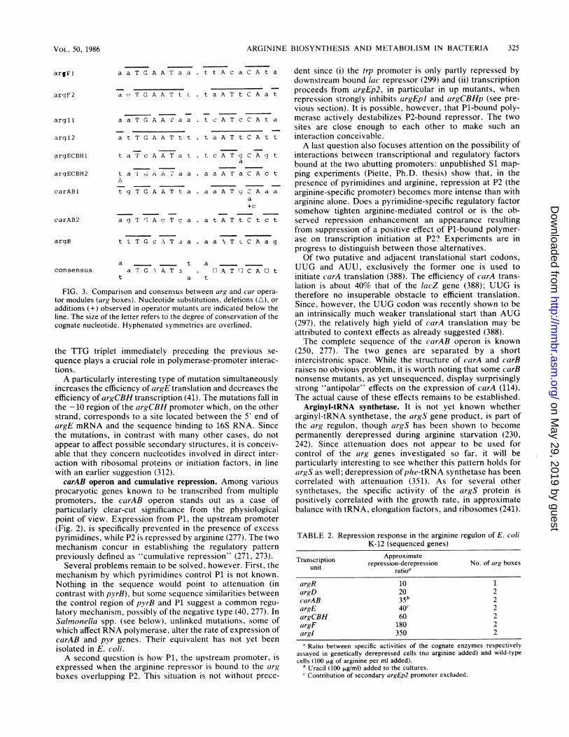
(including those of argD and -G [not shown here]) is thepresence of the so-called "arg box": an 18-base-pair-long,partly conserved, and distinctly palindromic sequence. Se-quencing operator mutations in argECBH (75, 274), argF(366), and carAB (Charlier and Roovers, unpublished exper-iments) established the role of the otrg box as the basicoperator module of the aiig regulon. A consensus derivedfrom the comparison of these sequences is given in Fig. 3.
In all cases, at least one org box overlaps the promoter.This suggests that steric hindrance between repressor andRNA polymerase is the basis of the repression response, inkeeping with the results of in vitro experiments varying theorder of addition of S30 extracts from argR' and argRstrains (319). argG, however, presents an additional putativeoperator site 110 nucleotides from the main start of tran-scription; the role of this site is unknown.There is a crude correlation between the number of arg
boxes present and the amplitude of the repression-derepression response. On the one hand, relatively lowvalues (10 to 15) obtain for one-box genes such as argH inthe suop102 deletion mutant (Fig. 2) and the autogenouslyregulated argR gene. On the other hand, argD, carAB (bothwith one "good" box and a more degenerate one), and geneswith two good boxes, i.e., argE (the contribution of thesecondary promoter argEp2 excluded [see below]),argCBH, argF, and argI, display increasingly higher values(Table 2). The tightest repression response characterizesorgF and orgI, where the overlap with the promoter is themost extensive of all and the extent of dyad symmetry is thehighest. Since the blc operon is expressed over an evenwider range and displays a relatively weak overlap, wewould assume the extent of symmetry to be the dominantfactor. A comparison of the trpEDCBA, trpR, and aroHpromoter-operator regions leads to the same conclusion(392). Moreover, constitutive mutations of carAB and argFwere found to alter two positions which are both highlyconserved and involved in symmetry (Fig. 2). Symmetry isnot the only factor involved, however; as in the lac operonthe tightness of the response must also depend on the actualcomposition of the box since argECBH 0c mutants mayresult from substitutions altering a position in the right-handpart of the box, a position which is neither conserved norinvolved in symmetry (75). Since the right-hand half of theconsensus olrg box is the lesser conserved one, it is conceiv-able that variations in this sequence play an important role indetermining the respective affinities of the different operatorsites for the repressor.
Considering the above correlation and the fact that allpairs of arg boxes are separated by the same number ofnucleotides (75), it is possible that repressor molecules bindat adjacent boxes in a cooperative way. In support of thissuggestion it can be mentioned that a deletion of 1 base pairbetween the two orgECBH boxes determines partialconstitutivity, as though efficient repression required the twoboxes to remain in register (75, 274).A notable characteristic of all of the above control regions
is the lack of typical attenuation features. Both orgCBH- andargG-coding sequences are, however, preceded on themRNA by leader sequences. Their functions, if any, are
VOL. 50, 1986 323
on May 29, 2019 by guest
http://mm
br.asm.org/
Dow
nloaded from
324 CUNIN ET AL.
.L rAsup 102---
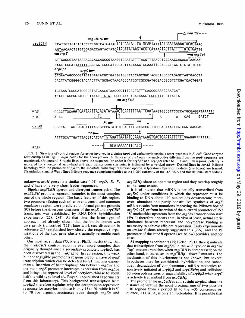
arg CBHp rIf 1
argECBH TCATTGTTGACACACCTCTGGTCATGATAGTATCAATATTCATGCAGTATTTATGAATAAAAATACACTAACAGTAACAACTGTGTGGAGACCAGTACTATCATAGTTATAAGTACGTCATAAATACTTATTTTTATGTGATTG-.aorgE --_argEp1GTTGAGCGTAATAAAACCCACCAGCCGTAAGGTGAATGTTTTACGTTTAACCTGGCAACCAGACATAAGAAG
CAACTCGCATTATTTTGGGTGGTCGGCATTCCACTTACAAAATGCAAATTGGACCGTTGGTCTGTATTCTTCargEpl argC -
GTGAATAGCCCCGATGTTGAATACGCTGATTGTGGGTACCAGCGGCTACGCTGGCGCAGAGCTAGTGACCTA
CACTTATCGGGGCTACAACTTATGCGACTAACACCCATGGTCGCCGATGCGACCGCGTCTCGATCACTGGAT
TGTAAATCGCCATCCGCATATGAACATAACCGCTTTGACTGTTTCAGCGCAAAGCAATGAT
ACATTTAGCGGTAGGCGTATACTTGTATTGGCGAAACTGACAAAGTCGCGTTTCGTTACTA-. argEp2
argF GGGGTTGCAAATGA4TAATTACACATATAAAGTGAATTTTAATTCAATAAGTGGCGTTCGCCATGCGAGGATAAAATGargI A AC C TC I IT T| A A CAG GATCT
IP1_ ttcarAB CGCCATTTAGTTGACTTTTAGCGCCCATATCTCCAGAATGCCGCCGTTTGCCAGAAATTCGTCGGTAAGCAG
P2_,.-ATTTGCATTGATTTACGTCATCAT1TGTGAATTAATATGCAAA AAAGTGAGTGAATATTCTC1 I9AGG TGTTTTG
argR ----JTTTGCATAAAAATTCATC-FIG. 2. Structure of control regions for genes involved in arginine (arg) and carbamoylphosphate (car) synthesis in E. coli. Gene-enzyme
relationship as in Fig. 1. argR codes for the aporepressor. In the case of argl only the nucleotides differing from the argF sequence arementioned. (Promoters) Straight lines above the sequence (or under it for argEpi and argEp2) refer to -35 and -10 regions; polarity isindicated by a horizontal arrowhead and each transcription startpoint is indicated by a vertical arrow. Dashed lines in carAB indicatehomology with the promoter of pyrBI, the aspartate carbamoyltransferase operon. (Operators) Operator modules (arg boxes) are framed.(Translation signals) Wavy lines indicate sequence complementarities to the 3'OH extremity of the 16S RNA and translational start codons.
unknown; aroH presents a similar case (404). argD, -E, -F,and -I have only very short leader sequences.
Bipolar argECBH operon and divergent transcription. TheargECBH promoter-operator complex is the most complexone of the whole regulon. The basic features of this region,two promoters facing each other over a central and commonregulatory region, were predicted on formal genetic grounds(97) before the divergent character of the argE and argCBHtranscripts was established by RNA-DNA hybridizationexperiments (258, 284). At that time the latter type ofapproach had already shown that the biotin operon wasdivergently transcribed (121); later work (see discussion inreference 274) established how closely the respective orga-nizations of the two gene clusters actually resemble eachother.Our most recent data (75; Piette, Ph.D. thesis) show that
the argECBH control region is even more complex thanoriginally thought since a secondary promoter, argEp2, hasbeen discovered in the argC gene. In repression, this weakbut not negligible promoter is responsible for a wave of argEtranscription which can be detected by Si mapping experi-ments. Insertion of bacteriophage Mu between argEp2 andthe main argE promoter interrupts expression from argEp2and brings the repressed level of acetylornithinase to abouthalf the wild-type level (A. Boyen, unpublished experimentsfrom this laboratory). This background transcription fromargEp2 therefore explains why the derepression-repressionresponse for acetylornithinase is only 15 to 20, while it is 50to 70 for argininosuccinase; even though argEp and
argCBHp share an operator region and they overlap roughlyto the same extent.
It is of interest that mRNA is actually transcribed fromargEp2 under conditions in which the repressor must bebinding to DNA about 110 nucleotides downstream; more-over, abundant and partly constitutive synthesis of argEmRNA results from mutations improving the Pribnow box ofargEp2 (75) or from insertion of the outward promoter of IS3180 nucleotides upstream from the argEpi transcription start(59). It therefore appears that, in vivo at least, actual sterichindrance between repressor and polymerase binding isnecessary to achieve efficient repression. Early experimentson trp-lac fusions already suggested this (299), and the P1promoter of the carAB operon (see below) provides anothercase.
Si mapping experiments (75; Piette, Ph.D. thesis) indicatethat transcription from argEp2 in the wild type or in argEp2"up" mutants vanishes when argCBH is derepressed; on theother hand, it increases in argCBHp "down" mutants. Themechanism of this interference is not known, but severalhypotheses may be considered: hybridization and subse-quent degradation of complementary mRNA molecules re-spectively initiated at argEp2 and argCBHp; and collisionsbetween polymerases or unavailability of argEp2 when argCis actively transcribed from argCBHp.The promoter for argCBH is at first sight atypical since the
distance separating the most proximal one of two possible-10 regions from a perfect fit to the -35 consensus se-quence, TTGACA, is only 15 nucleotides. It is possible that
MICROBIOL. REV.
on May 29, 2019 by guest
http://mm
br.asm.org/
Dow
nloaded from
ARGININE BIOSYNTHESIS AND METABOLISM IN BACTERIA
argF 1
arqF2
argI 1
arqI2
argECBH1
arqECBH2
carAB1
carAB2
argR
consensus
a a T G A A T a a . t t A c a
a a T G A A T t t . t a A T t
a a T G A A 'f a a . t c A T c
a t T G A A T tt . t a A T t
t a P c A A T a t .
t a 1 6 A A a a .
A
t g T G A A T t a .
a g T rl A cT T c: a .
t c A T ga
a a A r a
a a A T g
a t A T t
t t T G c A T a a . a a T t
a ta T G A A T a
t a
a[1 A T F]
t
FIG. 3. Comparison and consensus between arg andtor modules (arg boxes). Nucleotide substitutions, deleticadditions (+) observed in operator mutants are indicatedline. The size of the letter refers to the degree of conservacognate nucleotide. Hyphenated symmetries are overline
the TTG triplet immediately preceding the pre'
quence plays a crucial role in polymerase-promotetions.A particularly interesting type of mutation simult
increases the efficiency of argE translation and decrefficiency of argCBH transcription (41). The mutatithe -10 region of the argCBH promoter which, on
strand, corresponds to a site located between theargE mRNA and the sequence binding to 16S RNthe mutations, in contrast with many other case!
appear to affect possible secondary structures, it isable that they concern nucleotides involved in diraction with ribosomal proteins or initiation factorwith an earlier suggestion (312).carAB operon and cumulative repression. Amon
procaryotic genes known to be transcribed frompromoters, the carAB operon stands out as a
particularly clear-cut significance from the phypoint of view. Expression from P1, the upstream(Fig. 2), is specifically prevented in the presencepyrimidines, while P2 is repressed by arginine (277)mechanism concur in establishing the regulator,previously defined as "cumulative repression" (27
Several problems remain to be solved, however.mechanism by which pyrimidines control P1 is nc
Nothing in the sequence would point to attentcontrast with pyrB), but some sequence similaritiesthe control region of pyrB and P1 suggest a comnlatory mechanism, possibly of the negative type (4(Salmonella spp. (see below), unlinked mutations,which affect RNA polymerase, alter the rate of expcarAB and pyr genes. Their equivalent has notisolated in E. coli.A second question is how P1, the upstream prc
expressed when the arginine repressor is bound tboxes overlapping P2. This situation is not withc
C A t a dent since (i) the trp promoter is only partly repressed bydownstream bound lac repressor (299) and (ii) transcription
C A a t proceeds from argEp2, in particular in up mutants, whenrepression strongly inhibits argEpi and argCBHp (see pre-vious section). It is possible, however, that P1-bound poly-
C A t a merase actively destabilizes P2-bound repressor. The twosites are close enough to each other to make such an
C A t t interaction conceivable.A last question also focuses attention on the possibility of
C A g t interactions between transcriptional and regulatory factorsbound at the two abutting promoters: unpublished Si map-
C A c t ping experiments (Piette, Ph.D. thesis) show that, in thepresence of pyrimidines and arginine, repression at P2 (the
C A a a arginine-specific promoter) becomes more intense than witha arginine alone. Does a pyrimidine-specific regulatory factor
somehow tighten arginine-mediated control or is the ob-C t c t served repression enhancement an appearance resulting
from suppression of a positive effect of P1-bound polymer-C A a g ase on transcription initiation at P2? Experiments are in
progress to distinguish between those alternatives.Of two putative and adjacent translational start codons,
CT~A t UUG and AUU, exclusively the former one is used toinitiate carA translation (388). The efficiency of carA trans-lation is about 40% that of the lacZ gene (388); UUG is
car opera- therefore no insuperable obstacle to efficient translation.below the Since, however, the UUG codon was recently shown to betion ofthe an intrinsically much weaker translational start than AUG~d. (297), the relatively high yield of carA translation may be
attributed to context effects as already suggested (388).The complete sequence of the carAB operon is known
vious se- (250, 277). The two genes are separated by a shortr interac- intercistronic space. While the structure of carA and carB
raises no obvious problem, it is worth noting that some carBtaneously nonsense mutants, as yet unsequenced, display surprisinglyreases the strong "antipolar" effects on the expression of carA (114).ons fall in The actual cause of these effects remains to be established.the other Arginyl-tRNA synthetase. It is not yet known whether5' end of arginyl-tRNA synthetase, the argS gene product, is part of[A. Since the arg regulon, though argS has been shown to becomes, do not permanently derepressed during arginine starvation (230,conceiv- 242). Since attenuation does not appear to be used for
-ect inter- control of the arg genes investigated so far, it will be-s, in line particularly interesting to see whether this pattern holds for
argS as well; derepression ofphe-tRNA synthetase has beeng various correlated with attenuation (351). As for several othermultiple synthetases, the specific activity of the argS protein iscase of positively correlated with the growth rate, in approximate
siological balance with tRNA, elongation factors, and ribosomes (241).promoterof excessI. The twoy pattern71, 273).First, thet known.lation (ins betweennon regu-), 277). In
some ofression of
yet been
)moter, is;o the argiut prece-
TABLE 2. Repression response in the arginine regulon of E. coliK-12 (sequenced genes)
Transcription Approximateunit repression-derepression No. of arg boxes
ratioaargR 10 1argD 20 2carAB 35 2argE 40'c 2argCBH 60 2argF 180 2argl 350 2
aRatio between specific activities of the cognate enzymes respectivelyassayed in genetically derepressed cells (no arginine added) and wild-typecells (100 FLg of arginine per ml added).
b Uracil (100 ,ug/ml) added to the cultures.Contribution of secondary argEp2 promoter excluded.
325VOL. 50, 1986
on May 29, 2019 by guest
http://mm
br.asm.org/
Dow
nloaded from
326 CUNIN ET AL.
Other properties of the enzyme have been reviewed recently(Glansdorff, in press).
Concluding remarks. As the group of genes controlled bythe lex repressor, the arginine regulon of E. coli is one of thebest-known examples of a scattered set of structural genesrepressed by a unique aprepressor, recognizing with varyingefficiency the members of a family of related operators. Noother type of regulatory interaction could be demonstratedup to now within the system. The occurrence of long leadersequences with no apparent function in argC alnd (argG ispuzzling and requires further investigations; argA and -Mremain to be sequenced.
This "classical" type of gene regulation is now under-stood in its major characteristics, though repressor bindingremains to be studied in vitro. Several mechanistic problemswhich were uncovered en route remain to be solved: theargECBH RNA-enzyme discrepancy, the exact mechanismof interference between the convergent argCBH and argE'p2promoters, the P1-P2 interactions and the pyrimidine targetin carAB, and the regulatory mechanisms controlling thesynthesis of the permease (see below) and the arginyl-tRNAsynthetase. It is also clear that further studies with thearginine system will contribute to put to the test new ideason the effect of ppGpp on translational fidelity, while thec(arAB operon will provide an interesting system for studyingthe mechanism of translation initiation at the atypical codonUUG. Last but not least, several features of the org regulonare interesting to discuss from the evolutionary point ofview; this will be done in the last section of this review.
Control of Enzyme Synthesis in Other Procaryotes
On the basis of the information available, procaryotes maybe divided roughly into two categories according to thepattern of regulation which prevails for their arginine bio-synthesis (73, 354). Bacteria which possess an ornithineacetyltransferase and recycle the acetyl group fromacetylornithine to glutamate appear to rely mostly on feed-back inhibition by arginine of the second enzyme,acetylglutamate phosphotransferase, to control the pathway.In Enterobacteriaceae and Bacillacea(e which do not recy-cle the acetyl group, arginine represses the synthesis of all ormost of the enzymes and inhibits acetylglutamate synthe-tase, the first enzyme of the pathway. It is not knownwhether this correlation has biological significance or if itwill resist further screening. After a general survey of thesetwo groups, we shall deal separately with Pseudomonasspp., S. tvphirnirium, and B. slubtilis, organisms in whicharginine biosynthesis has been or is being studied systemat-ically.
Bacteria lacking or showing limited repression control.With the exception of Pseiadomsona(is spp., the current stateof knowledge of the regulation of arginine biosynthesis inbacteria of this group is still fragmentary, and caution shouldbe exercised when generalizing from observations relative toonly a few or even a single enzyme of the pathway.
In blue-green algae (cyanobacteria) and in several photo-synthetic bacteria, feedback inhibition of acetylglutamatephosphotransferase appears as the sole regulating mecha-nism controlling biosynthesis (51, 140, 141). Similarly, re-pression control is apparently absent in Corvnebacteriumigluitainicum and other micrococci (354, 397), N. gonorrh1elae(in which only CPSase has been investigated [240]), Agr-o-bacterium titinefaciens, and Rhizobilum spp. (in which onlyOTCase was studied [370; Vissers et al., manuscript inpreparation]).
In the extreme thermophile T. (aquaticuls, synthesis ofornithine acetyltransferase is repressed sevenfold by argi-nine (81).Data are now available about microbes from the third
kingdom of lif'e, archaebacteria, which are considered equi-distant from procaryotes and eucaryotes (106). Methano-genic bacteria possess an ornithine acetyltransferase andappear to lack acetylglutamate synthase (212). Acetyl-glutamate phosphotransferase is feedback inhibited by argi-nine. No repression by ar-ginine is observed (212).Pseudomnonas spp. In Pseadomnona(is spp., ornithine is pro-
duced from acetylornithine by an ornithine acetyltrans-ferase. It is not known whether the acetylornithinase activityalso found in P. ael(rginosa has biological significance (seeabove, "Enzymatic Steps and Regulation of MetabolicFlow").Carbamoylphosphate for both pyrimidine and arginine
biosyntheses is made by a single CPSase. Mutations in thecorresponding gene (cpa() result in a double auxotrophy(125). The genes for six of the nine biosynthetic enzymes andfor CPSase have been mapped in P. (leruiginosa. They arescattered on the chromosome (125).
Control of biosynthesis is achieved mainly through feed-back regulation of the activity of the first two enzymes of thepathway (126, 129, 354). However, regulation of synthesis isobserved for two enzymes in P. aeruiginosa: anabolicOTCase (aOTCase) and acetylornithine aminotransferase,respectively repressed and induced by arginine (148, 371).The latter enzyme, which is now referred to as suc-cinylornithine aminotransferase (see "Arginine Suc-cinyltransferase Pathway'") is involved both in biosynthesis,in which its basal uninduced level allows synthesis ofacetylornithine, and in degradation of arginine. A similarregulation of enzyme synthesis has been observed for P.putida, in which, in addition, the synthesis of acetylgluta-mate phosphotransferase or of the dehydrogenase or bothseems repressible too (63, 279). Repression of aOTCasesynthesis by arginine and induction of acetylornithine ami-notransferase seem to be mediated by a common regulatoryelement since a linear relationship is observed between thereciprocal of the acetylornithine aminotransferase specificactivity and that of aOTCase (373). Ornithine also inducesthe aminotransferase and represses aOTCase biosynthesis,which raises the question, in view of the key situation ofornithine in the metabolism, of which molecule (arginine orornithine) is the true effector of the genetic regulation. Thesyntheses of aOTCase and aminotransferase are also modu-lated by carbon sources (148, 373). The synthesis of theaminotransferase is repressed by citrate, a better carbonsource than arginine, while aOTCase synthesis is dere-pressed in the inverse ratio (148, 373). It was suggested (148)that the carbon source regulation functions indirectlythrough the arginine regulator, possibly via an inhibition ofarginine uptake by citrate and other good carbon sources.This hypothesis is supported by the characterization ofmutants able to derive proline from ornithine (via the ami-notransferase) in the presence of citrate (CITr mutants[3731). A single mutational event alters dramatically thecitrate regulation of both the aminotransferase and aOTCaseand abolishes arginine-specific regulation. Carbon sourceregulation does not involve cyclic adenosine monophosphate(cAMP). In P. pi,tida too, a considerable difference in theamplitude of control of aOTCase and aminotransferase syn-thesis is observed between cells grown on arginine as solecarbon or car-bon and nitrogen source and cells grown in thepresence of a more readily utilizable substrate (279). This
MICROBIOL. REV.
on May 29, 2019 by guest
http://mm
br.asm.org/
Dow
nloaded from
ARGININE BIOSYNTHESIS AND METABOLISM IN BACTERIA
difference is correlated to a modulation of arginine uptake bycatabolite repression (279).CPSase shows cumulative repression by arginine and
pyrimidines (7). The observed amplitude of repression ap-pears considerably smaller than in E. coli, ranging from 40%(7) to 75% (Vander Wauven and Pierard, unpublished data).Here too, the contribution of arginine to repression appearsto be modulated by carbon source regulation (VanderWauven and Pierard, unpublished data), which may accountto some extent for the low repressibility of CPSase synthe-sis.
Bacteria with extensive repression control. In several Ba-cillus species, OTCase synthesis is repressed at least 10-foldby arginine (354; see below). A comparable repression isobserved in the enteric bacteria K. aerogenes and Serratiamarcescens (354). The amplitude of repression by arginine isquite small in Proteus spp. (0. W. Prozesky, M.D. thesis,University of Pretoria, Pretoria, South Africa 1968) (287).As in E. coli, several genes of the biosynthetic pathway
are clustered in Proteus morganii, Proteus rutgeri, andProteus mirabilis, but in addition to argECBH, they includeargG (argECBGH) (Prozesky, M.D. thesis) (289). A similarclustering is found in Serratia marcescens (207). A clusteridentical to that of E. coli (ECBH) is present in Salmonellaspp. (309), Providentia stuartii (298), and K. pneumoniae(208). In B. subtilis also, extensive clustering is found, but itinvolves other arginine genes (235; see below).
S. typhimurium. The genetic organization of thebiosynthetic genes is similar to that of E. coli (309). There isonly one gene encoding OTCase, the equivalent of argI, asituation which prevails among E. coli strains other thanK-12. Repression by arginine is mediated by the product ofa regulatory gene, argR (174). Repressibility ratios are of thesame order of magnitude as those observed in E. coli (175).The regulatory regions (operator sites) must be homologousto their E. coli counterparts since almost normal repressionof the cognate genes is restored by the E. coli repressor inhybrid F' argR+ E. colilargR::TnJO S. tyhpimuriummerodiploids (112). The much more limited repression by theE. coli repressor reported earlier (175) for merodiploidsconstructed with an argR mutant of Salmonella sp. selectedas a suppressor of proline auxotrophy in the presence ofarginine (174) has been suggested to be the consequence ofthe formation of interspecific hybrid repressor molecules.The best-studied enzymes are OTCase and CPSase. Anti-
bodies raised against Salmonella OTCase cross-react withthe E. coli enzyme (11) and vice-versa (100). As in E. coli,the situation of CPSase at the interface of the pyrimidine andarginine pathways is mirrored in the array of enzymatic andgenetic controls to which it is submitted.
Genetic control takes the shape of a cumulative repressionby arginine and by the pyrimidines via a cytosine compound(1). Whether this compound acts through the arginine repres-sor or through a pyrimidine-specific aporepressor remains amatter of conjecture, but the interaction of a cytosinecompound with the arginine repressor might explain theincreased repression of OTCase when both cytosine andarginine are present in excess amounts (173). In E. coli,cumulative repression is achieved through the semi-independent regulation of transcription initiation at twotandem promoters (see above, "Control of Gene Expressionin E. coli").
Several mutant phenotypes originate from the interactionbetween arginine and pyrimidine biosyntheses. Similarly towhat is observed in E. coli, bradytrophic pyrH mutantsexhibit a partial arginine requirement due to the increase of
carbamoylphosphate consumption by the derepressed py-rimidine pathway, compounded by a limited carbamo-ylphosphate synthesis resulting from the inhibition ofCPSase by the increased UMP concentration (158). Somemissense mutations at the pyrA locus, which do not abolishcatalytic activity, cause uracil sensitivity, arginine sensitiv-ity (ARS) (9) or pseudo-arginine auxotrophy (AUX) (10).A particular uracil-sensitive phenotype (Use-) seems to
result from the hyperrepression of pyrA as well as of othergenes by uracil (47). Interestingly, the pyrimidine genesaffected are those presumed to be regulated by a cytosinecompound, which suggests that the use locus could encode aproduct acting pleiotropically as a regulatory element (47).This regulatory effect might, however, be an indirect one.Indeed, a fluorouracil-resistant mutant, in which two of theuridine-repressible pyr genes (pyrB, pyrE) are highly dere-pressed and pyrA is slightly derepressed, results from amutation in RNA polymerase (159). Should the expression ofpyrA be modulated in part by the size of the uridinetriphosphate and cytidine triphosphate pools through anattenuation type of mechanism analogous to that observedfor pyrBI in E. coli, the uracil sensitivity of the use mutantcould result from an alteration of a tRNA or a tRNA-modifying enzyme. However, in E. coli, where the sequenceof the corresponding locus (carAB) is known, no attenuationappears to operate.ARS and AUXpyrA mutants of S. typhimurium have been
studied in some detail. It has been proposed that the forma-tion of a complex between OTCase and CPSase is requiredfor correct subunit assembly of the latter enzyme in the ARSmutants but not in wild-type strains (9). The AUX phenotypeis often accompanied by cold sensitivity. Such a phenotypewould result from an antagonistic effect of acetylornithine oncorrect CPSase maturation at nonpermissive temperature,leading to the formation of an enzyme with altered catalyticand regulatory properties (10). This effect is abolished whenexogenous arginine curtails acetylornithine synthesis (10).
B. subtilis. Arginine biosynthesis in Bacillus spp. follows alinear pathway. There are two CPSases, one regulated bypyrimidines and one regulated by arginine (262, 283).Carbamoylphosphate is not channeled or compartmentalizedand mutants in each one of the loci result in arginine or uracilsensitivity (262). Arginine can be used as a carbon ornitrogen source or both. In B. subtilis it is degraded by anarginase pathway (82), whereas in B. licheniformis arginineis metabolized through either an arginase or an argininedeiminase pathway (45).The biosynthetic genes of B. subtilis are repressed by
arginine, while those of the arginase pathway are induced(82; see subsection, "The Arginase Pathway"). Mutantsunable to induce the catabolic enzymes were isolated on thebasis of their resistance to the arginine analog argininehydroxamate (29, 135). They map at four unlinked loci: ahrAto C (233) and ahrD (29). The current state of knowledge ofmetabolic interconnections does not allow us to distinguishbetween direct and indirect regulatory effects of these mu-tations. However, ahrC mutants also show derepression ofthe biosynthetic enzymes (135), which suggests the exist-ence of the same type of correlation between synthesis anddegradation of arginine as the one observed in yeasts (389).Seven of the arginine biosynthetic genes are clustered at
the argA-FicarAB locus (we apply here the nomenclatureused by Piggot and Hoch [278] where letters follow the orderof the biosynthetic steps); the other ones are clustered at theargGH locus (136, 203). Recently, the genes at argA-FlcarAB have been ordered by restriction-complementation
VOL. 50, 1986 327
on May 29, 2019 by guest
http://mm
br.asm.org/
Dow
nloaded from
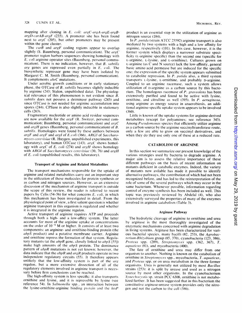
328 CUNIN ET AL.
mapping after cloning in E. (0oli: argC-argA-argE-argB-argD-carAB-argF (235). A promoter site has been foundnext to argC (205), but other promoters may be presentwithin the gene cluster.The carB and argF coding regions appear to overlap
slightly (S. Baumberg, personal communication). The argCpromoter region harbors sequences showing homology withE. coli arginine operator sites (Baumberg, personal commu-nication). There is no indication, however, that B. siibtilisar,g genes are repressed even partially in E. coli. Thebiosynthetic repressor gene may have been isolated byMargaret C. M. Smith (Baumberg, personal comunication).It complements ahrC mutations.Under aerobic growth conditions or in early stationary
phase, the OTCase of B. slibtilis becomes slightly inducibleby arginine (243; Stalon, unpublished data). The physiolog-ical relevance of this phenomenon is not evident since B.sulbtilis does not possess a deiminase pathway (243) andsince OTCase is not needed for arginine accumulation intospores (244). CPSase is also slightly inducible in stationarycells (263).Fragmentary nucleotide or amino acid residue sequences
are now available for the argF (R. Switzer, personal com-munication; Baumberg, personal communication) and argCand argH genes (Baumberg, personal communication) of B.siibtilis. Homologies were found by these authors betweenurgF and argF and argl of E.coli (366), ARG3 of Succharo-mynccs cerev'isiue (R. Huygen, unpublished experiments, thislaboratory), and human OTCase (143). alrgC shows homol-ogy with uirgC of E. coli (274) and urglg shows homologywith ARG4 of Succ/huromynces cere l isiue (30, 76) and argIlof E. coli (unpublished results, this laboratory).
Transport of Arginine and Related Metabolites
The transport mechanisms responsible for the uptake ofarginine and related metabolites carry out an important stepin the utilization of these molecules for protein synthesis oras carbon and nitrogen sources. However, as an extensivediscussion of the mechanism of arginine transport is outsidethe scope of this review, the reader is referred to recentpapers by Celis (54-56) for what concerns E. (oli, in whichthis mechanism has been investigated in detail. From thephysiological point of view, a first salient question is whetherarginine transport in this organism is regulated and whetherit is integrated in the arginine regulon.
Active transport of arginine requires ATP and proceedsthrough both a high- and a low-affiity system. The latteraccounts for most of the arginine uptake at concentrationson the order of 10-6 M or higher. It consists of at least twocomponents: an arginine- and ornithine-binding protein (theabpS product) and a putative membrane carrier. Arginineand ornithine repress the formation of that system. Regula-tory mutants (at the abpR gene, closely linked to abpS [55])make high amounts of the abpS protein. The dominancepattern of cubpR mutations is not yet known; however, thedata indicate that the abpR and argR products operate in twoindependent regulatory circuits (55). It therefore appearsunlikely that the low-affinity system is part of the argregulon, but a more extensive dissection of all of theregulatory elements involved in arginine transport is neces-sary before firm conclusions can be reached.The high-affinity system is less specific: it also transports
ornithine and lysine, and it is repressible by lysine (seereference 54). In Sal/none/l/l spp., an interaction betweenthe lysine-ornithine-arginine binding protein and the IiisP
product is an essential step in the utilization of arginine asnitrogen source (184).
In P. pultidal (strain ATCC 25592) arginine transport is alsomediated by two systems with a high and a low affinity forarginine, respectively (101). In this case, however, it is theformer system which displays a narrower substrate speci-ficity (L-arginine specific) than the second one (specific forL-arginine, L-lysine, and L-ornithine). Cultures grown onL-arginine (as C and N source) lack the low-affinity, generalbasic amino acid permease but are induced for the specificone; the expression of the specific system appears submittedto catabolite repression. In P. puMtida also, a third systemtransports L-lysine, L-ornithine, and probably D-arginine.Coupled to an arginine racemase, such a system allowsutilization of D-arginine as a carbon source by this bacte-rium. The homologous racemase of P. graveolens has beenextensively purified and found to be active with lysine,ornithine, and citrulline as well (395). In P. aeruginosa,using arginine as energy source in anaerobiosis, an addi-tional arginine-specific uptake system appears to be involved(360).
Little is known of the uptake systems for arginine-derivedmetabolites (except for polyamines; see reference 343).Among Pseuldornonas species that efficiently utilize arginineas a carbon source by the arginine succinyltransferase route,only a few are able to grow on succinyl derivatives, andwhen they do they use only one of these at a reduced rate.
CATABOLISM OF ARGININE
In this section we summarize our present knowledge of thevarious strategies used by bacteria to degrade arginine. Amajor aim is to assess the relative importance of thesedifferent pathways on the basis of recent information onmutants deficient in catabolic enzymes. Indeed, the varietyof mutants now avilable has made it possible to identifyalternative pathways, the contribution of which had not beenrecognized before, and has led to the reinterpretation of therole of some of the several pathways which may occur in thesame bacterium. Whenever possible, information regardingcontrol of enzyme synthesis has been included as well. Thistopic was previously reviewed by Abdelal (4), who alsoextensively surveyed the properties of many of the enzymesinvolved in arginine catabolism (Table 3).
Arginase Pathway
The hydrolytic cleavage of arginine to ornithine and ureaby arginase is the most thoroughly investigated of theenzymatic mechanisms concerned with arginine degradationin living systems. Arginase has been characterized for vari-ous bacterial species, many bacilli (82, 253), the Agrobac-terium-Rhizobiumi group (85, 370), cyanobacteria (123, 386),Pr-oteius spp. (289), Streptomyces spp. (362, 367), T.(aquaticlls (81), and mycobacteria (400).The fate of ornithine and urea may differ from one
organism to another. Nothing is known on the catabolism ofornithine in Streptomyces spp., mycobacteria, T. aquaticus,and Protelus spp. or on urea metabolism in the three formerorganisms. Urea is generally not utilized by most Bacillusstrains (253): it is split by urease and used as a nitrogensource by most other organisms. In the cyanobacteriumSvnechocystis sp. strain PCC 6308, ornithine is not metabo-lized further. It has been suggested that in this bacterium theconstitutive arginase-urease system provides only the nitro-gen and not the carbon to the cell (386).
MICROBIOL. REV.
on May 29, 2019 by guest
http://mm
br.asm.org/
Dow
nloaded from
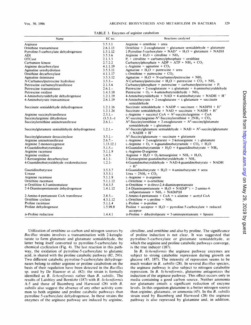
ARGININE BIOSYNTHESIS AND METABOLISM IN BACTERIA
TABLE 3. Enzymes of arginine catabolism
Name EC no. Reactions catalyzed
ArginaseOrnithine transaminasePyrroline-5-carboxylate dehydrogenaseADIOTCaseCarbamate kinaseArginine decarboxylaseAgmatine ureohydrolaseOrnithine decarboxylaseAgmatine deiminaseN-Carbamoylputrescine hydrolasePutrescine carbamoyltransferasePutrescine transaminasePutrescine oxidase4-Aminobutyraldehyde dehydrogenase4-Aminobutyrate transaminase
Succinate semialdehyde dehydrogenase
Arginine succinyltransferaseSuccinylarginine dihydrolaseSuccinylornithine aminotransferase
Succinylglutamate semialdehyde dehydrogenase
Succinylglutamate desuccinylaseArginine aminotransferaseArginine 2-monooxygenase4-GuanidinobutyramidaseArginine racemaseArginine oxidase2-Ketoarginine decarboxylase4-Guanidinobutyraldehyde oxidoreductase
GuanidinobutyraseUreaseArginine racemaseOrnithine racemaseD-Ornithine 4,5-aminomutase2-4-Diaminopentanoate dehydrogenase
2-Amino-4-pentanoate CoA transferaseOrnithine cyclaseProline racemaseProline dehydrogenase
D-Proline reductase
3.5.3.12.6.1.131.5.1.123.5.3.62.1.3.32.7.2.24.1.1.193.5.3.114.1.1.173.5.3.123.5.3.-2.1.3.62.6.1.-1.4.3. 101.2.1.192.6.1.19
1.2.1.161.2.1.242.3.1.-(3.5.3.-)2.6.1.-
1.2.1.-
3.5.1.-2.6.7.-1.13.12.15.1.5.-5.1.1.91.4.3-4.1.1-1.2.1-
3.5.3.73.5.3.15.1.1.95.1.1.125.4.3.51.4.1.12
2.8.3.-4.3.1.125.1.1.41.5.99.8
1.4.4.1
Arginine = ornithine + ureaOrnithine + 2-oxoglutarate = glutamate semialdehyde + glutamate1-Pyrroline-5-carboxylate + NAD+ + H20 = glutamate + NADHArginine + H20 = citrulline + NH3Pi + citrulline = carbamoylphosphate + ornithineCarbamoylphosphate + ADP = ATP + NH3 + CO2L-Arginine = agmatine + CO2Agmatine + H20 = putrescine + ureaL-Ornithine = putrescine + CO2Agmatine + H20 = N-carbamoylputrescine + NH3N-Carbamoylputrescine + H20 = putrescine + CO2 + NH3Carbamoylphosphate + putrescine = carbamoylputrescine + PiPutrescine + 2-oxoglutarate = L-glutamate + 4-aminobutyraldehydePutrescine + 02 = 4-aminobutyraldehyde + NH34-Aminobutyraldehyde + NAD = 4-aminobutyrate + NADH + H+4-Aminobutyrate + 2-oxoglutarate = L-glutamate + succinate
semialdehydeSuccinate semialdehyde + NADP = succinate + NADPH + H+Succinate semialdehyde + NAD = succinate + NADH + H+L-Arginine + succinyl CoA = N2-succinylarginine + CoAN2-succinylarginine-N2-Succinylornithine + 2NH3 + CO2N2-Succinylornithine + 2-oxoglutarate = N2-succinylglutamate
semialdehyde + L-glutamateN2-Succinylglutamate semialdehyde + NAD = N2-succinylglutamate
+ NADH + H+N2-Succinylglutamate = succinate + glutamateL-Arginine + 2-oxoglutarate = 2-ketoarginine + L-glutamateL-Arginine + 02 = 4-guanidinobutyramide + CO2 + H204-Guanidinobutyramide + H20 = 4-guanidinobutyrate + NH3L-Arginine-D-arginineArginine + H20 + 02-ketoarginine + NH3 + H2022-Ketoarginine-guanidinobutyraldehyde + NH34-Guanidinobutyraldehyde + NAD-4-guanidinobutyrate + NADH+ H+
4-Guanidinobutyrate + H20 = 4-aminobutyrate + ureaUrea = 2NH3 + CO2L-Arginine = D-arginineL-Ornithine = D-ornithineD-Ornithine = D-threo-2,4-diaminopentanoate2,4-Diaminopentanoate + H20 + NAD(P+) = 2-amino-4-
oxbpentanoate + NH3 + NAD(P)H2-Amino-4-pentanoate + CoA = L-alanine + acetyl CoAL-Ornithine = L-proline + NH3L-Proline = D-prolineProline + acceptor + H20 = pyrroline-5-carboxylate + reduced
acceptorD-Proline + dihydrolipoate = 5-aminopentanoate + lipoate
Utilization of ornithine as carbon and nitrogen sources byBacillus strains involves a transamination with 2-ketoglu-tarate to form glutamate and glutamate semialdehyde, thelatter being itself converted to pyrroline-5-carboxylate bychemical cyclization (Fig. 4). The last reaction in this path-way, the oxidation of pyrroline-5-carboxylate to glutamicacid, is shared with the proline catabolic pathway (82, 293).Two different catabolic pyrroline-5-carboxylate dehydroge-nases belong to either arginine or proline catabolism on thebasis of their regulation have been detected in the Bacillussp. used by De Hauwer et al. (82); the strain is formallyidentified as B. licheniforrnis rather than B. slubtilis. Theresults of Laishley and Bernlohr (187) with B. lichenfoirwnisA-S and those of Baumberg and Harwood (28) with B.subtilis also suggest the absence of any other activity com-mon to both arginine and proline catabolic pathways thanpyrroline 5-carboxylate dehydrogenase. In these strains theenzymes of the arginase pathway are induced by arginine,
citrulline, and ornithine and also by proline. The significanceof proline induction is not clear. It was suggested thatpyrroline-5-carboxylate or glutamate semialdehyde, atwhich the arginine and proline catabolic pathways converge,is the true inducer (187).
In B. licheniformins the arginase pathway enzymes aresubject to strong catabolite repression during growth onglucose (45, 187). The intensity of repression seems to bemuch weaker in B. siubtilis (28). In several Bacillus species,the arginase pathway is also subject to nitrogen cataboliterepression. In B. licheniformis, glutamine antagonizes theinduction of the arginase pathway. This effect occurs only inmedia containing a good carbon source. Neither ammonianor glutamate entails a significant reduction of enzymelevels. In this organism glutamine is a better nitrogen sourcethan arginine, glutamate, or ammonia (45). In the B. sbibtilisstrain used by Baumberg and Harwood (28) the arginasepathway is also repressed by glutamine and, in addition,
VOL. 50, 1986 329
on May 29, 2019 by guest
http://mm
br.asm.org/
Dow
nloaded from
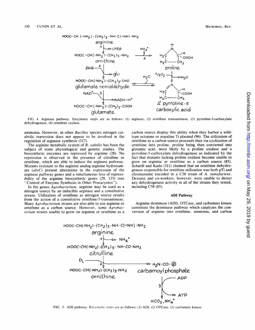
330 CUNIN ET AL.
HOOC-CH (-NH2)- (CH2)3 -NH-C(-NH)-NH2
arginineulrea
HOOC -CH(-NH2 )-(CH2)3-NH2orni'thine,
2KG 2.
gluHOOC-CH (-NH2) -(CH2 )2- CHO
glutamate. eeomialdehydeNAD 3
---AcNADH tH
HOOC -CH (-NH 2)- (CH2)2-COOF
glutamate,FIG. 4. Arginase pathway. Enzymatic steps are as follows: (1)
dehydrogenase, (4) ornithine cyclase.
ammonia. However, in other Bacillus species nitrogen cat-abolic repression does not appear to be involved in theregulation of arginase synthesis (317).The arginine metabolic system of B. subtilis has been the
subject of some physiological and genetic studies. Thebiosynthetic enzymes are repressed by arginine (28). Norepression is observed in the presence of citrulline orornithine, which are able to induce the arginase pathway.Mutants resistant to the arginine analog arginine hydroxam-ate (ahrC) present alterations in the expression of thearginase pathway genes and a simultaneous loss of repress-ibility of the arginine biosynthetic genes (29, 135) (see"Control of Enzyme Synthesis in Other Procaryotes").
In the genus Agrobacterium, arginine may be used as anitrogen source by an inducible arginase and a constitutiveurease. Utilization of ornithine as nitrogen source resultsfrom the action of a constitutive ornithine-5-transaminase.Many Agrobacterium strains are also able to use arginine orornithine as a carbon source. However, some Agrobac-terium strains unable to grow on arginine or ornithine as a
NH4+4
--D
H
H2CC"-COI I \COH2C OCH2
proline
H20 1/202 H2O
H20 'COOH
I+ M2C OCH2
11L pyrroline-5-carboxylic acid
arginase, (2) ornithine transaminase, (3) pyrroline-5-carboxylate
carbon source display this ability when they harbor a wild-type octopine or nopaline Ti plasmid (96). The utilization ofornithine as a carbon source proceeds thus via cyclization ofornithine into proline, proline being then converted intoglutamic acid, most likely by a proline oxidase and apyrroline-5-carboxylate dehydrogenase as indicated by thefact that mutants lacking proline oxidase become unable togrow on arginine or ornithine as a carbon source (85).Schardl and Kado (311) claimed that an ornithine dehydro-genase responsible for ornithine utilization was both pTi andchromosome encoded in a C58 strain of A. tumefaciens.Dessaux and co-workers, however, were unable to detectany dehydrogenase activity in all of the strains they tested,including C58 (85).
ADI Pathway
Arginine deiminase (ADI), OTCase, and carbamate kinaseconstitute the deiminase pathway which catalyzes the con-version of arginine into ornithine, ammonia, and carbon
HOOC-CH(-NH2)-(CH2)3- NH-C(=NH) -NH2
arginine,-1 NH4+
HOOC-CH(-NH2)- (CH2)3- NH-CO-NH2
citrul linetP _
HOOC-CH(-NH2)-(CH2)3-NH2
ornithine,
H2N-CO-@
carbamoyl phosphaEeADP
3
ATPHCO3NH4
FIG. 5. ADI pathway. Enzymatic steps are as follows: (1) ADI, (2) OTCase, (3) carbamate kinase.
MICROBIOL. REV.
I
on May 29, 2019 by guest
http://mm
br.asm.org/
Dow
nloaded from
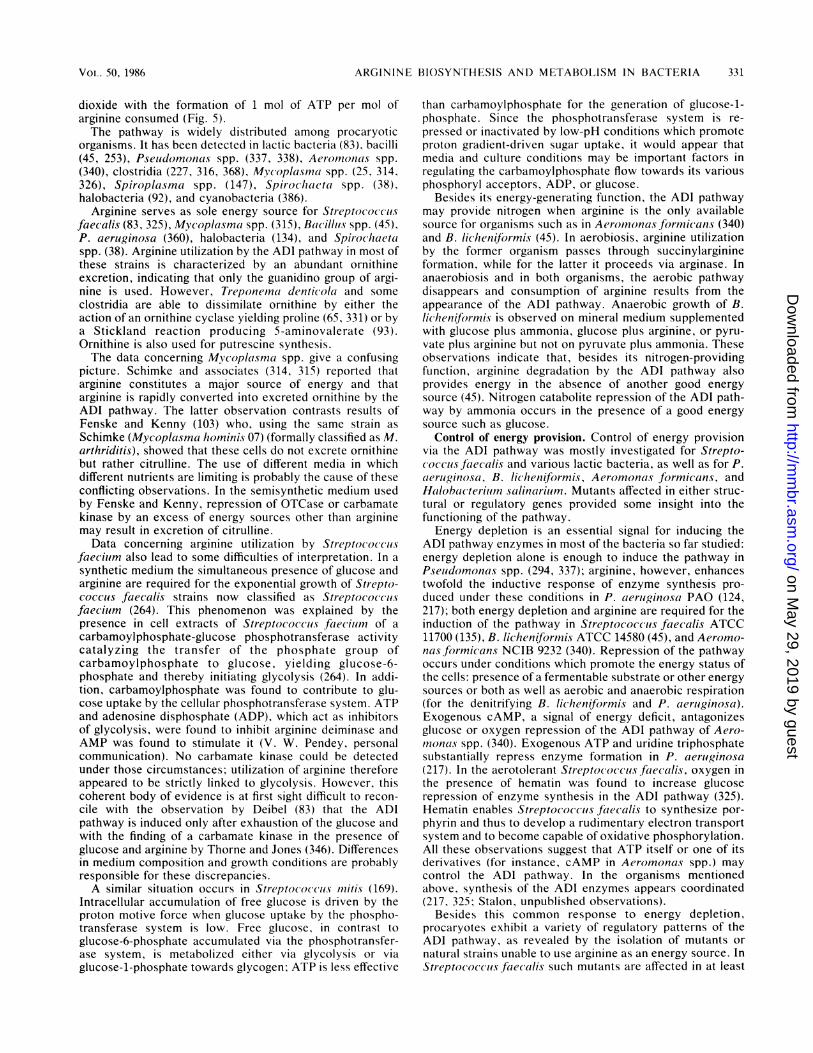
ARGININE BIOSYNTHESIS AND METABOLISM IN BACTERIA
dioxide with the formation of 1 mol of ATP per mol ofarginine consumed (Fig. 5).The pathway is widely distributed among procaryotic
organisms. It has been detected in lactic bacteria (83), bacilli(45, 253), Pseludomonas spp. (337, 338), Aermoionas spp.(340), clostridia (227, 316, 368), Mycoplasma spp. (25, 314,326), Spiroplassma spp. (147), Spirochlacta spp. (38),halobacteria (92), and cyanobacteria (386).
Arginine serves as sole energy source for Streptococcusfaecalis (83, 325), Mycoplasma spp. (315), Bacillus spp. (45),P. aeruginosa (360), halobacteria (134), and Spirochaetaspp. (38). Arginine utilization by the ADI pathway in most ofthese strains is characterized by an abundant ornithineexcretion, indicating that only the guanidino group of argi-nine is used. However, Treponema denticola and someclostridia are able to dissimilate ornithine by either theaction of an ornithine cyclase yielding proline (65, 331) or bya Stickland reaction producing 5-aminovalerate (93).Ornithine is also used for putrescine synthesis.The data concerning Mlvcoplasrna spp. give a confusing
picture. Schimke and associates (314, 315) reported thatarginine constitutes a major source of energy and thatarginine is rapidly converted into excreted ornithine by theADI pathway. The latter observation contrasts results ofFenske and Kenny (103) who, using the same strain asSchimke (Mycoplasma hominis 07) (formally classified as M.arthriditis), showed that these cells do not excrete ornithinebut rather citrulline. The use of different media in whichdifferent nutrients are limiting is probably the cause of theseconflicting observations. In the semisynthetic medium usedby Fenske and Kenny, repression of OTCase or carbamatekinase by an excess of energy sources other than argininemay result in excretion of citrulline.Data concerning arginine utilization by Streptococcus
faecium also lead to some difficulties of interpretation. In asynthetic medium the simultaneous presence of glucose andarginine are required for the exponential growth of Strepto-cocculs faecalis strains now classified as Streptococcusfaeciium (264). This phenomenon was explained by thepresence in cell extracts of Streptococcus faecium of acarbamoylphosphate-glucose phosphotransferase activitycatalyzing the transfer of the phosphate group ofcarbamoylphosphate to glucose, yielding glucose-6-phosphate and thereby initiating glycolysis (264). In addi-tion, carbamoylphosphate was found to contribute to glu-cose uptake by the cellular phosphotransferase system. ATPand adenosine disphosphate (ADP), which act as inhibitorsof glycolysis, were found to inhibit arginine deiminase andAMP was found to stimulate it (V. W. Pendey, personalcommunication). No carbamate kinase could be detectedunder those circumstances; utilization of arginine thereforeappeared to be strictly linked to glycolysis. However, thiscoherent body of evidence is at first sight difficult to recon-cile with the observation by Deibel (83) that the AD!pathway is induced only after exhaustion of the glucose andwith the finding of a carbamate kinase in the presence ofglucose and arginine by Thorne and Jones (346). Differencesin medium composition and growth conditions are probablyresponsible for these discrepancies.A similar situation occurs in Streptococcus m71itis (169).
Intracellular accumulation of free glucose is driven by theproton motive force when glucose uptake by the phospho-transferase system is low. Free glucose, in contrast toglucose-6-phosphate accumulated via the phosphotransfer-ase system, is metabolized either via glycolysis or viaglucose-i-phosphate towards glycogen; ATP is less effective
than carbamoylphosphate for the generation of glucose-i-phosphate. Since the phosphotransferase system is re-pressed or inactivated by low-pH conditions which promoteproton gradient-driven sugar uptake, it would appear thatmedia and culture conditions may be important factors inregulating the carbamoylphosphate flow towards its variousphosphoryl acceptors, ADP, or glucose.
Besides its energy-generating function, the ADI pathwaymay provide nitrogen when arginine is the only availablesource for organisms such as in Aerononas formicans (340)and B. lichenifr)rmis (45). In aerobiosis, arginine utilizationby the former organism passes through succinylarginineformation, while for the latter it proceeds via arginase. Inanaerobiosis and in both organisms, the aerobic pathwaydisappears and consumption of arginine results from theappearance of the ADI pathway. Anaerobic growth of B.lichenifjormis is observed on mineral medium supplementedwith glucose plus ammonia, glucose plus arginine, or pyru-vate plus arginine but not on pyruvate plus ammonia. Theseobservations indicate that, besides its nitrogen-providingfunction, arginine degradation by the ADI pathway alsoprovides energy in the absence of another good energysource (45). Nitrogen catabolite repression of the ADI path-way by ammonia occurs in the presence of a good energysource such as glucose.
Control of energy provision. Control of energy provisionvia the ADI pathway was mostly investigated for Strepto-coccus faecalis and various lactic bacteria, as well as for P.aeruiginosa, B. lichenifo)rmis, Aeromnonas fotrmicans, andHalobacteriu,m salinariurn. Mutants affected in either struc-tural or regulatory genes provided some insight into thefunctioning of the pathway.Energy depletion is an essential signal for inducing the
ADI pathway enzymes in most of the bacteria so far studied:energy depletion alone is enough to induce the pathway inPseuldomonas spp. (294, 337); arginine, however, enhancestwofold the inductive response of enzyme synthesis pro-duced under these conditions in P. aeruiginosa PAO (124,217); both energy depletion and arginine are required for theinduction of the pathway in Streptococcus fJecalis ATCC11700 (135), B. licheniformis ATCC 14580 (45), and Aeromo-nasformicans NCIB 9232 (340). Repression of the pathwayoccurs under conditions which promote the energy status ofthe cells: presence of a fermentable substrate or other energysources or both as well as aerobic and anaerobic respiration(for the denitrifying B. licheniformiis and P. aerliginosa).Exogenous cAMP, a signal of energy deficit, antagonizesglucose or oxygen repression of the ADI pathway of Aero-monoas spp. (340). Exogenous ATP and uridine triphosphatesubstantially repress enzyme formation in P. aerluginosa(217). In the aerotolerant Streptococcus ftecalis, oxygen inthe presence of hematin was found to increase glucoserepression of enzyme synthesis in the ADI pathway (325).Hematin enables Streptococcus ftaccalis to synthesize por-phyrin and thus to develop a rudimentary electron transportsystem and to become capable of oxidative phosphorylation.All these observations suggest that ATP itself or one of itsderivatives (for instance, cAMP in Aeromonas spp.) maycontrol the ADI pathway. In the organisms mentionedabove, synthesis of the ADI enzymes appears coordinated(217, 325; Stalon, unpublished observations).
Besides this common response to energy depletion,procaryotes exhibit a variety of regulatory patterns of theADI pathway, as revealed by the isolation of mutants ornatural strains unable to use arginine as an energy source. InStreptococcus faecalis such mutants are affected in at least
VOL. 50, 1986 331
on May 29, 2019 by guest
http://mm
br.asm.org/
Dow
nloaded from
332 CUNIN ET AL.
one of the three enzymes of the pathway or in the uptakesystem, or display a low level of all three enzymes, suggest-ing the existence of a common element for their induction. InStreptococcus sanguis, which seems to follow the sameregulatory pattern as Streptococcus faecalis, a barotolerantmutant has been isolated which escapes glucose repressionof all three enzymes (104). In Streptococcus lactis isolatedfrom a dairy, the synthesis of the first two enzymes of thepathway seems to be glucose controlled, the carbamatekinase activity being constitutive (72). Some Streptoco(cuslactis strains unable to use arginine are blocked in both ADIand OTCase. This observation is reminiscent of the naturaldairy strains of Streptococcus cremoris that have constitu-tive carbamate kinase but lack ADI or ADI and OTCase. Inthose strains, restoration of both ADI and OTCase to thelevel normally found in Streptococcus lactis may be ob-tained by a single point mutation (72). As the two species areclosely related, it is conceivable that Streptococcus cremorisis a variety of Streptococcus lactis which has lost the firsttwo enzymes of the ADI pathway or the possibility to inducethem.Mutants with mutations affecting the ADI route were first
obtained with P. aeruginosa as follows: (i) anabolic OTCase-deficient mutants were isolated (125); (ii) revertants wereselected in which the molecular and regulatory properties ofcatabolic OTCase were altered in such a way (see "Evolu-tionary Considerations") that the enzyme could fulfill ananabolic function (124); (iii) citrulline auxotrophs were se-lected in which no OTCase activity could be detected (124)(such strains, when induced for the ADI pathway, producedcitrulline from arginine); (iv) citrulline-nonproducing mu-tants were selected which lacked ADI activity (218).Of aerobic Pseudomonas spp., Haas and associates (360)
observed that P. aeruginosa can grow anaerobically witharginine as energy source. This property was used to selectstrains affected in the ADI route, using as phenotype the factthat arginine could not sustain the anaerobic growth of thesemutants any more. As for Streptococcus faecalis, thesemutants were found to be affected either in the anaerobicarginine uptake system or in at least one enzyme of thepathway or to be mutated in a control element required forpleiotropic enzyme induction. The mutations arcA (ADI),arcB (OTCase), arcC (carbamate kinase), and arcD(anaerobic uptake system) are all highly cotransducible inthe 17-min chromosome region (360). Interestingly, P. aeru-ginosa is the only fluorescent Pseudomonas sp. able to growfermentatively in the presence of arginine. P. putida, P.fluorescens, and P. mendocina are unable to do so (360).
For B. licheniformis, mutants deficient in carbamatekinase were found among strains unable to use arginine asenergy source in anaerobiosis (K. Broman, Ph.D. thesis,University of Brussels, Brussels, Belgium, 1978). Theyfailed to grow on pyruvate plus arginine in anaerobiosis butstill retained the ability to do so in aerobiosis due to thepresence of the arginase route (Broman, Ph.D. thesis). Thedeiminase pathway can fulfill the nitrogen requirement.Regulatory mutants with mutations affecting the ADI path-way were selected from a strain unable to induce botharginase and ornithine transaminase for their ability to usearginine in aerobiosis as the sole nitrogen source in thepresence of glucose (45). In these mutants, induction of thepathway by arginine is no longer repressed by nitrogen.Oxygen or nitrate are without effect and glucose repressionis largely reduced, thus indicating that one element is sharedby the mechanisms respectively responsible for nitrogen,glucose, 02, and NO3- repression. Secondary mutants of B.
licheniformis isolated for their ability to utilize arginine as acarbon source in aerobiosis either are revertants havingregained the ability to induce the synthesis of arginase andornithine transaminase or are inducible for ornithinetransaminase only, no arginase being synthesized; in theseorganisms the ornithine formed by the ADI route is used viathe transaminase (Stalon, unpublished experiments).
Halobacteria are able to grow anaerobically with the ATPderived from the light energy harvested by the bacterio-rhodopsin proton excretion system. This system is synthe-sized only under the condition of limited oxygen supply butnot in darkness or in the total absence of oxygen. Underthese conditions, arginine can replace oxygen or light asenergy sources by the ADI route. Mutants unable to usearginine as the energy source to grow in anaerobiosis havebeen selected, but their lesions have not yet been character-ized (134).The expression of the ADI pathway in clostridia is corre-
lated with sporulation (227, 369). While OTCase and carbam-ate kinase in Clostridiinm sporogenes appear constitutive,ADI seems to be regulated at the levels of both geneexpression and enzyme activity. Carbon sources known torepress sporulation also repress ADI synthesis (369).The catabolic OTCase of the fluorescent Pseudomonas
group as well as of P. mnendocina is also subject toenzymotropic regulation (Stalon, unpublished results). Co-operative binding of phosphate, citrulline, and carbamoyl-phosphate has been observed. In addition, the OTCases ofP. putida and P. aeriuginosa, the allosteric properties ofwhich have been thoroughly studied, are regulated by avariety of metabolic signals activating or inhibiting its activ-ity (334). Pyrimidine and purine triphosphonucleotides, aswell as the polyamines putrescine, spermine, and spermi-dine, inhibit the activity by increasing the threshold sub-strate concentration required to activate the enzyme,whereas the nucleotide monophosphate restores the normalkinetic behavior. Since the catabolic OTCase is integrated inan energy-producing pathway, the significance of the activa-tion by AMP, a signal of energy deficit, and the inhibition byATP, an index of the high energy status of the cell, are easilyunderstood. Since the putrescine content is high in prolifer-ating cells and low in starving cells, the inhibitors putrescine,spermidine, and spermine should be envisioned as signalsreflecting the availability of good carbon and nitrogensources rather than as end products of the ADI route.
Arginine Succinyltransferase PathwayThe recently discovered arginine succinyltransferase path-
way (335, 361) accounts for the dissimilation of the carbonskeleton of arginine in Aeromonas formicans, in K. aero-genes, and in most of the Pseiidomonas strains of group I (P.aeruginiosa, P. fliuorescens, P. putida, P. mendocina, and P.aureofaciens), group II (P. cepacia and P. caryophylii), andgroup V (P. indigofera and P. doudorofi) as defined byPalleroni et al. (256). Most of these organisms have, how-ever, other pathways for arginine dissimilation whose func-tion is to provide either polyamines when arginine is abun-dant (see the arginine decarboxylase pathway) or energy bythe ADI route (see preceding section). However, in P.cepacia, the arginine succinyltransferase route is the solearginine catabolic pathway. This route follows the fivereactions shown in Fig. 6. The first reaction in this sequenceis catalyzed by arginine succinyltransferase; N2-succinyl-arginine dihydrolase then converts N2-succinylarginine intoN2-succinylornithine, ammonia, and carbon dioxide. The
MICROBIOL. REV.
on May 29, 2019 by guest
http://mm
br.asm.org/
Dow
nloaded from
ARGININE BIOSYNTHESIS AND METABOLISM IN BACTERIA
HOOC-CH (-NH2) H(C=)5-HC(NH)-NH2arginine
CoASH ' ;
HOOC-CH-(CH2)3-NH-C(=NH)- NH2N H-CO- (CH2) 2 - COOH
2 .N 2 succinylarginine2 '-2NH4 ,C02
HOOC-CH-(CH2)3- NH2
NH-CO(C H2)2- COOH
N2_ succinylornithi ne2K(
3
C3gu
HOOC- CH- (CH 2)2-CHONH -CO- (CH2)2-COOH
N2 !nygIuamoe,Ysuccin e
semialdehyde
4
HOOC - (CH2)2 COSCoAsuccinylCoA
'HOOC-CO-(CH2)2-COOH2-ketoglutarate,
HOOC- CH (- NH2)- (CH2)2- COOHglutamate
5 csuccinate
HOOC-CH-(CH2)2 COOH
NH-CO-(CH2) 2-COOHNAD* N2- succinylgIutamate
FIG. 6. Arginine succinyltransferase pathway. Enzymatic steps are as follows: (1) arginine succinyltransferase, (2) succinylargininedihydrolase, (3) succinylornithine aminotransferase, (4) succinylglutamate semialdehyde dehydrogenase, (5) succinylglutamatedesuccinylase.
third step is catalyzed by N2-succinylornithine aminotrans-ferase, which transfers the delta nitrogen atom of N2-succinylornithine to 2-ketoglutarate, giving glutamate andN2-succinylglutamate semialdehyde. The latter is oxidizedby a dehydrogenase into N2-succinylglutamate, which iscleaved into succinate and glutamate. The presence of thearginine succinyltransferase route in K. aerogenes MK53brings into question the existence of the arginine trans-amidinase activity suggested by Friedrich and co-workers(107, 108) to explain arginine catabolism in this strain. Thepostulated existence of this activity was based on the obser-vations that (i) arginine donates its guanidino group tohydroxylamine and forms ornithine (108) and (ii) cells grownwith arginine synthesize an ornithine transaminase activity,this enzyme having, however, more affinity for acetyl-ornithine than for ornithine (107). In our hands, the arginine-inducible acetylornithine aminotransferase activity of Kleb-siella spp. may also transaminate succinylornithine, the Kmfor succinylornithine being four times lower than that ob-served for acetylornithine (335).As expected from their physiological role, the syntheses of
these enzymes in P. cepacia, P. aeruginosa, and P. putidaare subject not only to induction by arginine, but also tocarbon catabolite repression by succinate and nitrogencatabolite repression by glutamine. Ammonia has little effecton the synthesis of these enzymes.
P. aeruginosa mutants which cannot grow aerobically on
arginine have been isolated (292, 371); they are blocked instructural genes coding for at leat one activity of the pathway
or fail to induce synthesis of all of the enzymes (D. Haas, C.Vander Wauven, and V. Stalon, manuscript in preparation).All of these mutations are clustered in the 90-min region ofthe P. aeruginosa chromosome and are unlinked to the otherloci involved in arginine catabolism (Haas, personal commu-nication).For P. putida, a mutant impaired in the succinylarginine
dihydrolase activity can still grow on arginine but at a lowerrate than the wild-type strain. The growth yield is alsolargely reduced because 50% of the arginine consumed isrecovered in the culture fluid as N2-succinylarginine. Theability of a P. putida strain to grow on arginine in spite of adefect in the arginine succinyltransferase route is due to thepresence of another arginine catabolic pathway, the arginineoxidase route (see next section).
Arginine Transaminase, Oxidase, and Oxygenase Pathways
The coryneform bacterium Arthrobacter simplex cancatalyze the transamination between arginine and 2-ketoglutarate to produce 2-ketoarginine and glutamate (347).The arginine transaminase was extensively purified and wasfound to transaminate arginine (100%) but also citrulline(16%) and alanine (9.5%) (344). In this organism, 2-ketoarginine appears to undergo hydrolytic cleavage of theguanidino group to yield urea and 2-ketoornithine, the fate ofwhich remains unknown. This observation is at first sightdifficult to reconcile with the fact that arginine transaminaseactivity is not induced only by arginine and 2-ketoarginine
333VOL. 50, 1986
on May 29, 2019 by guest
http://mm
br.asm.org/
Dow
nloaded from
334 CUNIN ET AL.
C02
/arginineHOOC-CH(-NH2)-(CH2)3-NH-C(:NH)-NH2
10 NH4,
2 ketoarginineHOOC-CO.(CH2)3-NH-C(:NH)NH2
FC02
4-guanidinobutyraldehycieOHC -(CH2)3 -NH-C(. NH) -NH2
NAD*12' NADH + H'
4 -gue nidinobutyratQHOOC-(CH2)3-NH-C(:NH)-NH2
13
urea 4-aminobutyrateHOOC-(CH2)3- NH2
2 KG -8Cgtu
agmatineH2N- (CH2)4- NH-C(:NH)-NH2 >
3 NHH
N-car bamoylputrescine.H2N-(CH2)14-NH CO NH2
4 98N C02
put re scineH2N-(CH2)4- NH2
1/2 02 L-ll,- 2KG5 6 [
NH4 -'t g1 u
4-ami nobutyraldehydeH2N-(CH2)-3-CHO
?KJAD+NADHDH
2
urea
succinate semialdehydeHOOC- (CH2)2-CHO
NAD(P)- 9NAD(P)H. H
succinateHOOC- (CH2)2-COOH
FIG. 7. Arginine decarboxylase and arginine oxidase pathways. Enzymatic steps are as follows: (1) arginine decarboxylase, (2) agmatineureohydrolase, (3) agmatine deiminase, (4) N-carbamoylputrescine hydrolase, (5) putrescine oxidase, (6) putrescine transaminase, (7)4-aminobutyraldehyde dehydrogenase, (8) 4-aminobutyrate transaminase, (9) succinate semialdehyde dehydrogenase, (10) arginine oxidase,(11) 2-ketoarginine decarboxylase, (12) 4-guanidinobutyraldehyde oxidoreductase, (13) 4-guanidinobutyrase.
but also by guanidinobutyrate. Induction by the latter com-pound might of course be gratuitous, due to its structuralsimilarity to the true inducer.
2-Ketoarginine may also be produced by an arginineoxidase such as in the cyanobacterium Synecoccocus sp.strain PCC 6308 (or Anacystis nidlilans), in which thisenzyme, a flavoprotein, is responsible for the oxidation ofarginine in the dark (282). The native enzyme has an MW of98,000 and is composed of two identical subunits. L-Arginineoxidase also catalyzes the oxidation of lysine, ornithine, andhistidine. It is inhibited by divalent cations whose inhibitoryeffect can be reversed by ATP and ADP (281). The L-arginine oxidase of cyanobacteria is part of photosystem IIand may thus play a dual role, acting as either an electron-transferring protein in photosynthesis in the presence ofcertain metal ions or an L-amino acid oxidase in the absenceof them (282). The fate of 2-ketoarginine in Svynecoccocusnidutlans is not known. In other organisms such as P. pitidda,in which an arginine oxidase seems also to be involved in2-ketoarginine formation, this substrate may be used as solecarbon and nitrogen source (226). The pathway is described
in Fig. 7. A 2-ketoarginine decarboxylase yields guani-dinobutyraldehyde, which is oxidized into guanidinobuty-rate by the action of a dehydrogenase (359).
Actinomycetes and some corynebacteria may also pro-duce guanidinobutyrate from arginine by a pathway whichresembles that of P. pltida. Streptomyces griseius degradesarginine via 4-guanidinobutyramide, which is then oxidizedinto 4-guanidinobutyrate (363-365). The first step iscatalyzed by an arginine decarboxioxidase and the second iscatalyzed by a guanidinobutyramidase. The enzymes areinduced by growing the cells on arginine as sole carbon andnitrogen source (364). Induction of guanidinobutyramidaseby arginine in Brei'ihacteriurm helholum (ATCC 11822) sug-gests that this organism degrades arginine by the samepathway as Streptonyces griseius and the other actinomy-cetes (T. Yorifugi, personal communication).The pathway of guanidinobutyrate utilization is similar in
the various organisms examined so far. Guanidinobutyrasecleaves guanidinobutyrate into urea and 4-aminobutyrate,which in the presence of 2-ketoglutarate produces glutamateand succinate semialdehyde through the action of a
MICROBIOL. REV.
--a-
on May 29, 2019 by guest
http://mm
br.asm.org/
Dow
nloaded from
ARGININE BIOSYNTHESIS AND METABOLISM IN BACTERIA
transaminase. Succinate semialdehyde is then oxidized intosuccinate by a dehydrogenase (see below).
In P. putida studies on the regulation of the oxidasepathway suggest the existence of at least five independentregulatory entities: (i) the L-arginine-specific transport sys-
tem, which is induced by arginine (101); (ii) arginine oxidase;(iii) 2-ketoarginine decarboxylase; (iv) guanidinobutyralde-hyde dehydrogenase and guanidinobutyrase (359). (v) Thesubsequent enzymes of the pathway are induced by 4-aminobutyrate or its precursors guanidinobutyrate,agmatine, and putrescine and also by arginine (Legrain andStalon, manuscript in preparation) (see next section); suc-
cinic semialdehyde dehydrogenase is also controlled bysuccinic semialdehyde precursors such as parahydroxy-phenylacetate (24) or by hydroxybutyrate (246).When 2-ketoarginine and guanidinobutyrate are utilized
by P. putida as the sole carbon and nitrogen source,
guanidinobutyrase induction leads to an abundant release ofurea into the medium, 1 mol of urea being produced per molof substrate. When arginine is the sole carbon and nitrogensource, induction of guanidinobutyrase is only 40 to 60% ofthat observed with the substrate of the enzyme and about50% of the arginine is degraded to release urea in the culturemedium, the remainder of arginine being used via the argi-nine succinyltransferase route (Legrain and Stalon, in prep-
aration).Mutants of P. putida impaired in guanidinobutyrase activ-
ity are unable to use 2-ketoarginine or guanidinobutyrrate;they are still able to grow on arginine as sole carbon andnitrogen source, via the arginine succinyltransferase path-way, but with reduced rate and yield. 2-Ketoarginine and
guanidinobutyrate excretion is observed under these condi-tions (Legrain and Stalon, in preparation).Some induction of guanidinobutyrase in arginine-grown
cells was also observed with P. aureofaciens, P. chloro-raphis, and one P. fluorescens strain (394). Although P.
fluorescens ATCC 13525 and P. aeruginosa use 2-ketoarginine and guanidinobutyrate well, no induction ofguanidinobutyrase or urea excretion can be observed whenthese strains are grown on arginine; this suggests that theydo not possess the L-arginine oxidase and rather utilizearginine via the succinyltransferase pathway. In line withthis observation, mutants of P. aeruginosa blocked inguanidinobutyrase retain the ability to utilize arginine as the
wild-type strain does (Haas, personal communication).
Arginine Decarboxylase Pathway
Extensive work with both eucaryotes and procaryotes hasshown that putrescine, or its derivatives, is required foroptimal growth (see references 342 and 343). In addition,putrescine can be utilized by bacteria as both carbon andnitrogen source. Putrescine is formed by the decarboxyla-tion of either ornithine or arginine into agmatine, which isthen converted into putrescine either directly or indirectlyvia carbamoylputrescine. Which route is taken depends on
the regulatory status of the arginine pathway, since arginineblocks ornithine formation via feedback inhibition, and on
the type of arginine catabolism used (whether it producesornithine or not). Thus arginine prototrophic organismsgrowing on minimal medium and organisms that utilizearginine via arginase synthesize putrescine from ornithine.In arginine-supplemented medium and when arginine catab-olism does not produce ornithine, putrescine is derived bydecarboxylation of arginine rather than of ornithine.
Ornithine decarboxylase activity is present in enterobac-teria (228), Pseudomonas spp. (216), Agrobacterium spp.
(330), acetobacteria (260), mycobacteria (21, 200), Clostrid-iiin spp. (259), Lactobacillius spp. (122), andmethanobacteria (313). Arginine decarboxylase is found inenterobacteria (228), Aeromonas and Pseudomonas spp.(216, 337), and mycobacteria (400).
Further metabolism of agmatine and putrescine dependson the organism considered. Agmatine may be cleaved intoputrescine and urea by an agmatinase as in enterobacteriaand mycobacteria or hydrolyzed by an agmatine deiminasesinto carbamoylputrescine and ammonia as in Pseudomonasspp., Aeromonas spp., and lactic bacteria. Putrescine itself(as obtained directly via agmatinase or from carbamoyl-putrescine) may either accumulate in the medium (as inlactic bacteria) or be utilized further as a carbon or nitrogensource or both. The first step of putrescine metabolismrequires either oxidation of one nitrogen atom of putrescineto give ammonia and 4-aminobutyraldehyde or transamina-tion between putrescine and 2-ketoglutarate to give gluta-mate and 4-aminobutyraldehyde. The latter compound isthen oxidized into 4-aminobutyrate by a dehydrogenase. Thesubsequent steps of 4-aminobutyrate utilization were de-scribed previously for the arginine oxidase route: atransaminase and a dehydrogenase produce succinate via theformation of succinate semialdehyde.These various alternatives are depicted in Fig. 7. Details
are given in the subsections below.Lactic bacteria. In Streptococcus faecalis ATCC 11700
and some lactic bacteria devoid of arginine decarboxylase,agmatine may be used as an energy source by the consecu-tive action of a three-enzyme system analogous to the ADIroute occurring in the same organism (324, 325).Agmatine deiminase, putrescine carbamoyltransferase,
and carbamate kinase constitute the agmatine deiminaseroute, which catalyzes the conversion of agmatine intoputrescine, ammonia, and carbon dioxide with the concom-itant formation of 1 mol of ATP per mol of agmatineconsumed. Putrescine carbamoyltransferase from Strepto-coccus faecalis ATCC 11700 has been purified to homoge-neity; as the anabolic OTCase, it is a trimer of identicalsubunits of MW 38,000. In addition, this enzyme carbamoyl-ates ornithine at, however, one-tenth of the rate observedwith the substrate putrescine, whereas the Km for ornithineis 1.3 mM against 0.003 mM for putrescine (384).
E. coli. Two types of ornithine and arginine decarbox-ylases occur in some E. coli strains, the catabolic and thebiosynthetic enzymes. Catabolic ornithine and argininedecarboxylases are induced under special growth conditionssuch as rich media, semianaerobiosis, low pH, and thepresence of their respective substrates. These enzymesprobably constitute a defense mechanism against acidity(Becker, Fed. Proc. 26:812, 1967).The properties of E. coli arginine decarboxylase and
ornithine decarboxylase have been reviewed by Tabor andTabor (342, 343; Glansdorff, in press). They will not bediscussed here.
Wild-type E. coli cells do not use arginine or its deriva-tives as a carbon source but use them well as a nitrogensource. Agmatinase cleaves agmatine into urea andputrescine. No urease being present in E. coli, urea accu-mulates in the medium (231). Putrescine and 2-ketoglutaratetransaminate to give 4-aminobutyraldehyde and glutamate.The former compound is then oxidized into 4-amino-butyrate; the subsequent intermediates are succinicsemialdehyde and succinate. The entire pathway (Fig. 7)from arginine onwards is probably present in E. coli B (180,181), which can use arginine, putrescine, or 4-aminobutyrate
335VOL. 50, 1986
on May 29, 2019 by guest
http://mm
br.asm.org/
Dow
nloaded from
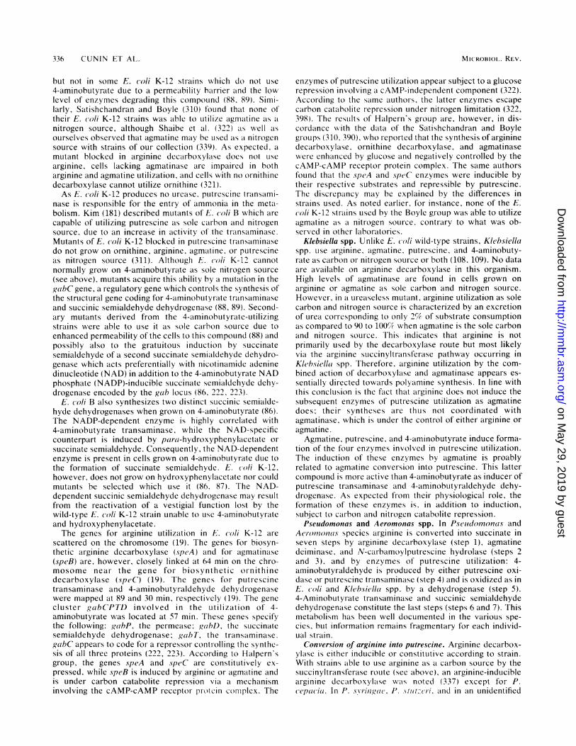
336 CUNIN ET AL.
but not in some E. coli K-12 strains which do not use
4-aminobutyrate due to a permeability barrier and the lowlevel of enzymes degrading this compound (88. 89). Simi-larly, Satishchandran and Boyle (310) found that none oftheir E. coli K-12 strains was able to utilize agmatine as a
nitrogen source, although Shaibe et al. (322) as well as
ourselves observed that agmatine may be used as a nitrogensource with strains of our collection (339). As expected. a
mutant blocked in arginine decarboxylase does not use
arginine, cells lacking agmatinase are impaired in botharginine and agmatine utilization, and cells with no ornithinedecarboxylase cannot utilize ornithine (321).As E. coli K-12 produces no urease, putrescine transami-
nase is responsible for the entry of ammonia in the meta-bolism. Kim (181) described mutants of E. coli B which are
capable of utilizing putrescine as sole carbon and nitrogensource, due to an increase in activity of the transaminase.Mutants of E. coli K-12 blocked in putrescine transaminasedo not grow on ornithine, arginine, agmaltine, or putrescineas nitrogen source (311). Although E. coli K-12 cannotnormally grow on 4-aminobutyrate as sole nitrogen source
(see above), mutants acquire this ability by a mutation in thegabC gene, a regulatory gene which controls the synthesis ofthe structural gene coding for 4-aminobutyrate transaminaseand succinic semialdehyde dehydrogenase (88, 89). Second-ary mutants derived from the 4-aminobutyrate-utilizingstrains were able to use it as sole carbon source due toenhanced permeability of the cells to this compound (88) andpossibly also to the gratuitous induction by succinatesemialdehyde of a second succinate semialdehyde dehydro-genase which acts preferentially with nicotinamide adeninedinucleotide (NAD) in addition to the 4-aminobutyrate NADphosphate (NADP)-inducible succinate semialdehyde dehy-drogenase encoded by the gab locus (86, 222, 223).
E. coli B also synthesizes two distinct succinic semialde-hyde dehydrogenases when grown on 4-aminobutyrate (86).The NADP-dependent enzyme is highly correlated with4-aminobutyrate transaminase, while the NAD-specificcounterpart is induced by para i-hydroxyphenylacetate or
succinate semialdehyde. Consequently, the NAD-dependentenzyme is present in cells grown on 4-aminobutyrate due tothe formation of succinate semialdehyde. E. co/i K-12,however, does not grow on hydroxyphenylacetate nor couldmutants be selected which use it (86. 87). Tfhe NAD-dependent succinic semialdehyde dehydrogenase may resultfrom the reactivation of a vestigial function lost by thewild-type E. co/i K-12 strain unable to use 4-aminobutyrateand hydroxyphenylacetate.The genes for arginine utilization in E. coli K-12 are
scattered on the chromosome (19). The genes for biosyn-thetic arginine decarboxylase (speA) and for agmatinase(speB) are, however, closely linked at 64 min on the chro-mosome near the gene for biosynthetic ornithinedecarboxylase (speC) (19). The genes for putrescinetransaminase and 4-aminobutyraldehyde dehydrogenasewere mapped at 89 and 30 min, respectively (19). The gene
cluster gaibCPTD involved in the utilization of 4-aminobutyrate was located at 57 min. These genes specifythe following: gabP, the permease; gabD, the succinate
semialdehyde dehydrogenase; gabT, the transaminase.gabC appears to code for a repressor controlling the synthe-sis of all three proteins (222, 223). According to Halpern'sgroup, the genes speA and speC are constitutively ex-
pressed, while speB is induced by arginine or agmatine andis under carbon catabolite repression via a mechanisminvolving the cAMP-cAMP receptor protein conmplex. The
enzymes of putrescine utilization appear subject to a glucoserepression involving a cAMP-independent component (322).According to the same authors, the latter enzymes escapecarbon catabolite repression under nitrogen limitation (322,398). The results of Halpern's group are, however, in dis-cordance with the data of the Satishchandran and Boylegroups (310, 390), who reported that the synthesis of argininedecarboxylase, ornithine decarboxylase, and agmatinasewere enhanced by glucose and negatively controlled by thecAMP-cAMP receptor protein complex. The same authorsfound that the speA and speC enzymes were inducible bytheir respective substrates and repressible by putrescine.The discrepancy may be explained by the differences instrains used. As noted earlier, for instance, none of the E.coli K-12 strains used by the Boyle group was able to utilizeagmatine as a nitrogen soul-ce, contrary to what was ob-served in other laboratories.
Klebsiella spp. Unlike E. coli wild-type strains, Klebsiellaspp. use arginine, agmatine, putrescine, and 4-aminobuty-rate as carbon or nitrogen source or both (108, 109). No dataare available on arginine decarboxylase in this organism.High levels of agmatinase are found in cells grown onarginine or agmatine as sole carbon and nitrogen source.However, in a ureaseless mutant, arginine utilization as solecarbon and nitrogen source is characterized by an excretionof urea corresponding to only 21% of substrate consumptionas compared to 90 to 100% when agmatine is the sole carbonand nitrogen source. This indicates that arginine is notprimarily used by the decarboxylase route but most likelyvia the arginine succinyltransferase pathway occurring inKlebsiella spp. Therefore, arginine utilization by the com-bined action of decarboxylase and agmatinase appears es-sentially directed towards polyamine synthesis. In line withthis conclusion is the fact that arginine does not induce thesubsequent enzymes of putrescine utilization as agmatinedoes, their syntheses are thus not coordinated withagmatinase. which is under the control of either arginine oragmatine.
Agmatine, putrescine, and 4-aminobutyrate induce forma-tion of the four enzymes involved in putrescine utilization.The induction of these enzymes by agmatine is proablyrelated to agmatine conversion into putrescine. This lattercompound is more active than 4-aminobutyrate as inducer ofputrescine transaminase and 4-aminobutyraldehyde dehy-drogenase. As expected from their physiological role, theformation of these enzymes is, in addition to induction,subject to carbon and nitrogen catabolite repression.Pseudomonas and Aeromnonas spp. In Pseudomonas and
Aelrotinoni(s species arginine is converted into succinate inseven steps by arginine decarboxylase (step 1), agmatinedeiminase, and N-carbamoylputrescine hydrolase (steps 2and 3), and by enzymes of putrescine utilization: 4-aminobutyraldehyde is produced by either putrescine oxi-dase or putrescine transaminase (step 4) and is oxidized as inE. coli and Klebsiella spp. by a dehydrogenase (step 5).4-Aminobutyrate transaminase and succinic semialdehydedehydrogenase constitute the last steps (steps 6 and 7). Thismetabolism has been well documented in the various spe-cies, but information remains fragmentary for each individ-ual strain.
Conversion of arginine into putrescine. Arginine decarbox-ylase is either inducible or constitutive according to strain.With strains able to use arginine as a carbon source by thesuccinyltransferase route (see above), an arginine-induciblearginine decarboxylase was noted (337) except for P.cepacia. In P. s.yringae, P. stItzcri. and in an unidentified
MIC ROBIOIL. REV.
on May 29, 2019 by guest
http://mm
br.asm.org/
Dow
nloaded from
ARGININE BIOSYNTHESIS AND METABOLISM IN BACTERIA
Pseidownonas species using arginine only as a nitrogensource (306, 337), arginine decarboxylase appears to beconstitutive; in these organisms, as well as in P. alcaligenes,the decarboxylase route is the only arginine catabolic path-way. In the unidentified Pselidomonas sp. isolated byRosenfeld and Roberts (306), the constitutive argininedecarboxylase is inhibited by its products agmatine,putrescine, and spermidine like the corresponding E. colienzyme. In contrast, in P. aerul,ginosa, arginine elicits induc-tion of the arginine decarboxylase; it is repressed byagmatine and its derivatives. This enzyme shows unusualproperties: it is inhibited by agmatine, but its activity isstimulated by putrescine, spermidine, and spermine. Thesesubstances were also found to control negatively theOTCase belonging to the ADI pathway (see correspondingsection). Polyamines may replace the Mg2' ion required forfull activity of the arginine decarboxylase. These moleculesstabilize the quaternary structure of the enzyme (whichdissociates upon dilution with loss of activity [A. Mercenier,Ph.D. thesis, University of Brussels, Brussels, Belgium,1981]). By controlling in opposite fashion the ADI andarginine decarboxylase pathways, polyamines may serve asa signal for routing the metabolic flow from arginine alongpathways whose physiological functions are different. For allPseiudoinonas spp. so far tested, agmatine or carbam-oylputrescine or both used either as sole carbon or nitrogensource elicit agmatine deiminase and N-carbamoylputres-cine hydrolase synthesis, but little or no induction by argi-nine is noted for these enzymes (216, 337). They are re-pressed by putrescine and its derivatives (216). Pseidoino-nas strains able to use arginine as a carbon source have thearginine succinyltransferase pathway (see above) whose lossby mutation (in P. aerluginoisa, for example) results in theability to grow on arginine. Thus the arginine decarboxylaseroute serves mainly to produce polyamines when arginine isabundant. Supporting this view is the observation that P.aeruiginosa mutants defective in agmatine deiminase orN-carbamoylputrescine hydrolase become unable to growon agmatine and carbamoylputrescine as carbon and nitro-gen sources but still retain their ability to grow on arginine assole carbon and nitrogen source (130).Enzymes of putrescine utilization. A transaminase, an oxi-
dase, or both are involved in the formation of 4-aminobutyraldehyde. Putrescine transaminase activity ispresent in all Pseludoinotnas species (224). Putrescinetransaminase and putrescine oxidase participate in putres-cine catabolism in P. fluorescens and P. (aerulginosa (224,296). In P. aeruoginosa and unidentified Pseiadornonas spe-cies, putrescine transaminase and 4-aminobutyratetransaminase activities may be supported by the same en-zyme molecule (44, 372, 393).The synthesis of 4-aminobutyraldehyde dehydrogenase is
induced under conditions of putrescine or pyrrolidine utili-zation and is repressed by the enzyme product 4-aminobutyrate (155, 225, 247). Mutants of P. aeriuginosadefective in this enzyme become impaired in agmatine,carbamoylputrescine, and putrescine utilization; in addition,they do not grow on 2-ketoarginine. Therefore, a singlealdehyde dehydrogenase seems to be involved in bothguanidinobutyraldehyde and 4-aminobutyraldehyde oxida-tion in this organism (Haas, personal communication).By contrast, distinct and separate aldehyde dehydrogen-
ases are present in P. puitida (Yorifugi, personal communi-cation): an arginine-inducible, specific guanidinobutyralde-hyde dehydrogenase and a putrescine-inducible 4-amino-butyraldehyde dehydrogenase showing a broad substrate
specificity. The latter enzyme oxidizes guanidinobutyralde-hyde, 5-aminovaleraldehyde, butyraldehyde, and propo-inaldehyde in addition to 4-aminobutyraldehyde. Althoughthese two dehydrogenases have the same subunit MW of57,000, they differ in their quaternary structure: theguanidinobutyraldehyde-specific enzyme is a dimer,whereas the other enzyme is a tetramer (Yorifugi, personalcommunication).The 4-aminobutyraldehyde dehydrogenase purified by
Tchen and associates also shows a broad specificity since itoxidizes also 3-aminopropionaldehyde and succinicsemialdehyde. It differs from the above-mentioned enzymesince it appears as a trimeric molecule (49) of subunit MW75,000.The next enzyme, 4-aminobutyrate transaminase, is in-
duced by 4-aminobutyrate or its metabolic precursors(guanidinobutyrate, agmatine, carbamoylputrescine, andputrescine). Induction by arginine occurs in P. putida mainlyvia intermediates of the oxidase pathway and not viaagmatine since P. aeruiginosa, which does not have theoxidase pathway, displays only weak induction. The broadspecificity of this enzyme allows it to participate inputrescine utilization (372, 383) and also in arginine biosyn-thesis: the uninduced level of this enzyme allows mutants ofP. aeriuginosa lacking acetylornithine aminotransferase togrow at a normal rate (372).
Certain Pseludoinonas species contain both a NAD- and aNADP-specific succinic semialdehyde dehydrogenase; inaddition, some of them have NAD-linked aminoaldehydedehydrogenases with variable substrate specificities whichare able to act on succinic semialdehyde (155, 238, 246, 254,255). As carbon and nitrogen source 4-aminobutyrate in-duces the synthesis of a NADP-dependent succinatesemialdehyde dehydrogenase in P. fluor-escens ATCC 13430(155) and P. piitida IRC 204 (Legrain and Stalon, unpub-lished data) and of a NAD-specific enzyme in the Pseudomo-nas sp. isolated by Michaels and Tchen (225). The latterenzyme is composed of three subunits of MW 55,000 (304),which, in vivo, hybridize with the 75,000-MW subunit ofeither 4-aminobutyraldehyde dehydrogenase or 3-amino-propionaldehyde dehydrogenase, which are also trimericproteins (49, 50). Immunological cross-reaction and hybrid-ization between the last two enzymes suggest a close evolu-tionary relationship, but succinate semialdehyde dehydroge-nase appears more distantly related since no immunologicalcross-reactions could be detected between this enzyme andthe two previous ones (50, 305).
Other Pathways of Arginine, Citrulline, and OrnithineUtilization
Clostridia, which are obligate anaerobes, are able to utilizecertain pairs of amino acids as energy sources by couplingthe oxidation of one of them to the reduction of another oneby the so-called Strickland reaction. In these organisms,ornithine may either be used as a hydrogen acceptor toproduce 5-aminovalerate or an electron donor or it may befermented as a single substrate.
Ornithine is produced from arginine by the ADI pathwayin C. boti(linium (227), C. peifringens (316), C. sporogenes(368), and other clostridia (331). The ability of Clostridiumspp. to use arginine has been quoted also by Mead (211),who classified the clostridia into four groups according toamino acid utilization and fermentation end product. Organ-isms of group I produce 5-aminovalerate in arginine fermen-tation, group II utilizes arginine without producing 5-
337VOL. 50, 1986
on May 29, 2019 by guest
http://mm
br.asm.org/
Dow
nloaded from
338 CUNIN ET AL.
H2N-(CH2)3- CH(-NH2)-COOH 4 D
ornitUineA +
CH3- CH(-NH2) -CH2-CH (-NH2)- COOH
2-4-aminopentanoate,
H
H2C COHH2C CH2prol ne
NAD(P)H+H-'I 5 +> NAD(P)
NAD(P)r 2 H2N - (CH2)4 - COOK\ ---; NAD(P)H+ H I-aminovaleraLe
CH5-CO-CH2- CH(-NH2)- COOH acetylCoA+aIanine2-amino 4-ketopentanoaLe C
CoASHFIG. 8. Ornithine utilization by Clostridilun spp. Steps: (1) D-ornithine-4,5-aminomutase, (2) 2,4-aminopentanoate dehydrogenase, (3)
2-amino-4-pentanoate CoA transferase, (4) ornithine cyclase, (5) D-proline reductase.
aminovalerate, group III is unable to utilize arginine, andgroup IV, consisting of one species, C. putrefaciens, utilizesit more slowly than groups I and II.
Ornithine oxidations is well documented. In C. sticklandii,ornithine oxidation starts with its isomerization by a
racemase (352) followed by the migration of the amino groupfrom C5 to C4, forming D-threo-2,4-aminopentanoate (93,328, 352). This is oxidized to produce 2-amino-4-ketopentanoate and ammonia (329) (Fig. 8).
In a last step, 2-amino-4-ketopentanoate undergoes a
thiolytic cleavage with CoA to form alanine and acetyl-CoA(157). The NADH produced during D-threo-2,4-aminopent-anoate oxidation reduces D-proline to yield 5-aminovalerate(320). Proline racemase and D-proline reductase of C.sticklandii have been characterized (320, 332).
In C. sticklandii and C. botulinuim the utilization ofornithine as a hydrogen acceptor involves the conversion ofL-ornithine to proline by an ornithine cyclase (65, 66). Thereaction proceeds by oxidation of ornithine to 2-keto-pentanoic acid with the reduction of enzyme-bound NADand release of ammonia. 2-Ketopentanoic acid undergoesring closure to form 5-pyrroline-2-carboxylate, and the pyr-rolidine ring is then reduced to form L-proline, using theenzyme-bound NADH as reactant (236). NAD prolinereductase and 5-pyrroline-2-carboxylate dehydrogenase ac-
tivities are found associated in C. sporogenes (332). Thephysiological role of these activities is thought to be theconversion of proline to glutamate (64).
Besides clostridia, ornithine cyclase has been found inaerobic A. tlimefaciens (85), P. cepacia, and P. putida(Legrain and Stalon, unpublished data). In both Agrohac-terium spp. and P. putida the enzyme is responsible for thedissimilation of the carbon skeleton since mutants of theseorganisms impaired in either ornithine cyclase or prolineutilization cannot use ornithine as a carbon source.
In other Pseiudomonas species, ornithine utilization was
thought at first to occur by transamination with 2-ketoglutarate to produce glutamic semialdehyde since a
transaminase activity is induced on ornithine-grown cells(63, 292, 371, 373). Besides, P. aeruginosa cells with a
reduction in this activity were impaired in ornithine utiliza-tion (292, 372, 373). However, it is now clear that thetransaminase in question is the succinylornithinetransaminase involved in the arginine succinyltransferasepathway. Therefore, in this organism ornithine utilizationfirst involves succinylation. Also, the arginine and ornithine
succinyltransferase activities seem to be supported by thesame protein molecule (Stalon, unpublished data).An ornithine-specific succinyltransferase was found
among some of the bacteria that possess the arginine suc-cinyltransferase pathway, namely, P. aeruginosa, P.aureofaciens, P. indigofera, P. doudoroffii, K. aerogenes,and Aeromonas formicans. A very low level of this enzymeis found in P. plutida, and no activity can be detected in P.cepacia. In these strains, however, ornithine catabolismoccurs via proline formation catalyzed by an ornithinecyclase.
Citrulline utilization as a carbon source is restricted torelatively few species: P. piutida, P. indigofera, and some P.cepacia strains (337). Mutants of P. putida impaired inornithine cyclase activity fail to grow on citrulline, indicatingthat citrulline catabolism involves ornithine as an intermedi-ate. Little is known of the conversion of citrulline intoornithine in these organisms. The catabolic OTCase belong-ing to the ADI pathway may fulfill this function in P. putida,the anabolic activity being repressed by citrulline and unableto catalyze in vitro the phosphorolysis of citrulline. In P.cepacia devoid of catabolic OTCase, the constitutive ana-bolic OTCase might catalyze the phosphorolysis of citrulline(Stalon, unpublished data). P. indigofera, when grown oncitrulline, induces the arginine succinyltransferase and theenzymes associated with this route. Citrulline utilization inthis bacterium, which lacks ornithine cyclase, may thusproceed via formation of arginine by the terminal step of thebiosynthetic pathway. However, another possibility re-mains: conversion into ornithine and its utilization via thesuccinyl derivatives. The isolation of mutants in the varioussegments of this metabolism could solve this problem.
Bacteria with Multiple Pathways
The possession of different ways of dissimilating particularorganic compounds as sources of carbon, nitrogen, or en-ergy may have had a survival value in fluctuating environ-mental conditions. As a matter of fact, multiple argininecatabolic pathways frequently appear in the same bacterium,especially among members of the genus Pseudomonas. P.pultida has four arginine catabolic pathways, while otherPseudomonas spp. of the fluorescent group and Aeromonasspp. have at least three ways of arginine dissimilation. Threeroutes have also been observed in cyanobacteria; at leasttwo occur in the same organism: arginase and ADI, as in
MICROBIOL. REV.
on May 29, 2019 by guest
http://mm
br.asm.org/
Dow
nloaded from
ARGININE BIOSYNTHESIS AND METABOLISM IN BACTERIA
some Bacillus strains. The roles of these divergent argininecatabolic pathways are largely independent. They are notused equally in all conditions and therefore there is littleredundancy between them: in Pselidornonas spp. of thefluorescent group the ADI pathway serves to generate ATPunder energy depletion conditions and the arginine suc-cinyltransferase route is responsible for the dissimilation ofthe carbon skeleton in aerobic or anaerobic respiration,whereas the arginine decarboxylase route mainly meetspolyamine requirements. An exception is P. plitida, in whichboth the arginine succinyltransferase and the arginine oxi-dase routes are involved in the aerobic dissimilation of thearginine carbon skeleton.Lack of redundancy also holds between the various cata-
bolic pathways on one side and arginine biosynthesis on theother. Indeed, in the case of intermediates being common toa degradative route and to biosynthesis, regulatory mecha-nisms have evolved which prevent the operation of wastefulrecycling by controlling OTCase activity (85, 150, 339; seeabove, arginine biosynthesis, OTCases, and CPSases). Inthe case of the succinyltransferase pathway, it is the succinyl"label" which plays that role.
EVOLUTIONARY CONSIDERATIONS
Biological Significance of Gene OrganizationAre there constraints which define the map position of
genes or is the topography we observe today largely, if notexclusively, the result of a series of historical accidents?Some constraints are likely to operate: e.g., the clusteringnear the E. coli replication origin of most of the genesrequired for macromolecular synthesis has been noted (20).In line with this observation, a correlation was observed(327) between the relative abundance of an amino acid in cellproteins and the position (distance from the origin) of thegene whose product was assumed to be rate limiting in thesystem (in this case the argA gene coding for the feedback-inhibited acetylglutamate synthetase). To strengthen thecase, similar studies should be undertaken with a largenumber of bacteria.
In the particular instance of a scattered regulon such as inE. coli, it has been repeatedly emphasized that this type oftopography (rather than the exact map position) allows finertuning of gene control than clustering into a single operon.This consideration is of course much less relevant for thosebacteria (such as P. piitida and P. aeru-iginosa) in which thereis little control of arginine biosynthetic genes.There is indeed a marked lack of coordination between the
individual repression responses of E. (oli arg genes. Themolecular basis of this pattern is becoming clear (see "Struc-ture of control regions and the repression response"), butwhether these widely different repression responses have aphysiological significance remains a pending question. Itcould be advantageous or even necessary to the cell to adjustindividually the rate of synthesis of enzymes working atmetabolic branch points (such as the unique CPSase ofEnterobacteriaceae and OTCase) or involved in both catab-olism and anabolism (such as the succinylornithine amino-transferase of Pseiudomonas spp.) or yet at steps usingsubstrates available in limiting concentrations; there is,however, not enough quantitative evidence regarding meta-bolic fluxes on which to base a discussion of this topic.Besides, it is clear that, even within the E. c(li species itself,different strains may display very different repression pat-terns: in E. coli B, which is weakly inducible by excess
arginine but displays moderate derepression at low arginineconcentrations, the intrinsically low level of enzyme makescontrol of the flux of arginine precursors by feedback inhi-bition no less efficient than in strain K-12 (99).The naive idea that an exclusive genetic pattern (not in
terms of exact topography but in terms of grouping andresulting coordination) could be the preferred one for mod-ulation of gene expression in the arginine regulon is alreadyundermined by the variations encountered among the En-terobacteriaceae, in which argG is sometimes found clus-tered with argE, -C, -B, and -H. It becomes frankly incon-sistent with the pattern found in B. sulbtilis; there, eightarginine biosynthetic genes, which are modulated over awide range as in E. (oli, are clustered in a 12-kilobasefragment. The evidence at hand (235) is not sufficient toestimate their degree of coordination, but at least the genesencoding OTCase (argF) and the arginine-specific CPSaseare part of the same operon. argG and argH map outside thecluster but the data suggest that argH is coordinated withargF (135).
If one compares B. siubtilis with E. coli it makes sense, ofcourse, that the genes for the arginine-specific CPSase of theformer are part of the cluster, whereas the unique andcumulatively repressed CPSase of E. coli is encoded by aseparate locus.The occurrence of divergent transcription from converg-
ing promoters such as in (IrgECBH and bio also raises thequestion of the biological significance of this pattern. Does itreflect a physiological advantage, a type of rearrangementmechanism repeatedly used in evolution, or both? Oneobvious physiological advantage is that, from an internalcontrol region, the whole operon is expressed more quicklythan from one end. But then, why is the long his operon notorganized in this way? On the other hand, rearrangementscreating divergent operons (inverted repeats of argE) havebeen observed (58, 60). One could envision inverted dupli-cations of whole operons, followed by selective inactivationand loss of duplicated genes on either side of the controlregion. Duplicative inversions caused by IS elements mayalso have been instrumental in the formation of divergentoperons.
Deciding on the face of specific activities and sequencedata whether different genetic arrangements and coordina-tion patterns must be regarded as equally successful solu-tions arrived at under stringent selection or as merelytolerable ones will probably never be possible. This is, ofcourse, a central question in many evolutionary debates.However, for microbes the tools are now available todeliberately engineer various types of arrangements and testtheir adequacy for survival over a large number of genera-tions.
Lack of Attenuation ControlFrom the evidence discussed in the subsection, "Struc-
ture of control regions and repression response," in E. coli,it appears that a classical type of attenuation control is notused in the ar-g regulon of this organism. Since argA andargS are not yet sequenced, this statement remains provi-sional but will be taken as such in the following discussion.There are basically three ways of understanding a lack ofattenuation control. (i) An attenuation mechanism based onarginyl-tRNA could be inefficient (discussed in reference75), which is indeed not contradicted by the weak attenua-tion effect displayed by the atrg codon present in the leadersequence of the trp operon (403). This view has the advan-tage of lending itself to experiments; it also provides an
VOL. 50, 1986 339
on May 29, 2019 by guest
http://mm
br.asm.org/
Dow
nloaded from
340 CUNIN ET AL.
incentive for the screening of other bacteria. (ii) Attenuationis absent in E. coli because, in this particular species (butmaybe not in other ones) the arginine regulon never used thismechanism. (iii) It is absent today because it was lost, thisloss having occurred at each locus individually or resulted(as in E. coli?) from scattering of a hypothetical ancestralarginine operon by chromosome rearrangements (see nextsection). The questions raised in (ii) and (iii) may, of course,never be answered; on the other hand, it is clear that theanalysis of the arginine regulon in other bacteria will behighly relevant in this respect. Preliminary sequence data(Baumberg, unpublished data) suggest that attenuation is notused for the control of at least some arg genes in B. slubtilis.
Chromosomal Rearrangements Involving Arginine Genes
Studies on rearrangements affecting the expression ofarginine genes have led to original observations of generalinterest from the evolutionary point of view. Tandem dupli-cations of argE, argH, or argB and -H were shown to occurin recA as well as in Rec+ strains, calling attention to thepossible role of illegitimate recombination between the twoarms of a replication fork in this important type of evolu-tionary event (31). Subsequent work established that in Rec'strains, short tandem duplications (about 2 kilobases long)are, on a proportional basis, much more stable than expectedfrom the rate at which longer duplications are lost (58).Duplications consisting of inverted repeats of argE arrangedin divergent operons were also shown to occur spontane-ously, forming single (and stable) or multiple (and unstable)palindromes (58, 60). High-copy-number amplification ofargF was described recently (160); the occurrence of thisrearrangement depends on the presence of an F factorintegrated near the argF gene (161). The mechanism in-volved remains to be established: it appears to involve an"activation" of the ISI repeats flanking argF, followed by arecA-dependent amplification of the IS1-bordered fragment.
IS2 elements integrated in the control region of a gene canturn it on constitutively (307). In the cases investigated up tonow this "turning on" effect is due either to the associationof a polymerase recognition site at extremity II of theelement with a Pribnow box on the other side of theIS-chromosome junction (35, 116, 138, 156) or to DNArearrangements creating a promoter near extremity I (113).In contrast, IS3 was shown to carry a complete outwardpromoter able to reactivate a silent argE gene (59). A recentdiscussion of this topic may be found in reference 78.
Cryptic argM Gene of E. coli
The occurrence of cryptic genes in bacteria and the roletheir periodic activation may play in the evolution of bacte-rial populations have been reviewed recently (132). E. coliargM is a gene for an inducible but normally silentacetylornithine transaminase; it can be reactivated underselective pressure in argD strains (see "Enzymatic Stepsand Regulation of Metabolic Flow"). The nature of thisreactivation is presently under study.That the argM protein is inducible by arginine, like the
catabolic succinylornithine aminotransferase of K. aero-genes, and is similar to it in structure (107) suggests thatargM is a cryptic element of a degradative pathway that issilent and possibly incomplete in current E. coli isolates.
Active argM has, however, been selected for under condi-tions which did not imply a catabolic function but rather thetransamination of glutamate with acetylglutamate semial-dehyde to produce acetylornithine and 2-ketoglutarate. Asthe inducible succinylornithine aminotransferase from Pseu-dornonas, Aeromonas, or Klebsiella spp. (335), the argMproduct transaminates succinylornithine, displaying an evenhigher affinity for this substrate than for acetylornithine orornithine (Stalon, unpublished observations). In the formerorganisms, the arginine-inducible succinylornithinetransaminase is involved in the arginine succinyltransferasecatabolic pathway (see above). It would therefore be inter-esting to see whether in E. coli successive mutations couldnot revive the very same pathway in its entirety. Since theargM protein is also able to transaminate ornithine toproduce the semialdehyde of glutamate, another type ofinvolvement in catabolism appears theoretically possible:argM might be part of a cryptic pathway leading fromarginine to glutamate via ornithine. Clearly, much could belearned about metabolic evolution from further studies onthis question.
Origin of CPSases
The properties of the microbial CPSases are typical ofthose of glutamine amidotransferases, a dozen enzymesdisplaying similar features (for reviews, see Buchanan [46],Meister [213], and a symposium on enzymes of glutaminemetabolism [2911). (i) All can utilize ammonia as nitrogendonor in place of glutamine. (ii) All have separate bindingsites for these two substrates, often located on distinctsubunits. (iii) All have a glutamine binding site which con-tains a cysteine residue, never a serine, for example. (iv) Allexhibit a glutaminase activity which is located on the samesubunit as the glutamine binding site. Therefore, a commonmechanism has been proposed to account for the use ofglutamine by these various enzymes (197, 133, 350). Itinvolves the covalent binding of glutamine to the cysteineresidue in the glutamine binding site, the release ofNH3 fromthe amide group of glutamine, and its transfer, probablythrough a nonaqueous pocket, to an NH3-binding site oftenlocated on a separate subunit where it is used for anamination reaction.
In view of these general properties of amidotransferases, ithas been considered that these enzymes evolved by thecombination of a common ancestral glutaminase with dis-tinct ammonia-dependent aminases (197, 237, 290, 348).Evolution by duplication and mutation, and in some cases bygene fusion, of the gene for that ancestral glutaminase wouldthus be responsible for the various contemporary glutamin-ase genes. Substantial support for this evolutionary pathwaywas provided recently by the comparison of the primarysequences of the small subunits of E. coli and yeast CPSaseto the glutaminase domains of other amidotransferases (102,251, 277, 387). Both CPSases do indeed share significanthomologies with the glutaminase components of anthranilatesynthase of E. coli (245) and other sources (see references inreference 387) and of p-aminobenzoate synthase (167). Threehighly conserved regions may be distinguished in theseproteins and were recently found to be present also in thesmall component of E. coli guanosine monophosphate syn-thetase (399). The most highly conserved of these regionscontains an invariant cysteine residue, which in the case ofanthranilate synthase (168, 257) has been identified as theactive site residue involved in glutamine binding. Interest-ingly, all three regions of homology are rich in hydrophobic
MICROBIOL. REV.
on May 29, 2019 by guest
http://mm
br.asm.org/
Dow
nloaded from
ARGININE BIOSYNTHESIS AND METABOLISM IN BACTERIA
residues and might consequently contribute to the formationof the nonaqueous pocket necessary to protect NH3 fromprotonation during its transfer to the NH3-binding site.These various data strongly argue that the glutaminasesubunits of CPSase, anthranilate synthase, p-aminobenzoatesynthase, and guanosine monophosphate synthase were
derived from a common ancestral glutaminase.The elucidation of the primary sequence of both subunits
of microbial CPSase has allowed us to address to theproblem of the origin of CPSase I, the ammonia-dependentCPSase of mammalian liver. The idea that this acetylglutam-ate-requiring synthetase originated from the subsequent ev-
olution of a glutamine-dependent enzyme was initially sug-gested by Trotta et al. (349, 350), who based their argumenton MW comparisons. An elegant attempt to reconstruct theevents involved in the emergence of the ornithine cycle fromthe enzymes of procaryotic metabolism of arginine has beenpresented by Paulus (261). This author has in particularanalyzed the steps by which the actual ammonia-dependentCPSase may have evolved by fusion of the genes of the twosubunits of an ancestral glutamine-dependent CPSase.The complete sequence of rat CPSase I, as deduced
recently from the sequence of the cognate mRNA, showshomology with the E. coli enzymes which encompasses theentire sequence of both the small and large subunits (248,249). This confirms that the mitochondrial enzyme is indeeda hybrid polypeptide encoded by a gene resulting from thefusion of the two genes of the microbial enzymes. Theglutamine domain of the microbial enzymes is still recogniz-able in the sequence of rat carbamoylphosphate synthetase,but the cysteine which is considered as participating in thebinding of glutamine is replaced by a serine. Whether in thecourse of evolution this domain has retained a function in thebinding of N-acetylglutamate, the effector of the mammalianenzyme, is unknown. However, this appears unlikely sincecarbamoylphosphate III of the teleost fish Micropteriussalmoides, a synthetase which uses glutamine but requiresacetylglutamate for activity, binds glutamine and N-acetylglutamate at separate but interacting sites (53). It willbe of special interest to compare the amino acid sequence ofthis teleost enzyme with those of the microbial and mamma-
lian liver CPSases.
Genetic Control of Carbamoylation: Origin ofCarbamoyltransferases
A present, the most instructive evidence derived fromstudies of the arg genetic system and bearing on metabolicevolution concerns the genetic control of carbamoylation inE. coli, Pseudomonas spp., and a few other bacteria.From the complete sequence of F and I E. coli OTCases
(see reference 366) and of the catalytic subunit of ATCase(142), it is clear that the early suggestion based on structuralconsiderations (189) and later supported by heteroduplexanalysis (179) and partial sequencing data (115, 276) was
correct: the two carbamoyltransferases are evolutionarilyrelated. With an overall homology of 35 to 40%, concen-
trated in similarly structured domains (144, 366), the twoenzymes provide a particularly eloquent case of enzymedifferentiation following the duplication of an ancestralcarbamoylase gene. The duplication may have occurred in
tandem as suggested by the strong linkage existing betweenargI and pyrB in E. coli (152). The emergence of these twometabolic functions was probably a primordial event in
metabolic evolution. If arginine was a "late intruder" in the
genetic code, having replaced ornithine in the course ofevolution (163), the OTCase gene may have been recruitedfrom a gene encoding a substrate-ambiguous transferaseprimarily involved in the carbamoylation of aspartate.The presence between two IS] elements of a duplicate
OTCase gene (argF) in E. coli K-12 (145, 396) opens thepossibility that this gene was translocated from a relatedspecies into E. coli K-12. This idea is supported by the factthat argF and argIexhibit greater amino acid than nucleotidehomology, whereas, in general, the reverse obtains forduplicated genes in the same genome (300). Comparinghomologous enzymes among Enterobacteriaceae rendersthe actual homology between argF and argI also consistentwith this idea (366). However, these data alone cannotexclude the possibility that argF originated from argl byIS-promoted duplication in an ancestral E. coli cell, withpossible interstrain transfer having taken place later on.
In Pseudomonas spp. possessing the ADI pathway, thebiosynthesis and catabolism of citrulline are catalyzed bydifferent proteins extremely specialized in their functionsince the anabolic OTCase is unable to catalyze thephosphorolysis of citrulline (due to the formation of an
abortive citrulline-enzyme complex) (336), while the cata-bolic OTCase does not carbamoylate ornithine at phyisologi-cal concentrations of carbamoylphosphate (334, 339). Arethese enzymes nevertheless related?A study of the immunological relatedness between several
catabolic OTCases from G- and G+ bacteria and some oftheir anabolic counterparts brings to light a surprising affinitybetween these catabolic enzymes and the argFOTCase of E.coli; in contrast, the same catabolic enzymes display littlecross-reaction with anabolic OTCases from organisms otherthan Enterobacteriaceae (100). Limited sequence data are inkeeping with this pattern: N-terminal amino acids from argFOTCase display, respectively, 45 and 28% homology withthe corresponding residues of catabolic OTCases from Aero-monasformicans and P. piutida. It is therefore possible thatthe argF enzyme is ancestrally related to some catabolicOTCase.
Despite their lack of immunological cross-reactivity, ana-
bolic OTCases are probably related to each other also.Indeed, though the anabolic OTCases of E. coli and both B.subtilis and B. licheniformis share no antigenic determinants(100), a comparison between the respective amino acidsequences from B. sufbtilis (153 residues known) and E. colidiscloses an overall homology of 32% (Baumberg, personalcommunication; R. Switzer, personal communication). Weassume that most of the homologous residues are buried inthe molecule.
Considering all these results together, it appears that bothanabolic and catabolic OTCases on the one hand and thecatalytic subunit of ATCase on the other share a common
ancestral gene; this is also reflected in the sequence of yeastand human OTCases (Huygen, unpublished experiments,this laboratory; 143). The putrescine carbamoyltransferasefunctioning in the agmatine deiminase route of Streptococ-cus faecalis probably belongs to the same family since theN-terminal amino acid sequences of this enzyme and ofcatabolic OTCase from the same organism display 30 to 35%homology (P. Falmagne and V. Stalon, unpublished data).The agmatine deiminase route may represent an interestingcase of evolution by pathway recruitment. The lack ofimmunological cross-reactivity between catabolic and ana-
bolic OTCases in one and the same organism may at least inpart be related to the necessity of not forming coaggregateswhich might be inactive.
341VOL. 50, 1986
on May 29, 2019 by guest
http://mm
br.asm.org/
Dow
nloaded from
342 CUNIN ET AL.
Acquisition of the Ability to Use Anabolic or CatabolicOTCases in the Reverse Direction of Their Normal
Operation
In the previous section we mentioned that the biosyntheticOTCases of P. plitida and P. elrFliginlosa are kineticallyunable to work in the reverse direction in vitro, while thecatabolic enzyme cannot synthesize citrulline in vivo be-cause of its extreme cooperativity towards carbam-oylphosphate: mutants lacking the anabolic activity requirecitrulline or arginine for growth (124, 294). It is possible,however, to select for revertants in which the anabolicactivity is still absent but the catabolic OTCase now fulfillsthe anabolic function. These revertants possess an alteredcatabolic OTCase with reduced or no cooperativity towardscarbamoylphosphate (124, 339). The altered enzyme is ki-netically very similar to E. coli OTCase. It is thus notunreasonable to see in the acquisition of a modified OTCasea reversal of the selection process that produced allostericproteins with a quaternary structural design apt to generateextreme cooperativity. The catabolic OTCase of Aeromonasfirrnicans, which is closely related antigenically to E. coliOTCase and to the catabolic enzyme of Pseudomonas spp.,appears as an intermediate in this process. Indeed, thisenzyme may occur as an active trimeric form like E. coliOTCase, but also as an active nonameric species (193). In P.aeruginiosai the latter form is the only one to be active(Stalon, unpublished data). Therefore, in keeping with thedata discussed in the preceding section, it appears that anancestral OTCase gene may have duplicated itself and,under natural selection, produced the two metabolicallyspecialized proteins we observe in today Pselidornonias spp.
Conversely, it is possible to reverse the metabolic role ofan anabolic OTCase in vivo so as to use it for the synthesisof carbamoylphosphate. The enzyme itself is not modified,however, but its rate of synthesis is increased. For E. (oliK-12, a mutant lacking CPSase requires both arginine anduracil for growth. Cells of this mutant grown in a mediumcontaining citrulline can synthesize arginine by the finalsteps of arginine biosynthesis, but the only possibility theyhave to produce carbamoylphosphate for the synthesis ofpyrimidines is phosphorolysis of citrulline. The wild typegrows very slowly under these conditions, but faster-growing mutants display elevated levels of OTCase (192);these mutants express either argF or argl constitutively (asthe result of operator mutations or gene amplification [160,192]) or have a partial defect in argininosuccinate synthetasewhich allows citrulline to accumulate (192).These metabolic reconversions illustrate the two main
strategies of acquisitive metabolic evolution: direct alter-ations of the properties of an enzyme (in this case itsallosteric properties) or regulatory mutations making it avail-able in high amounts (62, 232).
Evolutionary Significance and Origin of Multiple CatabolicPathways in Pseudomonas Species
The capacity of Pseldoomonas spp. to synthesize the ADIpathway under oxygen limitation even in the absence ofarginine may reflect the remnants of an ancient pathwayhaving lost inductive control by arginine. It is obvious that inthe course of evolution, microorganisms were faced with theproblem of generating energy under anaerobic conditions.Possession of the ADI pathway may have been important inthis respect, as attested to by its distribution among twomajor bacterial kingdoms, the archachacteria (halobcacteria)
and five of the eight groups of the eubacterial line as definedby Fox et al. (106). Among these organisms the strictlyaerobic P. piitidai, P. fluores(cens, and P. inendocina appearas exceptions since they are able to ferment arginine butcannot use it for anaerobic growth. The ADI pathway mayhave a single evolutionary origin (at least in eubacteria) sincethe catabolic ornithine carbamoyltransferases of the variousgroups of organisms investigated display common antigenicdeterminants (see above). Such cross-reactions indicate astructural uniformity and suggest that there has been ratherlittle evolution during the history of these enzymes. If weaccept that the scheme of evolution began with the processof fermentation and progressed through a series of stepsleading to the development of the contemporary aerobicorganisms, the aerobic Pseludomonias spp. may then repre-sent a form that lost its fermentative metabolism during thecourse of evolution.The participation of molecular oxygen is required for the
reaction catalyzed by arginine oxidase in P. plitida. Thisactivity is restricted to very few organisms and occurs in P.pltida together with other arginine catabolic pathways. Yet,the mutational loss of this activity does not prevent theutilization of arginine as carbon source due to the presenceof the succinyltransferase pathway. It is difficult to decidewhether this enzyme is a recent acquisition or an ancientactivity lost by the other Pseiudoinoncas spp. during thecourse of evolution. However, the occurrence of this activ-ity in the cyanobacterial group suggests that it arose soonafter the appearance of oxygen on earth.Does the possession of different pathways indicate a lack
of evolutionary affinities'? A comparison of P. plutida and P.aeruiginosa suggests that such an assumption is not valid.Indeed, both bacteria are physiologically related and bearstrong homologies at the level of 16S ribosomal RNA. Bothorganisms use the arginine succinyltransferase, ADI, andarginine decarboxylase pathways. However, P. aeruiginosauses arginine as an energy source for anaerobic growth butP. pittidai does not; arginine oxidase is present in P. piitidabut is absent in P. aerliginosa.
ACKNOWLEDGMENTS
We thank all of our colleagues who provided unpublished infor-mation during the preparation of this review: S. Baumberg, E.Canale-Parola, R. Celis, J. Charlier, T. Eckhardt, D. Haas, V. W.Pendey, P. Rogers. R. L. Switzer, S. Udaka, M. G. Williams, and T.Yorifugi. The dedication of P. Stalon in typing and correcting themanuscript is gratefully acknowledged. Thanks are due to R. Claesand J. P. ten Have for preparing the figures.Work done in our laboratory has been supported over the years by
several Belgian foundations for scientific research (FNRS-NFWO,FRFC-FKFO, IRSIA-IWONL) and by a concerted action betweenthe Belgian state and two Brussels universities (Universite Libre deBruxelles and Vrije Universiteit Brussel). V.S. is a ChercheurQualifi6 of the F.N.R.S.
LITERATURE CITED1. Abdelal, A., and J. L. Ingraham. 1969. Control of carbamyl
phosphate synthesis in SalmtIonella tYphimuriumi. J. Biol.Chem. 244:4033-4038.
2. Abdelal, A., and J. L. Ingraham. 1969. Cold sensitivity andother phenotypes resulting from mutations in pyrA gene. J.Biol. Chem. 244:4039-4045.
3. Abd-el-al, A., D. P. Kessler, and J. L. Ingraham. 1969. Argi-nine-auxotrophic phenotype resulting from a mutation in thePvrA gene of Eschericlija (oli B/c. J. Bacteriol. 97:466-468.
4 Abdelal, A. T. 1979. Arginine catabolism by microorganisms.Annu. Rev. Microbiol. 33:139-168.
MICROBIOL. REV.
on May 29, 2019 by guest
http://mm
br.asm.org/
Dow
nloaded from
ARGININE BIOSYNTHESIS AND METABOLISM IN BACTERIA
5. Abdelal, A. T., and 0. Nainan. 1979. Regulation of N-acetyl-glutamate synthesis in Salmonella typhimurium. J. Bacteriol.137:1040-1042.
6. Abdelal, A. T., W. F. Bibb, and 0. Nainan. 1982. Carbamatekinase from Pseudomonas aeruginosa: purification, character-ization, physiological role, and regulation. J. Bacteriol.151:1411-1419.
7. Abdelal, A. T., L. Bussey, and L. Vickers. 1983. Carbam-oylphosphate synthetase from Pseudomonas aeruginosa:subunit composition, kinetic analysis and regulation. Eur. J.Biochem. 129:697-702.
8. Abdelal, A. T. H., and J. L. Ingraham. 1975. Carbamylphosph-ate synthetase from Salmonella typhimurium. Regulation,subunit composition and function of the subunits. J. Biol.Chem. 250:4410-4417.
9. Abdelal, A. T. H., E. Griego, and J. L. Ingraham. 1976.Arginine-sensitive phenotype of mutations in pyrA of Salmo-nella typhimurium: role of ornithine carbamyltransferase in theassembly of mutant carbamylphosphate synthetase. J. Bacte-riol. 128:105-113.
10. Abdelal, A. T. H., E. Griego, and J. L. Ingraham. 1978.Arginine auxotrophic phenotype of mutations in pyrA of Sal-monella typhimurium: role of N-acetylornithine in the matura-tion of mutant carbamoylphosphate synthetase. J. Bacteriol.134:528-536.
11. Abdelal, A. T. H., E. H. Kennedy, and 0. Nainan. 1977.Ornithine transcarbamylase from Salmonella typhimurium:purification, subunit composition, kinetic analysis, and immu-nological cross-reactivity. J. Bacteriol. 129:1387-1396.
12. Albrecht, A. A., and H. J. Vogel. 1964. Acetylornithine-b-transaminase. Partial purification and repression behavior. J.Biol. Chem. 239:1872-1876.
13. Anderson, P. M. 1977. Evidence that the catalytic and regula-tory functions of carbamoylphosphate synthetase from Esche-richia coli are not dependent on oligomer formation. Biochem-istry 16:583-586.
14. Anderson, P. M. 1977. Binding of allosteric effectors tocarbamoylphosphate synthetase of Escherichia coli. Biochem-istry 16:587-592.
15. Anderson, P. M., and S. V. Marvin. 1970. Effect of allostericeffectors and adenosine triphosphate on the aggregation andrate of inhibition by N-ethyl-maleimide of carbamoylphosphatesynthetase of Escherichia coli. Biochemistry 9:171-178.
16. Anderson, P. M., and A. Meister. 1965. Evidence for anactivated form of carbon dioxide in the reaction catalyzed byEscherichia coli carbamoylphosphate synthetase. Biochemis-try 4:2803-2809.
17. Anderson, P. M., and A. Meister. 1966. Bicarbonate-dependentcleavage of adenosine triphosphate and other reactionscatalyzed by Escherichia coli carbamoylphosphate synthetase.Biochemistry 5:3157-3163.
18. Anderson, P. M., and A. Meister. 1966. Control of Escherichiacoli carbamoylphosphate synthetase by purine and pyrimidinenucleotides. Biochemistry 5:3164-3167.
19. Bachmann, B. 1983. Linkage map of Escherichia coli K-12,edition 7. Microbiol. Rev. 47:180-230.
20. Bachmann, B. J., K. Brooks Low, and A. L. Tampler. 1976.Recalibrated linkage map of Escherichia coli K-12. Bacteriol.Rev. 30:116-167.
21. Bachrach, V., S. Persky, and S. Razin. 1969. Metabolism ofamines. 2. The oxidation of natural polyamines by Mycobac-terium smegmatis. Biochem. J. 76:306-310.
22. Bacon, D. F., and H. J. Vogel. 1963. A regulatory genesimultaneously involved in repression and induction. ColdSpring Harbor Symp. Quant. Biol. 28:437-438.
23. Baich, A., and H. J. Vogel. 1962. N-acetyl-,y-glutamokinase andN-acetylglutamic-y-semialdehyde dehydrogenase: repressibleenzymes of arginine synthesis in Escherichia coli. Biochem.Biophys. Res. Commun. 7:491-496.
24. Barbour, M. G., and R. C. Bayly. 1977. Regulation of themetacleavage of 4-hydroxyphenylacetate by Psei(domonasputida. Biochem. Biophys. Res. Commun. 76:565-571.
25. Barile, M. F., R. T. Schimke, and D. B. Riggs. 1966. Presence
of the arginine dihydrolase pathway in Mycoplasma. J. Bacte-riol. 91:189-192.
26. Baumberg, S. 1970. Acetylhistidine as substrate for acetyl-ornithinase: a new system for the selection of arginine regula-tion mutants in Escherichia coli. Mol. Gen. Genet. 106:162-173.
27. Baumberg, S. 1976. Genetic control of arginine metabolism inprokaryotes, p. 369-388. In K. D. McDonald (ed.), SecondSymposium on the Genetics of Industrial Microorganisms.Academic Press, Inc., New York.
28. Baumberg, S., and C. R. Harwood. 1979. Carbon and nitrogenrepression of arginine catabolic enzymes in Bacillus subtilis. J.Bacteriol. 137:189-196.
29. Baumberg, S., and A. Mountain. 1984. Bacillus subtilis 168mutants resistant to arginine hydroxamate in the presence ofornithine or citrulline. J. Gen. Microbiol. 130:1247-1252.
30. Beacham, I. R., B. W. Schweitzer, H. M. Warrick, and J.Carbon. 1984. The nucleotide sequence of the yeast ARG4gene. Gene 29:271-279.
31. Beeftink, F., R. Cunin, and N. Glansdorff. 1974. Arginine geneduplication in recombination proficient and deficient strains ofEscherichia coi K12. Mol. Gen. Genet. 132:244-253.
32. Bencini, D. A., J. E. Houghton, T. A. Hoover, K. F. Fol-termann, J. R. Wild, and G. A. O'Donovan. 1983. The DNAsequence of argI from Escherichia coli K12. Nucleic AcidsRes. 11:8509-8518.
33. Beny, G., A. Boyen, D. Charlier, W. Lissens, A. Feller, and N.Glansdorff. 1982. Promoter mapping and selection of operatormutants by using insertion of bacteriophage Mu in theargECBH divergent operon of Escherichia coli K-12. J. Bac-teriol. 151:62-67.
34. Beny, G., R. Cunin, N. Glansdorff, A. Boyen, J. Charlier, andN. Kelker. 1982. Transcription of regions within the divergentargECBH operon of Escherichia coli: evidence for lack of anattenuation mechanism. J. Bacteriol. 151:58-61.
35. Besemer, J., I. Gortz, and D. Charlier. 1980. Deletions andDNA rearrangements within the transposable element IS2: amodel for the creation of palindromic DNA by DNA repairsynthesis. Nucleic Acids Res. 8:5825-5833.
36. Billheimer, J. T., H. N. Carnevale, T. Leisinger, T. Eckhardt,and E. E. Jones. 1976. Ornithine-5-transaminase activity inEscherichia coli: its identity with acetylornithine-B-trans-aminase. J. Bacteriol. 127:1315-1323.
37. Billheimer, J. T., N. Y. Chen, H. N. Carnevale, H. R. Horton,and E. E. Jones. 1979. Isolation and characterization ofacetylornithine-8-transaminase of wild-type Escherichia coliW. Comparison with arginine-induced acetylornithine-6-transaminase. Arch. Biochem. Biophys. 105:401-413.
38. Blackmore, R. P., and E. Canale-Parola. 1976. Arginine catab-olism by Treponema denticola. J. Bacteriol. 128:616-622.
39. Boettcher, B. R., and A. Meister. 1980. Covalent modificationof the active site of carbamoylphosphate synthetase by 5'-fluorosulfonylbenzoyladenosine. Direct evidence for two func-tionally different ATP binding sites. J. Biol. Chem. 255:7129-7133.
40. Bouvier, J., J. C. Patte, and P. Stragier. 1984. Multipleregulatory signals in the control region of the Escherichia colicarAB operon. Proc. Natl. Acad. Sci. USA 81:4139-4143.
41. Boyen, A., J. Piette, R. Cunin, and N. Glansdorff. 1982.Enhancement of translation efficiency in Escherichia coli bymutations in a proximal domain of messenger RNA. J. Mol.Biol. 162:715-720.
42. Brenchley, J. E., and L. S. Williams. 1975. Transfer RNAinvolvement in the regulation of enzyme synthesis. Annu. Rev.Microbiol. 29:251-274.
43. Bretscher, A. P., and S. Baumberg. 1976. Divergent transcrip-tion of the argECBH cluster of Escherichia coli K12. Muta-tions which alter the control of enzyme synthesis. J. Mol. Biol.102:205-220.
44. Brohn, F., and T. T. Tchen. 1971. A single transaminase for 1-4diaminobutane and 4 aminobutyrate in a Pseudomonas spe-cies. Biochem. Biophys. Res. Commun. 45:573-582.
45. Broman, K., N. Lauwers, V. Stalon, and J. M. Wiame. 1978.
VOL. 50, 1986 343
on May 29, 2019 by guest
http://mm
br.asm.org/
Dow
nloaded from
344 CUNIN ET AL.
Oxygen and nitrate in utilization by Bacillus licheniformnis ofthe arginase and arginine deiminase routes of arginine catabo-lism and other factors affecting their synthesis. J. Bacteriol.134:920-927.
46. Buchanan, J. M. 1973. The amidotransferases. Adv. Enzymol.39:1-90.
47. Bussey, L. B., and J. L. Ingraham. 1982. A regulatory gene(u.se) affecting the expression of pytrA and certain other pyrim-idine genes. J. Bacteriol. 151:144-152.
48. Callewaert, D. M., M. S. Rosemblatt, K. Suzuki, and T. T.Tchen. 1973. Succinic semialdehyde dehydrogenase from aPseludotnonas species. I. Purification and chemical properties.J. Biol. Chem. 248:6009-6013.
49. Callewaert, D. M., M. S. Rosemblatt, and T. T. Tchen. 1974.Purification and properties of 4 aminobutanal dehydrogenasefrom a Pseudo,nonas species. J. Biol. Chem. 249:1737-1741.
50. Callewaert, D. M., M. S. Rosemblatt, and T. T. Tchen. 1974.Purification and properties of 3 aminopropanol dehydrogenasefrom a Pseutdomonas species. Biochemistry 13:4181-4184.
51. Carr, N. G. 1973. Metabolic control and auxotrophic physiol-ogy, p. 39. In N. G. Carr and B. A. Whitton (ed.), The biologyof blue-green algae. Blackwell Scientific Publications, Oxford,England.
52. Casadaban, M., and S. N. Cohen. 1979. Lactose genes fused toexogenous promoters in one step using Mu-lac bacteriophageas in vivo probe for transcriptional control sequences. Proc.Natl. Acad. Sci. USA 76:4520-4533.
53. Casey, C. A., and P. M. Anderson. 1983. Glutamine- andN-acetyl-L-glutamate dependent carbamoylphosphate synthe-tase from Micropterus suilmtioides. Purification, properties, andinhibition by glutamine analogs. J. Biol. Chem. 258:8723-8732.
54. Celis, R. 1981. Chain-terminating mutants affecting a periplas-mic binding protein involved in the active transport of arginineand ornithine in Escherichia coli. J. Biol. Chem. 256:773-779.
55. Celis, R. 1982. Mapping of two loci affecting the synthesis andstructure of a periplasmic protein involved in arginine andornithine transport in Escherichiia coli K-12. J. Bacteriol.151:1314-1319.
56. Celis, R. 1984. Phosphorylation in vivo and in vitro of thearginine-ornithine periplasmic transport protein of Escherichiacoli. Eur. J. Biochem. 145:403-411.
57. Celis, T. F. R., and W. K. Maas. 1971. Studies on themechanism of repression of arginine biosynthesis in Esche-richia coli. IV. Further studies on the role of arginine transferRNA in repression of the enzymes of arginine biosynthesis. J.Mol. Biol. 62:179-188.
58. Charlier, D., M. Crabeel, R. Cunin, and N. Glansdorff. 1979.Tandem and inverted repeats of arginine genes in Esclherichiacoli K12. Mol. Gen. Genet. 174:75-88.
59. Charlier, D., J. Piette, and N. Glansdorff. 1982. IS3 canfunction as a mobile promoter in E. co/i. Nucleic Acids Res.10:5935-5948.
60. Charlier, D., Y. Severne, M. Zafarullah, and N. Glansdorff.1983. Turn-on of inactive genes by promoter recruitment inEscherichia coli: inverted repeats resulting in artificial diver-gent operons. Genetics 105:469-488.
61. Charlier, J., and E. Gerlo. 1976. Arginyl-tRNA synthetasefrom Escherichia coli. Influence of arginine biosynthetic pre-cursors on the charging of arginine-acceptor tRNA with[14C]arginine. Eur. J. Biochem. 70:137-145.
62. Clarke, P. H. 1978. Experiments in microbial evolution, p.137-218. In J. R. Sokatch and L. N. Ornston (ed.), Thebacteria, vol. 6. Academic Press, Inc., New York.
63. Condon, S., J. K. Collins, and G. A. O'Donovan. 1976. Regu-lation of arginine and pyrimidine biosynthesis in PseuidottiontuMspitida. J. Gen. Microbiol. 92:375-383.
64. Costilow, R. N., and D. Cooper. 1978. Identity of prolinedehydrogenase and Al-pyrroline-5-carboxylic acid reductase inClostridiuni sporogenes. J. Bacteriol. 134:139-146.
65. Costilow, R. N., and L. Laycock. 1969. Reactions involved inthe conversion of ornithine to proline in clostridia. J. Bacteriol.100:622-667.
66. Costilow, R. N., and L. Laycock. 1971. Oirnithine cyclase
(deaminating): purification of a protein that converts ornithineto proline and definition of the optimal assay conditions. J.Biol. Chem. 246:6655-6660.
67. Crabeel, M., D. Charlier, A. Boyen, R. Cunin, and N.Glansdorff. 1974. Mutant selection in the control region of theargECBH bipolar operon of Escherichia coli. Arch. Int. Phys-iol. Biochim. 82:973-974.
68. Crabeel, M., D. Charlier, R. Cunin, A. Boyen, N. Glansdorff,and A. Pierard. 1975. Accumulation of arginine precursors inEscherichia coli: effects on growth, enzyme repression, andapplication to the forward selection of arginine auxotrophs. J.Bacteriol. 123:898-904.
69. Crabeel, M., D. Charlier, R. Cunin, and N. Glansdorff. 1979.Cloning and endonuclease restriction analysis of argE and ofthe control region of the argECBH bipolar operon in Esche-richia co/i. Gene 5:207-231.
70. Crabeel, M., D. Charlier, G. Weyens, A. Feller, A. Pierard, andN. Glansdorff. 1980. Use of gene cloning to determine polarityof an operon: genes carAB of Escherichia coli. J. Bacteriol.143:921-925.
71. Crane, C. J., and A. T. Abdelal. 1980. Regulation of carbam-oylphosphate synthesis in Serrati(i maircescens. J. Bacteriol.143:588-593.
72. Crow, V. L., and T. D. Thomas. 1982. Arginine metabolism inlactic streptococci. J. Bacteriol. 150:1024-1032.
73. Cunin, R. 1983. Regulation of arginine biosynthesis inprokaryotes, p. 53-79. In K. M. Hermann and R. L. Somerville(ed.), Biotechnology series 3. Amino acids: biosynthesis andgenetic regulation. Addison-Wesley Publishing Co., Inc.,Reading, Mass.
74. Cunin, R., A. Boyen, P. Pouwels, N. Glansdorff, and M.Crabeel. 1975. Parameters of gene expression in the bipolarargECBH operon of E. coli K12. The question of translationalcontrol. Mol. Gen. Genet. 140:51-60.
75. Cunin, R., T. Eckhardt, J. Piette, A. Boyen, A. Pierard, and N.Glansdorff. 1983. Molecular basis for modulated regulation ofgene expression in the regulon of Escherichia coli K12. Nu-cleic Acids Res. 11:5007-5019.
76. Cunin, R., R. Huygen, M. Crabeel, A. M. Jacobs, S. Seneca, N.Glansdorff, M. A. Eldarov, and K. Skryabin. 1983. Nucleotidesequence of the 5' and 3' non-coding regions of the arg4 geneof Saccharominces cerei'isiae. Arch. Int. Physiol. Biochim.91:1B88-B89.
77. Cunin, R., N. Kelker, A. Boyen, H. Lang-Yang, G. Zubay, N.Glansdorff, and W. K. Maas. 1976. Involvement of arginine inin vitro repression of transcription of arginine genes C, B andH in Esc/herichia coli K12. Biochem. Biophys. Res. Commun.69:377-382.
78. Dalrymple, B., and W. Arber. 1985. Promotion of RNA tran-scription on the insertion element IS30 of E. coli K12. EMBOJ. 4:2687-2693.
79. De Deken, R. H. 1963. Biosynthese de l'arginine chez la levure.1. Le sort de la N-c-ac6tylornithine. Biochim. Biophys. Acta78:606-616.
80. Degryse, E. 1974. Evidence that yeast acetylornithinase is acarboxypeptidase. FEBS Lett. 43:285-288.
81. Degryse, E., N. Glansdorff, and A. Pierard. 1976. Argininebiosynthesis and degradation in an extreme thermophile, strainZO5. Arch. Int. Physiol. Biochim. 84:599-601.
82. De Hauwer, G., R. Lavalle, and J. M. Wiame. 1964. Etude dela pyrroline dehydrogenase et de la regulation du catabolismede l'arginine et de la proline chez Bacillus subtilis. Biochim.Biophys. Acta 81:257-269.
83. Deibel, R. H. 1964. Utilization of arginine as an energy sourcefor the growth of Streptococcus fitecalis. J. Bacteriol. 87:988-992.
84. Denes, G. 1970. Acetylornithine-8-aminotransferase (Chlam-Nvdotmonasieinilriadii). Methods Enzymol. 17A:277-281.
85. Dessaux, Y., A. Petit, J. Tempe, M. Demarez, C. Legrain, andJ. M. Wiame. 1986. Arginine catabolism in Agrobacteriuimstrains: role of the Ti plasmid. J. Bacteriol. 166:44-50.
86. Donnelly, M. I., and R. A. Cooper. 1981. Two succinicsemialdehyde dehydrogenase- alre induced when Escherichia
MICROBIOL. REV.
on May 29, 2019 by guest
http://mm
br.asm.org/
Dow
nloaded from
ARGININE BIOSYNTHESIS AND METABOLISM IN BACTERIA
coli K-12 is grown on -y-aminobutyrate. J. Bacteriol. 145:1425-1427.
87. Donnelly, M. I., and R. A. Cooper. 1981. Succinic semi-aldehyde dehydrogenases of E. co/i. Their role in the degrada-tion of p-hydroxyphenylacetate and 6-aminobutyrate. Eur. J.Biochem. 113:555-561.
88. Dover, S., and Y. S. Halpern. 1972. Utilization of -aminobutyric acid as the sole carbon and nitrogen source byEscherichia coli K-12 mutants. J. Bacteriol. 109:835-843.
89. Dover, S., and Y. S. Halpern. 1972. Control of the pathway ofy-aminobutyrate breakdown in Eschrcichia coli K-12. J. Bac-teriol. 110:165-170.
90. Dover, S., and Y. S. Halpern. 1974. Genetic analysis of the-y-aminobutyrate utilization pathway in Escherich/ia coli K-12.J. Bacteriol. 117:494-501.
91. Duchange, N., M. M. Zakin, P. Ferrara, I. Saint-Girons, I.Park, S. V. Tran, M. C. Py, and G. N. Cohen. 1983. Sequenceof the metB structural gene and of the 5' and 3' flanking regionsof the metBL operon. J. Biol. Chem. 258:14868-14871.
92. Dundas, I. E., and 0. H. Halvorson. 1966. Arginine metabolismin Halobacterirmn salinariirn, an obligately halophilic bacte-rium. J. Bacteriol. 91:113-119.
93. Dyer, J. K., and R. N. Costilow. 1968. Fermentation ofornithine by Clostridiui,n sticklandii. J. Bacteriol. 96:1617-1622.
94. Dyer, J. K., and R. N. Costilow. 1970. 2,4-Diaminovaleric acid:an intermediate in the anaerobic oxidation of ornithine byClostridile.stionlcklandii. J. Bacteriol. 101:77-83.
95. Eckhardt, T., and T. Leisinger. 1975. Isolation and character-ization of mutants with a feedback resistant N-acetylglutamatesynthase in Escheric/hia coli K12. Mol. Gen. Genet. 138:1225-1232.
96. Ellis, J. G., A. Kerr, J. Tempe, and A. Petit. 1979. Argininecatabolism: a new function of both octopine and nopaline Tiplasmid of Agrobacterium. Mol. Gen. Genet. 173:263-269.
97. Elseviers, D., R. Cunin, N. Glansdorff, S. Baumberg, and E.Ashcroft. 1972. Control regions within the alrgECBH genecluster of Escherichia coli K12. Mol. Gen. Genet. 117:349-366.
98. Emery, T. 1971. Hydroxamic acids of natural origin, p.135-186. In A. Meister (ed.). Advances in enzymology, vol. 35.John Wiley & Sons, Inc., New York.
99. Ennis, H. L., and G. Gorini. 1961. Control of arginine biosyn-thesis in strains of scherichia coli not repressible by arginitle. J.Mol. Biol. 3:439-446.
100. Falmagne, P., D. Portetelle, and V. Stalon. 1985. Immunologi-cal and structural relatedness of catabolic ornithine carbamoyl-transferases and the anabolic enzymes of enterobacteria. J.Bacteriol. 161:714-719.
101. Fan, C. L., D. L. Miller, and V. W. Rodwell. 1972. Metabolismof basic amino acids in Pselidomttonas pihtidac: transport oflysine, ornithine and arginine. J. Biol. Chem. 247:2283-2288.
102. Feller, A., M. Werner, and A. Pierard. 1983. Sequence homol-ogies between the small subunit of carbamoylphosphate syn-thetase of Sacchzarornices cerei'isiae and other glutamineamidotransferases. Arch. Int. Physiol. Biochim. 91:1B100-B101.
103. Fenske, J. D., and G. E. Kenny. 1976. Role of argininedeiminase in growth of Mycoplastna lo,ninis. J. Bacteriol.126:501-510.
104. Ferro, K. J., G. R. Bender, and R. Marquis. 1983. Coordinatelyrepressible arginine deiminase system in Streptococcussangiuis. Curr. Microbiol. 9:145-150.
105. Forsyth, G. W., E. C. Theil, and E. E. Jones. 1970. Isolationand characterization of arginine-inducible acetylornithine 8-transaminase from Esch,erichia co/i. J. Biol. Chem. 245:5354-5359.
106. Fox, G. E., E. Stackebrandt, R. B. Hespell, J. Gibson, J.Maniloff, T. A. Dyer, R. S. Wolfe, W. E. Balch, R. S. Tanner,L. J. Magrum, L. B. Zablen, R. Blakemore, R. Gupta, L.Bonen, B. J. Lewis, D. A. Stahl, K. L. Luehrsen, K. N. Chen,and C. R. Woese. 1980. The phylogeny of prokaryotes. Science209:457-463.
107. Friedrich, B., C. G. Friedrich, and B. Magasanik. 1978. Cata-
bolic N2-acetylornithine-5-aminotransferase of Klebsiella aero-genIes: control of synthesis by induction, catabolite repression,and activation by glutamine synthase. J. Bacteriol. 133:686-691.
108. Friedrich, B., and B. Magasanik. 1978. Utilization of arginineby Klebsiel/a aerogenes. J. Bacteriol. 133:680685.
109. Friedrich, B., and B. Magasanik. 1979. Enzymes of agmatinedegradation and the control of their synthesis in Klebsiellaareo-ogenes. J. Bacteriol. 137:1127-1133.
110. Fruh, H., and T. Leisinger. 1981. Properties and localization ofN-acetylglutamate deacetylase from Pseudomnonas aeriugi-nsaloo. J. Gen. Microbiol. 125:1-10.
111. Gallant, J. A. 1979. Stringent control in E. coli. Annu. Rev.Genet. 13:393-415.
112. Gardner, M. M., D. 0. Hennig, and R. A. Kelln. 1983. Controlof arg gene expression in Salinonell(/ tvphimurium by thearginine repressor from Eschierichia (o/i K-12. Mol. Gen.Genet. 189:458-462.
113. Ghosal, D., and H. Saedler. 1978. DNA sequence of themini-insertion IS2-6 and its relation to the sequence of IS2.Nature (London) 275:611-614.
114. Gigot, D., M. Crabeel, A. Feller, D. Charlier, W. Lissens, N.Glansdorff, and A. Pierard. 1980. Patterns of polarity in theEsc/heric/hia coli carAB gene cluster. J. Bacteriol. 127:302-308.
115. Gigot, D., N. Glansdorff, C. Legrain, A. Pierard, V. Stalon, W.Konigsberg, 1. Caplier, A. D. Strosberg, and G. Herve. 1977.Comparison of the N-terminal sequences of aspartate andornithine carbamoyltransfeiase of Esc/herichia co/i. FEBSLett. 81:28-32.
116. Glansdorff, N., D. Charlier, and M. Zafarullah. 1980. Activa-tion of gene expression by lS2 and IS3. Cold Spring HarborSymp. Quant. Biol. 45:153-156.
117. Glansdorff, N., C. Dambly, S. Palchaudhuri, M. Crabeel, A.Pierard, and P. Halleux. 1976. Isolation and heteroduplexmapping of a X transducing phage carrying the structural genesfor Escherichia coli K-12 carbamoylphosphate synthetase:regulation of enzyme synthesis in lysogens. J. Bacteriol. 127:302-308.
118. Glansdorff, N., G. Sand, and C. Verhoef. 1967. The dualgenetic control of ornithine transcarbamylase synthesis inEsc/heric/hia coli K-12. Mutat. Res. 4:743-751.
119. Gorini, L. 1960. Antagonism between substrate and repressorcontrolling the formation of a biosynthetic enzyme. Proc. Natl.Acad. Sci. USA 46:682490.
120. Gorini, L., W. Gundersen, and M. Burger. 1961. Genetics ofregulation of enzyme synthesis in the arginine biosyntheticpathway of Esch/lric/hia coli. Cold Spring Harbor Symp. Qu-ant. Biol. 26:173-182.
121. Guha, A., Y. Saturen, and W. Szybalski. 1971. Divergentorientation of transcription from the biotin locus of Escheric/iia(oli. J. Mol. Biol. 56:53-62.
122. Guirard, B. M., and E. E. Snell. 1980. Purification and prop-erties of ornithine decarboxylase from Lactobacillus sp.30a. J.Biol. Chem. 255:5960-5964.
123. Gupta, M., and N. G. Carr. 1981. Enzymology of argininemetabolism in heterocyst forming cyanobacteria. FEMSMicrob. Lett. 12:179-181.
124. Haas, D., R. Evans, A. Mercenier, J. P. Simon, and V. Stalon.1979. Genetic and physiological characterization of Pseuidomo-nas aieriuginosa mutants affected in the catabolic ornithinecarbamoyltransferase. J. Bacteriol. 139:713-720.
125. Haas, D., B. W. Holloway, A. Schambock, and T. Leisinger.1977. The genetic organization of arginine biosynthesis inPseludomonas aictiruginosa. Mol. Gen. Genet. 154:7-22.
126. Haas, D., V. Kurer, and T. Leisinger. 1972. N-acetylglutamatesynthetase of Pseudomtonas aeru,ginosa. An assay in vitro andfeedback inhibition by arginine. Eur. J. Biochem. 31:290-295.
127. Haas, D., and T. Leisinger. 1974. Multiple control of N-acetylglutamate synthetase from Pseiudomonas aieruginosa:synergistic inhibition by acetylglutamate and polyamines.Biochem. Biophys. Res. Commun. 60:42-47.
128. Haas, D., and T. Leisinger. 1975. N-acetylglutamate 5-phosphotransferase of Pseit(lomonais (eruiginosa. Purification
VOL. 50, 1986 345
on May 29, 2019 by guest
http://mm
br.asm.org/
Dow
nloaded from
346 CUNIN ET AL.
and ligand-directed association-dissociation. Eur. J. Biochem.52:365-375.
129. Haas, D., and T. Leisinger. 1975. N-acetylglutamate 5-phosphotransferase of Pseudomonas aeruginosa. Catalyticand regulatory properties. Eur. J. Biochem. 52:377-383.
130. Haas, D., H. Matsumoto, P. Moretti, V. Stalon, and A.Mercenier. 1984. Arginine degradation in Pseudoinonas aera-ginosa mutants blocked in two arginine catabolic pathways.Mol. Gen. Genet. 193:437-444.
131. Hall, B. G., and J. A. Gallant. 1973. On the rate of messengerdecay during amino acid starvation. J. Mol. Biol. 73:121-124.
132. Hall, B. G., S. Yokoyama, and D. H. Calhoun. 1983. Role ofcryptic genes in microbial evolution. Mol. Biol. Evol. 1:109-124.
133. Hartman, S. C. 1973. Relationship between glutamineamidotransferase and glutaminase, p. 319-330. In S. Prusinerand E. R. Stadtman (ed.), The enzymes of glutamine metabo-lism. Academic Press, Inc., New York.
134. Hartmann, R., H. D. Sickinger, and D. Oesterhelt. 1980.Anaerobic growth of halobacteria. Proc. Natl. Acad. Sci. USA77:3821-3825.
135. Harwood, C. R., and S. Baumberg. 1977. Arginine hydroxam-ate-resistant mutants of Bacillus subtilis with altered control ofarginine metabolism. J. Gen. Microbiol. 100:177-188.
136. Henner, D. J., and J. A. Hoch. 1980. The Bacillus subtilischromosome. Microbiol. Rev. 44:57-82.
137. Hilger, F., J. P. Simon, and V. Stalon. 1979. Yeast argininosuc-cinate synthetase. Purification, structural and kinetic proper-ties. Eur. J. Biochem. 94:153-163.
138. Hinton, D., and R. Musso. 1982. Transcription initiation siteswithin an IS2 insertion in a Gal-constitutive mutant of Esche-richia coli. Nucleic Acids Res. 10:5015-5031.
139. Hirschfield, I. N., R. H. De Deken, P. C. Horn, D. A. Hopwood,and W. K. Haas. 1968. Studies on the mechanism of repressionof arginine biosynthesis in Escherichia coli. IIl. Repression ofenzymes of arginine biosynthesis in arginyl-tRNa synthetasemutants. J. Mol. Biol. 35:83-93.
140. Hoare, D. S., and S. L. Hoare. 1966. Feedback regulation ofarginine biosynthesis in blue-green algae and photosyntheticbacteria. J. Bacteriol. 92:375-379.
141. Hood, W., and N. G. Carr. 1971. Apparent lack of control byrepression of arginine metabolism in blue-green algae. J. Bac-teriol. 107:365-367.
142. Hoover, T. A., W. D. Roof, K. F. Foltermann, G. A.O'Donovan, D. A. Bencini, and J. R. Wild. 1983. Nucleotidesequence of the structural gene (pyrB) that encodes the cata-lytic polypeptide of aspartate carbamoyltransferase of Esche-richia coli. Proc. Natl. Adac. Sci. USA 80:2462-2466.
143. Horwich, A. L., W. A. Fenton, K. R. Williams, F. Kalousek,J. P. Kraus, R. F. Doolittle, W. Konigsberg, and L. E.Rosenberg. 1984. Structure and expression of a complementaryDNA for the nuclear coded precursor of human mitochondrialornithine transcarbamoylase. Science 224:1068-1074.
144. Houghton, J. E., D. E. Bencini, G. A. O'Donovan, and J. R.Wild. 1984. Protein differentiation: a comparison of aspartatetranscarbamoylase and ornithine transcarbamoylase fromEscherichia coli K12. Proc. Natl. Acad. Sci. USA 81:4864-4868.
145. Hu, M., and R. C. Deonier. 1981. Mapping of ISI elementsflanking the argF region of the Escherichia coli K-12 chromo-some. Mol. Gen. Genet. 181:222-229.
146. Hutson, J. Y., and M. Downing. 1968. Pyrimidine biosynthesisin Lactobacillus leichmannii. J. Bacteriol. 96:1249-1254.
147. Igwebe, E. C. K., and C. Thomas. 1978. Occurrence of en-zymes of arginine dihydrolase pathway in Spiroplasma citri. J.Gen. Appl. Microbiol. 84:261-269.
148. Isaac, J. H., and B. W. Holloway. 1972. Control of argininebiosynthesis in Pseudomonas aeruginosa. J. Gen. Microbiol.73:427-438.
149. Issaly, I. M., and A. S. Issaly. 1974. Control of ornithinecarbamoyltransferase activity by arginase in Bacillus subtilis.Eur. J. Biochem. 49:485-495.
150. Issaly, 1. M., A. S. Issaly, and J. L. Reissig. 1970. Carbam-
oylphosphate biosynthesis in Bacillus subtilis. Biochim. Bio-phys. Acta 198:482-494.
151. Itikawa, H., S. Baumberg, and H. J. Vogel. 1968. Enzymicbasis for a genetic suppression: accumulation and deacylationof N-acetylglutamate--y-semialdehyde in enterobacterial mu-tants. Biochim. Biophys. Acta 159:547-550.
152. Jacoby, G. A. 1971. Mapping the gene determining ornithinetranscarbamylase and its operator in Escherichia coli. J. Bac-teriol. 108:645-651.
153. Jacoby, G. A. 1972. Control of the argECBH cluster in Esch-erichia coli. Mol. Gen. Genet. 177:337-348.
154. Jacoby, G. A., and L. Gorini. 1969. A unitary account of therepression mechanism of arginine biosynthesis in Escherichiacoli. 1. The genetic evidence. J. Mol. Biol. 39:73-87.
155. Jakoby, W. B., and J. Frederiks. 1959. Pyrroline andputrescine metabolism: y-aminobutyraldehyde dehydroge-nase. J. Biol. Chem. 234:2145-2150.
156. Jaurin, B., and S. Normark. 1983. Insertion of IS2 creates anovel ampC promoter in Escherichia coli. Cell 32:809-816.
157. Jeng, I. M., R. Somal, and H. A. Barker. 1974. Ornithinedegradation in Clostridium sticklandii: pyridoxal phosphateand coenzyme A dependent thiolytic cleavage of 2 amino 4pentanoate to alanine and acetyl CoA. Biochemistry 13:2898-2903.
158. Jenness, D. D., and H. K. Schachman. 1980. pyrB mutations assuppressors of arginine auxotrophy in Salmonella typhi-murium. J. Bacteriol. 141:33-40.
159. Jensen, K. F., J. Neuhard, and L. Schack. 1982. RNA poly-merase involvement in the regulation of expression of Salmo-nella typhimurium pyr genes. Isolation and characterization ofa fluorouracil-resistant mutant with high, constitutive expres-sion of the pyrB and pyrE genes due to a mutation in rpoBC.EMBO J. 1:69-74.
160. Jessop, A., and C. Clugston. 1985. Amplification of the argFregion in strain HfrP4X of E. coli K12. Mol. Gen. Genet.201:347-350.
161. Jessop, A., and N. Glansdorff. 1980. Genetic factors affectingrecovery of nonpoint mutations in the region of a gene codingfor ornithine transcarbamylase: involvement of both the Ffactor in its chromosomal state and the recA gene. Genetics96:779-799.
162. Jones, M. E., L. Spector, and F. Lipmann. 1955. Carbam-oylphosphate, the carbamyl donor in enzymatic citrulline syn-thesis. J. Am. Chem. Soc. 77:819-820.
163. Jukes, T. M. 1973. Arginine as an evolutionary intruder intoprotein synthesis. Biochem. Biophys. Res. Commun. 53:709-714.
164. Kadner, R., and W. K. Maas. 1971. Regulatory gene mutationsaffecting arginine biosynthesis in Escherichia coli. Mol. Gen.Genet. 111:1-14.
165. Kalman, S. M., P. H. Duffield, and T. Brzozowski. 1965.Identity in Escherichia coli of carbamylphosphokinase and anactivity which catalyzes amino group transfer from glutamineto ornithine in citrulline synthesis. Biochem. Biophys. Res.Commun. 18:530-537.
166. Kalman, S. M., P. H. Duffield, and T. Brzozowski. 1966.Purification and properties of a bacterial carbamoylphosphatesynthetase. J. Biol. Chem. 241:1871-1877.
167. Kaplan, J. B., and B. P. Nichols. 1983. Nucleotide sequence ofEscherichia coli pabA and its evolutionary relationship totrp(G)D. J. Mol. Biol. 168:451-468.
168. Kawamura, M., P. S. Keim, Y. Goto, H. Zalkin, and R.Heinrikson. 1978. Anthranilate synthetase component II fromPseudomonas putida. Covalent structure and identification ofthe cysteine residue involved in catalysis. J. Biol. Chem.253:4659-4668.
169. Keevil, C. W., P. D. Marsh, and D. C. Ellwood. 1984. Regula-tion of glucose metabolism in oral streptococci through inde-pendent pathways of glucose-6-phosphate and glucose-i-phosphate formation. J. Bacteriol. 157:560-567.
170. Kelker, N., and T. Eckhardt. 1977. Regulation of argA operonexpression in Escherichia coli K-12: cell-free synthesis of3-galactosidase under argA control. J. Bacteriol. 132:67-72.
MICROBIOL. REV.
on May 29, 2019 by guest
http://mm
br.asm.org/
Dow
nloaded from
ARGININE BIOSYNTHESIS AND METABOLISM IN BACTERIA
171. Kelker, N., W. K. Maas, H. L. Yang, and G. Zubay. 1976. Invitro synthesis and repression of argininosuccinase in Esche-richia coli; partial purification of the arginine repressor. Mol.Gen. Genet. 144:17-20.
172. Kelley, R. L., and C. Yanofsky. 1982. trp aporepressor produc-tion is controlled by autogenous regulation and inefficienttranslation. Proc. Natl. Acad. Sci. USA 79:3120-3124.
173. Kelin, R. A., J. J. Kinahan, K. F. Foltermann, and G. A.O'Donovan. 1975. Pyrimidine biosynthetic enzymes of Sal/mzo-nella typhimnurium repressed specifically by growth in thepresence of cytidine. J. Bacteriol. 124:764-774.
174. KelIn, R. A., and G. A. O'Donovan. 1976. Isolation and partialcharacterization of an argR mutant of Sallmonella typhi-inurilum. J. Bacteriol. 128:528-535.
175. KelIn, R. A., and V. L. Zak. 1978. Arginine regulon control ina Salmonella typhimnuriutm-Escherichiia c.oli hybridmerodiploid. Mol. Gen. Genet. 161:333-335.
176. Kersten, H. 1984. On the biological significance of modifiednucleosides in tRNA. Prog. Nucleic Acid Res. Mol. Biol.31:59-113.
177. Khedouri, E., P. M. Anderson, and A. Meister. 1966. Selectiveinactivation of the glutamine binding site of Escherichia colicarbamoylphosphate synthetase by 2-amino-4-oxo-5-chloro-pentanoic acid. Biochemistry 5:3552-3557.
178. Khramov, V. A., and E. N. Kontratyeva. 1981. Production ofcarbamoylphosphate and citrulline by phototrophic bacteria.Mikrobiologiya 50:932-934.
179. Kikuchi, A., and L. Gorini. 1975. Similarity of genes argF andargI. Nature (London) 256:621-623.
180. Kim, K., and T. T. Tchen. 1962. Putrescine s-ketoglutaratetransaminase in E. coli. Biochem. Biophys. Res. Commun.9:99-102.
181. Kim, R. 1963. Isolation and properties of a putrescine-degradating mutant of Escherichia coli. J. Bacteriol. 86:320-323.
182. Krzyzek, R. A., and P. Rogers. 1976. Effect of arginine on thestability and size of argECBH messenger ribonucleic acid inEschzerichia 0oli. J. Bacteriol. 126:365-376.
183. Kuo, T., and B. A. D. Stocker. 1969. Suppression of prolinerequirement of proA and proAB deletion mutants in Sallmo-nella typhimuriuim by mutation to arginine requirement. J.Bacteriol. 98:593-598.
184. Kustu, S. G., and G. Ferro-Luzzi Ames. 1973. The IiisP protein,a known histidine transport component in Salmnonellatvphimluriium, is also an arginine transport component. J.Bacteriol. 116:107-113.
185. Lacroute, F., A. Pierard, M. Grenson, and J. M. Wiame. 1965.The biosynthesis of carbamoylphosphate in Saccharomycescerevisiae. J. Gen. Microbiol. 40:127-142.
186. Laishley, E. J., and R. W. Bernlohr. 1968. The regulation andkinetics of the two ornithine transcarbamylase enzymes ofBacillius licheniformnis. Biochim. Biophys. Acta 167:547-554.
187. Laishley, E. J., and R. W. Bernlohr. 1968. Regulation ofarginine and proline catabolism in Bacilluis licheniformi.s. J.Bacteriol. 96:322-329.
188. Lavalle, R. 1970. Regulation at the level of translation in thearginine pathway of Escherichia coli K12. J. Mol. Biol.51:449-451.
189. Legrain, C., P. Halleux, V. Stalon, and N. Glansdorff. 1972.The dual genetic control of ornithine carbamoyltransferase inEscherichia co/i: a case of bacterial hybrid enzymes. Eur. J.Biochem. 27:93-102.
190. Legrain, C., and V. Stalon. 1976. Ornithine carbamoyltransfer-ase from Escherichia coli W. Purification, structure, andsteady-state kinetic analysis. Eur. J. Biochem. 63:289-301.
191. Legrain, C., V. Stalon, and N. Glansdorff. 1976. Escherichiacoli ornithine carbamoyltransferase isoenzymes: evolutionarysignificance and the isolation of XargF and XargI transducingphages. J. Bacteriol. 128:35-48.
192. Legrain, C., V. Stalon, N. Glansdorff, D. Gigot, A. Pierard, andM. Crabeel. 1976. Structural and regulatory mutations allowingutilization of citrulline or carbamoylaspartate as a source ofcarbamoylphosphate in Escherichia coli K-12. J. Bacteriol.
128:39-48.193. Legrain, C., V. Stalon, J. P. Noullez, A. Mercenier, J. P. Simon,
K. Broman, and J. M. Wiame. 1977. Structure and function ofornithine carbamoyltransferases. Eur. J. Biochem. 80:401-409.
194. Leisinger, T., and D. Haas. 1975. N-acetylglutamate synthaseof Esc/herichia coli: regulation of synthesis and activity byarginine. J. Biol. Chem. 250:1690-1693.
195. Leisinger, T., D. Haas, and M. P. Hegarty. 1972. Indospicine asan arginine antagonist in Escherichia coli and Psedolmnonasaier-luginlosa. Biochim. Biophys. Acta 262:214-219.
196. Leisinger, T., and H. J. Vogel. 1969. Repression by arginine inEscherichia coli: a comparison of arginyl transfer RNA pro-files. Biochim. Biophys. Acta 182:577-578.
197. Li, H. C., and J. M. Buchanan. 1971. Biosynthesis of thepurines. XXIII. Catalytic properties of the glutamine site offormylglycinamide ribonucleotide amidotransferase fromchicken liver. J. Biol. Chem. 246:4713-4719.
198. Lin, T., M. Goman, and J. Scaife. 1979. Lambda transducingbacteriophage carrying deletions of the argECBH-rpoBC re-gion of the Escherichia coli chromosome. J. Bacteriol. 140:479-489.
199. Lissens, W., R. Cunin, N. Kelker, N. Glansdorff, and A.Pierard. 1980. In vitro synthesis of Escherichia coli carbam-oylphosphate synthase: evidence for a participation of thearginine repressor in cumulative repression. J. Bacteriol. 141:58-66.
200. Loutit, J. S. 1952. Studies on mutationally deficient strains ofPseuidottIotiasaeieruigitnosat. I. The production by X-rays and theisolation of mutationally deficient strains. Aust. J. Exp. Biol.Med. 30:287-294.
201. Maas, W. K. 1961. Studies on repression of arginine biosyn-thesis in Escherichia (c/li. Cold Spring Harbor Symp. Quant.Biol. 26:183-191.
202. Maas, W. K., and A. J. Clark. 1964. Studies on the mechanismof repression of arginine biosynthesis in Escherichia coli. II.Dominance of repressibility in diploids. J. Mol. Biol. 8:365-370.
203. Mahler, I., J. Neumann, and J. Marmur. 1963. Studies ofgenetic units controlling arginine biosynthesis in Bacillus sulb-tilis. Biochim. Biophys. Acta 72:69-79.
204. Makoff, A. J., and A. Radford. 1978. Genetics and biochemis-try of carbamoylphosphate biosynthesis and its utilization inthe pyrimidine biosynthetic pathway. Microbiol. Rev. 42:307-328.
205. Mann, N. H., A. Mountain, R. N. Munton, M. C. M. Smith, andS. Baumberg. 1984. Transcription analysis of a Bacillus suibtilisCll-g gene following cloning in Eschieric/ia (oli in an initiallyunstable hybrid plasmid. Mol. Gen. Genet. 197:75-81.
206. Marvil, D. K., and T. Leisinger. 1977. N-acetylglutamatesynthase of Escherichia coli. Purification, characterization,and molecular properties. J. Biol. Chem. 252:3295-3303.
207. Matsumoto, H., S. Hosogaya, K. Suzuki, and T. Tazaki. 1975.Arginine gene cluster of Seraatial marcescens. Jpn. J. Micro-biol. 19:35-44.
208. Matsumoto, H., and T. Tazaki. 1970. Genetic recombination inKlebsiella aetrogenes: an approach to genetic linkage mapping.Jpn. J. Microbiol. 14:129-141.
209. McLellan, W., and H. J. Vogel. 1970. Translation repression inthe arginine system of Eschierichia coli. Proc. Natl. Acad. Sci.USA 67:1703-1707.
210. McLellan, W., and H. J. Vogel. 1973. Stability of argECBHmessenger RNA under arginine excess or restriction.Biochem. Biophys. Res. Commun. 55:1385-1389.
211. Mead, G. G. 1971. The amino acid fermenting Clostridia. J.Gen. Microbiol. 67:47-56.
212. Meile, L., and T. Leisinger. 1984. Enzymes of arginine biosyn-thesis in methanogenic bacteria. Experientia 40:899-900.
213. Meister, A. 1975. Structure-function relationships in glutamineamidotransferases: carbamoylphosphate synthetase. Pan Am.Assoc. Biochem. Soc. Rev. 4:273-286.
214. Meister, A., and S. G. Powers. 1978. Glutamine-dependentcarbamoylphosphate synthetase: catalysis and regulation.Adv. Enzyme Regul. 16:289-315.
347VOL. 50, 1986
on May 29, 2019 by guest
http://mm
br.asm.org/
Dow
nloaded from
348 CUNIN ET AL.
215. Menninger, J. R., A. B. Caplan, P. K. E. Gingrich, and A. G.Atherly. 1983. Test of the ribosome editor hypothesis. Mol.Gen. Genet. 190:215-221.
216. Mercenier, A., J. P. Simon, D. Haas, and V. Stalon. 1980.Catabolism of L-arginine by Pseudornonas (leruginosa. J. Gen.Microbiol. 116:381-389.
217. Mercenier, A., J. P. Simon, C. Vander Wauven, D. Haas, andV. Stalon. 1980. Regulation of enzyme synthesis in the argininedeiminase pathway of Pseudom(onas aerigitiosa. J. Bacteriol.144:159-163.
218. Mercenier, A., V. Stalon, J. P. Simon, and D. Haas. 1982.Mapping of the arginine deiminase gene in Pselado0ntlonsaeralginosa. J. Bacteriol. 149:787-788.
219. Mergeay, M., D. Gigot, J. Beckmann, N. Glansdorff, and A.Pierard. 1974. Physiology and genetics of carbamoylphosphatesynthesis in Escherichia(coli K-12. Mol. Gen. Genet. 133:299-316.
220. Messenguy, F. 1979. Concerted repression of the synthesis ofthe arginine biosynthetic enzymes by amino acids: A compar-ison between the regulatory mechanisms controlling aminoacid biosyntheses in bacteria and yeast. Mol. Gen. Genet.169:85-95.
221. Messenguy, F., and J. M. Wiame. 1969. The control ofornithine transcarbamylase activity by arginase in Sacchlaro-mnvces cereivisiae. FEBS Lett. 3:47-49.
222. Metzer, E., and Y. S. Halpern. 1980. Mutations affecting theregulation of y-aminobutyrate utilization in E. (c.li K12. Curr.Microbiol. 4:51-55.
223. Metzer, E., R. Levitz, and Y. S. Halpern. 1979. Isolation andproperties of Escheric/hia coli K-12 mutants impaired in theutilization of y-aminobutyrate. J. Bacteriol. 137:1111-1118.
224. Michaels, R., and R. H. Kim. 1965. Comparative studies ofputrescine degradation by microorganisms. Biochim. Biophys.Acta 115:59-64.
225. Michaels, R., and T. T. Tchen. 1971. Constitutive putrescinedegradation in a Pseidomnonas species and its possible physi-ological significance. Biochem. Biophys. Res. Commun. 42:545-550.
226. Miller, D. L., and V. W. Rodwell. 1971. Metabolism of basicamino acids in Pseuidononas puidda. J. Biol. Chem. 246:5053-5058.
227. Mitruka, B. M., and R. N. Costilow. 1967. Arginine andornithine catabolism by Clostridiuiiii botl/litlitn. J. Bacteriol.93:295-301.
228. M0ller, V. 1955. Simplified tests for some amino aciddecarboxylases and for the arginine dihydrolase system. ActaPathol. Microbiol. Scand. 36:158-172.
229. Moore, S., R. Garvin, and E. James. 1981. Nucleotide se-quence of the argF regulatory region of Escherichia coli K12.Gene 16:119-132.
230. Morgan, D. S., and D. Soll. 1978. Regulation of the biosynthe-sis of amino acids: tRNA ligases and tRNA. Prog. NucleicAcids Res. Mol. Biol. 21:181-207.
231. Morris, D. R., and C. M. Jorstad. 1970. Isolation of condition-ally putrescine-deficient mutant of Escherichia co/i. J. Bacte-riol. 101:731-737.
232. Mortlock, R. P. 1982. Metabolic acquisitions through labora-tory selection. Annu. Rev. Microbiol. 36:259-289.
233. Mountain, A., and S. Baumberg. 1980. Map location of somemutations conferring resistance to arginine hydroxamate inBacillus subtilis 168. Mol. Gen. Genet. 178:691-701.
234. Mountain, A., N. H. Mann, R. N. Munton, and S. Baumberg.1984. Cloning a Bacillus siabtilis restriction fragment comple-menting auxotrophic mutants of eight Esc/herichia coli genes ofarginine biosynthesis. Mol. Gen. Genet. 197:82-89.
235. Mountain, A., J. McChesney, M. C. M. Smith, and S.Baumberg. 1986. Gene sequence within a cluster in Bacillussubtilis encoding early enzymes of arginine synthesis as re-vealed by cloning in Escheric/hia coli. J. Bacteriol. 165:1026-1028.
236. Muth, W. L., and R. N. Costilow. 1974. Ornithine cyclase. Ill.Mechanism of the conversion of ornithine to proline. J. Biol.Chem. 249:7463-7467.
237. Nagano, H., H. Zalkin, and E. J. Henderson. 1970. Theanthranilate synthetase-anthranilate-5-phosphoribosyl-pyrophosphate phosphoribosyltransferase aggregate. On thereaction mechanism of anthranilate synthetase from Salmo-nella typhitnuirium. J. Biol. Chem. 245:3810-3820.
238. Nakamura, K. 1960. Separation and properties of DNP andTPN-linked succinic semialdehyde dehydrogenase from Pseu-domnonas aeruginosa. Biochim. Biophys. Acta 45:554-560.
239. Nakamura, Y., and H. Uchida. 1983. Isolation of conditionallylethal amber mutations affecting synthesis of the nusA proteinof Escherichia coli. Mol. Gen. Genet. 190:196-203.
240. Nash Shinners, E., and B. W. Catlin. 1982. Arginine andpyrimidine biosynthetic defects in Neisseria gonorrhoeaestrains isolated from patients. J. Bacteriol. 151:295-302.
241. Neidhardt, F. C., P. L. Bloch, S. Pedersen, and R. Reeh. 1977.Chemical measurement of steady-state levels of ten aminoacyl-transfer ribonucleic acid synthetases in Escherichia coli. J.Bacteriol. 129:378-387.
242. Neidhardt, F. C., J. Parker, and W. G. McKeever. 1975.Function and regulation of aminoacyl-tRNA synthetase inprokaryotic and eukaryotic cells. Annu. Rev. Microbiol. 29:215-250.
243. Neway, J. O., and R. L. Switzer. 1983. Purification, character-ization, and physiological function of Bacillus subtilis ornithinecarbamoyltransferase. J. Bacteriol. 155:512-521.
244. Neway, J. O., and R. L. Switzer. 1983. Degradation of ornithinetranscarbamylase in sporulating Bacillus subtilis cell. J. Bac-teriol. 155:522-530.
245. Nichols, B. P., G. F. Miozzari, M. van Cleemput, G. N. Bennett,and C. Yanofsky. 1980. Nucleotide sequence of the trpGregions of Esc/lerichia coli, Shigella dysenteriae, Salmonellatyphimurium and Serratia marcescens. J. Mol. Biol. 142:503-517.
246. Nirenberg, M. W., and W. B. Jakoby. 1960. Enzymatic utili-zation of y-hydroxybutyric acid. J. Biol. Chem. 235:954-960.
247. Noe, F. F., and W. J. Nikerson. 1958. Metabolism of 2-pyrrolidone and y-aminobutyric acid by Pseudomonas aerugi-nosa. J. Bacteriol. 75:674-681.
248. Nyunoya, H., K. E. Broglie, and C. J. Lusty. 1985. The genecoding for carbamoylphosphate synthetase I was formed byfusion of an ancestral glutaminase gene and a synthetase gene.Proc. Natl. Acad. Sci. USA 82:2244-2246.
249. Nyunoya, H., K. E. Broglie, E. E. Windgren, and C. J. Lusty.1985. Characterization and derivation of the gene coding formitochondrial carbamoylphosphate synthetase I of rat. J. Biol.Chem. 260:9346-9353.
250. Nyunoya, H., and C. J. Lusty. 1983. The carB gene of Esche-richia coli: a duplicated gene coding for the large subunit ofcarbamoylphosphate synthetase. Proc. Natl. Acad. Sci. USA80:4629-4633.
251. Nyunoya, H., and C. J. Lusty. 1984. Sequence of the smallsubunit of yeast carbamoylphosphate synthetase and identifi-cation of its catalytic domain. J. Biol. Chem. 259:9790-9798.
252. O'Farrell, P. H. 1978. The suppression of defective translationby ppGpp and its role in the stringent response. Cell14:545-557.
253. Ottow, J. C. G. 1974. Arginine dihydrolase activity in speciesof the genus Bacillus revealed by thin-layer chromatography.J. Gen. Microbiol. 84:209-213.
254. Padmanabhan, R., and T. T. Tchen. 1969. Nicotinamide ade-nine dinucleotide and nicotinamide adenine dinucleotide phos-phate-linked succinic semialdehyde dehydrogenase in a Pseu-domnonas species. J. Bacteriol. 100:398-402.
255. Padmanabhan, R., and T. T. Tchen. 1972. Aminoaldehydedehydrogenases from a Pseudomonas species grown onpolyamines. Arch. Biochem. Biophys. 150:531-541.
256. Palleroni, N. J., R. Kunisawa, R. Contopoulou, and M.Doudoroff. 1974. Nucleic acid homologies in the genus Pseu-domonas. Int. J. Syst. Bacteriol. 23:333-339.
257. Paluh, J. L., H. Zalkin, D. Betsch, and H. L. Weith. 1985.Study of anthranilate synthase function by replacement ofcysteine 84 using site-directed mutagenesis. J. Biol. Chem.260: 1889-1894.
MICROBIOL. REV.
on May 29, 2019 by guest
http://mm
br.asm.org/
Dow
nloaded from
ARGININE BIOSYNTHESIS AND METABOLISM IN BACTERIA
258. Panchal, C. J., S. N. Bagehee, and A. Guha. 1974. Divergentorientation of transcription from the argECBH operon ofEscherichia coli. J. Bacteriol. 117:675-680.
259. Paulin, L., H. Ruohola, I. Nykanen, and M. Poso. 1983. Theincorporation of 1-3 diaminopropane into thermine by anextreme thermophile: a novel route for the biosynthesis ofpolyamines. FEMS Microbiol. Lett. 19:299-302.
260. Paulin, L., J. Vehmaanpera, I. Nykanen, and H. Poso. 1983.GTP insensitive ornithine decarboxylase in acetobacteria ableto synthesize spermine. Biochem. Biophys. Res. Commun.114:779-784.
261. Paulus, H. 1983. The evolutionary history of the ornithinecycle as a determinant of its structure and regulation. Curr.Top. Cell Regul. 22:177-200.
262. Paulus, T. J., and R. L. Switzer. 1979. Characterization ofpyrimidine-repressible and arginine-repressible carbamoyl-phosphate synthetases from Bacillus subtilis. J. Bacteriol.137:82-91.
263. Paulus, T. J., and R. L. Switzer. 1979. Synthesis and inactiva-tion of carbamylphosphate synthetase isozymes of Bacillussubtilis during growth and sporulation. J. Bacteriol. 140:769-773.
264. Pendey, V. W. 1980. Interdependence of glucose and argininein catabolism of Streptococcus faecalis ATCC 3043. Biochem.Biophys. Res. Commun. 96:1480-1487.
265. Penninckx, M. 1980. The "illicit" uptake of antimetabolites:potential use in antimicrobial chemotherapy. Trends Phar-macol. Sci. 1:271-272.
266. Penninckx, M., and D. Gigot. 1978. Synthesis and interactionwith Escherichia coli L-ornithine carbamoyltransferase of twopotential transition state analogs. FEBS Lett. 88:94-96.
267. Penninckx, M., and D. Gigot. 1979. Synthesis of a peptide fromN-8-(phosphonoacetyl-L-ornithine. Its antibacterial effectthrough the specific inhibition of Escherichia coli carbamoyl-transferase. J. Biol. Chem. 254:6392-6396.
268. Penninckx, M., J. P. Simon, and J. M. Wiame. 1974. lnterac-tions between arginase and L-ornithine carbamoyltransferasein Saccharomyces cerevisiae. Eur. J. Biochem. 49:429-442.
269. Pierard, A. 1966. Control of the activity of Escherichia colicarbamoylphosphate synthetase by antagonistic allosteric ef-fectors. Science 154:1572-1573.
270. Pierard, A. 1983. Evolution des systemes de synthese etd'utilisation du carbamoylphosphate, p. 55-61. In G. Herve(ed.), L'evolution des proteines. Masson, Paris.
271. Pierard, A., N. Glansdorff, M. Mergeay, and J. M. Wiame.1965. Control of the biosynthesis of carbamoylphosphate inEscherichia coli. J. Mol. Biol. 14:23-36.
272. Pierard, A., W. Lissens, P. Halleux, R. Cunin, and N.Glansdorf. 1980. Role of transcriptional regulation and enzymeinactivation in the synthesis of Escherichia colicarbamoylphosphate synthetase. J. Bacteriol. 141:382-385.
273. Pierard, A., and J. M. Wiame. 1964. Regulation and mutationaffecting a glutamine-dependent formation of carbam-oylphosphate in Escherichia coli. Biochem. Biophys. Res.Commun. 15:76-81.
274. Piette, J., R. Cunin, A. Boyen, D. Charlier, M. Crabeel, P. VanVliet, N. Glansdorff, C. Squires, and C. L. Squires. 1982. Theregulatory region of the divergent argECBH operon in Esche-richia coli K12. Nucleic Acids Res. 10:8031-8048.
275. Piette, J., R. Cunin, M. Crabeel, and N. Glansdorff. 1981. Theregulatory region of the argF gene of Escherichia coli K12.Arch. Int. Physiol. Biochim. 89:B127-B128.
276. Piette, J., R. Cunin, F. Van Vliet, D. Charlier, M. Crabeel, Y.Ota, and N. Glansdorff. 1982. Homologous control sites andDNA trnscription starts in the related argF and argI sites ofEscherichia coli k12. EMBO J. 1:853-857.
277. Piette, J., H. Nyunoya, C. J. Lusty, R. Cunin, G. Weyens, M.Crabeel, D. Charlier, N. Glansdorff, and A. Pierard. 1984.DNA sequence of the carA gene and the control region ofcarAB: tandem promoters, respectively controlled by arginineand the pyrimidines, regulate the synthesis of carbam-oylphosphate synthetase in Escherichia coli K12. Proc. Natl.Acad. Sci. USA 81:4134-4138.
278. Piggot, P. J., and J. A. Hoch. 1985. Revised genetic linkagemap of Bacillus subtilis. Microbiol. Rev. 49:158-179.
279. Piggott, R. P., and S. Condon. 1982. Correlation betweencatabolite repression of arginine transport and repression ofanabolic ornithine carbamoyltransferase in Pseudomonasputida. J. Gen. Microbiol. 128:2291-2296.
280. Pinkus, L., and A. Meister. 1972. Identification of a reactivecysteine residue at the glutamine binding site of carbam-oylphosphate synthetase. J. Biol. Chem. 247:6119-6127.
281. Pistorius, E. K., and H. Voss. 1980. Some properties of basicL-amino acid oxidase from Anacystis nidulans. Biochim. Bio-phys. Acta 611:227-240.
282. Pistorius, E. K., and H. Voss. 1982. Presence of an aminoacidoxidase in photosystem II of Anacystis nidulans. Eur. J.Biochem. 126:203-204.
283. Potvin, B., and H. Gooder. 1975. Carbamylphosphate synthesisin Bac illus subtilis. Biochem. Genet. 13:125-143.
284. Pouwels, P., R. Cunin, and N. Glansdorff. 1974. Divergenttranscription in the argECBH cluster of genes in Escherichiacoli K-12. J. Mol. Biol. 83:421-424.
285. Powers, S. G., and A. Meister. 1978. Mechanism of the reactioncatalyzed by carbamoylphosphate synthetase. Binding of ATPto the two functionally different ATP sites. J. Biol. Chem.253:800-803.
286. Prozesky, 0. W. 1967. Arginine synthesis in Proteus mirabilis.J. Gen. Microbiol. 49:325-334.
287. Prozesky, 0. W. 1969. Regulation of the arginine pathway inProteus mirabilis. J. Gen. Microbiol. 55:89-102.
288. Prozesky, 0. W., and J. N. Coetzee. 1966. Linked transductionin Proteus mirabilis. Nature (London) 209:1262.
289. Prozesky, 0. W., W. 0. K. Grabow, S. van der Merwe, andJ. N. Coetzee. 1973. Arginine cluster in Proteus Providencegroup. J. Gen. Microbiol. 77:237-240.
290. Prusiner, S. 1973. Glutaminase of Escherichia coli. Properties,regulation and evolution, p. 293-318. In S. Prusiner and E. R.Stadtman (ed.), The enzymes of glutamine metabolism. Aca-demic Press, Inc., New York.
291. Prusiner, S., and E. R. Stadtman (ed.). 1973. The enzymes ofglutamine metabolism. Academic Press, Inc., New York.
292. Rahman, M., P. D. Laverack, and P. Clarke. 1980. Thecatabolism of arginine by Pseudomonas aeruginosa. J. Gen.Microbiol. 116:371-380.
293. Ramaley, R. F., and R. W. Bernlohr. 1966. Postlogarithmicphase metabolism of sporulating microorganisms. III. Break-down of arginine to glutamic acid. Arch. Biochem. Biophys.117:34-43.
294. Ramos, F., V. Talon, A. Pierard, and J. M. Wiame. 1967. Thespecialization of the two ornithine carbamoyltransferases ofPseudomonas. Biochim. Biophys. Acta 139:98-106.
295. Ratner, S. 1976. Enzymes of arginine and urea synthesis, p.181-220. In S. Grisolia, R. Bagena, and F. Mayor (ed.), Theurea cycle. Academic Press, Inc., New York.
296. Razin, S., I. Gery, and U. Bachrach. 1959. The degradation ofnatural polyamines and diamines by bacteria. Biochem. J.71:551-558.
297. Reddy, P., A. Peterkofsky, and K. McKenney. 1985. Transla-tional efficiency of the Escherichia coli adenylate cyclase gene:mutating the UUG initiation codon to GUG or AUG results inincreased gene expression. Proc. Natl. Acad. Sci. USA 82:5656-5660.
298. Reed, D. E., and N. Lukens. 1966. Observations on theconversion of N-acetylglutamate to proline in extracts ofEscherichia coli. J. Biol. Chem. 241:264-270.
299. Reznikoff, W. S., J. H. Miller, J. G. Scaife, and J. R. Beckwith.1969. A mechanism for repressor action. J. Mol. Biol. 43:201-213.
300. Riley, M. 1984. Arrangement and rearrangement of bacterialgenomes, p. 285-315. In R. P. Mortlock (ed.), Microorganismsas model systems for studying evolution. Plenum PublishingCorp., New York.
301. Riley, M., and N. Glansdorff. 1983. Cloning the Escherichiacoli K-12 argD gene specifying acetylornithine 8-transaminase.
349VOL. 50, 1986
on May 29, 2019 by guest
http://mm
br.asm.org/
Dow
nloaded from
350 CUNIN ET AL.
Gene 24:335-339.302. Robin, Y., and B. Marescau. 1985. Natural guanidino com-
pounds, p. 383-438. In A. Mori. B. D. Cohen, and A.Loewental (ed.), Guanidines. Plenum Publishing Corp.. NewYork.
303. Rogers, P., T. M. Kaden, and J. Toth. 1975. Repression of orgmRNA synthesis by L-arginine in cell-free extracts of Eschel-richia coli. Biochem. Biophys. Res. Commun. 65:1284-1291.
304. Rosemblatt, M. S., D. M. Callewaert, and T. T. Tchen. 1973.Succinic semialdehyde dehydrogenase from a Pseiudomtionias.II. Physical and immunochemical properties of the enzymes. J.Biol. Chem. 248:6014-6018.
305. Rosemblatt, M. S., D. M. Callewaert, and T. T. Tchen. 1974. Invivo subunit hybridization of succinic semialdehyde and 4-aminobutanal dehydrogenase from a Psedoltdnonas species.Biochemistry 13:4176-4180.
306. Rosenfeld, M. J., and J. Roberts. 1976. Arginine decarboxylasefrom a Pselidomonas species. J. Bacteriol. 125:601.
307. Saedler, H., J. Reif, S. Hu, and N. Davidson. 1974. IS2, agenetic element for turn-off and turn-on of gene activity in E.coli. Mol. Gen. Genet. 132:265-289.
308. #aint-Girons, I., N. Duchange, M. Zakin, I. Park, D.Margarita, P. Ferrara, and G. N. Cohen. 1983. Nucleotidesequence of rnetF, the E. coli structural gene for 5-10 methy-lene tetrahydrofolate reductase and of its control region.Nucleic Acids Res. 11:6723-6732.
309. Sanderson, K. E., and J. R. Roth. 1983. Linkage map ofSal,nonella tvphirniriltn, edition VI. Microbiol. Rev. 47:410-453.
310. Satishchandran, C., and S. M. Boyle. 1984. Antagonistic tran-scriptional regulation of the putrescine biosynthetic enzymeagmatine ureohydrolase by cyclic AMP and agmatine in Esch-eric/ia coli. J. Bacteriol. 157:552-559.
311. Schardl, C. L., and C. I. Kado. 1983. Ti plasmid chromosomalornithine catabolism genes of A grobacteriumn titinef'aciens. J.Bacteriol. 155:196-202.
312. Scherer, G. F. F., M. D. Walkinshaw, S. Arnott, and D. J.Morre. 1980. E. coli ribosome have regions with signal char-acter in both the leader and protein coding segments. NucleicAcids Res. 8:3895-3907.
313. Scherer, P., and H. Kneiffel. 1983. Distribution of polyaminesin methanogenic bacteria. J. Bacteriol. 154:1315-1322.
314. Schimke, R. T., and M. F. Barile. 1963. Arginine metabolism inpleuropneumonia-like organisms isolated from mammalian cellcultures. J. Bacteriol. 86:195-206.
315. Schimke, R. T., C. M. Berlin, E. W. Sweeney, and W. R.Carroll. 1966. The generation of energy by the argininedihydrolase pathway in Mycoplasmna liomtinis 07. J. Biol.Chem. 241:2228-2236.
316. Schmidt, G. C., M. A. Logan, and A. A. Tytell. 1952. Thedegradation of arginine by Clostridium pefifingetns (BPGK). J.Biol. Chem. 198:771-783.
317. Schreirer, H. J., T. M. Smith, and R. M. Bernlohr. 1982.Regulation of nitrogen catabolism in Bacil/us spp. J. Bacteriol.151:971-975.
318. Sens, D., W. Natter, R. T. Garvin, and E. James. 1977.Transcription of the argF and argI genes of the argininebiosynthetic regulon of E. coli. Mol. Gen. Genet. 155:7-18.
319. Sens, D., W. Natter, and E. James. 1977. In vitro transcriptionof the Escherichia coli K-12 argA, argE, and argCBH operons.J. Bacteriol. 130:642-655.
320. Seto, B., and T. C. Stadtman. 1976. Purification and propertiesof proline reductase from Clostridliumi sticklandii. J. Biol.Chem. 251:2435-2439.
321. Shaibe, E., E. Metzer, and Y. S. Halpern. 1985. Metabolicpathway for the utilization of L-arginine, L-ornithine, agmatine.and putrescine as nitrogen source in Esch/erichia coli K-12. J.Bacteriol. 163:933-937.
322. Shaibe, E., E. Metzer, and Y. S. Halpern. 1985. Control ofutilization of L-arginine, L-ornithine. agmatine, and putrescineas nitrogen source in Escherichia coli K-12. J. Bacteriol.163:938-942.
323. Shepherdson, M., and A. B. Pardee. 1960. PrlodLuction and
crystallization of aspartate transcarbamylase. J. Biol. Chem.235:3233-3237.
324. Simon, J. P., and V. Stalon. 1982. Enzymes of agmatinedegradation and the control of their synthesis in Streptococcusfaecalis. J. Bacteriol. 152:676-681.
325. Simon, J. P., B. Wargnies, and V. Stalon. 1982. Control ofenzyme synthesis in the arginine deiminase pathway of Strep-lococcu(s faccalis. J. Bacteriol. 150:1085-1090.
326. Smith, P. F. 1957. Conversion of citrulline to ornithine bypleuropneumonia-like organisms. J. Bacteriol. 74:801-806.
327. Snellings, K., and C. W. Vermeulen. 1982. Non-random layoutof the amino acid loci on the genome of Escherichia co/i. J.Mol. Biol. 157:687-688.
328. Somack, R., and R. N. Costilow. 1973. Purification and prop-erties of a pyridoxal phosphate and coenzyme B12 dependentD-os-ornithine 5-4 aminomutase. Biochemistry 12:2597-2604.
329. Somack, R., and R. N. Costilow. 1973. 2,4-Diamino-pentanoicacid C4 dehydrogenase purification and properties ofthe protein. J. Biol. Chem. 248:385-388.
330. Speranza, A., and N. Bagni. 1977. Putrescine biosynthesis inAgrobacteriumn tunefaciens and in normal and crown galltissues of Scorzonera Iiispacnic a L. Z. Pflanzenphysiol.81:S226-S233.
331. Stadtman, T. C. 1954. On the metabolism of an amino acidfermenting Clostridiuem. J. Bacteriol. 67:314-320.
332. Stadtman, T. C., and P. Elliot. 1957. Studies on the enzymicreduction of amino acids. ll. Purification and properties of aD-proline reductase and a proline racemase from Clostridiumsticklandii. J. Biol. Chem. 228:983-997.
333. Stadtman, T. C., and J. R. White. 1954. Traces studies onornithine, lysine, and formate metabolism in an amino acidfermenting Clostr-idiiutni. J. Bacteriol. 67:651-657.
334. Stalon, V. 1972. Regulation of the catabolic ornithine carbam-oyltransferase of Pseudo(motnais fluorescens: a study of theallosteric properties. Eur. J. Biochem. 29:36-46.
335. Stalon, V. 1985. Evolution of arginine metabolism, p. 277-308.In H. K. Schleifer and E. Stackebrandt (ed.), Evolution ofprocaryotes. Academic Press, Inc., New York.
336. Stalon, V., C. Legrain, and J. M. Wiame. 1977. Anabolicornithine carbamoyltransferase of Pselidomonas: the bases ofits functional specialization. Eur. J. Biochem. 74:319-327.
337. Stalon, V., and A. Mercenier. 1984. L-arginine utilization byPseuidotinonatis species. J. Gen. Microbiol. 130:69-76.
338. Stalon, V., F. Ramos, A. Pierard, and J. M. Wiame. 1967. Theoccurrence of a catabolic and an anabolic ornithine carbamo-yltransferase in Pseudomonas fluorescens. Biochim. Biophys.Acta 139:91-97.
339. Stalon, V., F. Ramos, A. Pierard, and J. M. Wiame. 1972.Regulation of the catabolic ornithine carbamoyltransferase inPseiudomtion(ais fluorescens: a comparison with the anabolictransferase and with a mutationally modified catabolic trans-ferase. Eur. J. Biochem. 29:25-35.
340. Stalon, V., J. P. Simon, and A. Mercenier. 1982. Enzymes ofarginine utilization and their formation in Aeromonasfoinmicans NCIB 9232. Arch. Microbiol. 133:295-299.
341. Stragier, P., D. Olivier, and J. C. Patte. 1983. Regulation ofdiaminopimelate decarboxylase synthesis in Eschlerichia coli.II. Nucleotide sequence of the /NsA gene and its regulatoryregion. J. Mol. Biol. 168:321-331.
342. Tabor, C. W., and H. Tabor. 1984. Polyamines. Annu. Rev.Biochem. 53:749-770.
343. Tabor, C. W., and H. Tabor. 1985. Polyamines in microorgan-isms. Microbiol. Rev. 49:81-99.
344. Tachiki, T., H. Kohno, K. Sugiyama, T. Masubara, and T.Tochikura. 198(1. Purification, properties and formation ofarginine-cx-ketoglutarate transaminase in Arthrobactersimplex. Biochim. Biophys. Acta 615:79-89.
345. Tesh, M. J., and R. D. Miller. 1983. Arginine biosynthesis inLegionella pneu,nlophliil: absence of N-acetylglutamate syn-thetase. Can. J. Microbiol. 29:1230-1233.
346. Thorne, K. J. I., and M. E. Jones. 1963. Carbamyl andacetylphosphokinase activities of Streptococcus faecalis and
MICROBIOL. REV.
on May 29, 2019 by guest
http://mm
br.asm.org/
Dow
nloaded from
ARGININE BIOSYNTHESIS AND METABOLISM IN BACTERIA
Escherichia coli. J. Biol. Chem. 238:2992-2998.347. Tochikura, T., K. Sugiyana, M. Bunno, T. Matsubara, and T.
Tachiki. 1980. Degradation of arginine via cx-keto-b-guanidinovalerate and ax-keto-8-aminovalerate in arginine-grown Arthrobacter simplex. Agric. Biol. Chem. 44:1773-1778.
348. Trotta, P. P., M. E. Burt, R. H. Hashemeyer, and A. Meister.1971. Reversible dissociation of carbamoylphosphate synthe-tase into a regulated synthesis subunit and a subunit requiredfor glutamine utilization. Proc. Natl. Acad. Sci. USA 68:2599-2604.
349. Trotta, P. P., L. M. Pinkus, R. H. Hashemeyer, and A. Meister.1974. Reversible dissociation of the monomer of glutamine-dependent carbamoylphosphate synthetase into catalyticallyactive heavy and light subunits. J. Biol. Chem. 249:492-499.
350. Trotta, P. P., L. M. Pinkus, V. P. Wellner, L. Estis, R. J.Hashemeyer, and A. Meister. 1973. Structure-function relation-ships in glutamine-dependent carbamoylphosphate synthetase,p. 431-482. In S. Prusiner and E. R. Stadtman (ed.), Theenzymes of glutamine metabolism. Academic Press, Inc., NewYork.
351. Trudel, M., M. Springer, M. Graffe, G. Fayat, S. Blanquet, andM. Grunberg-Manago. 1984. Regulation of E. coliphenylalanyl-tRNA synthetase operon in vivo. Biochim. Bio-phys. Acta 782:10-17.
352. Tsuda, Y., and H. C. Friedman. 1970. Ornithine metabolism byClostridium sticklandii. J. Biol. Chem. 245:5914-5926.
353. Turnbough, C. L., Jr. 1983. Regulation of Escherichia coliaspartate transcarbamylase synthesis by guanosine tetra-phosphate and pyrimidine ribonucleoside triphosphate. J. Bac-teriol. 153:998-1007.
354. Udaka, S. 1966. Pathway-specific pattern of control of argininebiosynthesis in bacteria. J. Bacteriol. 91:617-621.
355. Udaka, S. 1970. Isolation of the arginine repressor in Esche-richia coli. Nature (London) 228:336-338.
356. Udaka, S, and S. Kinoshita. 1958. Studies on L-ornithinefermentation. I. The biosynthetic pathway of L-ornithine inMicrococcus glutamicus. J. Gen. Appl. Microbiol. 4:272-282.
357. Udaka, S., and S. Kinoshita. 1958. Studies on L-ornithinefermentation. II. The change of fermentation product by afeedback type mechanism. J. Gen. Appl. Microbiol. 4:283-288.
358. Urm, E., H. Y. Lang, G. Zubay, N. Kelker, and W. K. Maas.1973. In vitro repression of N-o-acetylornithinase synthesis inEscherichia coli. Mol. Gen. Genet. 121:1-7.
359. Vanderbilt, A. S., N. S. Gaby, and V. W. Rodwell. 1975.Metabolism of basic amino acid in P. putida. Intermediates andenzymes of L-arginine catabolism between 2-ketoarginine andy-guanidinobutyrate. J. Biol. Chem. 250:5322-5329.
360. Vander Wauven, C., A. Pierard, M. Kley-Raymann, and D.Haas. 1984. Pseudomonas aeruginosa mutants affected inanaerobic growth on arginine: evidence for a four-gene clusterencoding the arginine deiminase pathway. J. Bacteriol. 160:928-934.
361. Vander Wauven, D., and V. Stalon. 1985. Occurrence ofsuccinyl derivatives in the catabolism of arginine in Pseudomo-nas cepacia. J. Bacteriol. 164:882-886.
362. Van Thoai, N., J. L. Hatt, T. T. An, and J. Roche. 1956.Mtabolisme des derives guanidyles. VI. Degradation desderives guanidiques chez Streptomyces griseus. Biochim. Bio-phys. Acta 22:337.
363. Van Thoai, N., and A. Olomucki. 1962. Arginine decar-boxyoxydase. I. Catarctere et nature de l'enzyme. Biochim.Biophys. Acta 59:533-544.
364. Van Thoai, N., F. Thome-Beau, and A. Olomucki. 1966. Induc-tion et specificite des enzymes de la nouvelle voie cataboliquede l'arginine. Biochim. Biophys. Acta 115:73-80.
365. Van Thoai, N., F. Thome-Beau, and D. B. Pho. 1962. R6lebiologique de la guanidinobutyramide chez Streptomycesgriseus. Biochim. Biophys. Acta 63:128-135.
366. Van Vliet, F., R. Cunin, A. Jacobs, J. Piette, D. Gigot, M.Lauwereys, A. Pierard, and N. Glandsdorff. 1984. Evolutionarydivergence of genes for ornithine and aspartate carbam-oyltransferases-complete sequence and mode of regulation ofthe Escherichia coli argF gene; comparison of argF with argI
and pyrB. Nucleic Acids Res. 12:6277-6289.367. Vargha, G., T. Karsai, an G. Szabo. 1983. A conditional aerial
mycelium mutant of Streptomyces fradiae with deficientornithine carbamoyltransferase activity. J. Gen. Microbiol.129:539-542.
368. Venugopal, V., S. N. D. Harikumar, and 0. S. Kohta. 1975.Regulatory response of arginine deiminase in whole cells ofClostridium sporogenes. Biochim. Biophys. Acta 403:521-529.
369. Venugopal, V., and G. B. Nadrarni. 1977. Regulation of thearginine dihydrolase pathway in Clostridium sporogenes. J.Bacteriol. 131:693-694.
370. Vissers, S., Y. Dessaux, C. Legrain, and J. M. Wiame. 1981.Feedback inhibition by arginine of ornithine carbamoyltrans-ferase of Agrobacterium tumefaciens. Arch. Int. Physiol.Biochim. 98:B83-B84.
371. Voellmy, R., and T. Leisinger. 1975. The dual role for N2-acetylornithine-5-aminotransferase from Pseudomonas aerugi-nosa in arginine biosynthesis and arginine catabolism. J. Bac-teriol. 122:799-809.
372. Voellmy, R., and T. Leisinger. 1976. Role of 4-aminobutyrateaminotransferase in the arginine metabolism of Pseudomonasaeruginosa. J. Bacteriol. 128:722-729.
373. Voellmy, R., and T. Leisinger. 1978. Regulation of enzymesynthesis in the arginine biosynthetic pathway of Pseudomo-nas aeruginosa. J. Gen. Microbiol. 109:25-35.
374. Vogel, H. J. 1955. On the glutamate-proline-ornithine interre-lation in various microorganisms, p. 335-353. In W. D.McElroy and H. B. Glass (ed.), A symposium on amino acidmetabolism. Johns Hopkins Press, Baltimore.
375. Vogel, H. J. 1957. Repression and induction as control mech-anisms of enzyme biogenesis: the "adaptative" formation ofacetylornithinase, p. 276-289. In W. D. McElroy and B. Glass(ed.), The chemical basis of heredity. Johns Hopkins Press,Baltimore.
376. Vogel, H. J. 1970. Arginine biosynthetic system in Escherichiacoli. Methods Enzymol. 17A:249-251.
377. Vogel, H. J., D. F. Bacon, and A. Baich. 1963. Induction ofacetylornithine 8-transaminase during pathway-wide repres-sion, p. 293-300. In H. J. Vogel, V. Bryson, and J. 0. Lampen(ed.), Informational macromolecules. Academic Press, Inc.,New York.
378. Vogel, H. J., and D. M. Bonner. 1956. Acetylornithinase ofEscherichia coli: partial purification and some properties. J.Biol. Chem. 218:97-106.
379. Vogel, H. J., and W. L. McLellan. 1970. N-acetyl--y-gluta-minokinase (Escherichia coli). Methods Enzymol. 17A:251-255.
380. Vogel, H. J., and W. L. McLellan. 1970. N-acetylglutamic--y-semialdehyde dehydrogenase (Escherichia coli). MethodsEnzymol. 17A:255-260.
381. Vogel, H. J., and W. L. McLellan. 1970. Acetylornithinase(Escherichia coli). Methods Enzymol. 17A:265-269.
382. Vogel, H. J., and R. H. Vogel. 1974. Enzymes of argininebiosynthesis and their respective controls. Adv. Enzymol.40:65-90.
383. Vyas, S., and W. K. Maas. 1963. Feedback inhibition ofacetylglutamate synthetase by arginine in Escherichia coli.Arch. Biochem. Biophys. 100:542-546.
384. Wargnies, B., N. Lauwers, and V. Stalon. 1979. Structure andproperties of putrescine carbamoyltransferase of Streptococ-cusfaecalis. Eur. J. Biochem. 101:145-152.
385. Wargnies, B., C. Legrain, and V. Stalon. 1978. Anabolicornithine carbamoyltransferase of Escherichia coli and cata-bolic ornithine carbamoyltransferase of Pseudomonas putida:steady-state kinetic analysis. Eur. J. Biochem. 89:203-212.
386. Weathers, P. S., H. L. Chee, and M. M. Allen. 1978. Argininecatabolism in Aphonocapsa 6308. Arch. Microbiol. 118:1-6.
387. Werner, M., A. Feller, and A. Pierard. 1985. Nucleotidesequence of yeast gene CPA] encoding the small subunit ofarginine-pathway carbamoylphosphate synthetase. Homologyof the deduced amino acid sequence to other glutamineamidotransferases. Eur. J. Biochem. 146:371-381.
388. Weyens, G., K. Rose, P. Falmagne, N. Glansdorff, and A.
VOL. 50, 1986 351
on May 29, 2019 by guest
http://mm
br.asm.org/
Dow
nloaded from
352 CUNIN ET AL.
pentaphosphate and B-r-methylenyl-guanosine pentaphos-phate on gene expression in Escher/icio (coi in vitro. Proc.Nat!. Acad. Sci. USA 71:63-67.
392. Yanofsky, C. 1983. Comparison of regulatory and structuralregions of genes of tryptophan metabolisnm. Mol. l3iol. Evol.1:149-161.
393. Yonaha, K., and S. Toyama. 1980. y-Aminobutyrate-x-ketoglutarate aminotransferase from P,SeudotnonaS sp. F126:purification, crystallization and enzymologic properties. Arch.Biochem. Biophys. 200:156-164.
394. Yorifugi, T., T. Kobayashi, H. Tabuchi, Y. Shiritani, and K.Yonaha. 1983. Distribution of anmidinohydrolases anmong P.seu-dlomtiona.s and comparative studies of some purified enzymes byone dimensional peptide mapping. Agric. Biol. Chem. 47:2825-2830.
395. Yorifuji, T., K. Ogata, and K. Soda. 1971. Arginine racemaseof Pseudonionas gratweolenis. 1. Pur-ificattion, crystallization andproperties. J. Biol. Chem. 246:5085-5092.
396. York, M. K., and M. Stodolsky. 1981. Characterization ofplargF derivatives from Escherichia coli K-12 transduction. 1.IS1 elements flank the argF gene segment. Mol. Gen. Genet.181:230-240.
397. Yoshida, H., K. Araki, and K. Nakayama. 1979. N-acetyl-glutamate-acetylornithine acetyltransferatse deficient arginineauxotroph of Corvnebacteriumz glutainicum. Agric. Biol.Chem. 43:1899-1903.
398. Zaboura, M., and Y. S. Halpern. 1978. Regulation of -y-aminobutyr-ic acid degradation in ENshelri(chia co/i by nitrogenmetabolism enzymes. J. Bacteriol. 133:447-451.
399. Zalkin, H., P. Argos, S. V. L. Narayama, A. A. Tiedeman, andJ. NM. Smith. 1985. Identification of a ti-pG-related glutaminemidie transfer domnain in LNC/wr-iChitl coli GMP synthetase. J.
B3iol. Chem. 260:3350-3354.400. Zeller, A., L. S. Van Orden, and A. Vogtli. 1954. Enzymology
of mycobacteria. VII. Degrcadation of guanidine derivatives. J.Biol. Chem. 209:429-455.
4t)1. Zidwick, NI. J., G. Keller, and P. Rogers. 1984. Regulation andcoupling of argECBH mRNA and enzyme synthesis in cellextracts of Esce/lric/i(l (coli. J. Bacteriol. 159:640-646.
4(02. Zidwick, M. J., J. Korshus, and P. Rogers. 1984. Positivecontrol of expression of the argECBH gene cluster in vitro byguaInosine-5'-diphosphatte-3'-diphosphatte. J. Bacteriol. 159:647-651.
403. Zurawski, G., D. Elseviers, G. V. Stauffer, and C. Yanofsky.1978. Translational control of transcription termination at the.attenuaMtion of the Escherichia coli tryptophan operon. Proc.Natl. Acad. Sci. USA 75:5988-5992.
404. Zurawski, G., R. P. Gunsalus, K. D. Brown, and C. Yanofsky.1981. Structure and regulation of aroIl, the structural gene forthe tryptophaln-r-epressible 3-deoxy-D-ar-abino-heptulonic acid-7-phosphate synthetase of Lsche/ric/iao (coi. J. Mol. Biol.145:47-73.
MICKRIOBio REV.
on May 29, 2019 by guest
http://mm
br.asm.org/
Dow
nloaded from


![blog. · Web viewANSWER: B ANSWER: C [CI`(H2O)4C1(NO2)]CI COON HOOC-CH2\N_CCH~_CH___N/H Ml ` | ` \' ' CH2 CH2 -COOH HOOC' HOOC`.."CHZ CH2"COOH \ I /N-CH2-CH2-N\ HOOC""CH2 CH2-COOH](https://img.pdfslide.net/doc/110x75/5ab561c67f8b9a0f058cbd1a/blog-viewanswer-b-answer-c-cih2o4c1no2ci-coon-hooc-ch2ncchchnh.jpg)