Embed Size (px)
Citation preview
Psychiatry Research: Neuroimaging 182 (2010) 281–283
Contents lists available at ScienceDirect
Psychiatry Research: Neuroimaging
j ourna l homepage: www.e lsev ie r.com/ locate /psychresns
Brief report
Blind rage? Heightened anger is associated with altered amygdala responses tomasked and unmasked fearful faces
Joshua Michael Carlson⁎, Tsafrir Greenberg, Lilianne R. Mujica-ParodiDepartment of Biomedical Engineering, State University of New York at Stony Brook, Stony Brook, NY, USA
⁎ Corresponding author. Laboratory for the StudyDepartment of Biomedical Engineering, BioengineeringStony Brook, NY 11794, USA. Tel.: +1 631 632 1911.
E-mail address: [email protected] (J.M. Carlso
0925-4927/$ – see front matter. Published by Elsevierdoi:10.1016/j.pscychresns.2010.02.001
a b s t r a c t
a r t i c l e i n f oArticle history:Received 2 July 2009Received in revised form 19 January 2010Accepted 5 February 2010
Keywords:AmygdalaAngerFunctional magnetic resonance imaging
We investigated anger-related variability in the BOLD fMRI response to crude/masked and detailed/unmasked fearful faces. Anger expression positively covaried with amygdala activation to crude fear, whiletrait anger negatively covaried with amygdala responses to detailed fear. This differential processing maytrigger aggression without the subsequent inhibition associated with distress cues.
of Emotion and Cognition,Building, SUNY Stony Brook,
n).
Ireland Ltd.
Published by Elsevier Ireland Ltd.
1. Introduction
Given the high incidences of aggression and violence that stemfrom the expression of anger, it is important to understand thefunctional neuroanatomy underlying individual differences withinthis variable. The amygdala is critically involved in the recognition offearful facial expressions (Adolphs et al., 1994), which are salientthreat and distress cues that are thought to reduce aggression/angerin healthy populations, thereby stabilizing social interactions (Marshand Blair, 2008). Aggression-related traits are associated withimpairments in recognizing fearful faces and hypoactive amygdalaresponses to fearful faces (Gordon et al., 2004; Marsh and Blair, 2008).In addition to emotion recognition, the amygdala is also involved inmodulating the expression of emotions, including anger (LeDoux,1996). Yet, the relationship between amygdala reactivity and thedisposition for aggressive behavior has not been examined.
Evidence from animal (LeDoux, 1996) and neuroimaging (Liddellet al., 2005; Vuilleumier and Pourtois, 2007) research indicates thatthreat information is relayed to the amygdala through two pathways.These include a subcortical route (via the thalamus) for rapidresponses to crude threats and a slower cortical route (including thefusiform gyrus for visual stimuli) for more discriminative/detailedresponses. Here we investigated how amygdalar reactivity to bothcrude (initial sensory processing interrupted and restricted bybackward masking) and detailed (unmasked) facial cues is associated
with trait anger (i.e., disposition to feel angry) and anger expression(i.e., disposition for aggressive behavior).
2. Methods
2.1. Participants
Fifteen (female=7; 19–48 years old, M=26.60, S.D.=7.41) healthyconsenting adults participated in the study. Thirteen reportedbeing right-handed and two left-handed. Participants completed the State-TraitAnger Expression Inventory-2 (Spielberger, 1999) and the State-TraitAnxiety Inventory (Spielberger et al., 1970). Participants' Trait Anger (TA;11–23, M=14.8, S.D.=3.21), Anger Expression-Out (AE-O; 9–24,M=13.87, S.D.=4.09), and Trait Anxiety (20–41, M=32.33, S.D.=5.73) scores were within the normal range. TA and AE-O weresignificantly correlated with each other (r=0.66, P=0.008), but notwith age or Trait Anxiety (P'sN0.10).
2.2. Experimental setup and procedure
Theexperimentwas programmedand runwith E-prime (PsychologySoftware Tools, Pittsburgh, PA, USA). An MRI-compatible 60-Hzprojector with a 1024×768 resolution reflected stimuli onto a mirrorattached to the head coil. Facial stimuli (Gur et al., 2002) were grey-scaled and cropped to eliminate hair and other extraneous features.Each trial started with a 2300-ms fixation cue (+) centered on a blackbackground. Next, the initial face was briefly (33 ms) presented andthen immediately masked by a new face for 167 ms. Finally, a jitteredintertrial interval (M=5.5 s, 2.5–17.5 s) followed the face pairs. Trialtypes were determined by the order and expression of the initial face-mask face pairing, where masked fearful=fearful–neutral (FN),
282 J.M. Carlson et al. / Psychiatry Research: Neuroimaging 182 (2010) 281–283
unmasked fearful=neutral–fearful (NF), andneutral=neutral–neutral(NN). There were a total of 132 trials (44 of each type) presentedpseudorandomly in a 12-min run. The mask face was offset from theinitial face by approximately 1° of visual angle on either the Y- or X-axisto reduce apparent motion (Liddell et al., 2005). We do not claim thatbackward masking rendered the initial image subliminal per se, but itdid restrict fearful face processing during FN relative to NF trials.Participants were instructed to always maintain fixation in the centerof the screen and to pay close attention to the faces.
2.3. Functional image acquisition and analysis
A 3 T Philips whole body scanner was used to acquire 288 T2*-weighted scans with an EPI sequence using the following parameters:Repetition Time=2500 ms, Echo Time=22 ms, Flip Angle=83°, MatrixDimensions=96×96, Field of View=224×224mm, Slices=36, SliceThickness=3.5 mm, Gap=0. Standard preprocessing procedures wereperformed in SPM5, including image realignment corrections formovement, slice timing corrections, normalization to standard2×2×2mmMontrealNeurological Institute space, and spatial smoothingwith a Gaussian full-width-at-half-maximum 6-mm filter. First-levelsingle subject SPMs were created for each condition (FN, NF, and NN).Second-level analyses of FN vs. NN and NF vs. NN with TA and AE-Oregressors were created. Bilateral amygdala, thalamus, and posteriorfusiform gyrus (y≤−36) region-of-interest (ROI) analyses were per-formed in SPM5 using the Masks for ROI Analysis (Walter et al., 2003)with a cluster-level search volume corrected (SVC) α=0.05 and extent
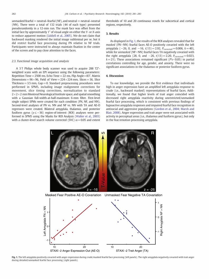
Fig. 1. The left amygdala positively covaried with anger expression during crude/masked fearduring detailed/unmasked fearful face processing (right panels).
thresholds of 10 and 20 continuous voxels for subcortical and corticalregions, respectively.
3. Results
As displayed in Fig. 1, the results of the ROI analyses revealed that formasked (FNNNN) fearful faces AE-O positively covaried with the leftamygdala (−26, 0, and −16, t(13)=2.92, Pcorrected=0.006, k=40),while for unmasked (NFNNN) fearful faces TA negatively covaried withthe right amygdala (28, 0, and −26, t(13)=2.20, Pcorrected=0.023,k=21). These associations remained significant (P'sb0.05) in partialcorrelations controlling for age, gender, and anxiety. There were nosignificant associations in the thalamus or posterior fusiform gyrus.
4. Discussion
To our knowledge, we provide the first evidence that individualshigh in anger expression have an amplified left amygdala response tocrude (i.e., backward masked) representations of fearful faces. Addi-tionally, we found that higher levels of trait anger coincided withdecreased right amygdala reactivity during unrestricted/unmaskedfearful face processing, which is consistent with previous findings ofhypoactive amygdala responses and impaired fearful face recognition inantisocial and aggressive populations (Gordon et al., 2004; Marsh andBlair, 2008). Anger expression and trait anger were not associated withactivity in perceptual areas (i.e., thalamus and fusiform gyrus), but onlyin the fear/emotion processing amygdala.
ful face processing (left panels). The right amygdala negatively covaried with trait anger
283J.M. Carlson et al. / Psychiatry Research: Neuroimaging 182 (2010) 281–283
The observed hyperactive left amygdala response to crude fearexpressions in individuals with higher levels of anger expression mayreflect a mechanism that triggers aggressive responses, while thehypoactive right amygdala response to detailed fear expressions mayreflect deficits in fearful face processing, which result in dismissal ofthese distress cues. In extreme cases, this differential amygdalareactivity may lead to “blind rage” or aggressive behavior withoutappropriate distress processing and subsequent withdrawal. Interest-ingly, the observed amygdala asymmetries are consistentwithmodelsof affective asymmetry where the left hemisphere is thought to beinvolved in approach-related behaviors (e.g., lashing out), whereasthe right hemisphere is associated with withdrawal behaviors andnegatively valenced perceptual processing (Demaree et al., 2005).Given our sample size and lack of a recognition test, future research inthis area is needed. Nevertheless, the current findings lead us tospeculate that two processes are associated with aggression—a rapidreactivity to crude threat/distress that facilitates the aggressiveresponse and a deficit in processing detailed threat/distress cuesthat maintains it.
Acknowledgements
This research was supported by the Office of Naval Research#N0014-04-1-005 (LRMP) and the National Institutes of Health#5MO1-RR-10710 (General Clinical Research Center).
References
Adolphs, R., Tranel, D., Damasio, H., Damasio, A., 1994. Impaired recognition of emotionin facial expressions following bilateral damage to the human amygdala. Nature372, 669–672.
Demaree, H.A., Everhart, D.E., Youngstrom, E.A., Harrison, D.W., 2005. Brain laterali-zation of emotional processing: historical roots and a future incorporating“dominance”. Behavioral and Cognitive Neuroscience Reviews 4 (1), 3–20.
Gordon, H.L., Baird, A.A., End, A., 2004. Functional differences among those high and lowon a trait measure of psychopathy. Biological Psychiatry 56, 516–521.
Gur, R.C., Sara, R., Hagendoorn, M., Marom, O., Hughett, P., Macy, L., 2002. A method forobtaining 3-dimensional facial expressions and its standardization for use inneurocognitive studies. Journal of Neuroscience Methods 115, 137–143.
LeDoux, J.E., 1996. The Emotional Brain: The Mysterious Underpinnings of EmotionalLife. Weidenfeld and Nicholson, London.
Liddell, B.J., Brown, K.J., Kemp, A.H., Barton, M.J., Das, P., Peduto, A., Gordon, E., Williams,L.M., 2005. A direct brainstem–amygdala–cortical ‘alarm’ system for subliminalsignals of fear. Neuroimage 24, 235–243.
Marsh, A.A., Blair, R.J., 2008. Deficits in facial affect recognition among antisocialpopulations: a meta-analysis. Neuroscience and Biobehavioral Reviews 32, 454–465.
Spielberger, C.D., 1999. State-Trait Anger Expression Inventory-2. PsychologicalAssessment Resources Inc., Lutz, FL.
Spielberger, C.D., Gorsuch, R.L., Lushene, R.E., 1970. Manual for the State-Trait AnxietyInventory (Self-Evaluation Questionnaire). Consulting Psychology Press, Palo Alto, CA.
Vuilleumier, P., Pourtois, G., 2007. Distributed and interactive brain mechanisms duringemotion face perception: evidence from functional neuroimaging. Neuropsycho-logia 45, 174–194.
Walter, B., Blecker, C., Kirsch, P., Sammer, G., Schienle, A., Stark, R., Vaitl, D., 2003.MARINA: an easy to use tool for the creation of MAsks for Region of INterestAnalyses. Paper presented at the 9th International Conference on FunctionalMapping of the Human Brain, New York, NY.


![[Shinobi] Bleach - Ulquiorra UNMASKED](https://img.pdfslide.net/doc/110x75/568c51f01a28ab4916b4b8ab/shinobi-bleach-ulquiorra-unmasked.jpg)


![Vaticanism Unmasked [1878]](https://img.pdfslide.net/doc/110x75/577cd0ed1a28ab9e7893488a/vaticanism-unmasked-1878.jpg)