Embed Size (px)
Citation preview
J. med. Genet. (I966). 3, 148.
Bloch-Sulzberger Syndrome(Incontinentia Pigmenti)NINA SHOTTS and ALAN E. H. EMERY
From the Dental Hospital of Manchester and the Department of Medical Genetics, Manchester Royal Infirmary
The Bloch-Sulzberger syndrome is a rare disease.It presents at or soon after birth with erythema andblistering of the skin, which leave pigmented scarsof characteristic swirling pattern, which graduallyfade (Bloch, I926; Sulzberger, I927). In addition,a number of other abnormalities occur with varyingfrequency. These include dental aberrations (Gorlinand Anderson, I960), eye defects (Scott, Friedmann,Chitters, and Pepler, I955), mental deficiency(Carney, ig5i), and paralyses.A case is reported of a young girl who presented
with this syndrome, showing skin, tooth, and hairdefects. The family came to the notice of one of us(N.S.) because of dental troubles in the proband,and to the other (A.E.H.E.) through a maternal auntwho sought genetic counselling advice.
Case ReportPersonal History. M.A.C., a female child born
February 20, 1959, was first seen in November I962when she was referred from an infant welfare clinicbecause she lacked some teeth. Her previous medicalhistory revealed that she was born at term (birth weight357Ig.), and that she developed blisters on her trunkand neck within 24 hours. The blistering, accompaniedby an increasing degree of pigmentation, continued forabout 5 weeks. The vesiculation then subsided. Duringinfancy some degree of verrucosity was apparent at thesites of the original lesions. The child was examined onApril 24 by a consultant paediatrician, who reported thather general development appeared to be normal. Sincethen she has continued to grow and develop normally.The child is now a pleasant little girl of apparentlyaverage intelligence. She attends her local infants' schooland is thought by her mother to be slow but notbackward.
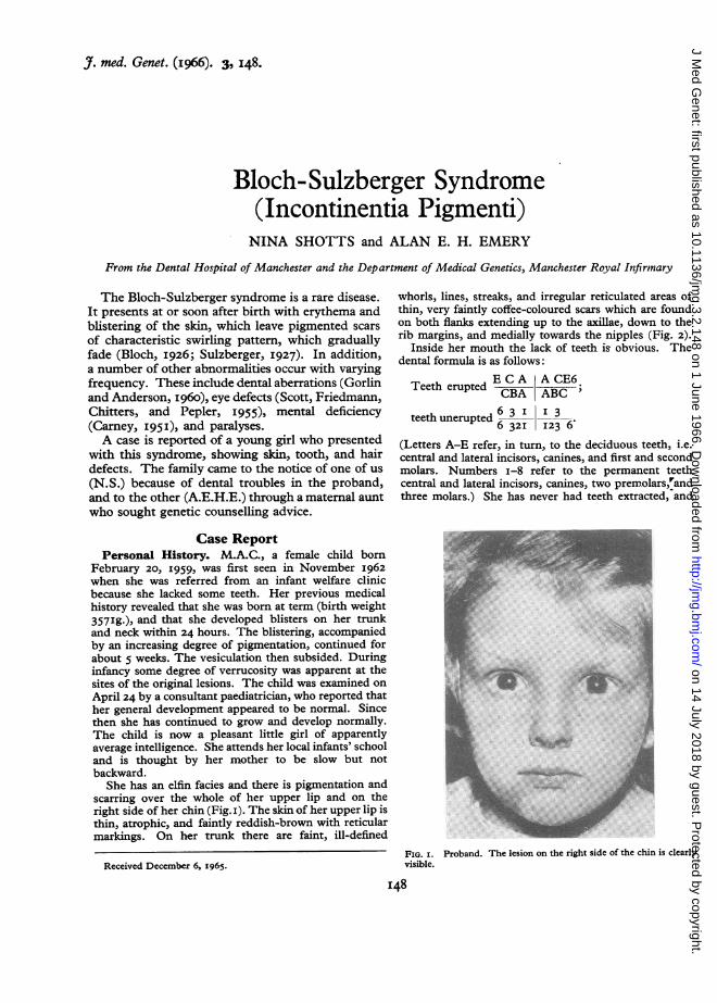
She has an elfin facies and there is pigmentation andscarring over the whole of her upper lip and on theright side of her chin (Fig. i). The skin of her upper lip isthin, atrophic, and faintly reddish-brown with reticularmarkings. On her trunk there are faint, ill-defined
Received December 6, 1965.
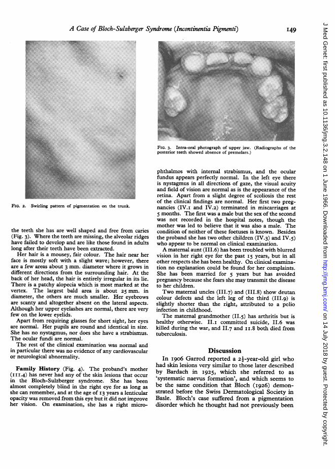
whorls, lines, streaks, and irregular reticulated areas ofthin, very faintly coffee-coloured scars which are foundon both flanks extending up to the axillae, down to therib margins, and medially towards the nipples (Fig. 2).
Inside her mouth the lack of teeth is obvious. Thedental formula is as follows:
ECA ACE6Teeth eruptedCB ACPCBA ABC;
teeth unerupted 6 3 I I 36 32I 123 6
(Letters A-E refer, in turn, to the deciduous teeth, i.e.central and lateral incisors, canines, and first and secondmolars. Numbers i-8 refer to the permanent teethcentral and lateral incisors, canines, two premolars,randthree molars.) She has never had teeth extracted, and
FIG. I. Proband. The lesion on the right side of the chin is clearlyvisible.
I48
on 14 July 2018 by guest. Protected by copyright.
http://jmg.bm
j.com/
J Med G
enet: first published as 10.1136/jmg.3.2.148 on 1 June 1966. D
ownloaded from
A Case of Bloch-Sulzberger Syndrome (Incontinentia Pigmenti)
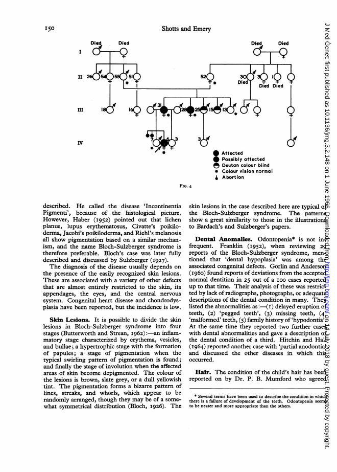
FIG. 3. Intra-oral photograph of upper jaw. (Radiographs of theposterior teeth showed absence of premolars.)
FIG. 2. Swirling pattern of pigmentation on the trunk.
the teeth she has are well shaped and free from caries
(Fig. 3). Where the teeth are missing, the alveolar ridgeshave failed to develop and are like those found in adults
long after their teeth have been extracted.
Her hair is a mousey, fair colour. The hair near her
face is mostly soft with a slight wave; however, there
are a few areas about 3 mm. diameter where it grows in
different directions from the surrounding hair. At the
back of her head, the hair is entirely irregular in its lie.
There is a patchy alopecia which is most marked at the
vertex. The largest bald area is about 25 in
diameter, the others are much smaller. Her eyebrowsare scanty and altogether absent on the lateral aspects.
Although her upper eyelashes are normal, there are very
few on the lower eyelids.
Apart from requiring glasses for short sight, her eyesare normal. Her pupils are round and identical in size.She has no nystagmus, nor does she have a strabismus.The ocular fundi are normal.
The rest of the clinical examination was normal and
in particular there was no evidence of any cardiovascularor neurological abnormality.
Famitly History (Fig. 4). The proband's mother
(II114) has never had any of the skin lesions that occurin the Bloch-Sulzberger syndrome. She has been
almost completely blind in the right eye for as long as
she can remember, and at the age Of I3 years a lenticularopacity was removed from this eye but it did not improveher vision. On examination, she has a right micro-
phthalmos with internal strabismus, and the ocularfundus appears perfectly normal. In the left eye thereis nystagmus in all directions of gaze, the visual acuityand field of vision are normal as is the appearance of theretina. Apart from a slight degree of scoliosis the restof the clinical findings are normal. Her first two preg-nancies (IV.i and IV.2) terminated in miscarriages at5 months. The first was a male but the sex of the secondwas not recorded in the hospital notes, though themother was led to believe that it was also a male. Thecondition of neither of these foetuses is known. Besidesthe proband she has two other children (IV.3) and IV.5)who appear to be normal on clinical examination.A maternal aunt (III.6) has been troubled with blurred
vision in her right eye for the past 15 years, but in allother respects she has been healthy. On clinical examina-tion no explanation could be found for her complaints.She has been married for 5 years but has avoidedpregnancy because she fears she may transmit the diseaseto her children.Two maternal uncles (III.7) and (III.8) show deutan
colour defects and the left leg of the third (III.9) isslightly shorter than the right, attributed to a polioinfection in childhood.The maternal grandmother (II.s) has arthritis but is
healthy otherwise. II.i committed suicide, II.6 waskilled during the war, and II.7 and ii.8 both died fromtuberculosis.
DiscussionIn I906 Garrod reported a 21-year-old girl who
had skin lesions very similar to those later describedby Bardach in I925, which she referred to as'systematic naevus formation', and which seems tobe the same condition that Bloch (I926) demon-strated before the Swiss Dermatological Society inBasle. Bloch's case suffered from a pigmentationdisorder which he thought had not previously been
I49
on 14 July 2018 by guest. Protected by copyright.
http://jmg.bm
j.com/
J Med G
enet: first published as 10.1136/jmg.3.2.148 on 1 June 1966. D
ownloaded from
Shotts and Emery
I
IV
* :AffectedPossibly affectedDeuton colour blind
* Colour vision normal
I Abortion
FIG. 4
described. He called the disease 'IncontinentiaPigmenti', because of the histological picture.However, Haber (I952) pointed out that lichenplanus, lupus erythematosus, Civatte's poikilo-derma, Jacobi's poikiloderma, and Riehl's melanosisall show pigmentation based on a similar mechan-ism, and the name Bloch-Sulzberger syndrome istherefore preferable. Bloch's case was later fullydescribed and discussed by Sulzberger (1927).The diagnosis of the disease usually depends on
the presence of the easily recognized skin lesions.These are associated with a variety of other defectsthat are almost entirely restricted to the skin, itsappendages, the eyes, and the central nervoussystem. Congenital heart disease and chondrodys-plasia have been reported, but the incidence is low.
Skin Lesions. It is possible to divide the skinlesions in Bloch-Sulzberger syndrome into fourstages (Butterworth and Strean, I962)):-an inflam-matory stage characterized by erythema, vesicles,and bullae; a hypertrophic stage with the formationof papules; a stage of pigmentation when thetypical swirling pattern of pigmentation is found;and finally the stage of involution when the affectedareas of skin become depigmented. The colour ofthe lesions is brown, slate grey, or a dull yellowishtint. The pigmentation forms a bizarre pattern oflines, streaks, and whorls, which appear to berandomly arranged, though they may be of a some-what symmetrical distribution (Bloch, 1926). The
skin lesions in the case described here are typical ofthe Bloch-Sulzberger syndrome. The patternsshow a great similarity to those in the illustrationsto Bardach's and Sulzberger's papers.
Dental Anomalies. Odontopenia* is not in-frequent. Franklin (I952), when reviewing 29reports of the Bloch-Sulzberger syndrome, men-tioned that 'dental hypoplasia' was among theassociated congenital defects. Gorlin and Anderson(I960) found reports of deviations from the acceptednormal dentition in 25 out of a ioo cases reportedup to that time. Their analysis of these was restric-ted by lack of radiographs, photographs, or adequatedescriptions of the dental condition in many. Theylisted the abnormalities as:-(i) delayed eruption ofteeth, (2) 'pegged teeth', (3) missing teeth, (4)'malformed' teeth, (5) family history of 'hypodontia'.At the same time they reported two further caseswith dental abnormalities and gave a description ofthe dental condition of a third. Hitchin and Hall(1964) reported another case with 'partial anodontia'and discussed the other diseases in which thisoccurred.
Hair. The condition of the child's hair has beenreported on by Dr. P. B. Mumford who agreed
* Several terms have been used to describe the condition in whichthere is a failure of development of the teeth. Odontopenia seemsto be neater and more appropriate than the others.
150
on 14 July 2018 by guest. Protected by copyright.
http://jmg.bm
j.com/
J Med G
enet: first published as 10.1136/jmg.3.2.148 on 1 June 1966. D
ownloaded from
A Case of Bloch-Sulzberger Syndrome (Incontinentia Pigmenti)
that the lie of the hair was strange but could findno abnormality of its structure. There appears tobe no other published report of irregular lie of thehair in the Bloch-Sulzberger syndrome. The areasof alopecia in our case are probably the result ofsecondary infections of the early lesions of the scalp.There is no sign of the pseudo-pelade type ofalopecia described in other reports of the Bloch-Sulzberger syndrome. Bardach (1925) reported theretention of lanugo hair in the twins she described.She did not, however, see them after 6 weeks of age,when they were transferred to the care of anotherdepartment. Various skeletal and neurologicalabnormalities have occasionally been described inpatients with the Bloch-Sulzberger syndrome aswell as mental retardation and epilepsy (Jacksonand Nigam, I962).
Ocular Lesions. Eye defects are found quitefrequently in association with the syndrome.Bloch and Sulzberger's case was referred to themby an ophthalmologist to whom the child was sentfor treatment of retrobulbar glioma (Bloch, 1926) orretinal detachment (Sulzberger, I927). Bardach'stwins are described as having star-shaped irises.More recently Findlay (I952) found eye defectsdescribed in 8 of 25 reported cases, and Scott et al.in I955 found eye defects in 24 out of 92 reportedcases, and these included nystagmus, strabismus,cataract, optic nerve atrophy, glioma and pseudo-glioma, and retinal detachment.
Genetic Aspects. There are numerous reportsof several members of a family being affected withthe Bloch-Sulzberger syndrome (Bardach, I925;Franklin, I952; Calnan, I952; Rozehnal andVodicka, I957; Marty, Bechtel, and Wood, I958;Jackson and Nigam, I962; Kiister and Olbing, I964);and Jackson and Nigam (I962) have reviewed manyof the earlier reports of familial cases. The diseaseis clearly familial, but the mode of inheritance isuncertain, and there are several possibilities.
(i) It has been suggested that the syndrome isinherited as an X-linked dominant trait withlethality in the hemizygous male (Lenz, I963). Infavour of this idea are the observations that thedisease very rarely affects males, and is transmittedby affected females who have a deficiency of malesamong their offspring, and an unusually high fre-quency of abortions that are possibly hemizygousmales. So far, about ioo cases have been reportedof which no more than 5 have been males (Oldfelt,I959). The characteristic pattern of pigmentation
in affected females might be explained in terms ofthe Lyon hypothesis concerning gene action in theX chromosome (Lyon, I962). Possibly the pig-mented areas represent those cells in which theactive X chromosome is the one bearing the genefor the Bloch-Sulzberger syndrome (McKusick,I962). Random inactivation of the X chromosomemay be a possible explanation of the variation inphenotypic manifestations in affected females. Somefemales are minimally affected whereas others maybe so severely affected that they die in infancy(Kiister and Olbing, I964).
(ii) Other possible explanations for the pedigreepatterns in the Bloch-Sulzberger syndrome includeautosomal dominance with sex limitation to thefemale and lethality in the male, or a cytoplasmicfactor which is lethal in males.
(iii) The early stages of the disease resemble aninflammatory reaction in the skin and the findingof eosinophil leucocyte deposits in the skin, liver,and spleen (Kuster and Obling, I964) suggest thatthe disease may be due to an infective agent. In factcytoplasmic inclusions similar to those in molluscumcontagiosum have been identified in this disease(Murrell, I962). If infection occurs in utero thenpossibly susceptibility is genetically determined. Ina pair of twins Hitchin and Hall (I964) found thatwhereas the female twin was affected the male twinwas healthy, perhaps because he was not hemizygous(or heterozygous) for the Bloch-Sulzberger gene.
In this report, the proband's mother had a rightmicrophthalmos and was almost completely blind inthis eye. There have been several reports of ocularmalformations in the relatives of cases with Bloch-Sulzberger syndrome (Scott et al., I955), and it ispossible that in this report the proband's motherwas a carrier of the mutant gene. It is difficult toassess the situation with regard to the maternal auntwhose only complaint was of blurred vision in theright eye. If she was affected then the maternalgrandmother must also carry the gene, but sheshowed no manifestations of the disease and hadbeen healthy apart from arthritis. On the presentevidence it seems that the maternal aunt was notaffected and that the disease in this family was theresult of a new mutation in a gamete from one ofthe maternal grandparents.
SummaryA child with Bloch-Sulzberger syndrome is
described. Her mother (and perhaps a maternalaunt) may also be affected to a mild degree. Theclinical manifestations and possible mode of inherit-ance are discussed.
I51
on 14 July 2018 by guest. Protected by copyright.
http://jmg.bm
j.com/
J Med G
enet: first published as 10.1136/jmg.3.2.148 on 1 June 1966. D
ownloaded from

Shotts and Emery
Our thanks are due to Professor Miller, now atCardiff, for bringing this case to the notice of one of us(N.S.) and to Dr. Holloway for constant encouragement.The Department of Medical Illustration at ManchesterRoyal Infirmary prepared the illustrations.
REFERENCES
Bardach, M. (I925). Systematisierte Naevusbildungen bei einemeineiigen Zwillingspaar. Ein Beitrag zur Naevusatiologie. Z.Kinderheilk., 39, 542.
Bloch, B. (I926). Eigentumliche, bisher nicht beschriebenePigmentaffektion (Incontinentia pigmenti). Schweiz. med. Wschr.,7, 404-
Butterworth, T., and Strean, L. P. (I962). Clinical Genodermatology.Williams and Wilkins, Baltimore.
Calnan, C. D. (I952). Incontinentia pigmenti (Bloch-Sulzberger).Brit. 7. Derm., 64, 20I.
Carney, R. G. (I95I). Incontinentia pigmenti: a report of five casesand review of the literature. Arch. Derm. Syph. (Chic.), 64, 126.
Findlay, G. H. (I952). On the pathogenesis of incontinentia pig-menti. With observations on associated eye disturbance resemblingretrolental fibroplasia. Brit. J7. Derm., 64, I41.
Franklin, A. W. (1952). Incontinentia pigmenti; report of a case,with summary of 29 cases from the literature. Brit. med. J., I, 75.
Garrod, A. E. (I906). Peculiar pigmentation of the skin in an infant.Trans. clin. Soc. Lond., 39, 2I6.
Gorlin, R. J., and Anderson, J. A. (I960). The characteristicdentition of incontinentia pigmenti. Pediat., 57, 78.
Haber, H. (I952). Bloch-Sulzberger syndrome (incontinentiapigmenti). Brit. J. Derm., 64, I29.
Hitchin, A. D., and Hall, D. C. (I964). Incontinentia pigmenti(Block-Sulzberger syndrome) and its dental manifestations. Brit.dent. J., II6, 239.
Jackson R., and Nigam S. (I962). Incontinentia pigmenti: a reportof 3 cases in one family. Pediatrics, 30, 433.
Kuster, F. von, and Olbing, H. (I964). Incontinentia pigmenti:Bericht uber neun Erkrankungen in einer Familie und einenObduktions befund. Ann. paediat. (Basel), 202, 92.
Lenz, W. (I963). Medical Genetics. University of Chicago Press,Chicago.
Lyon, M. F. (I962). Sex chromatin and gene action in themammalian X-chromosome. Amer. J. hum. Genet., 14, I35.
McKusick, V. A. (I962). On the X chromosome of man. Quart.Rev. Biol., 37, 69.
Marty, S. D., Bechtel, H. B., and Wood, C. E. (I958). Incontinentiapigmenti: report of a family. A.M.A. Arch. Derm., 78, 607.
Murrell, T. W., Jr. (I962). Personal communication to McKusick(i962).
Oldfelt, V. (I959). Incontinentia pigmenti. J. Pediat., 54, 446.Rozehnal, V., and Vodicka, F. (I957). Incontinentia pigmenti in
four girls of the same family. [In Czech.] Cs. Pediat., 12, 549.
Scott, J. G., Friedmann, A. I., Chitters, M., and Pepler, W. J. (I955).Ocular changes in the Bloch-Sulzberger syndrome (incontinentiapigmenti). Brit. 7. Ophthal., 39, 276.
Sulzberger, M. B. (I927). Uber eine bisher nicht beschriebenecongenitale Pigmentanomalie (Incontinentia pigmenti). Arch.Derm. Syph., 154, 19.
I52
on 14 July 2018 by guest. Protected by copyright.
http://jmg.bm
j.com/
J Med G
enet: first published as 10.1136/jmg.3.2.148 on 1 June 1966. D
ownloaded from


![Tnfa Signaling Through Tnfr2 Protects Skin Against ...eprints.whiterose.ac.uk/81541/1/Tnfa signaling through tnfr2 protects... · genodermatosis incontinentia pigmenti (IP) [17]](https://img.pdfslide.net/doc/110x75/5f3bedf6651a4c137761035c/tnfa-signaling-through-tnfr2-protects-skin-against-signaling-through-tnfr2-protects.jpg)
![First IKBKG Gene Mutation Study in Serbian Incontinentia ... · Incontinentia pigmenti (IP; Bloch-Sulzberg-er syndrome; MIM 308300) is a rare X-linked dominant genodermatosis [5]](https://img.pdfslide.net/doc/110x75/5f3bedf5651a4c1377610355/first-ikbkg-gene-mutation-study-in-serbian-incontinentia-incontinentia-pigmenti.jpg)