Embed Size (px)
Citation preview
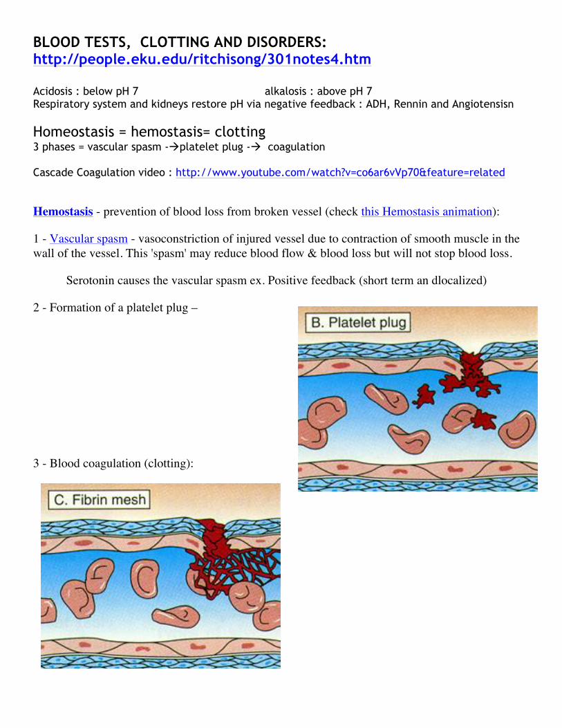
BLOOD TESTS, CLOTTING AND DISORDERS: http://people.eku.edu/ritchisong/301notes4.htm Acidosis : below pH 7 alkalosis : above pH 7 Respiratory system and kidneys restore pH via negative feedback : ADH, Rennin and Angiotensisn Homeostasis = hemostasis= clotting 3 phases = vascular spasm -àplatelet plug -à coagulation Cascade Coagulation video : http://www.youtube.com/watch?v=co6ar6vVp70&feature=related
Hemostasis - prevention of blood loss from broken vessel (check this Hemostasis animation):
1 - Vascular spasm - vasoconstriction of injured vessel due to contraction of smooth muscle in the wall of the vessel. This 'spasm' may reduce blood flow & blood loss but will not stop blood loss.
Serotonin causes the vascular spasm ex. Positive feedback (short term an dlocalized)
2 - Formation of a platelet plug –
3 - Blood coagulation (clotting):
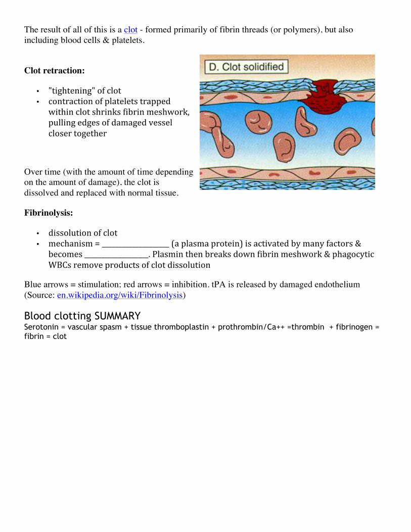
The result of all of this is a clot - formed primarily of fibrin threads (or polymers), but also including blood cells & platelets.
Clot retraction:
• "tightening" of clot • contraction of platelets trapped
within clot shrinks fibrin meshwork, pulling edges of damaged vessel closer together
Over time (with the amount of time depending on the amount of damage), the clot is dissolved and replaced with normal tissue.
Fibrinolysis:
• dissolution of clot • mechanism = ____________________ (a plasma protein) is activated by many factors &
becomes ___________________. Plasmin then breaks down fibrin meshwork & phagocytic WBCs remove products of clot dissolution
Blue arrows = stimulation; red arrows = inhibition. tPA is released by damaged endothelium (Source: en.wikipedia.org/wiki/Fibrinolysis)
Blood clotting SUMMARY Serotonin = vascular spasm + tissue thromboplastin + prothrombin/Ca++ =thrombin + fibrinogen = fibrin = clot
Disorders of hemostasis or Inappropriate clotting:
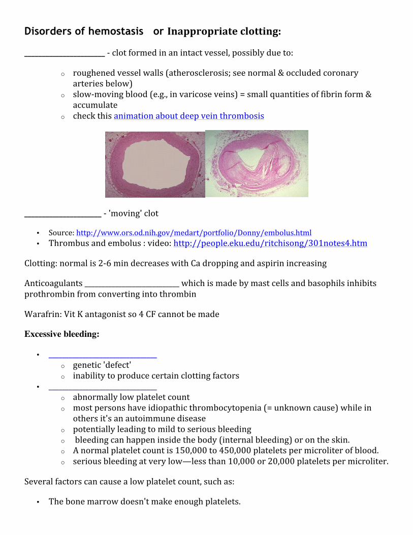
_______________________ -‐ clot formed in an intact vessel, possibly due to:
o roughened vessel walls (atherosclerosis; see normal & occluded coronary arteries below)
o slow-‐moving blood (e.g., in varicose veins) = small quantities of fibrin form & accumulate
o check this animation about deep vein thrombosis
______________________ -‐ 'moving' clot
• Source: http://www.ors.od.nih.gov/medart/portfolio/Donny/embolus.html • Thrombus and embolus : video: http://people.eku.edu/ritchisong/301notes4.htm
Clotting: normal is 2-‐6 min decreases with Ca dropping and aspirin increasing
Anticoagulants ____________________________ which is made by mast cells and basophils inhibits prothrombin from converting into thrombin
Warafrin: Vit K antagonist so 4 CF cannot be made
Excessive bleeding:
• ________________________________ o genetic 'defect' o inability to produce certain clotting factors
• ________________________________ o abnormally low platelet count o most persons have idiopathic thrombocytopenia (= unknown cause) while in
others it's an autoimmune disease o potentially leading to mild to serious bleeding o bleeding can happen inside the body (internal bleeding) or on the skin. o A normal platelet count is 150,000 to 450,000 platelets per microliter of blood. o serious bleeding at very low—less than 10,000 or 20,000 platelets per microliter.
Several factors can cause a low platelet count, such as:
• The bone marrow doesn't make enough platelets.
• The bone marrow makes enough platelets, but the body destroys them (autoimmunity) or uses them up.
• The spleen holds onto too many platelets. Or a combo of these (Source: NHLBI).
Blood tests: know how to perform these, read then and interpret 1. Blood typing 2. Talliquist: preliminary quantitative % Hb. Used as an indicator for anemia 3. Erythrocyte Sedimentation rate: http://medical-dictionary.thefreedictionary.com/Blood+sedimentation
Normal results
A normal value does not rule out disease. Normal values for the Westergren method are: Men 0 mm/hour-‐15 mm/hour; women 0 mm/hour-‐20 mm/hour; and children 0 mm/hour-‐10 mm/hour.
Abnormal results
Lower rate : RBC abnormalities such as sickle cell
Higher Rate: Menses, pregnant, anemic
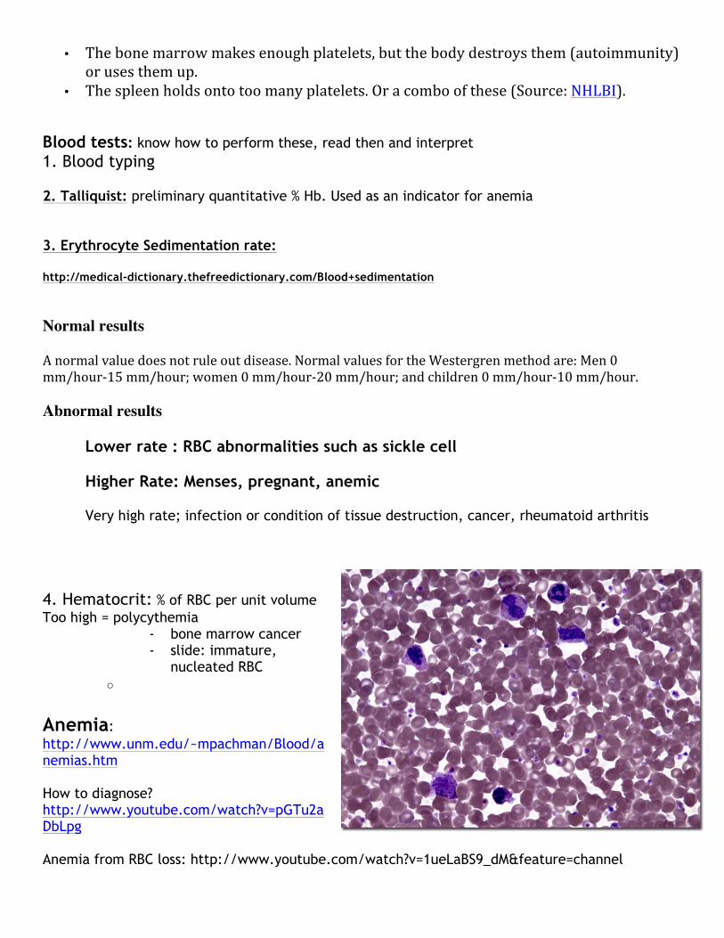
Very high rate; infection or condition of tissue destruction, cancer, rheumatoid arthritis 4. Hematocrit: % of RBC per unit volume Too high = polycythemia
- bone marrow cancer - slide: immature,
nucleated RBC o
Anemia: http://www.unm.edu/~mpachman/Blood/anemias.htm How to diagnose? http://www.youtube.com/watch?v=pGTu2aDbLpg Anemia from RBC loss: http://www.youtube.com/watch?v=1ueLaBS9_dM&feature=channel
Types of Anemias a. decrease in RBC number
b. inadequate Hb in RBC
c. Abnormal Hb in RBC
Liver and jaundice: due to a buildup of bilirubin Macrophage phagocytizes RBC in spleen, liver and bone marrow.
1. heme is separated from globin 2. Fe is removed from heme by transferrin 3. Biliverdin is green inside the macrophage and converted into bilirubin in the blood 4. Bilirubin in the blood goes to liver 5. Liver secretes bilirubin into bile 6. Bile goes to LI where bacteria convert bile into urobilinogen
a. Goes back to blood as urobilin (yellow pigment) and is reoved by kidneys b. sterobilin (brown pigment feces)
Ex. Neonatal due to immature liver, blue wavelengths of light breaks it up Ex. Hepatitis: liver infected Ex. Alcohol: liver destruction Cord Blood: http://www.youtube.com/user/cordbloodregistry?v=96DItviGvKI