Embed Size (px)
Citation preview
BioMed CentralBMC Musculoskeletal Disorders
ss
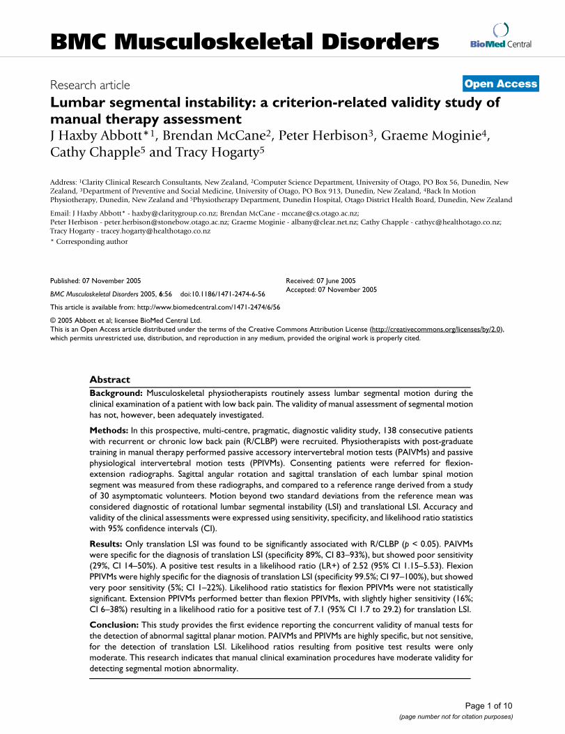
Open AcceResearch articleLumbar segmental instability: a criterion-related validity study of manual therapy assessmentJ Haxby Abbott*1, Brendan McCane2, Peter Herbison3, Graeme Moginie4, Cathy Chapple5 and Tracy Hogarty5Address: 1Clarity Clinical Research Consultants, New Zealand, 2Computer Science Department, University of Otago, PO Box 56, Dunedin, New Zealand, 3Department of Preventive and Social Medicine, University of Otago, PO Box 913, Dunedin, New Zealand, 4Back In Motion Physiotherapy, Dunedin, New Zealand and 5Physiotherapy Department, Dunedin Hospital, Otago District Health Board, Dunedin, New Zealand
Email: J Haxby Abbott* - [email protected]; Brendan McCane - [email protected]; Peter Herbison - [email protected]; Graeme Moginie - [email protected]; Cathy Chapple - [email protected]; Tracy Hogarty - [email protected]
* Corresponding author
AbstractBackground: Musculoskeletal physiotherapists routinely assess lumbar segmental motion during theclinical examination of a patient with low back pain. The validity of manual assessment of segmental motionhas not, however, been adequately investigated.
Methods: In this prospective, multi-centre, pragmatic, diagnostic validity study, 138 consecutive patientswith recurrent or chronic low back pain (R/CLBP) were recruited. Physiotherapists with post-graduatetraining in manual therapy performed passive accessory intervertebral motion tests (PAIVMs) and passivephysiological intervertebral motion tests (PPIVMs). Consenting patients were referred for flexion-extension radiographs. Sagittal angular rotation and sagittal translation of each lumbar spinal motionsegment was measured from these radiographs, and compared to a reference range derived from a studyof 30 asymptomatic volunteers. Motion beyond two standard deviations from the reference mean wasconsidered diagnostic of rotational lumbar segmental instability (LSI) and translational LSI. Accuracy andvalidity of the clinical assessments were expressed using sensitivity, specificity, and likelihood ratio statisticswith 95% confidence intervals (CI).
Results: Only translation LSI was found to be significantly associated with R/CLBP (p < 0.05). PAIVMswere specific for the diagnosis of translation LSI (specificity 89%, CI 83–93%), but showed poor sensitivity(29%, CI 14–50%). A positive test results in a likelihood ratio (LR+) of 2.52 (95% CI 1.15–5.53). FlexionPPIVMs were highly specific for the diagnosis of translation LSI (specificity 99.5%; CI 97–100%), but showedvery poor sensitivity (5%; CI 1–22%). Likelihood ratio statistics for flexion PPIVMs were not statisticallysignificant. Extension PPIVMs performed better than flexion PPIVMs, with slightly higher sensitivity (16%;CI 6–38%) resulting in a likelihood ratio for a positive test of 7.1 (95% CI 1.7 to 29.2) for translation LSI.
Conclusion: This study provides the first evidence reporting the concurrent validity of manual tests forthe detection of abnormal sagittal planar motion. PAIVMs and PPIVMs are highly specific, but not sensitive,for the detection of translation LSI. Likelihood ratios resulting from positive test results were onlymoderate. This research indicates that manual clinical examination procedures have moderate validity fordetecting segmental motion abnormality.
Published: 07 November 2005
BMC Musculoskeletal Disorders 2005, 6:56 doi:10.1186/1471-2474-6-56
Received: 07 June 2005Accepted: 07 November 2005
This article is available from: http://www.biomedcentral.com/1471-2474/6/56
© 2005 Abbott et al; licensee BioMed Central Ltd. This is an Open Access article distributed under the terms of the Creative Commons Attribution License (http://creativecommons.org/licenses/by/2.0), which permits unrestricted use, distribution, and reproduction in any medium, provided the original work is properly cited.
Page 1 of 10(page number not for citation purposes)
BMC Musculoskeletal Disorders 2005, 6:56 http://www.biomedcentral.com/1471-2474/6/56
BackgroundMusculoskeletal physiotherapists routinely assess lumbarspinal segmental motion and choose interventions on thebasis of the findings of those assessments. However, thevalidity of clinical tests used to assess segmental motionhas not been established. When physiotherapists examinethe lumbar spine, common assessments include passiveaccessory intervertebral motion tests (PAIVMs) and pas-sive physiological intervertebral motion tests (PPIVMs)[1,2]. Movement abnormalities, such as hypermobility,are believed to be detected by these assessments [1].
To date, the only evidence for the concurrent validity ofmanual testing for the presence of lumbar segmentalinstability (LSI) comes from two studies in which the pres-ence of spondylolysis was considered a proxy for the pres-ence of segmental hypermobility. The first comprised of avery small subgroup analysis (6 patients) of patients withspondylolysis, within a sample of 62 patients with non-specific LBP [3]. The results of that investigation indicatedthat PAIVMs and PPIVMs could identify the symptomaticlevel with 83% sensitivity and 98% specificity [3]. In thesecond study, manual assessment (combined informationfrom both PPIVMs and PAIVMs) was 69% sensitive and96% specific for detection of the lytic segment [4]. Whenanalysis was restricted to subjects who reported visualanalogue pain scores of greater than 4/10, sensitivity andspecificity rose to 100% [4]. In addition, some prelimi-nary evidence indicates that PAIVM testing may have pre-dictive validity for the purpose of classifying patients in a'stabilisation' category, who respond better to an exerciseintervention intended to increase lumbar segmental sta-bility [5].
As there is currently no evidence in the literature to estab-lish the concurrent validity of manual therapy tests for thedetection of excessive sagittal planar motion of the lum-bar spine, the aims of this study were to estimate the accu-racy of three common clinical assessment items for thedetection of lumbar segmental hypermobility (PAIVMs,flexion PPIVMs, and extension PPIVMs), compared to acriterion standard of radiographic measurement of sagit-tal segmental rotation and translation.
MethodsDesignPhysiotherapists with post-graduate training in muscu-loskeletal manual therapy recruited consecutive eligiblepatients presenting with a new episode of recurrent orchronic low back pain (R/CLBP). Recruiting took place inthe physiotherapists' own clinics, between October 2001and August, 2003. Patients were included if i) they pre-sented with a new episode of low back pain and, ii) theyhad experienced similar low back pain before, the firstepisode of which was at least three months prior to the
date of recruitment, or iii) they were experiencing persist-ent low back pain of at least three months duration.Patients were excluded if they i) had spinal surgery withinthe previous six months, or ii) had a history of traumaticfracture of the spine which resulted in permanent neuro-logical deficit, iii) had a history of serious neurological orpsychiatric disease, iv) were under 20 years of age, or v)were pregnant. This research was approved by the Otagoand Canterbury Regional Ethics Committees (reference #01/05/030 & 01/10/095) of the New Zealand Ministry ofHealth.
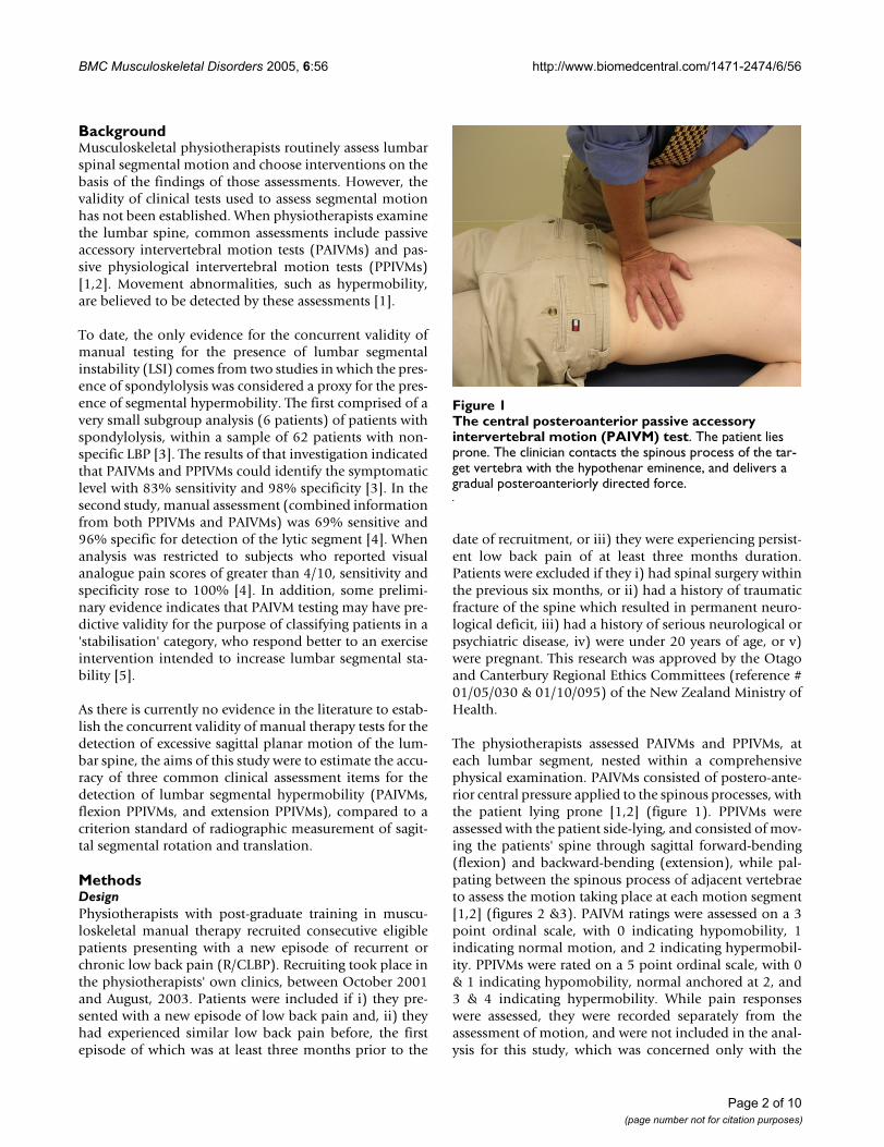
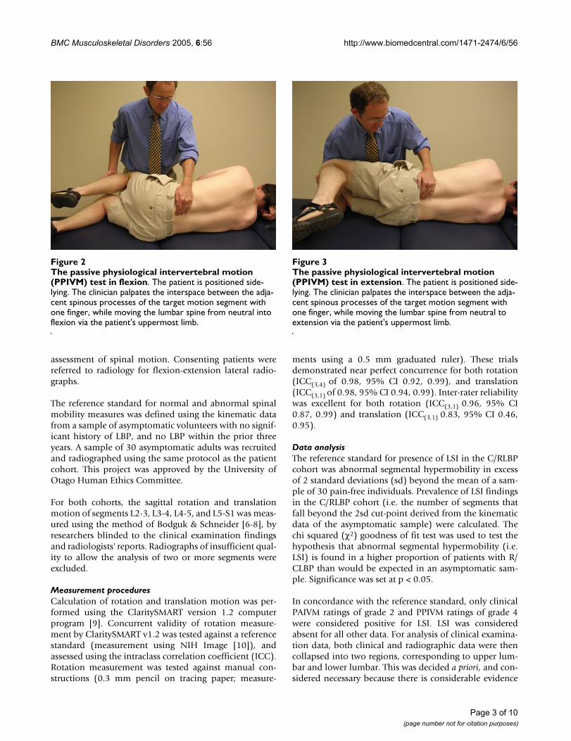
The physiotherapists assessed PAIVMs and PPIVMs, ateach lumbar segment, nested within a comprehensivephysical examination. PAIVMs consisted of postero-ante-rior central pressure applied to the spinous processes, withthe patient lying prone [1,2] (figure 1). PPIVMs wereassessed with the patient side-lying, and consisted of mov-ing the patients' spine through sagittal forward-bending(flexion) and backward-bending (extension), while pal-pating between the spinous process of adjacent vertebraeto assess the motion taking place at each motion segment[1,2] (figures 2 &3). PAIVM ratings were assessed on a 3point ordinal scale, with 0 indicating hypomobility, 1indicating normal motion, and 2 indicating hypermobil-ity. PPIVMs were rated on a 5 point ordinal scale, with 0& 1 indicating hypomobility, normal anchored at 2, and3 & 4 indicating hypermobility. While pain responseswere assessed, they were recorded separately from theassessment of motion, and were not included in the anal-ysis for this study, which was concerned only with the
The central posteroanterior passive accessory intervertebral motion (PAIVM) testFigure 1The central posteroanterior passive accessory intervertebral motion (PAIVM) test. The patient lies prone. The clinician contacts the spinous process of the tar-get vertebra with the hypothenar eminence, and delivers a gradual posteroanteriorly directed force.
Page 2 of 10(page number not for citation purposes)
BMC Musculoskeletal Disorders 2005, 6:56 http://www.biomedcentral.com/1471-2474/6/56
assessment of spinal motion. Consenting patients werereferred to radiology for flexion-extension lateral radio-graphs.
The reference standard for normal and abnormal spinalmobility measures was defined using the kinematic datafrom a sample of asymptomatic volunteers with no signif-icant history of LBP, and no LBP within the prior threeyears. A sample of 30 asymptomatic adults was recruitedand radiographed using the same protocol as the patientcohort. This project was approved by the University ofOtago Human Ethics Committee.
For both cohorts, the sagittal rotation and translationmotion of segments L2-3, L3-4, L4-5, and L5-S1 was meas-ured using the method of Bodguk & Schneider [6-8], byresearchers blinded to the clinical examination findingsand radiologists' reports. Radiographs of insufficient qual-ity to allow the analysis of two or more segments wereexcluded.
Measurement proceduresCalculation of rotation and translation motion was per-formed using the ClaritySMART version 1.2 computerprogram [9]. Concurrent validity of rotation measure-ment by ClaritySMART v1.2 was tested against a referencestandard (measurement using NIH Image [10]), andassessed using the intraclass correlation coefficient (ICC).Rotation measurement was tested against manual con-structions (0.3 mm pencil on tracing paper; measure-
ments using a 0.5 mm graduated ruler). These trialsdemonstrated near perfect concurrence for both rotation(ICC(3,4) of 0.98, 95% CI 0.92, 0.99), and translation(ICC(3,1) of 0.98, 95% CI 0.94, 0.99). Inter-rater reliabilitywas excellent for both rotation (ICC(3,1) 0.96, 95% CI0.87, 0.99) and translation (ICC(3,1) 0.83, 95% CI 0.46,0.95).
Data analysisThe reference standard for presence of LSI in the C/RLBPcohort was abnormal segmental hypermobility in excessof 2 standard deviations (sd) beyond the mean of a sam-ple of 30 pain-free individuals. Prevalence of LSI findingsin the C/RLBP cohort (i.e. the number of segments thatfall beyond the 2sd cut-point derived from the kinematicdata of the asymptomatic sample) were calculated. Thechi squared (χ2) goodness of fit test was used to test thehypothesis that abnormal segmental hypermobility (i.e.LSI) is found in a higher proportion of patients with R/CLBP than would be expected in an asymptomatic sam-ple. Significance was set at p < 0.05.
In concordance with the reference standard, only clinicalPAIVM ratings of grade 2 and PPIVM ratings of grade 4were considered positive for LSI. LSI was consideredabsent for all other data. For analysis of clinical examina-tion data, both clinical and radiographic data were thencollapsed into two regions, corresponding to upper lum-bar and lower lumbar. This was decided a priori, and con-sidered necessary because there is considerable evidence
The passive physiological intervertebral motion (PPIVM) test in extensionFigure 3The passive physiological intervertebral motion (PPIVM) test in extension. The patient is positioned side-lying. The clinician palpates the interspace between the adja-cent spinous processes of the target motion segment with one finger, while moving the lumbar spine from neutral to extension via the patient's uppermost limb.
The passive physiological intervertebral motion (PPIVM) test in flexionFigure 2The passive physiological intervertebral motion (PPIVM) test in flexion. The patient is positioned side-lying. The clinician palpates the interspace between the adja-cent spinous processes of the target motion segment with one finger, while moving the lumbar spine from neutral into flexion via the patient's uppermost limb.
Page 3 of 10(page number not for citation purposes)
BMC Musculoskeletal Disorders 2005, 6:56 http://www.biomedcentral.com/1471-2474/6/56
that therapists are not sufficiently accurate in identifyingspecific segmental levels by palpation, although they areusually within one level (up or down) and are generallyreliable at locating again a segment they had previouslylocated [11-13]. This inaccuracy presented an unaccepta-ble risk of misclassification, that collapsing into regionswould attenuate. Furthermore, it is also clear that somephysical assessment procedures affect mobility at multiplesegments [14] and that segmental specificity does notappear to be important with regard to application of phys-ical therapies for LSI, including manual therapy [5,15-22](although one study has found otherwise [23]). Data werethus collapsed into the 2 × 2 tables. By-segment resultsare, however, provided [see Additional file 1] for readersto compare.
Missing data resulted in list-wise deletion of the clinicaland radiographic data, on a per-lumbar region, per-analy-sis basis. The accuracy of the clinical examination itemswas tested by calculating sensitivity and specificity from 2× 2 contingency tables. Likelihood ratios were then calcu-lated from these data. These statistics were calculated inMicrosoft Excel, using a program written by the primaryinvestigator (JHA). The program calculated 95% confi-dence intervals (CI) using Wilson's method for sensitivity& specificity, and the score method for likelihood ratios[24]. Methods and results were reported according to theSTARD guideline checklist [25].
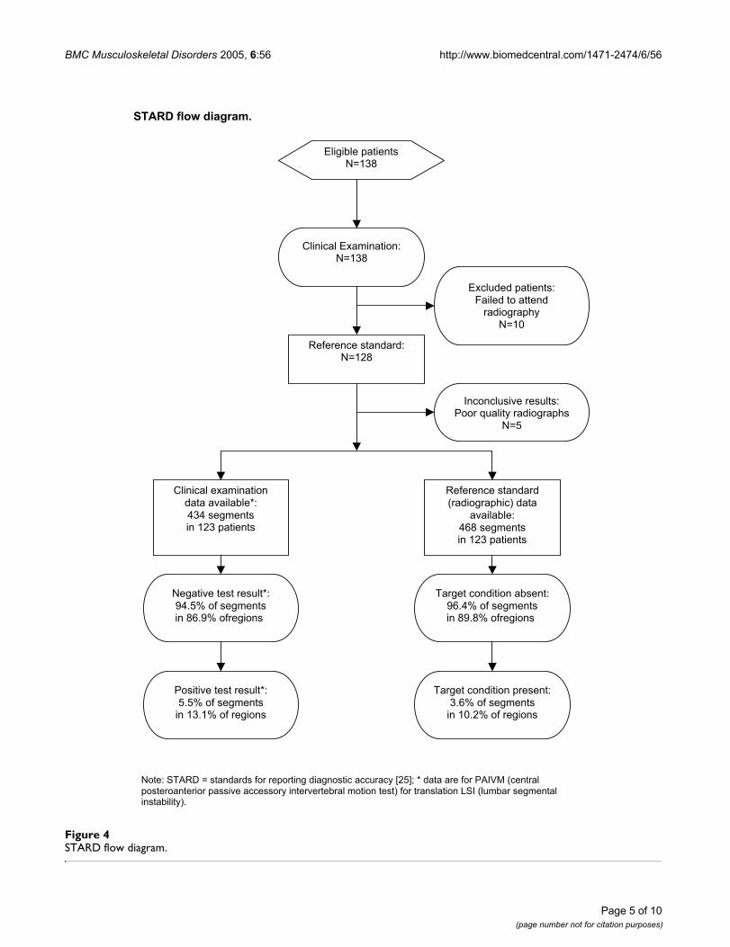
ResultsOne hundred and thirty eight (138) consenting patientswere recruited for clinical examination. One hundred andeight (108) were recruited in primary care; the remaining30 presented to a hospital outpatient physiotherapydepartment. Ten patients failed to present to radiology forflexion-extension radiographs. Five sets of radiographswere of insufficient quality for analysis. Of the 123included participants, 68 (55%) were males and 55 (45%)females. Further characteristics are described in Table 1. A
STARD flow chart is provided in figure 4. No adverseevents were reported.
Nine males and 24 females were available for recruitmentinto the asymptomatic sample. Three participants vio-lated the exclusion criteria with regard to low back painhistory, and were therefore ineligible. The asymptomaticsample therefore comprised of 9 males and 21 females,aged 23 to 60 years (mean 41.3, sd 12.8).
The 27 clinicians who collaborated on this study gradu-ated with their first professional physiotherapy qualifica-tion between 1974 and 1996 (mean years sincegraduation 17, range 6 to 29). All had gained at least onepost-graduate qualification in musculoskeletal physio-therapy which included training in manual therapy proce-dures for the spine, between 1983 to 2000 (mean yearssince graduation 8.7, range 2 to 19). They spent an averageof 31 hours (interquartile range 21 to 40) per week treat-ing patients, with LBP patients comprising, on average,30% of their patient load (interquartile range 20 to 40).
Prevalence of lumbar segmental instabilitySagittal rotation LSI was not found in statistically signifi-cant numbers (6 of 468 segments, or 1.3%), which issmaller than the number that would be expected bychance alone in a normally distributed sample of this size.Sagittal translation LSI was found at a prevalence of 3.6%(17 of 468 segments) (χ2 p < 0.05). In this cohort, 5.6%of individuals had rotation LSI at least one segment, and12.0% had translation LSI at least one segment.
Accuracy of manual therapy assessmentPAIVMs and PPIVMs were specific for the diagnosis ofboth rotation LSI and translation LSI, but showed poorsensitivity. The accuracy statistics for PAIVM and PPIVMtests appear in Tables 2 &3. Full 2 × 2 contingency tablesare also provided [see Additional file 1]. A positive PAIVMtest (grade 2 on a scale from 0 to 2) results in likelihood
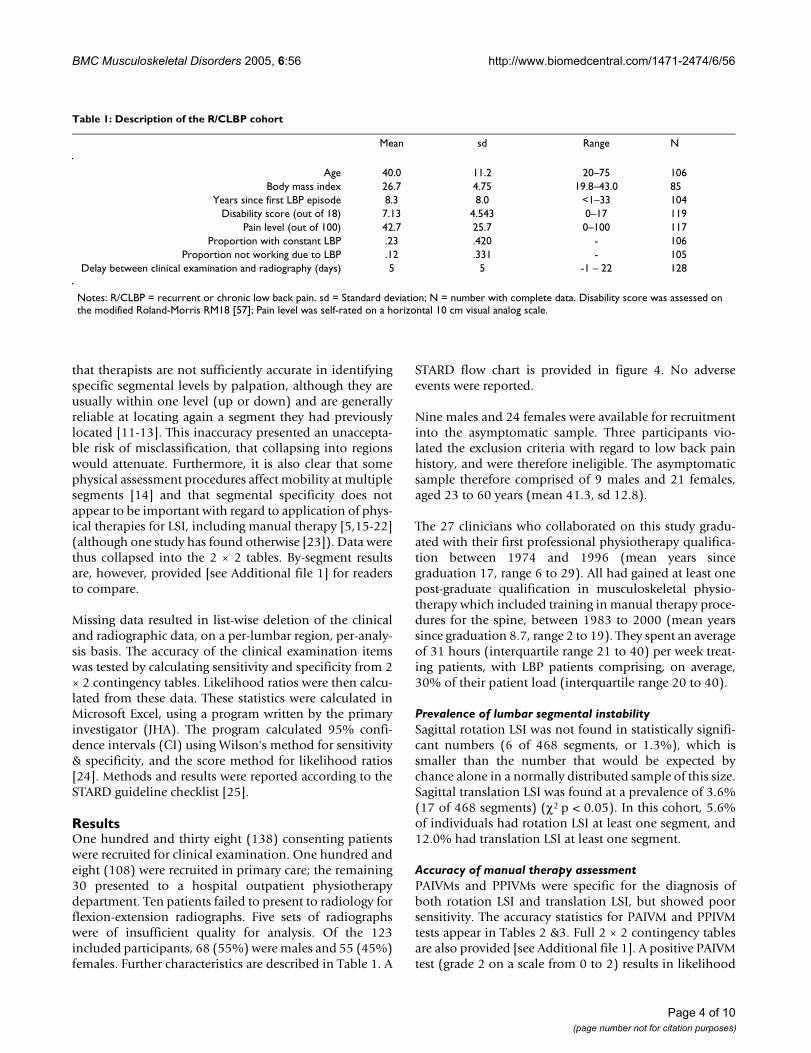
Table 1: Description of the R/CLBP cohort
Mean sd Range N
Age 40.0 11.2 20–75 106Body mass index 26.7 4.75 19.8–43.0 85
Years since first LBP episode 8.3 8.0 <1–33 104Disability score (out of 18) 7.13 4.543 0–17 119
Pain level (out of 100) 42.7 25.7 0–100 117Proportion with constant LBP .23 .420 - 106
Proportion not working due to LBP .12 .331 - 105Delay between clinical examination and radiography (days) 5 5 -1 – 22 128
Notes: R/CLBP = recurrent or chronic low back pain. sd = Standard deviation; N = number with complete data. Disability score was assessed on the modified Roland-Morris RM18 [57]; Pain level was self-rated on a horizontal 10 cm visual analog scale.
Page 4 of 10(page number not for citation purposes)
BMC Musculoskeletal Disorders 2005, 6:56 http://www.biomedcentral.com/1471-2474/6/56
Page 5 of 10(page number not for citation purposes)
STARD flow diagramFigure 4STARD flow diagram.
STARD flow diagram.
Excluded patients:Failed to attend
radiographyN=10
Eligible patientsN=138
Inconclusive results:Poor quality radiographs
N=5
Clinical Examination:N=138
Reference standard:N=128
Positive test result*:5.5% of segments
in 13.1% of regions
Negative test result*:94.5% of segmentsin 86.9% ofregions
Clinical examinationdata available*:434 segmentsin 123 patients
Target condition present:3.6% of segments
in 10.2% of regions
Target condition absent:96.4% of segmentsin 89.8% ofregions
Reference standard(radiographic) data
available:468 segmentsin 123 patients
Note: STARD = standards for reporting diagnostic accuracy [25]; * data are for PAIVM (centralposteroanterior passive accessory intervertebral motion test) for translation LSI (lumbar segmentalinstability).
BMC Musculoskeletal Disorders 2005, 6:56 http://www.biomedcentral.com/1471-2474/6/56
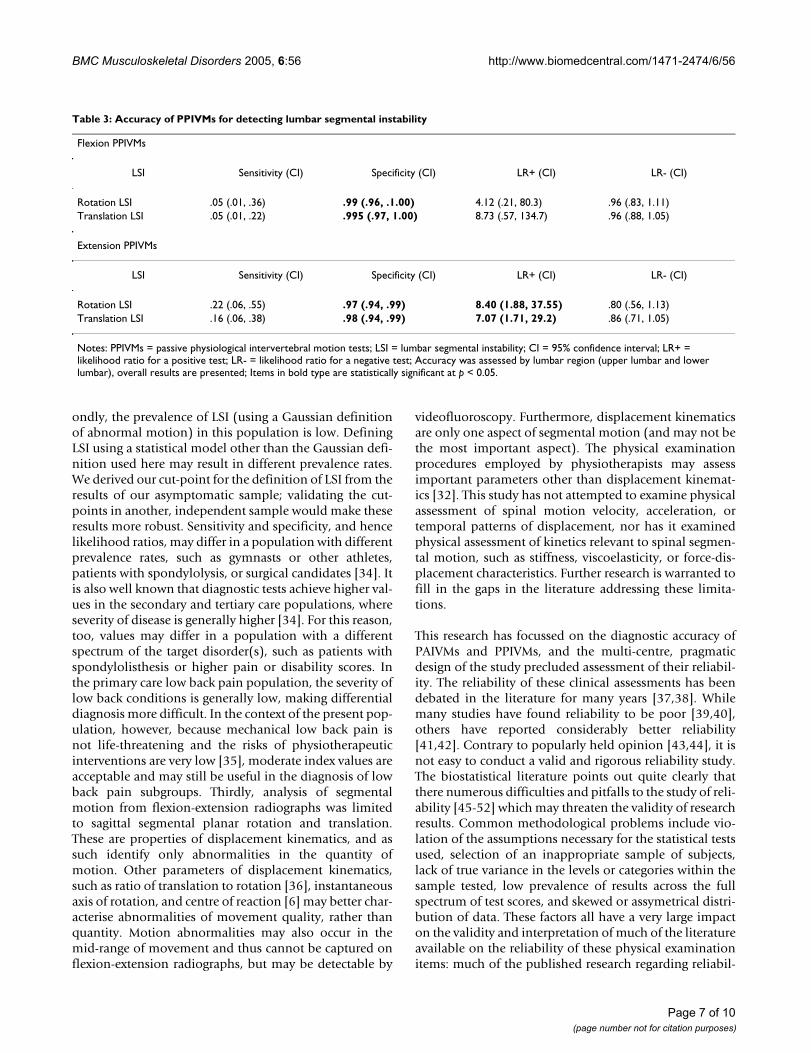
ratios (LR+) of 2.74 and 2.52 for rotation LSI and transla-tion LSI respectively. Extension PPIVMs performed betterthan flexion PPIVMs due to their slightly higher sensitiv-ity. A positive extension PPIVM test (grade 4 on a scalefrom 0 to 4) results in LR+ of 8.4 and 7.1 for rotation LSIand translation LSI, respectively. Likelihood ratios forflexion PPIVMs were not statistically significant.
DiscussionDespite their widespread use, the validity of PAIVMs andPPIVMs for assessing abnormal sagittal planar motion hasnot been previously established. We have found PAIVMsand PPIVMs to have high specificity, but poor sensitivity,for the diagnosis of both rotation LSI and translation LSI.
Like sensitivity and specificity, the likelihood ratio for apositive test (LR+) is more powerful when its value ishigh. Because of the many factors which must be takeninto account when applying a diagnostic test to an indi-vidual patient (such as the setting the test is used in, pur-pose of applying the test, prevalence of the disorder,consequences of missing a diagnosis, and risk of harmfrom the indicated therapy), there are no set cut-off valuesfor sensitivity, specificity, or likelihood ratios, howeversome authors provide general guidelines [26]. Testsreturning LR+ values of 2 to 5 produce small but oftenuseful changes in probability [26], while LR+ values of 5to 10 (and greater) are more powerful. A test with a likeli-hood ratio of one is of no clinical utility. The results of thisstudy indicate that a segment testing positive with aPAIVM test is approximately two-and-a-half times morelikely to be hypermobile than not [27]. The results forPPIVMs were higher, indicating that a segment testingpositive with an extension PPIVM test is approximatelyseven times more likely to be hypermobile than it is to benormal or hypomobile.
Likelihood ratios for negative tests from this research wereless impressive than were the LR+ values, with valuesbetween 0.76 and 0.96. None were statistically significant.A LR- closer to zero is more powerful, whereas a LR- of onehas no discriminative power. Tests returning LR- values of0.2 to 0.5 produce small but useful changes in probability,while those with values less than 0.2 are more powerful[26]. This research indicates that a negative result for
hypermobility with PAIVM or PPIVM tests is clinicallyuninformative.
The low prevalence of rotation LSI in this non-surgical,mostly primary care cohort indicate that sagittal rotationhypermobility does not appear to be associated with R/CLBP, as the number of segments hypermobile in rotationis less than the number that would be expected in a sam-ple from a normally distributed asymptomatic popula-tion. Sagittal translation hypermobility was found in asignificantly higher than expected proportion of patientswith R/CLBP (12.0%), and therefore using a Gaussian def-inition of abnormality (i.e. beyond 2sd from a referencemean) [28] can be considered a valid clinical disorder.Only a small proportion of segments (3.6%) satisfied thisGaussian definition for sagittal translational LSI, however,indicating that it is neither common in this populationnor strongly associated with C/RLBP. This may be consid-ered surprising in the light of the emphasis on sagittaltranslation in the LSI literature [29,30]. This proportiondoes, however, compare well with clinicians' judgementusing PAIVM tests. In the present study, therapists consid-ered 5% of lumbar segments to have manual tests findingspositive for LSI. This figure compares well to the 12% ofpatients with LBP reported to be hypermobile by thera-pists using PAIVM testing in other research [5]. Withregard to the physical examination, though, it is also rec-ognised that assessment of displacement kinematics alonemay not be a sufficient basis for the diagnosis of LSI[31,32].
This study has a number of limitations which limit theinterpretation of these results. Firstly, while the assess-ments were nested within a comprehensive clinical exam-ination, and performed in the physiotherapists' ownclinical setting, only these three physical assessments werestudied in isolation. No attempt was made to identifyclusters of assessments that may multiplicatively improvediagnostic accuracy. It is likely that these assessmentswould have much greater clinical utility within a cluster ofother valid signs, symptoms, and history items [16,19].Furthermore, it may be necessary to adjust the likelihoodratios of these and other tests researched in the future, toremove the influence of conditional dependence, usingstatistical methods such as logistic regression [33]. Sec-
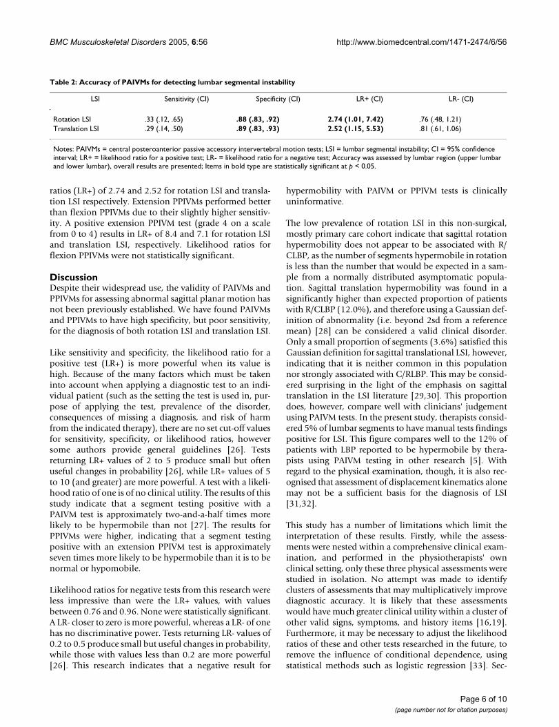
Table 2: Accuracy of PAIVMs for detecting lumbar segmental instability
LSI Sensitivity (CI) Specificity (CI) LR+ (CI) LR- (CI)
Rotation LSI .33 (.12, .65) .88 (.83, .92) 2.74 (1.01, 7.42) .76 (.48, 1.21)Translation LSI .29 (.14, .50) .89 (.83, .93) 2.52 (1.15, 5.53) .81 (.61, 1.06)
Notes: PAIVMs = central posteroanterior passive accessory intervertebral motion tests; LSI = lumbar segmental instability; CI = 95% confidence interval; LR+ = likelihood ratio for a positive test; LR- = likelihood ratio for a negative test; Accuracy was assessed by lumbar region (upper lumbar and lower lumbar), overall results are presented; Items in bold type are statistically significant at p < 0.05.
Page 6 of 10(page number not for citation purposes)
BMC Musculoskeletal Disorders 2005, 6:56 http://www.biomedcentral.com/1471-2474/6/56
ondly, the prevalence of LSI (using a Gaussian definitionof abnormal motion) in this population is low. DefiningLSI using a statistical model other than the Gaussian defi-nition used here may result in different prevalence rates.We derived our cut-point for the definition of LSI from theresults of our asymptomatic sample; validating the cut-points in another, independent sample would make theseresults more robust. Sensitivity and specificity, and hencelikelihood ratios, may differ in a population with differentprevalence rates, such as gymnasts or other athletes,patients with spondylolysis, or surgical candidates [34]. Itis also well known that diagnostic tests achieve higher val-ues in the secondary and tertiary care populations, whereseverity of disease is generally higher [34]. For this reason,too, values may differ in a population with a differentspectrum of the target disorder(s), such as patients withspondylolisthesis or higher pain or disability scores. Inthe primary care low back pain population, the severity oflow back conditions is generally low, making differentialdiagnosis more difficult. In the context of the present pop-ulation, however, because mechanical low back pain isnot life-threatening and the risks of physiotherapeuticinterventions are very low [35], moderate index values areacceptable and may still be useful in the diagnosis of lowback pain subgroups. Thirdly, analysis of segmentalmotion from flexion-extension radiographs was limitedto sagittal segmental planar rotation and translation.These are properties of displacement kinematics, and assuch identify only abnormalities in the quantity ofmotion. Other parameters of displacement kinematics,such as ratio of translation to rotation [36], instantaneousaxis of rotation, and centre of reaction [6] may better char-acterise abnormalities of movement quality, rather thanquantity. Motion abnormalities may also occur in themid-range of movement and thus cannot be captured onflexion-extension radiographs, but may be detectable by
videofluoroscopy. Furthermore, displacement kinematicsare only one aspect of segmental motion (and may not bethe most important aspect). The physical examinationprocedures employed by physiotherapists may assessimportant parameters other than displacement kinemat-ics [32]. This study has not attempted to examine physicalassessment of spinal motion velocity, acceleration, ortemporal patterns of displacement, nor has it examinedphysical assessment of kinetics relevant to spinal segmen-tal motion, such as stiffness, viscoelasticity, or force-dis-placement characteristics. Further research is warranted tofill in the gaps in the literature addressing these limita-tions.
This research has focussed on the diagnostic accuracy ofPAIVMs and PPIVMs, and the multi-centre, pragmaticdesign of the study precluded assessment of their reliabil-ity. The reliability of these clinical assessments has beendebated in the literature for many years [37,38]. Whilemany studies have found reliability to be poor [39,40],others have reported considerably better reliability[41,42]. Contrary to popularly held opinion [43,44], it isnot easy to conduct a valid and rigorous reliability study.The biostatistical literature points out quite clearly thatthere numerous difficulties and pitfalls to the study of reli-ability [45-52] which may threaten the validity of researchresults. Common methodological problems include vio-lation of the assumptions necessary for the statistical testsused, selection of an inappropriate sample of subjects,lack of true variance in the levels or categories within thesample tested, low prevalence of results across the fullspectrum of test scores, and skewed or assymetrical distri-bution of data. These factors all have a very large impacton the validity and interpretation of much of the literatureavailable on the reliability of these physical examinationitems: much of the published research regarding reliabil-
Table 3: Accuracy of PPIVMs for detecting lumbar segmental instability
Flexion PPIVMs
LSI Sensitivity (CI) Specificity (CI) LR+ (CI) LR- (CI)
Rotation LSI .05 (.01, .36) .99 (.96, .1.00) 4.12 (.21, 80.3) .96 (.83, 1.11)Translation LSI .05 (.01, .22) .995 (.97, 1.00) 8.73 (.57, 134.7) .96 (.88, 1.05)
Extension PPIVMs
LSI Sensitivity (CI) Specificity (CI) LR+ (CI) LR- (CI)
Rotation LSI .22 (.06, .55) .97 (.94, .99) 8.40 (1.88, 37.55) .80 (.56, 1.13)Translation LSI .16 (.06, .38) .98 (.94, .99) 7.07 (1.71, 29.2) .86 (.71, 1.05)
Notes: PPIVMs = passive physiological intervertebral motion tests; LSI = lumbar segmental instability; CI = 95% confidence interval; LR+ = likelihood ratio for a positive test; LR- = likelihood ratio for a negative test; Accuracy was assessed by lumbar region (upper lumbar and lower lumbar), overall results are presented; Items in bold type are statistically significant at p < 0.05.
Page 7 of 10(page number not for citation purposes)
BMC Musculoskeletal Disorders 2005, 6:56 http://www.biomedcentral.com/1471-2474/6/56
ity may be biased toward the null. It has been argued thattests can be useful for clinical decision-making, in spite ofostensibly low reliability [53], and that it is more impor-tant to establish validity of a test or measure [46]. Forthese reasons, it can be argued that reliability should onlybe studied in the context of validity [53]. Further researchis warranted into these issues.
The first research published in the peer-reviewed literatureto test the concurrent validity of these manual assess-ments for the detection of abnormal segmental rotationappeared in the literature only recently [54], andaddressed lumbar segmental hypomobility. The findingsof that research indicated that PAIVMs were moderatelysensitive (75%) but not specific (35%) for the detection ofhypomobility, while flexion PPIVMs were found to bespecific (89%) but not sensitive (42%), with a LR+ of 3.9[54]. Those findings, and others from the literature onpredictive validity of hypomobility [16,55,56], are gener-ally consistent with the present results, and represent agathering body of evidence supporting the validity andclinical utility of these manual clinical assessments.
While the LR+ values reported in the present research areonly of moderate strength, they may have some clinicalutility. If a patient returns a positive test using the exten-sion PPIVM, this would increase the probability that thelumbar segment being tested has translation LSI from3.6% (the proportion of lumbar segments found to haveLSI in this study) to 20.9%. Even assuming conditionalindependence of the tests, if the patient then returns apositive test using the central P-A PAIVM, post-test proba-bility that the segment is hypermobile would rise to only40%. This is, however, still too low for clinical or researchusefulness, without further improvement in diagnosticcertainty being available from other components of theclinical examination (such as the patients history andinterview findings, other patient-derived information,and other physical signs). Research investigating the pre-dictive validity of clinical examination findings has foundthat manual assessments of a similar nature to be a signif-icantly useful addition to a clinical prediction rule, whencombined in a test item cluster with other findings[16,55,56]. These factors mean that the LR+ values foundin this study may be of a magnitude sufficient to be usefulin clinical practice when combined with other informa-tion from the clinical examination.
ConclusionThis study provides the first evidence reporting the con-current validity of manual assessments for detecting theexcessive sagittal planar motion associated with LSI invivo. PAIVMs and PPIVMs were specific, but not sensitive,for the detection of rotation LSI and translation LSI. Posi-tive PAIVM and extension PPIVM tests had statistically sig-
nificant likelihood ratios for identifying translational LSI.The validity of the manual therapists' assessments ofexcessive sagittal planar motion was only moderate, but asthese results do not take into account other importantparameters of segmental mobility, such as stiffness or vis-coelasticity, this level of validity is still encouraging. Fur-ther investigation into the validity of the clinicalexamination for the detection of lumbar segmentalmotion disorders is warranted, such as whether greateraccuracy may be achieved from clinical examination whenmanual assessments are combined with other informa-tion from the patients' history and physical examination.
Competing interestsThe authors declare that they have no competing interests.
Authors' contributionsJHA conceived, designed and coordinated the study,recruited the clinicians, recruited and examined some ofthe patients, carried out data analysis and prepared themanuscript. JHA retains copyright on all contents. BMcCassisted with measurement technology & data analysis,and manuscript preparation. PH provided statistical sup-port. GM, CC, and TH assisted in clinician recruitment,patient recruitment and examination, data collection, andprovided facilities. All authors read and approved the finalmanuscript.
Additional material
AcknowledgementsThis project was supported in part by grants from the Department of Anat-omy & Structural Biology, the Otago School of Medical Sciences, the Uni-versity of Otago Research Fund, and the New Zealand Society of Physiotherapists Scholarship Trust Fund. JHA was supported in part by a University of Otago PhD Scholarship. Many thanks to Dr Susan Mercer for assistance with project design and coordination. Thanks to Barry Donald-son, Deidre Johnson, Carole Stevens, Sally Lovell-Smith, Geoff Anderson, Jane Ashby, Mary Connors, Karen Elliot, Rachael Hopkins, Richard Hop-kins, Lindsay Jago, Karl Koch, Karl McDonald, Nicola Newlands, Robyn Owen, Michelle Sintmaartensdyk, Mike Stewart, and Sean Wilson, who all recruited & examined two or more patients. Thanks to Drs Georgia Ste-fanko & Richard Walsh for contributing to radiograph analysis. Thanks also to Marion de Lambert, Rachael Walker, Maggie James, and Karen Wilson for radiography, Sue Wallace, Pat Robertson, and Lesley Dixon for preg-nancy screening, as well as consultant radiologists Drs Brett Lyons Andrew Slaven, and Neil Morrison for their willing collaboration.
Additional file 1
Click here for file[http://www.biomedcentral.com/content/supplementary/1471-2474-6-56-s1.pdf]
Page 8 of 10(page number not for citation purposes)
BMC Musculoskeletal Disorders 2005, 6:56 http://www.biomedcentral.com/1471-2474/6/56
References1. Maitland GD, Edwards BC: Vertebral manipulation. 5th edition. Lon-
don; Boston: Butterworths; 1986. 2. Magee DJ: Orthopedic physical assessment. 3rd edition. Phila-
delphia: W.B. Saunders; 1997. 3. Phillips DR, Twomey LT: comparison of manual diagnosis with
a diagnosis established by a uni-level lumbar spinal block pro-cedure. Manual Therapy 1996, 1(2):82-87.
4. Avery AF: The reliability of manual physiotherapy palpationtechniques in the diagnosis of bilateral pars defects in sub-jects with chronic low back pain. In Master of Applied Science the-sis Perth, Western Australia: Curtin University of Technology; 1996.
5. Fritz JM, Childs JD, Flynn TW, Whitman JM, Wainner RS: Segmentalmobility testing in the classification of low back pain. J OrthopSports Phys Ther 2004, 34(1):A7.
6. Bogduk N, Amevo B, Pearcy M: biological basis for instantaneouscentres of rotation of the vertebral column. Proceedings of theInstitute of Mechanical Engineers Part H: Journal of Engineering in Medicine1995, 209(3):177-183.
7. Schneider G: Instantaneous centres of rotation, centres ofreaction, and true translation of lumbar segments: Norma-tive data and reliability. In Master of Medical Science thesis New-castle, NSW, Australia: University of Newcastle; 1999.
8. Schneider G, Bogduk N: Evaluation of new method for deter-mining translation of lumbar spinal segments. Proceedings ofthe Spine Society of Australia Conference: Adelaide 2000.
9. ClaritySMART Spinal Motion Analysis Research Technology[http://www.claritysmart.com]
10. NIH Image [http://rsb.info.nih.gov/nih-image/Default.html]11. McKenzie AM, Taylor NF: Can physiotherapists locate lumbar
spinal levels by palpation? Physiotherapy 1997, 83(5):235-239.12. Downey B, Taylor N, Niere K: Can manipulative physiothera-
pists agree on which lumbar level to treat based on palpa-tion? Physiotherapy 2003, 89(2):74-81.
13. Harlick JC: Is spinal lumbar palpation a valid clinical measure?New Zealand Journal of Physiotherapy 2001, 29(1):33-34.
14. Kulig K, Landel R, Powers CM: Assessment of lumbar spine kin-ematics using dynamic MRI: a proposed mechanism of sagit-tal plane motion induced by manual posterior-to-anteriormobilization. J Orthop Sports Phys Ther 2004, 34(2):57-64.
15. Chiradejnant A, Maher CG, Latimer J, Stepkovitch N: Efficacy of"therapist-selected" versus "randomly selected" mobilisa-tion techniques for the treatment of low back pain: a ran-domised controlled trial. Australian Journal of Physiotherapy 2003,49(4):233-241.
16. Flynn T, Fritz J, Whitman J, Wainner R, Magel J, Rendeiro D, Butler B,Garber M, Allison S: A clinical prediction rule for classifyingpatients with low back pain who demonstrate short-termimprovement with spinal manipulation. Spine 2002,27(24):2835-2843.
17. Flynn TW, Fritz JM, Wainner RS, Whitman JM: The audible pop isnot necessary for successful spinal high-velocity thrustmanipulation in individuals with low back pain. Archives of Phys-ical Medicine & Rehabilitation 2003, 84(7):1057-1060.
18. Fritz JM, Whitman JM, Flynn TW, Wainner RS, Childs JD: Clinicalfactors related to the failure of individuals with low back painto improve with spinal manipulation. J Orthop Sports Phys Ther2003, 33:A4-A5.
19. Childs JD, Fritz JM, Flynn TW, Irrgang JJ, Delitto A, Johnson KK,Majkowski GR: Validation of a clinical prediction rule to iden-tify patients likely to benefit from spinal manipulation. JOrthop Sports Phys Ther 2004, 34(1):A9-A10.
20. Richardson CA, Jull GA, Hides JA, Hodges PW: Therapeutic exer-cise for spinal segmental stabilization in low back pain: scien-tific basis and clinical approach. Edinburgh; Sydney: ChurchillLivingstone; 1999.
21. O'Sullivan PB: Lumbar segmental 'instability': clinical presen-tation and specific stabilizing exercise management. ManualTherapy 2000, 5(1):2-12.
22. Mayer TG, Robinson R, Pegues P, Kohles S, Gatchel RJ: Lumbar seg-mental rigidity: can its identification with facet injections andstretching exercises be useful? Archives of Physical Medicine &Rehabilitation 2000, 81(9):1143-1150.
23. Chiradejnant A, Latimer J, Maher CG, Stepkovitch N: Does thechoice of spinal level treated during posteroanterior (PA)
mobilisation affect treatment outcome? Physiotherapy Theoryand Practice 2002, 18(4):165-174.
24. Altman DG, Machin D, Bryant TN, Gardner MJ, eds: Statistics withConfidence: Confidence Intervals and Statistical Guidelines.2nd edition. BMJ Books; 2000.
25. Bossuyt PM, Reitsma JB, Bruns DE, Gatsonis CA, Glasziou PP, IrwigLM, Lijmer JG, Moher D, Rennie D, de Vet HC: Towards completeand accurate reporting of studies of diagnostic accuracy: theSTARD initiative. BMJ 2003, 326(7379):41-44.
26. Jaeschke R, Guyatt GH, Sackett DL: Users' guides to the medicalliterature. III. How to use an article about a diagnostic test.B. What are the results and will they help me in caring formy patients? The Evidence-Based Medicine Working Group.JAMA 1994, 271(9):703-707.
27. Sackett DL, Richardson WS, Rosenberg W, Haynes RB: Evidence-Based Medicine: How to practice and teach EBM. Edinburgh:Churchill Livingstone; 1997.
28. Sackett DL, Straus SE, Richardson WS, Rosenberg W, Haynes RB:Evidence-Based Medicine: How to practice and teach EBM.2nd edition. Edinburgh: Churchill Livingstone; 2000.
29. Nachemson A: The role of spinal fusion: Question 8: How doyou define instability? How is it diagnosed, and what surgicaltreatment policy do you follow? Spine 1981, 6(3):306-307.
30. White A, Panjabi M: Clinical Biomechanics of the Spine. 2nd edi-tion. Philadelphia: JB Lippincott; 1990.
31. Landel RF, Kulig K, Powers CM: Accuracy of manual spinal seg-mental motion testing as determined by dynamic MRI. JOrthop Sports Phys Ther 2003, 33(1):A3-A4.
32. Maher CG, Simmonds M, Adams R: Therapists' conceptualiza-tion and characterization of the clinical concept of spinalstiffness. Physical Therapy 1998, 78(3):289-300.
33. Holleman DR Jr, Simel DL: Quantitative assessments from theclinical examination. How should clinicians integrate thenumerous results? Journal of General Internal Medicine 1997,12(3):165-171.
34. Jaeschke R, Guyatt G, Sackett DL: Users' guides to the medicalliterature. III. How to use an article about a diagnostic test.A. Are the results of the study valid? Evidence-Based Medi-cine Working Group. JAMA 1994, 271(5):389-391.
35. Flynn TW: Move it and move on. J Orthop Sports Phys Ther 2002,32(5):192-193.
36. Frobin W, Brinckmann P, Leivseth G, Biggemann M, Reikeras O: Pre-cision measurement of segmental motion from flexion-extension radiographs of the lumbar spine. Clinical Biomechan-ics 1996, 11(8):457-465.
37. Matyas T, Bach T: The reliability of selected techniques in clin-ical arthrometrics. Australian Journal of Physiotherapy 1985,31(5):175-195.
38. Riddle DL: Measurement of accessory motion: critical issuesand related concepts. Physical Therapy 1992, 72(12):865-874.
39. Maher CG, Latimer J, Adams R: An investigation of the reliabilityand validity of posteroanterior spinal stiffness judgmentsmade using a reference-based protocol. Physical Therapy 1998,78(8):829-837.
40. Inscoe EL, Witt PL, Gross MT, Mitchell RU: Reliability in evaluat-ing passive intervertebral motion of the lumbar spine. Journalof Manual & Manipulative Therapy 1995, 3(4):135-143.
41. Strender LE, Sjoblom A, Sundell K, Ludwig R, Taube A: Interexam-iner reliability in physical examination of patients with lowback pain. Spine 1997, 22(7):814-820.
42. Lundberg G, Gerdle B: The relationships between spinal sagit-tal configuration, joint mobility, general low back mobilityand segmental mobility in female homecare personnel. Scan-dinavian Journal of Rehabilitation Medicine 1999, 31(4):197-206.
43. Crosbie J: Physiotherapy research: A retrospective look at thefuture. Australian Journal of Physiotherapy 2000, 46(3):159-164.
44. Refshauge K: Reflections on the direction of research and PRI.Physiotherapy Research International 2002, 7(2):iii-v.
45. Streiner DL: Learning how to differ: agreement and reliabilitystatistics in psychiatry. Can J Psychiatry 1995, 40(2):60-66.
46. Portney LG, Watkins MP: Foundations of clinical research:Applications to practice. East Norwark, Connecticut: Appleton &Lange; 1993.
47. Bartko JJ: Measurement and reliability: statistical thinkingconsiderations. Schizophr Bull 1991, 17(3):483-489.
Page 9 of 10(page number not for citation purposes)
BMC Musculoskeletal Disorders 2005, 6:56 http://www.biomedcentral.com/1471-2474/6/56
Publish with BioMed Central and every scientist can read your work free of charge
"BioMed Central will be the most significant development for disseminating the results of biomedical research in our lifetime."
Sir Paul Nurse, Cancer Research UK
Your research papers will be:
available free of charge to the entire biomedical community
peer reviewed and published immediately upon acceptance
cited in PubMed and archived on PubMed Central
yours — you keep the copyright
Submit your manuscript here:http://www.biomedcentral.com/info/publishing_adv.asp
BioMedcentral
48. Brennan P, Silman A: Statistical methods for assessing observervariability in clinical measures. BMJ 1992,304(6840):1491-1494.
49. Feinstein AR, Cicchetti DV: High agreement but low kappa: I.The problems of two paradoxes. J Clin Epidemiol 1990,43(6):543-549.
50. Fleiss JL: The design and analysis of clinical experiments. NewYork: Wiley; 1986.
51. Lantz CA: Application and evaluation of the kappa statistic inthe design and interpretation of chiropractic clinicalresearch. J Manipulative Physiol Ther 1997, 20(8):521-528.
52. Cicchetti DV, Feinstein AR: High agreement but low kappa: II.Resolving the paradoxes. J Clin Epidemiol 1990, 43(6):551-558.
53. Wainner RS: Reliability of the clinical examination: How closeis close enough? J Orthop Sports Phys Ther 2003, 33(9):488-491.
54. Abbott JH, Mercer SR: Lumbar segmental hypomobility: Crite-rion-related validity of clinical examination items (a pilotstudy). New Zealand Journal of Physiotherapy 2003, 31(13-9 [http://nzsp.org.nz/index02/Publications/JournalPDF/31(1)p03-9.pdf].
55. Fritz JM, Whitman JM, Flynn TW, Wainner RS, Childs JD: Factorsrelated to the inability of individuals with low back pain toimprove with a spinal manipulation. Phys Ther 2004,84(2):173-190.
56. Childs JD, Fritz JM, Flynn TW, Irrgang JJ, Johnson KK, Majkowski GR,Delitto A: A clinical prediction rule to identify patients withlow back pain most likely to benefit from spinal manipula-tion: a validation study. Ann Intern Med 2004, 141(12):920-928.
57. Stratford PW, Binkley JM: Measurement properties of the RM-18. A modified version of the Roland-Morris Disability Scale.Spine 1997, 22(20):2416-2421.
Pre-publication historyThe pre-publication history for this paper can be accessedhere:
http://www.biomedcentral.com/1471-2474/6/56/prepub
Page 10 of 10(page number not for citation purposes)