Embed Size (px)
Citation preview
ARAŞTIRMALAR (Research Reports)
BONE MiNERAL DENSITY iN BETA THALASSEMIA MAJOR PATIENTS* Beta talassemi majörlü hastalarda kemik mineral dansitesi
Duran ARSLAN 1, Türkan PATIROGLU2, Mehmet AkifÖZDEMİR2, Mustafa KU LA
3,
Ali Fatih KISAARSLAN4, Celal GÜR4
Abstract Purpose : lnvesıigaıion of bone mineral density (BMD) in thalassemia major patients , and its relationship with anthropometric, hematolog ic, biochemical and hormona! parameters. Patients and Methods: Twenty-two transfusion dependent thalassemic ehi/dren aged between 2.5 and 18 years (13 female , 9 male) were studied. Their weight , height, triceps skinfold thickness and midarm circumference were measured and the body mass index was calculated. Hemoglobin, serum levels of caleium , phosphorııs, alkaline phosphatase , triglyeeride , cholesterol, vitamin A, vitamin E, ~carotene, andferritin, were studied in ali ehi/dren . Bone mineral density was measured with dua/ energy Xray absorbtiometry method /rom Ll-L4 spine and lefifemoral neck in alf pati ents. Results : Mean BMD for lumbar spine and left femoral neek was O. 53±0. 07 glcm2 (0.401- O. 617 glcm 2
) and 0.58 ± 0.08 glcm 2 (0.396 - 0.740 glc m2) respectively . These values were lower than BMD values of age and sex matched healthy ehi/dren. Age, height, weight, midarm eireıımferenee (not for femoral BMD) and triglyceride valııes correlated positively with BMD of both sites, but ferritin negatively correlated. Conclusion: BMD va/ııes of tha/assemic patients were signifıcantly lower than the values of healthy ehi/dren . Hypertransfusion, effective chelation and aggressive dietary management cou/d positively affect BMD in these patients .
Key Words: Anthropometry, Bone mineral densily , Thalassemia
*Presented in 1. Nationa/ Congress of Hemoglobinopathy, 25-27 Feb, 1999, Antalya
Erciyes Üniversitesi Tıp Fakültesi 38039 KA YSERl Pediyatri. Y.Doç.Dr.1, ProfDr.2
, Araş.Gör.Dr.'. Nükleer Tıp. Y. Doç. Dr. 3.
Geliş tarihi. 23 Şubat 2000
Özet Amaç : Beta talasemi major/ii hastala rda kemik mineral dansitesini ölçmek ve bunun antropometrik, biyokimyasal ve hormona! parametre lerle ilişkisini araştırmaktır.
Hastalar ve Yöntem: Yaşları 2. 5 ile J 8 arasında olan I 3 'ii kız, 9 'u erkek 22 talasemi majorlii hasta çalışmaya alındı. Hastaların ağırlık, boy, triseps cilt kıvrım kalınlığı,
artakal çevresi ölçüldü ve vücut kitle indeksi hesaplandı. Tüm hastalardan hemogl obin, serum kals iyum , fosfor, al kalen fosfataz, trigliserid, kolesterol, vitamin A, vitamin E, ~karaten veferritin, düzey leri çalışıldı. Kemik mineral dansitesi , dua/ enerji x-ray absorpsiy ometri (DEXA) yöntemi ile, Ll-L4 vertebralar ve sol .femur boynundan ö~~~ . Sonuçlar : Lomber vertebralar ve sol femur boynun dan ölçülen ortalama kemik mineral dansite leri sırasıyla :
0.53 ±0.07 g/cm2 (0.401- 0.617 g!cın2) ve 0.58±0.08 glcm2 (0.396 - O. 740 glcm 2
) bulundu. Bu değerler hastalara yaş ve cinsiyet olarak benzer sağlıklı çocukların değerlerinden düşük idi. Yaş, ağırlık, boy, artakal çevresi ve trigliserid düzeyleri her iki bölgeden elde edilen kemik mineral dansitesi değerleri ile pozit ıf kore lasyon gösterirken, ferritin düzeyleri negatıf korelasyon gös terdi. Soııuç: Talasemili hastaların kemik mineral dansitesi değerleri sağlıklı, çocuklardan düşük bıılundu. Hipertransfiizyon, etkil i şelasyon tedavisi ve yoğun nütrisyone/ desteğin bu hastalarda kemik mineral dansitesi değerlerini olumlu etkileyeceğini düşünüyoruz.
Aııalıtar Kelimeler: An tropo metr i, Kemik mineral dansitesi, Talasemi
Optimal acquisition of bone mineral durin g growth contributes to the adequecy of the bone mineral content (BMC) throughout life (!). Bon e minera l acquisition is affected by many factors including age, genetic determinants , sexu al maturation, amount of weight-bearing physical activi ty and dietary calcium (2-5). Measurement of BMC can be performed using several methods , includ ing
standard radiography, radiogrammetry, computed tomography, and single and dual photon absorbtionıetry ( 1 ,6). Dual energy X-ray absorbtionıetry (DEXA) has the advantage of requiring low radiation , and ability to take measurements from different sites and also allows discrimination of bone mineral from soft tissue and air interfaces (6).
Patients with beta thalassemia major (BTM) frequently have bone disorders of nıultifactorial
etiology. The main reason of the bone changes in BTM is chronic expansion ofred marrow leading to widening of the medullary space, cortical thinning and trabecular atrophia (7). Other possible factors which affect bones in BTM patients are hypoparathyroidisnı, changes in vitamin D (vit D) metabolisnı, defıciencies of calcitonin, osteocalcine, sex steroids, vitamin C and defective activity of growth hormone (GH) / insulin-like growth factor-1 (IGF-1) / insulin-like growth factor binding protein-3 (JGFBP-3) system and desferrioxamine (DFO) toxicity (8-19).
in this study, we investigated bone mineral densities (BMD) of BTM patients and the relationship with some anthropometric, hematologic, biochemical and hormona! factors.
Arslan , Patıroğlu, Özdemir, ve ark.
PATIENTS AND METHODS
This study was prospectiv ely conducted in pediat ric hematology department of Erciyes Un iversity Medical faculty between January and October 1999. The study group consisted of 22 transfusiondependent BTM patients aged between 2.5 and 18 years (13 fenıale, 9 male) . Their fırst transfusion ages were 3 months-3 years (] .17±0.76 year). They were on normal diet without addi t ional calcium or vitamins. They were on irregu]ar chelation therap y with DFO. They had required blood tran sfusion with packed red celi at 3 or 4 weeks. Written consen t was obtained fronı parent s of patients before study. Their weight, height, triceps skinfo ld thickness (TST) and nıidarm circumference (MC) were measured, and body mass index (BMl ) then calculated according to weight (kg)/ [height (m)J2 formula. Blood samples for ali analys is were obtained immediately before transfusion . Hemoglobin (Hb) levels and serum leve ls of calc iurn (Ca), phosphorus (P), alkaline phosphatase (ALP), triglyceride (TG) and choles tero l (CL S) were analysed by standard hematologic and biochemical methods. Ferritin was assayed by IRMA method (RADIM, ftaly). Serum levels of vitam in A (vit A), vitamin E (vit E) and ~-carotene were analyzed by spectrophotometric method (20. 21). Bone mineral
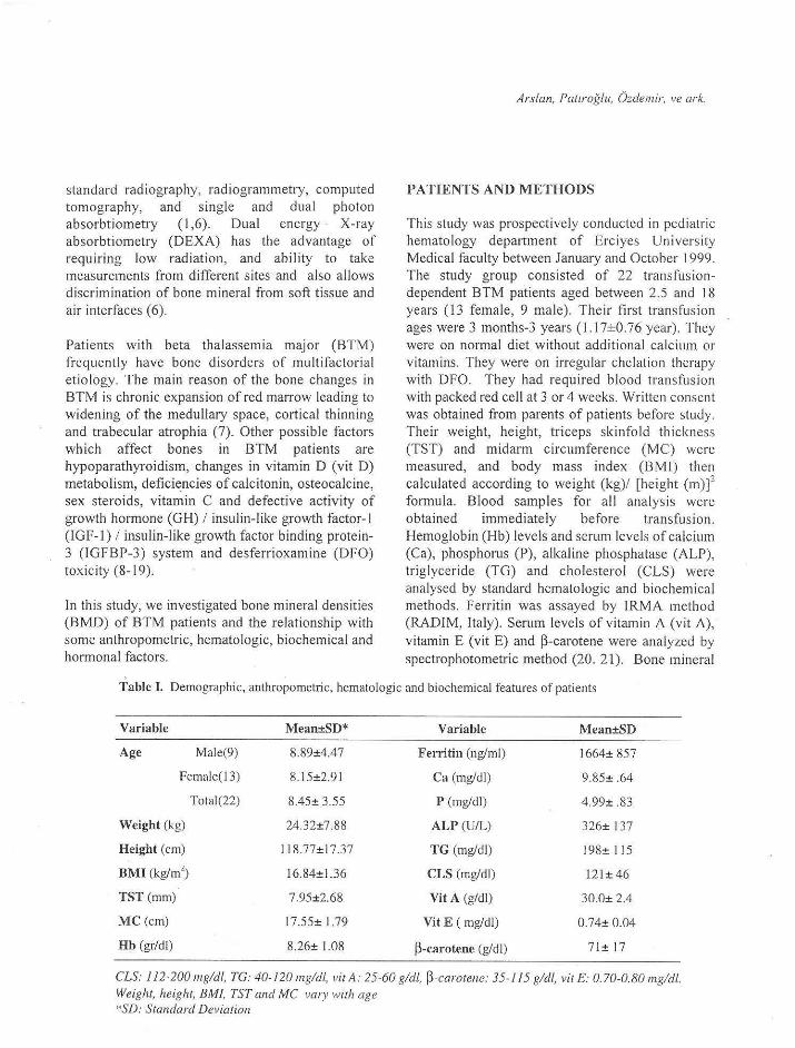
Table I. Demographic, anthropometric, hematologic and biochemical features of patients
Yariable Mean±SD* Yariable Mean±SD
Age Male(9) 8.89±4.47 Ferritin (ng/ml) 1664± 857
Female(13) 8.15±2 .91 Ca (mg/dl) 9.85± .64
Total (22) 8.45± 3.55 P (mg/dl) 4.99 ± .83
Weight (kg) 24.32±7.88 ALP (U/L) 326± 137
Height (cm) 118.77±17.37 TG (mg/dl) 198± 115
BMI (kg/m2) 16.84±1.36 CLS (mg/dl) 121± 46
TST(mm) 7.95±2.68 Yit A (gidi) 30.0± 2.4
MC (cm) 17.55± 1.79 Yit E ( mg/dl) 0.74± 0.04
Hb (gr/dl) 8.26± 1.08 ~-carotene (gidi) 7 1± 17
CLS: 112-200 nıgldl, TC: 40-120 mglcll, viı A: 25-60 gidi, ~-caroteııe: 35 -115 gidi, vit E: O. 70-0.80 ıııgldl. Weight, heighı, BM!, TST aııd MC vary with age *SD: Standard Deviatioıı
Bone mineral density in beta thalassemia major patients
density (BMD) was measured by DEXA method (Holog ic QDR-4500 A) from Ll to L4 spine, aııd
left femoral neck in all patients. The average of the L 1 to L4 spinal measurement was expressed as spinal BMD. Vertebral BMD values of the patients were compared with the densitometer's standards and BMD values in normal children , measured using the same method (DEXA) (22,23). Patients' left femoral neck BMDs were compared with young
adult standards and BMD values in healthy children measured by the same method (DEXA) ( 24 ). We could not obtain BMD measurements from left femoral neck for children less than 9 years old.
Statistical analysis were performed using SPSS for Windows Release 5.0 and relation ofthe parameters with BMD values were evaluated with correlation, p values below 0.05 were accep ted as signifıcant.
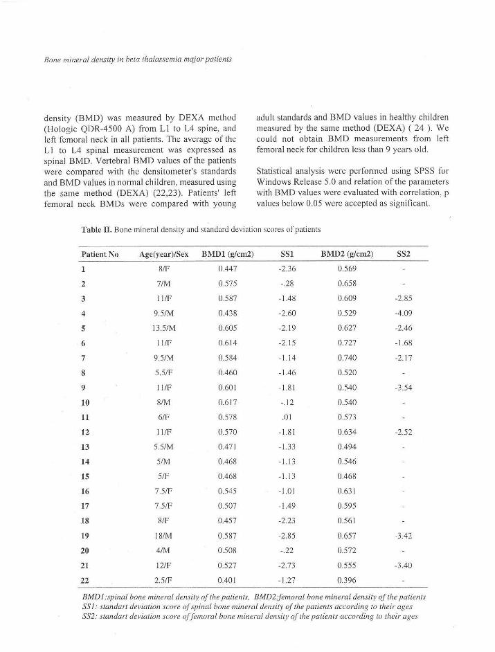
Table il . Bone mineral density and standard deviation scores of patients
Patient No Age(year)/Sex BMD1 (g/cm2) ss1 BMD2 (g/cm2) SS2
1 8/F 0.447 -2.36 0.569
2 7/M 0.575 -.28 0.658
3 11/F 0.587 -1 .48 0.609 -2.85
4 9.5/M 0.438 -2.60 0.529 -4.09
5 13.5/M 0.605 -2.19 0.627 -2.46
6 1 l/F 0.614 -2.15 0.727 -1.68
7 9.5/M 0.584 -1. i 4 0.740 -2.17
8 5.5/F 0.460 -1.46 0.520
9 11/F 0.601 -1.81 0.540 -3.54
10 8/M 0.617 -.12 0.540
11 6/F 0.578 .Ol 0.573
12 11/F 0.570 -1.81 0.634 -2.52
13 5.5/M 0.471 -1.33 0.494
14 5/M 0.468 -1.13 0.54 6
15 5/F 0.468 - 1.13 0.468
16 7.5/F 0.545 -1.01 0.631
17 7.5/F 0.507 -1.49 0.595
18 8/F 0.457 -2.23 0.561
19 18/M 0.587 -2.85 0.657 -3.42
20 4/M 0.508 -.22 0.572
21 12/F 0.527 -2.73 0.555 -3.40
22 2.5/F 0.401 -1.27 0.396
BMDl:spinal bone mineral deıısiıy of the patients, BMD2.femoral bone mineral deıısity of ıhe patienıs SSJ: standart deviation score of spinal bone mineral density of the patients accordiııg ıo their ages SS2: standart deviation score of feınoral hoııe mineral density of tlıe patieııts according to t/ıeir ages
Arsla n, Patır~ğlu, Özdemir, ve ark.
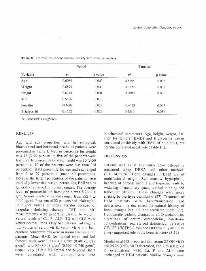
Table 111. Correlation of bone mineral density with some parameters
Spinal Femoral
Variable r* p value r* p value
Age 0.6069 0.003
Weight 0.6898 0.000
Height 0.6718 0.001
MC 0.5206 0.013
Ferritin -0.4689 0.028
Triglycerid 0.4652 0.029
*r: correlation coefficient
RESULTS
Age and sex properties, and hematological, biochemical and hormona! results of patients were presented in Table I. Median percentile for weight was 18 (3-90 percentile; five of the patients were less than 3rd percentile) and for height was 10 (3-50 percentile; 10 of the patients were less than 3rd percentile). BMI percentile for age and sex ranged from 3 to 97 percentile (mean 50 percentile). Because the height percentiles of the patients were markedly lower than weight percentiles, BMI values generally remained in normal ranges. The average level of pretransfusion hemoglobin was 8.26±1.8 gidi. Serum levels of ferritin ranged from 222.7 to 4000 ng/ınl. Fourteen of22 patients had 1500 ng/ml or higher values of serum ferritin because of irregular chelating therapy. TST and MC measurements were generally parallel to weight. Serum levels of Ca, P, ALP, TG and CLS were within normal limits. Only two patients had slightly low values of serum vit E. Serum vit A and beta carotene concentrations were in normal ranges in ali patients. Mean BMD for lumbal spine and left femoral neck were 0.53 ±0.07 g/cm 2 (0.401- 0.6 I 7 g/cm
2) and 0.58±0.08 g/cm 2 (0.396 - 0.740 g/cm 2
)
respectively (Table II). Spinal and femoral BMD were correlated with anthropometric and
0.5755 0.005
0.6190 0.002
0.7080 0.000
-0.4323 0.045
0.4530 0.034
biochemical parameters. Age, height, weight, MC (not for femoral BMD) and tr iglyceride values correlated positively with BMD of both sites, but fetTitin correlated negatively (Table III).
DISCUSSION
Patients with BTM frequently have osteopenia measured using DEXA and other method s (9,10,14,25,26). Bone changes in BTM are of multifactorial origin. Red marrow hype rplasia , because of chronic anemia and hypoxia, leads to widening of medullary space, cortica l thinning and trabecular atrophy. These changes were more striking before hypertransfusion (27). Treatınent of BTM patients with hypertransfusion and desferrioxamine decreased the natura ! history of bone changes but did not eradicate them (27). Hypoparathyroidism, changes in vit D metabolism , alterations of serum osteocalcine, calci tonin concentrations, sex steroid deficiency, defective GH/IGF-1/IGFBP-3 axis and DFO tox icity a lso play a very important role in the bone struc ture (8-1 9)
Moulas et al (13 ) reported that serum 25-0H vit D and 24,25 (OH) 2 vit D decreased , and 1,25 (OH) 2 vi t D, osteocalcine, PTH, Ca, P and ALP were unchanged in BTM patients. Simi lar changes were
Bone mineral density in beta thalassemia major patienıs
observed by Dandona et al ( 12 ) i.e. 25-0H vit D concentrations were lower than controls and l .25 (OH) 2 vit D, PTH and osteocalcine concentrations were simi lar. So I iman et al ( 14 ) reported that B TM patients have lower BMD than constitutional short stature (CSS) children . They noticed that five of their 30 thalassemic patients had hypocalcemia, two of the fıve had hypoparathyroidism and three had rickets. Aloia et al (8) reported both vit D and PTH defıciencies in thalassemic patients. Zamboni et al (16) demonstrated that serum Ca, P, PTH, 25-0H vit D, l ,25(0H) 2 vit D concentrations and urinary cAMP excretion were lower than the contro l group, whereas serum calcitonin and urinary P and OHprolin levels were higher. Canatan et a l ( 11) treated BTM patients with salmon calcitonin for one year and observed improvement in bone pain and osteoporosis.
In our patients, BMD values were low for same age and sex. Serum levels ofCa, P and ALP were within normal ranges. No parameter regarding vit D metabolism was studied in these patients.
Bone mineralization normally increases with growth and accelerates with puberty. Maximum bone mineral acquisition is achieved with the onset of puberty and most of the adult bone content is acquired during this period (5,9,28 ,29). Delayed growth and puberty is a major feature of BTM. Growth hormone and related proteins (IGF-1 and IGFBP-3), and sex steroids have been evaluated in several studies (14,17,30 ). So liman et al (17) showed that serum IGF-1 and IGFBP-3 concentrations were lower in BTM patients than in con tro ls. BMD was highly correlated with circulating IGF-1 and IGFBP -3 concentrations. Serum GH response to provocation was defective in 40 % patients. Serum ferritin concentration correlated with GH peak response to provocation and circulating IGF-1, and IGFBP-3 concentrations. in the IGF generation test after GH injection, thalassemic children had signifıcantly lower IGF-1 and IGFBP-3 levels than CSS and GHD patients. After one year therapy with hGH, there was a marked acceleration of growth velocity in GH
deficient short BTM patients, but this acceleration was slower than CSS and GHD patients . They concluded that some children with BTM have defective GH/IGF-1/JGFBP-3 axis and suggest the presence ofpartial resistance to GH.
Beta thalassemia major patients with hypogonadism had low BMD values and treatment with transdermal estrogen (for females) or hCG (for ma les) resulted in impairment in BMD (9). Filosa et al (25) repor ted that patients with thalass emia had lower BMD than contro ls, and severe form of the disea se corresponded to severe reduction in BMD and to hypogonadism. Güler et al (31) found that GH deficiency was 43.4 % and partial GH deficiency was 21.7 % in a previous study in our patients. Ali patients were at the prep ubertal age except for two, and both had pubertal problems. Eighty-two % of our patients have low levels of IGF-1 in the serum for same age and sex in a the previous study (31 ).
Desferrioxaınine therapy (toxicity) may induce some bone changes and growth failure in thalassem ic children. Flattening of the vertebral bodies, widened growth plates, circumferen tial metaphyseal osseous defect , sharp zones of provisional calcifıcation and rachitic-like changes in long bones were repor ted in DFO treated patients. These fındings were obse rved especially in early started treatment and in pat ients treated with high dose DFO (18, 19).
in this study we found that BTM patients had low BMD, measured from lumbar spine and left femoral neck. Seven of the 22 patients had L l -L4 lumbar spine BMD values of less than 2 standard deviation for same age and sex. Eleven had BMD values between (-1) and (-2 ) standard deviation. Our patients' BMDs were also compared to BMD va lues in Glastre (22), Pond er (23) and Maynard's (24) studies and similar resul ts were obtained. The re is very limited data for left femoral neck BMD for healthy children, so we coınpared the left fenıoral neck BMD values ofthe patients whose ages were 8 or above similarly to Maynard's report (24). Seven of the nine patients whose ages were 8 or above had BMD values of less than 2 standard deviation for
same age and sex. The other two patients had standard deviation between (-1) and (-2). Our patients' BMD values for tumba! spine and left: femoral neck correlated to each other similarly to Henderson's report (32). BMD values correlated with age weight, height , MC and serum triglyceride, and but not surprisingly, inversely correlated with serum ferritine. Spinal BMDs negatively (not significantly) correlated with pretransfusion hemoglobin values (p= .062). We conclude that BMD values from lumbal spine and upper femur of BTM patients were signifıcantly lower than those of healthy children. Age, weight, height, serum ferritin and pretransfusion hemoglobin (weak effect on spinal BMD) are effective parameters on BMD. Hypertransfusion (higher pretransfusion Hb ), effective chelation and aggressive dietary management could positively affect BMD in BTM children.
REFERENCES
1. Fassler ALJ, Bonjour JP. Osteoporosis as a pediatric problem. Pediatr Clin North Am 1995;42:811-824.
2. Jouanny P, Gııillemin F, Kuntz C, Jeandel C, Poıırel J. Enviromental and genetic factors ajfecting bone mass: Similarity of bone density among members of healthy families. Arthritis Rhuem 1995;38:61-67.
3. Lonzer MD, lmrie R, Rogers D, Worley D, Licata A, Secic M. Effects of heredity, age weight, pııberty, activity and caleium intake on bone mineral density in ehi/dren. Clin Pediatr 1996;35: 185-189.
4. Johnson ML, Gang G, Kimberling W, Recker SM, Kimmel DB, Reeker RB. Linkage of a gene eausing high bone mass ta human chromosome I 1 (1 lql 2-13). Am J Hum Genel 1997;60: 1326-1332.
5. Rubin K, Sehirduan V, Gendreau P, Sar/arazi M, Mendola R, Dalsky G. Predictors of axial and peripheral bone mineral density in healthy children and adoleseents, with speeial attention ta the role of puberty. J Pediatr !993;123 :863-870.
Arslan, Patıroğ/u, Özdemir, ve ark.
6. Kimmel P. Radiologic methods ta evaluate bone mineral content . Ann lntern Med !984;100:908-911.
7. Rioja l, Girot R, Garabedian M, Cournot-Witmer G. Bone disease in ehi/dren with homozygous beta-thalassemia. Bone Miner 1990;8:69-86.
8. Aloia JF, Ostuni JA, Yeh JK, Zaino EC. Combined vitamin D parathyroid defeet in thalassemia major . Arch lnt Med 1982; 142:831-832.
9. Anapliotou ML, Kastanias iT, Psara P, Evangelou EA, Liparak i M, Dimitrioıı P. The eontribution of hypogonadism to the development of osteoporosis in thalassemia major: new therapeuti c appro aches . Clin Endoerinol (Oxford) !995;42:279-287.
I O. Bisbocei D, Livorn o P, Modina P et al. Osteodystrophy in thalassemia major. Ann !tal Med lnt 1993;8: 224-226. (Abstraet)
I !. Canatan D, Akar N, Arcasoy A. Ejfects of ealeitonin therapy on osteoporosis in patients with thalassemia .Acta Haematol !995;93:20-24.
12. Dandona P, Menon RK, Houlder S, Thomas M, Hojjbrand AV, Flynn DM. Serum 1,25 dihydroxyvitamin D and osteocalcin eoneentrations in thalassemia major. Areh Dis Child !987; 62: 474-477.
13. Moulas A, Challa A, Cha/iasos N, lapatsanis PD. Vitamin D metabolites (25-hydroxyvitaınin D, 24,25-dihydroxyv itamin D and 1,25-dihydroxyvitamin D) and osteocafcin in betathalassemia. Aeta Paed iatr !997 ;86:594-599.
14. Sa/iman AT, El Banna N, Abde l Fattah M, E/Zafabani MM, Ansar i BM. Bone mineral density in prepuberta f ehifdren with betathafassemia: Correfation with growth and hormona! data. Metab o/ism 1998;47:541-548.
15. Zaino EC, Yeh JK, Afoia J. Defective vitamin D metabolism in thalassemia maj or. Ann N Y Acad Sci 1985;445:127-/34.
16. Zamboni G, Marradi P, Tagliaro F, Dorizzi R, Tato L. Parathyroid hormone, cafc iton in and vitamin D metabolites in beta-thalasseınia
major. EurJ Pediatr 1986; 145: 133-136. 17. Soliman AT, El Bann a N, Ansari BM. GH
Bone mineral density in beta thalassenıia nıajor patients
response to provoeation and eireulating IGF-1 and 1GFBP-3 eoneentrations, the 1GF-l generation test and clinieal response to GH therapy in ehi/dren with beta-thafassemia. Eur J Endoerinof 1998; 138:394-400.
l 8. Briff P W, Winehester P, G iardina P J, Cunningham-Rundfes S. Deferoxamine-indueed bone dysplasia in patients with thalassemia major. AJR Am J Roentgenof 1991;156:561-565.
l 9. 0/ivieri NF, Koren G, Harris J et af. Growth failure and bony ehanges indueed by deferoxamine . Am J Pediatr Hematof Oneof 1992; 14: 48-56.
20. Needf JB, Pearson WN. Maero and miero methods for the determination serum vitamin A using trifluoroaeetie aeid. J Nuir 1962; 79: 454-462.
21. Rindi G. Rapid eo/orimetrie method for the determination of toeopherol and toeopherylaeetat in pfasma. Speeial reprint of Internationaf Review of Vitamin Researeh. 1958; 28: 225-234.
22. Glastre C, Braillon P, David L, Coehat P, Meunier P J, De/mas P D. Measurement of bone mineral eontent of the, fumbar spine by dua! energy X-ray absorbtiometry in normal ehi/dren : Correlations with growth parameters. J Clin Endoerinol Metab /990;70: 1330-1333.
23. Ponder SW, MeCormiek DP, Faweett HD, Palmer JL, MeKernan MG, Brouhard BH. Spinal bone mineral density in ehi/dren aged 5.00 through 11.99 years. AJDC l 990; 144: I 346-1348.
24. May:nard LM, Guo SS, Chumlea WC et al. Totalbodv and regional bone mineral eontent and areal bone mineral density in ehi/dren aged 8-
l 8 y : The fels longitudina! study. Am J Clin Nutr 1998;68: I l l 1-1117.
25. Fifosa A, Di Maio S, Voeea S, Saviano A, Esposito G, Pagana L. Longitudinal monitoring of bone mineral density in thalassemie patients. Genetie strueture and osteoporosis . A ela Peadiatr /997;86:3 42-346.
26. Chao T, Hwang B. The eorrelation of serum ferritin !eve! to Ca-P metabolism and bone density in thalassemie patients. J Pedia tr Endoerinol Metab l 996; 9: 609-6 I l.
27. Orzineo/o C, Castaldi G, Bariani L, Seııtellari PN. The evolutionary effeets of therapy on skeletal lesions in beta-thalassemia . Radiol Med (Torino) 1994;87:381-388. (Abstraet)
28. Bonjoıır JP, Theintz G, Buehs B, Slosman D, Rizzoli R. Critieal years and stages of pııberıy
far spinal and femoral bone mass aeeumulation during adoleseen ee. J Clin Endoerinol Metab !991;73:555-563 .
29. MeKay HA, Bailey DA, Mirwald RL, Davison KS, Faulkner RA. Peak bone mineral acerual and age at menarehe in adoleseent girls: A 6-year longitudinaf study. J Pediatr l 998; J 33:682-68 7.
30. Cavaflo L, Gurrado R, Gafla F, Zacehino C. De Mattia D, Tato L. Growth defieiency in polytransfused beta-thafassemia patients is not growth hormone dependent. Clin Endoerinol (Oxf) 1997;46:701-706.
31. Güler E. Endoerine complications in beta thafassemia major patients . Doetoraf thesis, Kayseri, 1998.
32. Henderson RC The eorrelation between dıtalenergy X-ray absorbtiometry measures of bone density in the proximaf femur and lumbar spine of ehi/dren. Skeletal Radi of l 997; 26: 544-547.