Embed Size (px)
Citation preview
20
Bone Quality Assessment for Dental Implants
Ayse Gulsahi Baskent University Faculty of Dentistry, Ankara,
Turkey
1. Introduction
Dental implants have become a predictable treatment option for restoring missing teeth. The
purpose of tooth replacement with implants is to restore adequate function and esthetics
without affecting adjacent hard and/or soft tissue structures. The use of dental implants in
oral rehabilitation has currently been increasing since clinical studies with dental implant
treatment have revealed successful outcomes (Turkyilmaz et al., 2008a). The successful
outcome of any implant procedure depends on a series of patient-related and procedure-
dependent parameters, including general health conditions, biocompatability of the implant
material, the feature of the implant surface, the surgical procedure, and the quality and
quantity of the local bone. (Turkyilmaz et al., 2007)
Successfully providing dental implants to patients who have lost teeth and frequently the
surrounding bone relies on the careful gathering of clinical and radiological information, on
interdisciplinary communication and on detailed planning. One of the most important
factors in determining implant success is proper treatment planning. In the past, periapical
radiographs along with panoramic images were used as the sole determinants of implant
diagnosis and treatment planning. With the advancement of radiographic technology,
Computed tomography (CT), as well as cone-beam computed tomography (CBCT) is
increasingly considered essential for optimal implant placement, especially in the case of
complex reconstructions (Benson & Shetty, 2009; Chan et al., 2010; Resnik et al., 2008).
2. Radiologic examination
The objectives of diagnostic imaging depend on a number of factors, including the amount
and type of information required and the period of the treatment rendered. The desicion to
image the patient is based on the patient’s clinical needs. After a desicion has been made to
obtain images, the imaging modality is used that yields the necessary diagnostic information
related to the patient’s clinical needs and results in the least radiologic risk (Resnik et al., 2008).
The ideal imaging technique for dental implant care should have several essential
characteristics, including the ability to visualize the implant site in the mesiodistal, bucco-
lingual and superioinferior dimensions; the ability to allow reliable, accurate measurements;
a capacity to evaluate trabecular bone density and cortical thickness; reasonable access and
cost to the patient and minimal radiation risk (Benson & Shetty, 2009). Diagnostic imaging is
an integral part of dental implant therapy for preoperative planning, intraoperative
assessment, and postoperative assessment by use of a variety of imaging techniques.
www.intechopen.com
Implant Dentistry – The Most Promising Discipline of Dentistry
438
2.1 Selecting imaging technique for preoperative implant planning
The objectives of the preoperative implant imaging include all necessary surgical and prosthetic information to determine the quantity, quality and angulations of bone; selection of the potential implant sites, and to verify absence of pathology. However, there is no ideal imaging technique in the field of oral implantology that would be acceptable for all patients. All imaging techniques have inherent advantages and disadvantages (Resnik et al. 2008). In dental and medical radiology, a recommended principle when selecting the appropriate radiographic modality is based on radiologic dosage. Obviously, the goal is to choose a radiographic method providing sufficient diagnostic information for treatment planning with the least possible radiation dose (ALARA principle: as low as reasonably achievable) and costs for the patient. The preferred imaging procedure for this purpose seems to vary much among different parts of the world as well as among individual dentists.
2.1.1 Intraoral radiography
Traditionally, conventional radiographic images e.g., periapical and panoramic images have
been used to assist practitioners in planning implant treatment. Periapical radiographs
commonly are used to evaluate the status of adjacent teeth and remaining alveolar bone in
the mesiodistal dimension. In addition they have been used for determining vertical height,
architecture and bone quality (bone density, amount of cortical bone and amount of
trabecular bone). Although readily available and relatively inexpensive, periapical
radiography has geometric and anatomic limitations. If the paralleling technique is not used,
periapical radiographs create an image with foreshortening and elongation (Benson &
Shetty, 2009; Chan et al., 2010). When the x-ray beam is perpendicular to the film, but the
object is not parallel to the film, foreshortening will occur. If the x-ray beam is oriented
perpendicular to the object but not the film, elongation will occur. The most accurate
intraoral radiographic technique used for implant planning is the paralleling technique.
These principles in positioning will allow for an intraoral image with minimal distortion
and magnification. Therefore, standardized periapical radiographs with bite-blocks by using
paralling technique should be perform to the longitudinal studies (Benson & Shetty, 2009;
Resnik et al., 2008).
Because the periapical radiographs are unable to provide any cross-sectional information,
occlusal radiographs are used to determine bucco-lingual dimensions of the mandibular
alveolar ridge. However, the occlusal image records only the widest portion of the
mandible, which typically is located inferior to the alveolar ridge. This may give the
clinician the impression that more bone is available in the cross-sectional dimension than
actually exists. The occlusal technique is not useful for the maxillary arch because of the
anatomic limitations (Benson & Shetty, 2009).
2.1.2 Panoramic radiography
Panoramic radiographs have been used frequently as a radiographic method for preimplant
evaluation and the preparation of treatment protocols. Although the resolution and
sharpness of panoramic radiographs are less than those of intraoral radiographs, panoramic
radiographs is an excellent tool for the overview of the maxillofacial area, including many of
the vital structures, such as maxillary sinus, inferior alveolar nerve and nasal fossa.
Panoramic radiography units are widely available, making this imaging technique very
useful and popular as a screening (Benson & Shetty, 2009; Chan et al., 2010). (Figure 1)
www.intechopen.com
Bone Quality Assessment for Dental Implants
439
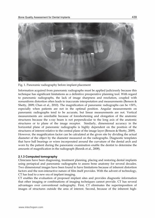
Fig. 1. Panoramic radiography before implant placement
Information acquired from panoramic radiographs must be applied judiciously because this
technique has significant limitations as a definitive preoperative planning tool. With regard
to panoramic radiographs, the lack of image sharpness and resolution, coupled with
nonuniform distortion often leads to inaccurate interpretation and measurements (Benson &
Shetty, 2009; Chan et al., 2010). The magnification of panoramic radiographs can be >30%,
especially when patients are not in the optimal position. Angular measurements on
panoramic radiographs tend to be accurate, but linear measurements are not. Vertical
measurements are unreliable because of foreshortening and elongation of the anatomic
structures because the x-ray beam is not perpendicular to the long axis of the anatomic
structures or to plane of the image receptor. Similarly, dimensional accuracy in the
horizontal plane of panoramic radiographs is highly dependent on the position of the
structures of interest relative to the central plane of the image layer (Benson & Shetty, 2009).
However, the magnification factor can be calculated at the given site by dividing the actual diameter of the object by the diameter measured on the radiographs. Diagnostic templates that have ball bearings or wires incorporated around the curvature of the dental arch and worn by the patient during the panoramic examination enable the dentist to determine the amounts of magnification in the radiograph (Resnik et al., 2008).
2.1.3 Computed tomography
Clinicians have been diagnosing, treatment planning, placing and restoring dental implants using periapical and panoramic radiographs to assess bone anatomy for several decades. Two dimensional images have been found to have limitations because of inherent distortion factors and the non-interactive nature of film itself provides. With the advent of technology, CT has lead to a new era of implant imaging. CT enables the evaluation of proposed implant sites and provides diagnostic information that other imaging or combinations of imaging techniques cannot provide. CT has several advantages over conventional radiography. First, CT eliminates the superimposition of images of structures outside the area of interest. Second, because of the inherent high-
www.intechopen.com
Implant Dentistry – The Most Promising Discipline of Dentistry
440
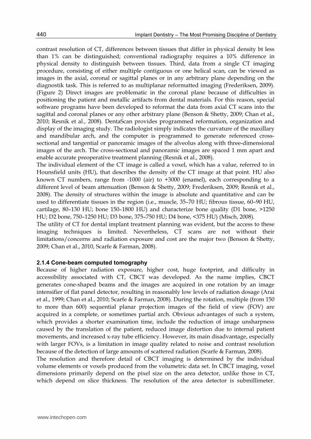
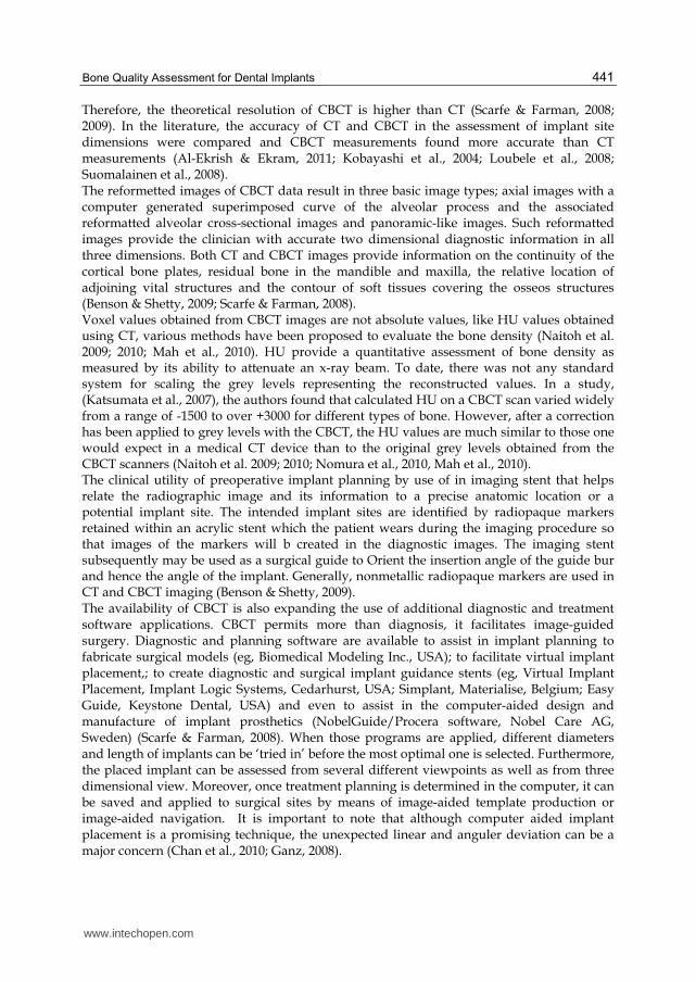
contrast resolution of CT, differences between tissues that differ in physical density bt less than 1% can be distinguished; conventional radiography requires a 10% difference in physical density to distinguish between tissues. Third, data from a single CT imaging procedure, consisting of either multiple contiguous or one helical scan, can be viewed as images in the axial, coronal or sagittal planes or in any arbitrary plane depending on the diagnostik task. This is referred to as multiplanar reformatted imaging (Frederiksen, 2009). (Figure 2) Direct images are problematic in the coronal plane because of difficulties in positioning the patient and metallic artifacts from dental materials. For this reason, special software programs have been developed to reformat the data from axial CT scans into the sagittal and coronal planes or any other arbitrary plane (Benson & Shetty, 2009; Chan et al., 2010; Resnik et al., 2008). DentaScan provides programmed reformation, organization and display of the imaging study. The radiologist simply indicates the curvature of the maxillary and mandibular arch, and the computer is programmed to generate referenced cross-sectional and tangential or panoramic images of the alveolus along with three-dimensional images of the arch. The cross-sectional and panoramic images are spaced 1 mm apart and enable accurate preoperative treatment planning (Resnik et al., 2008). The individual element of the CT image is called a voxel, which has a value, referred to in
Hounsfield units (HU), that describes the density of the CT image at that point. HU also
known CT numbers, range from -1000 (air) to +3000 (enamel), each corresponding to a
different level of beam attenuation (Benson & Shetty, 2009; Frederiksen, 2009; Resnik et al.,
2008). The density of structures within the image is absolute and quantitative and can be
used to differentiate tissues in the region (i.e., muscle, 35–70 HU; fibrous tissue, 60–90 HU,
cartilage, 80–130 HU; bone 150–1800 HU) and characterize bone quality (D1 bone, >1250
HU; D2 bone, 750–1250 HU; D3 bone, 375–750 HU; D4 bone, <375 HU) (Misch, 2008).
The utility of CT for dental implant treatment planning was evident, but the access to these
imaging techniques is limited. Nevertheless, CT scans are not without their
limitations/concerns and radiation exposure and cost are the major two (Benson & Shetty,
2009; Chan et al., 2010, Scarfe & Farman, 2008).
2.1.4 Cone-beam computed tomography
Because of higher radiation exposure, higher cost, huge footprint, and difficulty in
accessibility associated with CT, CBCT was developed. As the name implies, CBCT
generates cone-shaped beams and the images are acquired in one rotation by an image
intensifier of flat panel detector, resulting in reasonably low levels of radiation dosage (Arai
et al., 1999; Chan et al., 2010; Scarfe & Farman, 2008). During the rotation, multiple (from 150
to more than 600) sequential planar projection images of the field of view (FOV) are
acquired in a complete, or sometimes partial arch. Obvious advantages of such a system,
which provides a shorter examination time, include the reduction of image unsharpness
caused by the translation of the patient, reduced image distortion due to internal patient
movements, and increased x-ray tube efficiency. However, its main disadvantage, especially
with larger FOVs, is a limitation in image quality related to noise and contrast resolution
because of the detection of large amounts of scattered radiation (Scarfe & Farman, 2008).
The resolution and therefore detail of CBCT imaging is determined by the individual volume elements or voxels produced from the volumetric data set. In CBCT imaging, voxel dimensions primarily depend on the pixel size on the area detector, unlike those in CT, which depend on slice thickness. The resolution of the area detector is submillimeter.
www.intechopen.com
Bone Quality Assessment for Dental Implants
441
Therefore, the theoretical resolution of CBCT is higher than CT (Scarfe & Farman, 2008; 2009). In the literature, the accuracy of CT and CBCT in the assessment of implant site dimensions were compared and CBCT measurements found more accurate than CT measurements (Al-Ekrish & Ekram, 2011; Kobayashi et al., 2004; Loubele et al., 2008; Suomalainen et al., 2008). The reformetted images of CBCT data result in three basic image types; axial images with a computer generated superimposed curve of the alveolar process and the associated reformatted alveolar cross-sectional images and panoramic-like images. Such reformatted images provide the clinician with accurate two dimensional diagnostic information in all three dimensions. Both CT and CBCT images provide information on the continuity of the cortical bone plates, residual bone in the mandible and maxilla, the relative location of adjoining vital structures and the contour of soft tissues covering the osseos structures (Benson & Shetty, 2009; Scarfe & Farman, 2008). Voxel values obtained from CBCT images are not absolute values, like HU values obtained using CT, various methods have been proposed to evaluate the bone density (Naitoh et al. 2009; 2010; Mah et al., 2010). HU provide a quantitative assessment of bone density as measured by its ability to attenuate an x-ray beam. To date, there was not any standard system for scaling the grey levels representing the reconstructed values. In a study, (Katsumata et al., 2007), the authors found that calculated HU on a CBCT scan varied widely from a range of -1500 to over +3000 for different types of bone. However, after a correction has been applied to grey levels with the CBCT, the HU values are much similar to those one would expect in a medical CT device than to the original grey levels obtained from the CBCT scanners (Naitoh et al. 2009; 2010; Nomura et al., 2010, Mah et al., 2010). The clinical utility of preoperative implant planning by use of in imaging stent that helps relate the radiographic image and its information to a precise anatomic location or a potential implant site. The intended implant sites are identified by radiopaque markers retained within an acrylic stent which the patient wears during the imaging procedure so that images of the markers will b created in the diagnostic images. The imaging stent subsequently may be used as a surgical guide to Orient the insertion angle of the guide bur and hence the angle of the implant. Generally, nonmetallic radiopaque markers are used in CT and CBCT imaging (Benson & Shetty, 2009). The availability of CBCT is also expanding the use of additional diagnostic and treatment software applications. CBCT permits more than diagnosis, it facilitates image-guided surgery. Diagnostic and planning software are available to assist in implant planning to fabricate surgical models (eg, Biomedical Modeling Inc., USA); to facilitate virtual implant placement,; to create diagnostic and surgical implant guidance stents (eg, Virtual Implant Placement, Implant Logic Systems, Cedarhurst, USA; Simplant, Materialise, Belgium; Easy Guide, Keystone Dental, USA) and even to assist in the computer-aided design and manufacture of implant prosthetics (NobelGuide/Procera software, Nobel Care AG, Sweden) (Scarfe & Farman, 2008). When those programs are applied, different diameters and length of implants can be ‘tried in’ before the most optimal one is selected. Furthermore, the placed implant can be assessed from several different viewpoints as well as from three dimensional view. Moreover, once treatment planning is determined in the computer, it can be saved and applied to surgical sites by means of image-aided template production or image-aided navigation. It is important to note that although computer aided implant placement is a promising technique, the unexpected linear and anguler deviation can be a major concern (Chan et al., 2010; Ganz, 2008).
www.intechopen.com
Implant Dentistry – The Most Promising Discipline of Dentistry
442
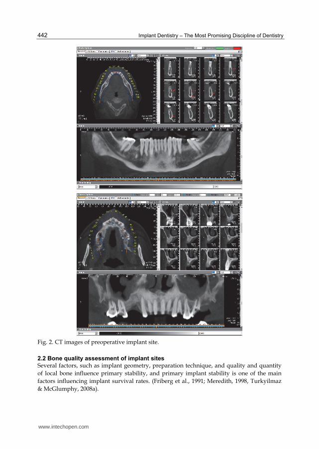
Fig. 2. CT images of preoperative implant site.
2.2 Bone quality assessment of implant sites
Several factors, such as implant geometry, preparation technique, and quality and quantity
of local bone influence primary stability, and primary implant stability is one of the main
factors influencing implant survival rates. (Friberg et al., 1991; Meredith, 1998, Turkyilmaz
& McGlumphy, 2008a).
www.intechopen.com
Bone Quality Assessment for Dental Implants
443
2.2.1 Implant stability measurements
Implant stability can be measured by non-invasive clinical test methods (i.e., insertion torque, the periotest, resonance frequency analysis). One of these quantitative methods is the insertion torque described by Johansson and Strid (1994). This method records the torque required to place the implant and provides valuable information about local bone quality. Another method, named Periotest, has been developed to measure the degree of the
periodontal integration of teeth and the stiffness of the bone/implant interface (Olive &
Aparicio, 1990; Turkyilmaz & McGlumphy, 2008b). The Periotest instrument measures the
deflection/deceleration of a tooth or implant that has been struck by a small pistil from
inside the instrument's hand piece. The contact time of the accelerated pistil to the implant,
which moves according to the strike, is calculated into a value called the Periotest value.
However, Periotest values include only a narrow range over the scale of the instrument and
thus, provide relatively less sensitive information about implant stability. Therefore, its
benefit on detection of osseointegration is a matter of debate.
Another method, resonance frequency analysis (RFA) has been introduced by Meredith and
coworkers (1996). In RFA, the stiffness of the bone/ implant interface is calculated from a
resonance frequency as a reaction to oscillations exerted onto the implant/bone system. The
implant is excited with an oscillating transducer screwed onto the implant and the
resonance specific to the resonance system 'implant/bone' is captured electronically over a
range of 5 to 15 kHz. RF values have clinically been correlated with changes in implant
stability during osseous healing, failure of implants to integrate and the supracrestal
dimensions of the implant. The results of a histomorphometric study suggested that RFA
values correlated well with the amount of bone-to-implant contact. These findings support
the use of RFA in evaluating changes in the bone healing and osseointegration process
following implant placement (Turkyilmaz & McGlumphy, 2008b).
2.2.2 Bone quality and quantity
The term bone quality is commonly used in implant treatment and in reports on implant
success and failure. Lindh et al. (2004) emphasized that bone density (Bone Mineral Density,
BMD) and bone quality are not synonymous. Bone quality encompasses factors other than
bone density such as skeletal size, the architecture and 3-dimensional orientation of the
trabeculea, and matrix properties. Bone quality is not only a matter of mineral content, but
also of structure. It has been shown that the quality and quantity of bone available at the
implant site are very important local patient factors in determining the success of dental
implants (Drage et al., 2007; Lindh et al., 2004).
The success rate obtained with dental implants depends to a great extent on the volume and quality of the surrounding bone. Therefore, it is important to know the bone quantity and quality of the jaws when planning implant treatment. Bone quantity of jawbone is broken down into five groups (from minimal to severe, A- E), based on residual jaw shape different rates of bone resorption following tooth extraction (Ribeiro-Rotta et al., 2010). During all stages of atrophy of the alveolar ridge, characteristic shapes result from the resorptive process. It is difficult to obtain implant anchorage in bone that is not very dense. Sufficient bone density and volume are therefore crucial factors for ensuring implant success (Lekholm & Zarb, 1985). Bone quality is broken down into four groups according to the proportion and stucture of
compact and trabecular bone tissue (Ribeiro-Rotta et al., 2010). Bone quality is categorized
into four groups: groups 1-4 or type I to IV (Bone Quality Index-BQI) (Figure 3).
www.intechopen.com
Implant Dentistry – The Most Promising Discipline of Dentistry
444
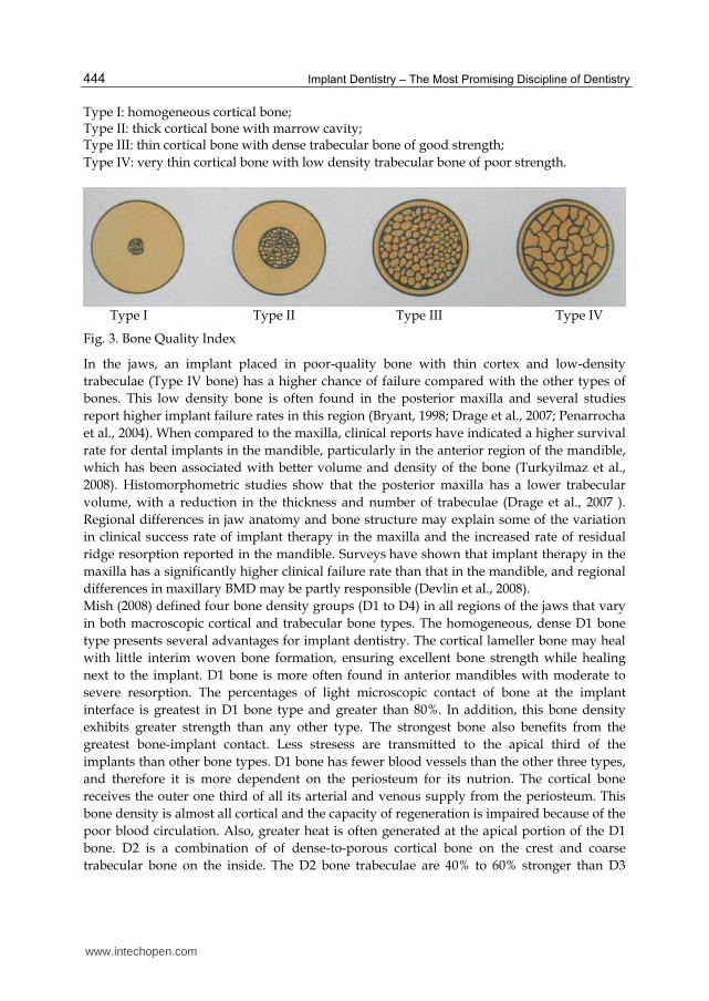
Type I: homogeneous cortical bone; Type II: thick cortical bone with marrow cavity; Type III: thin cortical bone with dense trabecular bone of good strength;
Type IV: very thin cortical bone with low density trabecular bone of poor strength.
Type I Type II Type III Type IV
Fig. 3. Bone Quality Index
In the jaws, an implant placed in poor-quality bone with thin cortex and low-density
trabeculae (Type IV bone) has a higher chance of failure compared with the other types of
bones. This low density bone is often found in the posterior maxilla and several studies
report higher implant failure rates in this region (Bryant, 1998; Drage et al., 2007; Penarrocha
et al., 2004). When compared to the maxilla, clinical reports have indicated a higher survival
rate for dental implants in the mandible, particularly in the anterior region of the mandible,
which has been associated with better volume and density of the bone (Turkyilmaz et al.,
2008). Histomorphometric studies show that the posterior maxilla has a lower trabecular
volume, with a reduction in the thickness and number of trabeculae (Drage et al., 2007 ).
Regional differences in jaw anatomy and bone structure may explain some of the variation
in clinical success rate of implant therapy in the maxilla and the increased rate of residual
ridge resorption reported in the mandible. Surveys have shown that implant therapy in the
maxilla has a significantly higher clinical failure rate than that in the mandible, and regional
differences in maxillary BMD may be partly responsible (Devlin et al., 2008).
Mish (2008) defined four bone density groups (D1 to D4) in all regions of the jaws that vary
in both macroscopic cortical and trabecular bone types. The homogeneous, dense D1 bone
type presents several advantages for implant dentistry. The cortical lameller bone may heal
with little interim woven bone formation, ensuring excellent bone strength while healing
next to the implant. D1 bone is more often found in anterior mandibles with moderate to
severe resorption. The percentages of light microscopic contact of bone at the implant
interface is greatest in D1 bone type and greater than 80%. In addition, this bone density
exhibits greater strength than any other type. The strongest bone also benefits from the
greatest bone-implant contact. Less stresess are transmitted to the apical third of the
implants than other bone types. D1 bone has fewer blood vessels than the other three types,
and therefore it is more dependent on the periosteum for its nutrion. The cortical bone
receives the outer one third of all its arterial and venous supply from the periosteum. This
bone density is almost all cortical and the capacity of regeneration is impaired because of the
poor blood circulation. Also, greater heat is often generated at the apical portion of the D1
bone. D2 is a combination of of dense-to-porous cortical bone on the crest and coarse
trabecular bone on the inside. The D2 bone trabeculae are 40% to 60% stronger than D3
www.intechopen.com
Bone Quality Assessment for Dental Implants
445
tarbeculae. This bone type occurs most frequently in the anterior mandible, followed by the
posterior mandible. On occasion it is observed in the anterior maxilla, especially for a single
missing tooth. D2 bone provides excellent implant interface healing, and osseointegration is
very predictable. The intrabony blood supply allows bleeding during the osteotomy, which
helps control overheating during preparation and is most beneficial for bone-implant
interface healing. D3 is composed of thinner porous cortical bone on the crest and fine
trabecular bone within the ridge. The trabecula are approximtely 50% weaker than those in
D2 bone. D3 bone is found most often in the anterior maxilla and posterior regions of the
mouth in either arch. The D3 anterior maxilla is usually of less width than its mandibular D3
counterpart. The D3 bone is not only 50% weaker than D2 bone, the bone-implant contact is
also less favorable in D3 bone. The additive factors can increase the risk of implant failure.
D4 bone has very little density and little or no cortical crestal bone. It is the opposite
spectrum of D1 (dense cortical bone). The most commen locations for this type of bone are
the posterior region of the maxilla. It is rarely observed in mandible. The bone trabeculae
may be up to 10 times weaker than the cortical bone of D1. The bone-implant contact after
initial loading is often less than 25%. Bone trabeculae are sparse and, as a result, initial
fixation of any implant design presents a surgical challenge (Misch, 2008).
2.2.3 Bone mineral density measurements
BMD is the amount of bone tissue in a certain volume of bone. Assessment of jaw BMD may
be considered useful in implant planning (Gulsahi et al., 2010). Several approaches have
been introduced to measure jawbones and skeletal bones density. Densitometric
measurements of panoramic and periapical radiographs have been used, as have more
advanced methods such as Dual Energy X-Ray Absorptiometry (DEXA), CT and CBCT.
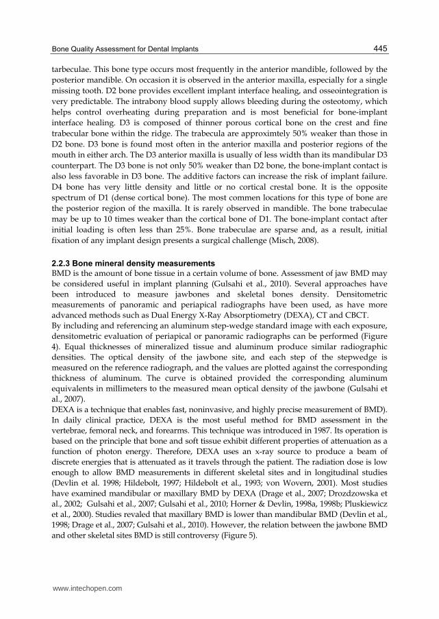
By including and referencing an aluminum step-wedge standard image with each exposure,
densitometric evaluation of periapical or panoramic radiographs can be performed (Figure
4). Equal thicknesses of mineralized tissue and aluminum produce similar radiographic
densities. The optical density of the jawbone site, and each step of the stepwedge is
measured on the reference radiograph, and the values are plotted against the corresponding
thickness of aluminum. The curve is obtained provided the corresponding aluminum
equivalents in millimeters to the measured mean optical density of the jawbone (Gulsahi et
al., 2007).
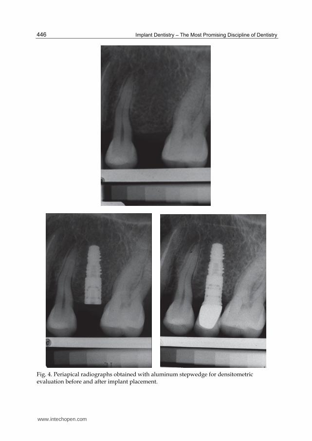
DEXA is a technique that enables fast, noninvasive, and highly precise measurement of BMD).
In daily clinical practice, DEXA is the most useful method for BMD assessment in the
vertebrae, femoral neck, and forearms. This technique was introduced in 1987. Its operation is
based on the principle that bone and soft tissue exhibit different properties of attenuation as a
function of photon energy. Therefore, DEXA uses an x-ray source to produce a beam of
discrete energies that is attenuated as it travels through the patient. The radiation dose is low
enough to allow BMD measurements in different skeletal sites and in longitudinal studies
(Devlin et al. 1998; Hildebolt, 1997; Hildebolt et al., 1993; von Wovern, 2001). Most studies
have examined mandibular or maxillary BMD by DEXA (Drage et al., 2007; Drozdzowska et
al., 2002; Gulsahi et al., 2007; Gulsahi et al., 2010; Horner & Devlin, 1998a, 1998b; Pluskiewicz
et al., 2000). Studies revaled that maxillary BMD is lower than mandibular BMD (Devlin et al.,
1998; Drage et al., 2007; Gulsahi et al., 2010). However, the relation between the jawbone BMD
and other skeletal sites BMD is still controversy (Figure 5).
www.intechopen.com
Implant Dentistry – The Most Promising Discipline of Dentistry
446
Fig. 4. Periapical radiographs obtained with aluminum stepwedge for densitometric evaluation before and after implant placement.
www.intechopen.com
Bone Quality Assessment for Dental Implants
447
Fig. 5. DEXA measurement before implant placement.
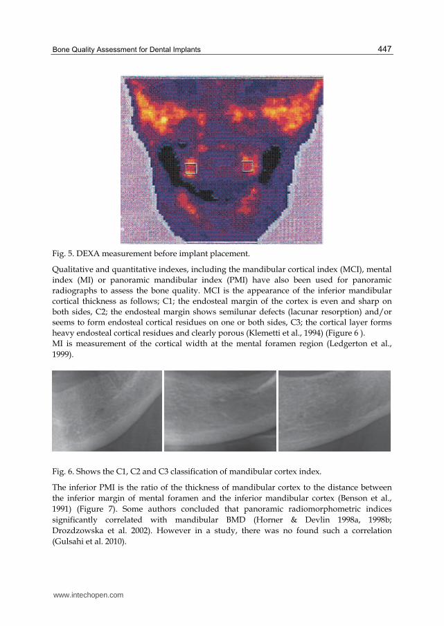
Qualitative and quantitative indexes, including the mandibular cortical index (MCI), mental
index (MI) or panoramic mandibular index (PMI) have also been used for panoramic
radiographs to assess the bone quality. MCI is the appearance of the inferior mandibular
cortical thickness as follows; C1; the endosteal margin of the cortex is even and sharp on
both sides, C2; the endosteal margin shows semilunar defects (lacunar resorption) and/or
seems to form endosteal cortical residues on one or both sides, C3; the cortical layer forms
heavy endosteal cortical residues and clearly porous (Klemetti et al., 1994) (Figure 6 ).
MI is measurement of the cortical width at the mental foramen region (Ledgerton et al.,
1999).
Fig. 6. Shows the C1, C2 and C3 classification of mandibular cortex index.
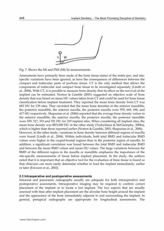
The inferior PMI is the ratio of the thickness of mandibular cortex to the distance between
the inferior margin of mental foramen and the inferior mandibular cortex (Benson et al.,
1991) (Figure 7). Some authors concluded that panoramic radiomorphometric indices
significantly correlated with mandibular BMD (Horner & Devlin 1998a, 1998b;
Drozdzowska et al. 2002). However in a study, there was no found such a correlation
(Gulsahi et al. 2010).
www.intechopen.com
Implant Dentistry – The Most Promising Discipline of Dentistry
448
Fig. 7. Shows the MI and PMI (MI/h) measurements.
Assessments have primarily been made of the bone tissue status of the entire jaw, and site-specific variations have been ignored, as have the consequences of differences between the compact and trabecular parts of jawbone tissue. CT is the only method that allows the components of trabecular and compact bone tissue to be investigated separately (Lindh et al., 2004). With CT, it is possible to measure bone density that its effect on the survival of the implant can be estimated. Norton & Gamble (2001) suggested an objective scale of bone density that was besed on mean HU values taken from CT and could be used for bone tissue classification before implant treatment. They reported the mean bone density from CT was 682 HU for 139 sites. They recorded that the mean bone densities in the anterior mandible, the posterior mandible, the anterior maxilla, the posterior maxilla were 970, 669, 696, and 417 HU respectively. Shapurian et al. (2006) reported that the average bone density values in the anterior mandible, the anterior maxilla, the posterior maxilla, the posterior mandible were 559, 517, 333 and 321 HU for 219 implant sites. When considering all implant sites, the mean bone density was 887±180 HU in the other study (Turkyilmaz & McGlumphy, 2008a), which is higher than those reported earlier (Norton & Gamble, 2001; Shapurian et al., 2006). However, in the other study, variations in bone density between different regions of maxilla
were found (Lindh et al., 2004). Within individuals, both total BMD and trabecular BMD
values were higher in the cuspid-frontal regions than in the posterior region of maxilla. In
addition, a significant correlation was found between the total BMD and trabecular BMD
and between the mean BMD values and mean HU values. The large variations between the
BMD of the different region in the maxilla or mandible emphasize the importance of the
site-specific measurements of tissue before implant placement. In the study, the authors
noted that it is important that an objective tool for the evaluation of bone tissue is found so
that clinicians can more easily determine whether to load the implant immediately, earlier
or later (Ericsson et al., 2002).
2.3 Intraoperative and postoperative assessments
Intraoral and panoramic radiographs usually are adequate for both intraoperative and
postoperative assessments. Intraoperative imaging may be required to confirm correct
placement of the implant or to locate a lost implant. The two aspects that are usually
assessed with time after implant placement are the alveolar bone height around the implant
and the appearance of the bone immediately adjacent to and surrounding the implant. In
general, periapical radiographs are appropriate for longitudinal assessments. The
www.intechopen.com
Bone Quality Assessment for Dental Implants
449
angulation of the x-ray beam must be within 9 degrees of the long axis of the fixture to open
the threads on the image on most threaded fixtures (Benson & Shetty, 2009).
In evaluating the the bone height around an implant, an effort should be made to reproduce the vertical angulation of the central ray of the x-ray beam as closely as possible between radiographs. Distal and mesial marginal bone height is measured from a collar of the implant, or in tha case of threaded implants by use of known interthreaded measurements and compared with bone levels in previous periapical radiographs. The presence of relatively distinct bone margins with a constant height relative to the implant suggests successful osseos integration. Any resorptive changes, if present, are evidenced by apical migration of the alveolar bone or distinct osseos margins. Radiographic studies suggest that the rate of marginal bone loss after successful implantation is approximately 1.2 mm in the first year, subsequently tapering off to about 0.1 mm in succeeding years (Benson & Shetty, 2009). The success of an implant can also be evaluated by the appearance of normal bone surrounding it and its apposition to the surface of the implant body. The development of a thin radiolucent area that closely follows the outline of the implant usually correlates to clinically detectable implant mobility, it is an important indicator of failed osseointegration. Changes in the periodontal ligament space of associated teeth are also useful in monitoring the functional competence of the implant-prostheses composite. Any widening of the periodontal ligament space compared with baseline radiographs indicates poor stress distribution and forecasts implant failure (Benson & Shetty, 2009). After successful implantation, radiographs may be made at regular intervals to assess the success or failure of the implant fixture (Benson & Shetty, 2009).
3. Conclusion
In summary, diagnostic imaging is an integral part of dental implant therapy for preoperative planning, intraoperative and posoperative assessment by use of variety of techniques. In general, good starting point would be proceed with panoramic radiograph and possibly intraoral radiographs if greater image detail is required. If images are required of all of the maxilla and mandible to evaluate possible implant sites, cross-sectional images assists to clinician. Today, CBCT is the best modality for the ease of acquisition and relatively low radiation risk even for single implants.
4. Acknowledgement
Special thanks to Dr İlker Cebeci for the CBCT images from his archieve.
5. References
Al-Ekrish, A.A., Ekram, M. (2011). A comparative study of the aacuracy and reliability of multidetector computed tomography and cone beam computed tomography in the assessment of dental implant site dimension. Dentomaxillofac Radiol, 40, 67-75.
Arai, Y., Tammisalo, E., Iwai, K. (1999). Development of a compact computed tomographic apparatus for dental use. Dentomaxillofac Radiol, 28, 245-8.
Benson BW, Prihoda TJ, Glass BJ. (1991). Variations in adult cortical bone mass as measured by a panoramic mandibular index. Oral Surg Oral Med Oral Pathol, 71, 349-356.
www.intechopen.com
Implant Dentistry – The Most Promising Discipline of Dentistry
450
Benson, B.W. & Shetty, V (2009). Dental Implants, In: Oral Radiology Principles and Interpretation, S.C. White & M. J. Pharoah, pp. 597-612, Mosby, Elsevier, ISBN 978-0-323-04983-2, St. Louis, Missouri.
Bryant, S.R. (1998). The effects of age, jaw site, and bone condition on oral implant outcomes. Int J Prosth, 11, 470-90.
Chan H-L., Misch K., Wang H-L. (2010). Dental Imaging in Implant Treatment Planning. Implant Dent, 19, 288-298.
Devlin H, Horner K, Ledgerton D. (1998). A comparison of maxillary and mandibular bone mineral densities. J Prosthet Dent, 79, 323-7.
Drage NA, Palmer RM, Blake G, Wilson R, Crane F, Fogelman I. (2007). A comparison of bone mineral density in the spine, hip and jaws of edentulous subjects. Clin Oral Impl Res, 18, 496-500.
Drozdzowska B., Pluskiewicz W., Tarnawska B. (2002). Panoramic based mandibular indices in relation to mandibular bone mineral density and skeletal status assessed by dual energy X-ray absorptiometry and quantitative ultrasound. Dentomaxillofac Radiol, 31, 361-7.
Ericsson, I., Nilner, K. (2002). Early functional loading using Branemark dental implants. J Periodont Restor Dent, 22, 9-19.
Frederiksen, N., L. (2009). Advanced Imaging, In: Oral Radiology Principles and Interpretation, S.C. White & M. J. Pharoah, pp. 207-224, Mosby, Elsevier, ISBN 978-0-323-04983-2, St. Louis, Missouri.
Friberg B, Jemt T, Lekholm U. (1991). Early failures in 4641 consecutively placed Branemark dental implants: a study from stage I surgery to the connection of completed prostheses. Int J Oral Maxillofac Implants, 6, 142–6.
Johansson, P., Strid K.G. (1994). Assessment of bone quality from placement resistance during implant surgery. Int J Oral Maxillofac Implants, 9, 279-88.
Ganz, S. (2008). Computer-aided Design/Computer-aided Manufacturing Applications Using CT and Cone Beam CT Scanning Technology. Dent Clin N Am, 52, 777-808.
Gulsahi A., Paksoy C.S., Yazicioglu N., Arpak N., Kucuk NO, Terzioglu H. The Assessment of Bone Density Differences between Conventional and Bone-Condensing Techniques using Dual Energy X-Ray Absorptiometry and Radiography. (2007). Oral Surg Oral Med Oral Pathol Oral Radiol Endod, 104, 692-98.
Gulsahi, A., Ozden, S., Paksoy, C.S., Kucuk, O., Cebeci, A.R.I, Genc Y. (2010). Assessment of Bone Mineral Density in The Jaws and Its Relationship to radiomorphometric Indices. Dentomaxillofac Radiol, 39, 284-89.
Hildebolt CF (1997). Osteoporosis and oral bone loss. Dentomaxillofac Radiol, ;26, 3-15. Hildebolt CF, Rupich RC, Vannier MW, Zerbolio DJ Jr, Shrout MK, Cohen S, et al. (1993).
Inter relationships between bone mineral content measures. Dual energy radiography (DER) and bitewing radiographs (BWX). J Clin Periodontol, 20, 739-45.
Horner K, Devlin H. (1998a). The relationship between mandibular bone mineral density and panoramic radiographic measurements. J Dent, 26, 337-343.
Horner K, Devlin H. (1998b). The relationships between two indices of mandibular bone quality and bone mineral density measured by dual energy x-ray absorptiometry. Dentomaxillofac Radiol, 27, 17-21.
Johansson, P., Strid, K.G. (1994). Assessment of bone quality from placement resistance during implant surgery. Int J Oral Maxillofac Implants, 9, 279-88.
www.intechopen.com
Bone Quality Assessment for Dental Implants
451
Katsumata, A., Hirukawa, A., Okumura, S., Naitoh, M., Fujishita, M., Ariji, E., et al. (2007). Effects of image artifacts on gray-value density in limited-volume-cone-beam Computerized tomography. Oral Surg Oral Med Oral Pathol Oral Radiol Endod, 104, 829-36.
Klemetti E, Kolmakov S, Kroger H. (1994). Pantomography in assessment of the osteoporosis risk group. Scand J Dent Res, 102, 68-72.
Kobayashi, K., Shimoda, S.,Nakagawa, Y., Yamamoto, A. (2004). Accuracy in measurement of distance using limited cone beam computerized tomography. Int J Oral Maxillofac Implants, 19, 228-31.
Ledgerton D, Horner K, Devlin H, Worthington H. (1999). Radiomorphometric indices of the mandible in a British female population. Dentomaxillofac Radiol, 28,173-181.
Lekholm U, Zarb G.A, (1985). Patient selection and preparation. In: Branemark PI, Zarb GA, Albrektsson T, editors. Tissue-integrated prostheses: osseointegration in clinical dentistry. pp. 199-209, Chicago: Quintessence.
Lindh C, Obrant K, Petersson A. (2004). Maxillary bone mineral density and its relationship to the bone mineral density of the lumbar spine and hip. Oral Surg Oral Med Oral Pathol Oral Radiol Endod, 98, 102-109.
Loubele, M., Van Assche, N., Carpentier, K., Maes, F., Jacobsi R., van Steenberghe, D., et al. (2008). Comparative localized linear accuracy of small-field cone beam CT and multislice CT for alveolar bone measurements. Oral Surg Oral Med Oral Pathol Oral Radiol Endod, 105, 512-18.
Mah, P., Reeves, T.E., McDavid, W.D. (2010). Deriving Hounsfield units using grey levels in cone-beam computed tomography. Dentomaxillofac Radiol, 39, 323-35.
Meredith N. Assessment of implant stability as a prognostic determinant. (1998). Int J Proshodont, 11, 491–501.
Meredith, N., Alleyne, D., Cawley P. (1996). Quantitative determination of the stability of the implant-tissue interface using resonance frequency analysis. Clin Oral Implants Res, 7, 261–7.
Misch C.,E. (2008). Density of Bone: Effects on surgical approach and healing, In: Contemporary Implant Dentistry, C.E. Misch (ed), pp. 645-667, Mosby, Elsevier, ISBN 978-0-323-04373-1, Canada.
Naitoh, M., Hirukawa, A., Katsumata, A., Ariji, E. (2009). Evaluation of voxel values in mandibular cancellous bone: Relationship between cone-beam computed tomography and multislice helical computed tomography. Clin Oral Implants Res, 20, 503-6.
Naitoh, M., Hirukawa, A., Katsumata, A., Ariji, E. (2010). Prospective study to estimate mandibular cancellous bone density using large-volume cone-beam computed tomography. Clin Oral Implants Res, 21, 1309-13.
Nomura, Y., Watanabe, H., Honda, E., Kurabayashi, T. (2010). Reliability of voxel values from cone-beam computed tomography for Dental use in evaluating bone mineral density. Clin Oral Implants Res, 21, 558-62.
Norton, M.R., Gamble, C. (2001). Bone classification: an objective scale of bone density using the computerized tomography scan. Clin Oral Impl Res, 12, 79-84.
Olive, J., Aparicio, C. (1990). Periotest method as a measure of osseointegrated oral implant stability. Int J Oral Maxillofac Implants, 5, 390-400.
www.intechopen.com
Implant Dentistry – The Most Promising Discipline of Dentistry
452
Penarrocha, M., Palomar, M., Sanchis, J.,M., Guarinos, J., Balaguer, J. (2004). Radiologic study of marginal bone loss around 108 dental implants and its relationship to smoking, implant location and morphology. Int J Oral Maxillofac Impl 19, 861-7.
Pluskiewicz W., Tarnawska B., Drozdzowska B. (2000). Mandibular bone mineral density measured using dual-energy X-ray absorptiometry: relationship to hip bone mineral density and quantitative ultrasound at calcaneus and hand phalanges. Br J Radiol, 73, 288-292.
Resnik, R.R., Kircos, L. and Misch C.,E. (2008). Diagnostic Imaging and Techniques, In: Contemporary Implant Dentistry, C.E. Misch (ed), pp. 38-67, Mosby, Elsevier, ISBN 978-0-323-04373-1, Canada.
Ribeiro-Rotta, R.F., Lindh, C., Pereira, A.C., Rohlin, M. Ambiguity in bone tissue characteristics as presentes in studies on dental implant planning and placement: a systematic review. Clin Oral Impl Res, (in-press)
Scarfe, W.C., Farman, A.G. (2008). What is Cone-Beam CT and How Does it Work? Dent Clin N Am, 52, 707-730.
Scarfe, W.C., Farman, A.G. (2009). Cone-Beam Computed Tomography, In: Oral Radiology Principles and Interpretation, S.C. White & M. J. Pharoah, pp. 225-243, Mosby, Elsevier, ISBN 978-0-323-04983-2, St. Louis, Missouri.
Shapurian, T., Damoulis, P.D., Reiser, G.M., Griffin, T.J., Rand. W.M. (2006). Quantitative evaluation of bone density using the Hounsfield Index. Int J Oral Maxillofac Implants, 21, 290–97.
Suomalainen, A., Vehmas, T., Kortesniemi, M., Robinson, S., Peltola, J. (2008). Accuracy of linear measurements using Dental cone beam and conventional multislice computed tomography. Oral Surg Oral Med Oral Pathol Oral Radiol Endod, 37, 10-17.
Turkyilmaz, I., McGlumphy, E.A. (2008a). Is there a lower threshold value of bone density for early loading protocols of dental implants? Journal of Oral Rehabilitation, 35, 775–81.
Turkyilmaz, I., McGlumphy, E.A. (2008b). Influence of bone density on implant stability parameters and implant success: a retrospective clinical study. BMC Oral Health, 8, 32.
Turkyilmaz, I., Tözüm, T.F., Tümer, C. (2007). Bone density assessments of oral implant sites using computerized tomography. J Oral Rehabil, 34, 267–72.
Turkyilmaz, I., Ozan, O., Yilmaz, B., Ersoy, A.E. (2008). Determination of Bone Quality of 372 Implant Recipient Sites Using Hounsfield Unit from Computerized Tomography: A Clinical Study. Clin Implant Dent Relat Res,10, 238-44.
von Wowern N. (2001). General and oral aspects of osteoporosis: a review. Clin Oral Investig, 5, 71-82.
www.intechopen.com
Implant Dentistry - The Most Promising Discipline of DentistryEdited by Prof. Ilser Turkyilmaz
ISBN 978-953-307-481-8Hard cover, 476 pagesPublisher InTechPublished online 30, September, 2011Published in print edition September, 2011
InTech EuropeUniversity Campus STeP Ri Slavka Krautzeka 83/A 51000 Rijeka, Croatia Phone: +385 (51) 770 447 Fax: +385 (51) 686 166www.intechopen.com
InTech ChinaUnit 405, Office Block, Hotel Equatorial Shanghai No.65, Yan An Road (West), Shanghai, 200040, China
Phone: +86-21-62489820 Fax: +86-21-62489821
Since Dr. Branemark presented the osseointegration concept with dental implants, implant dentistry haschanged and improved dramatically. The use of dental implants has skyrocketed in the past thirty years. Asthe benefits of therapy became apparent, implant treatment earned a widespread acceptance. The need fordental implants has resulted in a rapid expansion of the market worldwide. To date, general dentists and avariety of specialists offer implants as a solution to partial and complete edentulism. Implant dentistrycontinues to advance with the development of new surgical and prosthodontic techniques. The purpose ofImplant Dentistry - The Most Promising Discipline of Dentistry is to present a comtemporary resource fordentists who want to replace missing teeth with dental implants. It is a text that integrates common threadsamong basic science, clinical experience and future concepts. This book consists of twenty-one chaptersdivided into four sections.
How to referenceIn order to correctly reference this scholarly work, feel free to copy and paste the following:
Ayse Gulsahi (2011). Bone Quality Assessment for Dental Implants, Implant Dentistry - The Most PromisingDiscipline of Dentistry, Prof. Ilser Turkyilmaz (Ed.), ISBN: 978-953-307-481-8, InTech, Available from:http://www.intechopen.com/books/implant-dentistry-the-most-promising-discipline-of-dentistry/bone-quality-assessment-for-dental-implants