Embed Size (px)
Citation preview
Proc. Natl. Acad. Sci. USAVol. 90, pp. 9857-9861, November 1993Cell Biology
Brain-specific tropomyosins TMBr-1 and TMBr-3 have distinctpatterns of expression during development and in adult brainSTEFAN STAMM*, DIANA CASPERt, JAMES P. LEES-MILLER*, AND DAVID M. HELFMAN*t*Cold Spring Harbor Laboratory, Cold Spring Harbor, NY 11724; and tFishberg Research Center for Neurobiology, Mount Sinai School of Medicine, OneGustave L. Levy Place, New York, NY 10029
Communicated by James D. Watson, July 12, 1993
ABSTRACT In this study we report on the developmentaland regional expression oftwo brain-specific isoforms of tropo-myosin, TMBr-1 and TMBr-3, that are generated from the rata-tropomyosin gene via the use of alternative promoters andalternative RNA splicing. Western blot analysis using anexon-specific peptide polyclonal antibody revealed that the twoisoforms are differentially expressed in development withTMBr-3 appearing in the embryonic brain at 16 days ofgestation, followed by the expression of TMBr-1 at 20 daysafter birth. TMBr-3 was detected in all brain regions examined,whereas TMBr-1 was detected predominantly in brain areasthat derive from the prosencephalon. Immunocytochemicalstudies on mixed primary cultures made from rat embryonicmidbrain indicate that expression of the brain-specific epitopeis restricted to neurons. The developmental pattern and neu-ronal localization of these forms of tropomyosin suggest thatthese isoforms have a specialized role in the development andplasticity of the nervous system.
Due to their function as conductors ofinformation in a mostlyvectorial manner, neurons differ significantly from other celltypes in size and geometry. Their size and shape are reflectedin the neuronal cytoskeleton (1) composed of a unique set ofstructural proteins, such as neurofilaments, and a variety ofneuron-specific actin binding proteins, such as brain-specificisoforms of myosin (2), spectrin (3), and tropomyosin (TM)(4). The process of modifying, strengthening, or eliminatingsynaptic connections both during and well after embryonicdevelopment has ended is now hypothesized to be themechanism underlying such dynamic processes as learningand memory (5-8). Thus, there is a potential requirement fora changing repertoire of unique neuronal cytoskeletal ele-ments that confer differential capabilities upon the neuron atvarious times. Structural proteins unique to neurons aregenerated by several different mechanisms. Some of these,such as the neurofilament triplet (9), are transcribed fromdifferent genes. Other isoforms are created by alternativesplicing of a single pre-mRNA, an important mechanism togenerate multiple isoforms from one gene (4, 10-16).TMs are a group of actin-binding proteins that are ex-
pressed in most eukaryotic cells. TMs are encoded bymultiple genes, but diversity is also generated by alternativeRNA splicing (for a review, see ref. 17). Whereas TMfunctions in skeletal and cardiac muscles in association withthe troponin complex to regulate the calcium-sensitive actinand myosin interaction, the biological functions of TM innonmuscle and smooth muscle cells are not yet understood(18). Since nonmuscle and smooth muscle cells are devoid ofa troponin complex, they likely have a distinct functioncompared to TMs of skeletal and cardiac muscle.The rat a-TM gene expresses at least nine isoforms (17),
including three isoforms in the rat brain, which were named
The publication costs of this article were defrayed in part by page chargepayment. This article must therefore be hereby marked "advertisement"in accordance with 18 U.S.C. §1734 solely to indicate this fact.
TMBr-1, TMBr-2, and TMBr-3, and are 281, 251, and 245amino acids long with apparent molecular weights by SDS/PAGE of 36,000, 31,000, and 31,000, respectively (4).To study the localization and possible function of the
brain-specific TM isoforms, we have generated an isoform-specific antibody. Our data indicate that expression of exon9c in the a-TM gene is limited to neurons and is develop-mentally regulated.
MATERIALS AND METHODSNeuronal and Glial Cultures. Cultures from 16-day Spra-
gue-Dawley rat embryos were established from the midbrainas described (19) and in a similat way from the prosenceph-alon. Glial cultures were made from the primary cultures byadding epidermal growth factor (Collaborative Research, 10ng/ml), followed by trypsin treatment after 9 days andreplating on uncoated dishes in Dulbecco's modified Eagle'smedium/15% (vol/vol) fetal calf serum.Development of Anti-rTM9c, an Antibody Against an Epi-
tope in Exon 9c. The peptide CSH085 (Cys-Tyr-His-Gln-Leu-Glu-Gln-Asn-Arg-Arg-Leu-Thr-Asn-Glu-Leu-Lys-Leu-Ala-Leu-Asn-Glu-Asp) representing .the amino acid sequence ofbrain-specific exon 9c of the a-TM gene (Fig. 1), with theexception of the first Cys that was used for coupling, wascoupled to keyhole limpet hemocyanin as described (20). Theresulting polyclonal rabbit serum was affinity-purified (21).Immunoblot Analysis. Protein for immunoblot analysis was
prepared by homogenizing 0.25 g of tissue in 1 ml of samplebuffer [60 mM Tris*HCI, pH 6.8/2% (wt/vol) SDS/0.1 Mdithiothreitol] using a 20-gauge 1Y2 inch (1 inch = 2.54 cm)syringe needle, followed by boiling and centrifugation. Pro-tein was quantitated in sample buffer using a modification ofthe Bradford assay (Bio-Rad) (22). Western blot analysis wasperformed as described (20) using the ECL system (Amer-sham).Immunocytochemistry of Primary Neuronal and Glial Cul-
tures. Immunocytochemistry was performed as described(19). Primary antibodies were PHF-1, which recognized tauin rats (a gift from S. Greenberg, Burke Medical ResearchInstitute, White Plains, NY), anti-glial fibrillary acidic pro-tein (GFAP), and anti-neurofilament (heavy, medium, andlight chains) from Boehringer Mannheim. They were visual-ized using fluorescein-conjugated anti-mouse IgG or biotiny-lated anti-rabbit immunoglobulin (Amersham) and streptavi-din-conjugated tetramethylrhodamine (Molecular Probes).RNA Isolation and PCR Analysis. RNA from tissue was
isolated using the hot phenol/guadinium salt method (23).The sequences of the oligonucleotides (5' -) 3') were asfollows: AS1B01, ATGGCGGGTAGCAGCTCGCTGG;AS1A01, AAGAAGATGCAGATGCTGAAGCTC; AA501,CGTCTCGATGATGACCAGCTTAC; AS301, ACAGCTC-
Abbreviations: TM, tropomyosin; GFAP, glial acid fibrillary protein;E, embryonic day; P, postnatal day.iTo whom reprint requests should be addressed.
9857
9858 Cell Biology: Stamm et al. Proc. Natl. Acad. Sci. USA 90 (1993)
la 2a Zb lb 3 4 5 6a 6b 7 8 9a 9b 9c 9d~~~z~~~muu~~mm I!m
-> AS301 S <- AA501ASlAO1 ASiBO-> ZMXHQLEQNRRLTNQLKLALNEDAS1A1 AlB--{-> "
gene structure
TLr
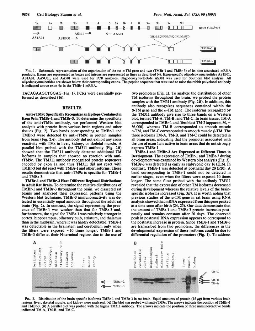
FIG. 1. Schematic representation of the organization of the rat a-TM gene and two (TMBr-1 and TMBr-3) of its nine associated mRNAproducts. Exons are represented as boxes and introns are represented as lines as described (4). Exon-specific oligodeoxynucleotides AS1BO1,AS1AO1, AA9CO1, and AA501 were used for PCR analyses. Oligodeoxynucleotide AS301 was used for Southern blot analysis. Alloligodeoxynucleotides are shown below their corresponding exons. The peptide sequence that was used to raise the rabbit polyclonal antibodyis indicated above exon 9c in the TMBr-1 mRNA.
TACAGAAGCTGGAG (Fig. 1). PCRs were essentially per-formed as described (16).
RESULTSAnti-rTM9c Specifically Recognizes an Epitope Contained in
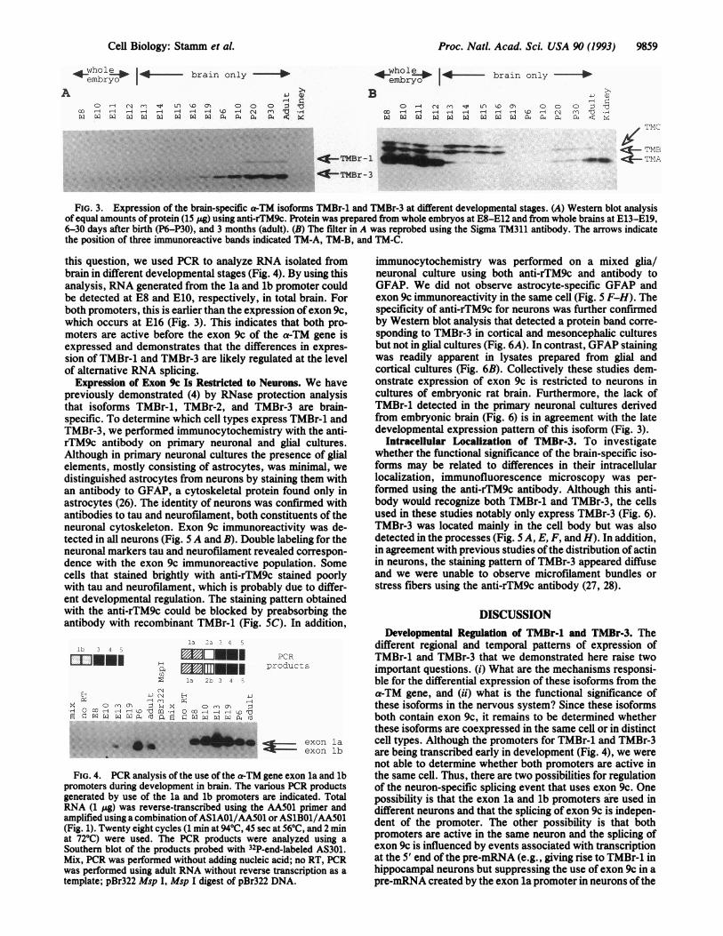
Exon 9c in TMBr-l and TMBr-3. To determine the specificityof the anti-rTM9c antibody, we performed Western blotanalysis with protein from various brain regions and othertissues (Fig. 2). Two bands corresponding to TMBr-1 andTMBr-3 were detected by anti-rTM9c in protein samplesfrom brain (Fig. 2A). The antibody did not exhibit any crossreactivity with TMs in liver, kidney, or skeletal muscle. Aparallel blot probed with the TM311 antibody (Fig. 2B)showed that the TM311 antibody detected additional TMisoforms in samples that showed no reaction with anti-rTM9c. The TM311 antibody recognized protein sequencesencoded by exon la and thus TM311 did not react withTMBr-3 but did react with TMBr-1 and other isoforms. Theseresults demonstrate that anti-rTM9c is specific for TMBr-1and TMBr-3.TMBr-l and TMBr-3 Have Different Regional Distributions
in Adult Rat Brain. To determine the relative distributions ofTMBr-1 and TMBr-3 throughout the brain, we dissected ratbrains and analyzed their expression patterns using theWestern blot technique. TMBr-3 immunoreactivity was de-tected in essentially equal amounts throughout the adult ratbrain (Fig. 2). In contrast, the signal representing the pres-ence of TMBr-1 was weaker than that for TMBr-3 and,furthermore, the signal for TMBr-1 was relatively stronger incortex, hippocampus, olfactory bulb, striatum, and thalamusthan in the midbrain, where it was hardly detectable. TMBr-1was detectable in the brainstem and cerebellum only whenthe filters were exposed =10 times longer. TMBr-1 andTMBr-3 differ at their N-terminal regions due to the use of
A
L)U)4)U)0r-H.sqR4
E.0
a)a)a)
xU)4-)C40C)
U)
0
.,H
0.U
IQ.0
>104)
0
o co
.4 0
U) U)co ~-L
C L).- UU) C) 0
~-1 r0.U)1
two promoters (Fig. 1). To analyze the distribution of otherTM isoforms throughout the brain, we probed the proteinsamples with the TM311 antibody (Fig. 2B). In addition, thisantibody also recognizes sequences contained within the/-TM gene and the a-TM gene. The isoforms recognized bythe TM311 antibody give rise to three bands on a Westernblot, termed TM-A, TM-B, and TM-C. In brain tissue, TM-Acorresponded to TMBr-1 and fibroblast TM-2 (apparent Mr =36,000), whereas TM-B corresponded to smooth musclea-TM, and TM-C corresponded to smooth muscle ,-TM. Thethree isoforms TM-A, TM-B, and TM-C could be detected inall brain areas, indicating that the promoter associated withthe use of exon la is active in brain areas that do not stronglyexpress TMBr-1.TMBr-l anid TMBr-3 Are Expressed at Different Times in
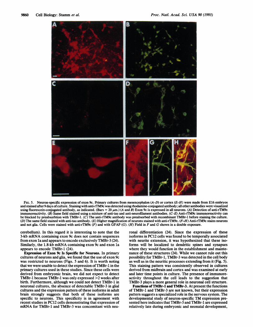
Development. The expression of TMBr-1 and TMBr-3 duringdevelopment was examined by Western blot analysis (Fig. 3).TMBr-3 was detected as early as embryonic day 16 (E16). Incontrast, TMBr-1 was detected at postnatal day 20 (P20). Aband corresponding to TMBr-1 could not be detected inearlier stages, even when the filters were exposed 10 timeslonger. The same filter probed with the antibody TM311revealed that the expression of other TM isoforms decreasedduring development whereas the relative levels of the brain-specific isoforms increased (Fig. 3B). It is worth noting thatprevious studies of the a-TM gene in rat brain using RNAanalysis showed thatmRNA expressed from this gene peakedat a time soon after birth (24, 25). Our data demonstrate thatthe amount of TMBr-1 and TMBr-3 protein increases post-natally and remains constant after 20 days. The observedpeak in postnatal RNA expression appears to correspond tothe postnatal increase in protein. Since TMBr-1 and TMBr-3are transcribed from two promoters, the differences in thedevelopmental expression of these isoforms could be due todifferential regulation of the promoters (Fig. 1). To address
B0
00
U) U)z R.H U)
Q0 C)
U1)4.)
0IL)
tn RU) .
a >1
X H 0 D DC) U 4-) 4.) 0~o C) C U)ee0 4 u) H M" IQ eT -1 -i
.H4 -H i 4v) .0 o U) D)
C))
U).,4 -D (3C).H ¶j *H
( TM-C
M=:: TM-BTM-AqC:* TMBr- 1
- TmBr-3
FIG. 2. Distribution of the brain-specific isoforms TMBr-1 and TMBr-3 in rat brain. Equal amounts of protein (15 ,ug) from various brainregions, liver, skeletal muscle, and kidney were analyzed. (A) The blot was probed with anti-rTM9c. The arrows indicate the position of TMBr-land TMBr-3. (B) A parallel blot was probed with the Sigma TM311 antibody. The arrows indicate the position of three immunoreactive bandsindicated TM-A, TM-B, and TM-C.
DEr",
111111WRI.... .:.'....
a ON
Proc. Natl. Acad. Sci. USA 90 (1993) 9859
whol |- Io - brain only --A embryo
O Nc L, 'IO O 0 0 0Oco 4 '-4 -4 '-4 '-4 '-4 D H-4 (N (n.X XW W X eX X X X 4X,ffi co 0ff a
-Fwhole;e -- brain only - -
B: : ~~~~~~~~~~~~~~~~~~~~~~~~~~~~~~~~~~~~~~~~~~~~~~. ...............,....TM Br- -1
-4--MBr-3
4-) G2)0 '-4 N rm sr U a' o0 0 D0
oo 4 4 ll -4 4 -4 1 -4 D -1 A m 10 .HX X X X X X X X X 04 a4 13 ai lz:C S
TMC
TMB_ -- TMA
FIG. 3. Expression of the brain-specific a-TM isoforms TMBr-1 and TMBr-3 at different developmental stages. (A) Western blot analysisof equal amounts of protein (15 pg) using anti-rTM9c. Protein was prepared from whole embryos at E8-E12 and from whole brains at E13-E19,6-30 days after birth (P6-P30), and 3 months (adult). (B) The filter in A was reprobed using the Sigma TM311 antibody. The arrows indicatethe position of three immunoreactive bands indicated TM-A, TM-B, and TM-C.
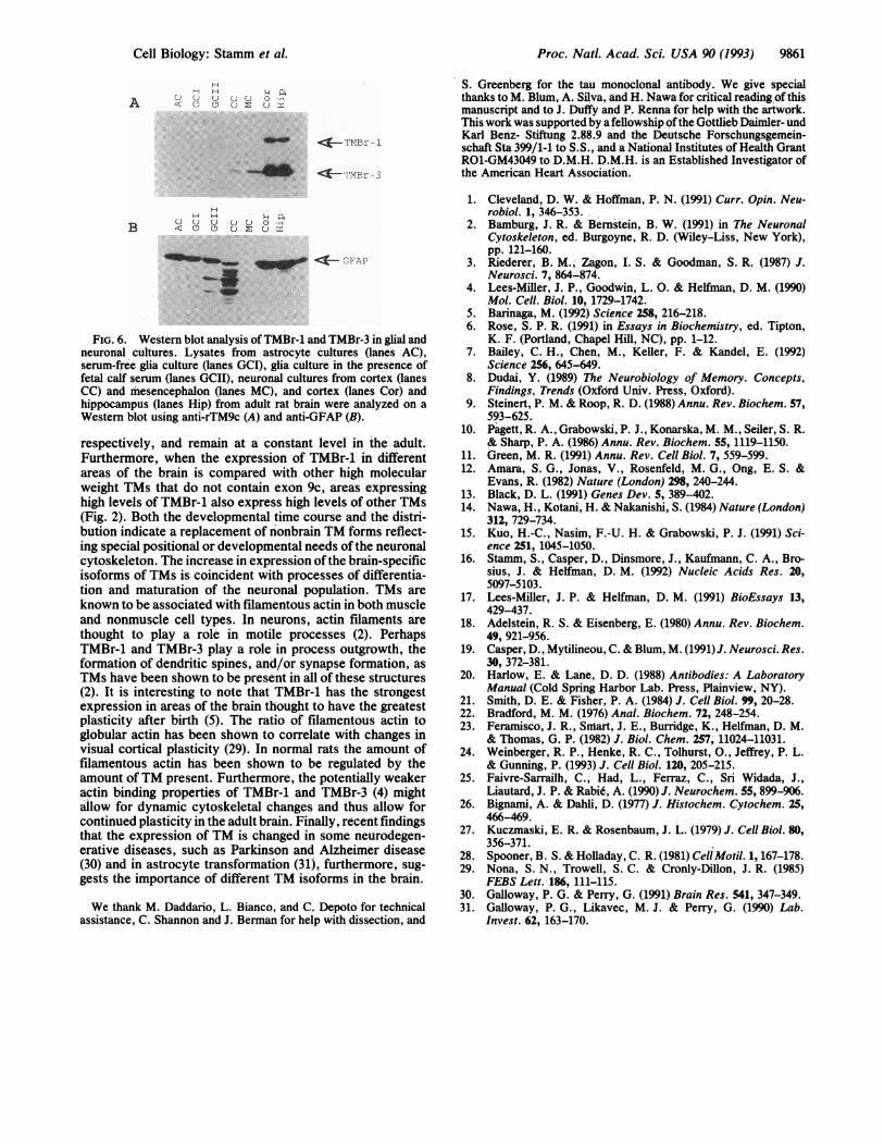
this question, we used PCR to analyze RNA isolated frombrain in different developmental stages (Fig. 4). By using thisanalysis, RNA generated from the la and lb promoter couldbe detected at E8 and E10, respectively, in total brain. Forboth promoters, this is earlier than the expression ofexon 9c,which occurs at E16 (Fig. 3). This indicates that both pro-moters are active before the exon 9c of the a-TM gene isexpressed and demonstrates that the differences in expres-sion of TMBr-l and TMBr-3 are likely regulated at the levelof alternative RNA splicing.
Expression of Exon 9c Is Restricted to Neurons. We havepreviously demonstrated (4) by RNase protection analysisthat isoforms TMBr-1, TMBr-2, and TMBr-3 are brain-specific. To determine which cell types express TMBr-1 andTMBr-3, we performed immunocytochemistry with the anti-rTM9c antibody on primary neuronal and glial cultures.Although in primary neuronal cultures the presence of glialelements, mostly consisting of astrocytes, was minimal, wedistinguished astrocytes from neurons by staining them withan antibody to GFAP, a cytoskeletal protein found only inastrocytes (26). The identity of neurons was confirmed withantibodies to tau and neurofilament, both constituents of theneuronal cytoskeleton. Exon 9c immunoreactivity was de-tected in all neurons (Fig. 5 A and B). Double labeling for theneuronal markers tau and neurofilament revealed correspon-dence with the exon 9c immunoreactive population. Somecells that stained brightly with anti-rTM9c stained poorlywith tau and neurofilament, which is probably due to differ-ent developmental regulation. The staining pattern obtainedwith the anti-rTM9c could be blocked by preabsorbing theantibody with recombinant TMBr-1 (Fig. SC). In addition,
1- -+ A
I --1I
NEI
EX O n'H O 00 -4 -H s
PCRproducts
t ,, _' A ~,
I
x la 'b, '. 4 :;
(N4
,-I (N)p -4
X -,o m
m Do rie H C5
exon la
exon lb
FIG. 4. PCR analysis of the use of the a-TM gene exon la and lbpromoters during development in brain. The various PCR productsgenerated by use of the la and lb promoters are indicated. TotalRNA (1 /&g) was reverse-transcribed using the AA501 primer andamplified using a combination ofASlAOl/AA50l or ASlBOl/AA50l(Fig. 1). Twenty eight cycles (1 min at 94°C, 45 sec at 56°C, and 2 minat 72°C) were used. The PCR products were analyzed using aSouthern blot of the products probed with 32P-end-labeled AS301.Mix, PCR was performed without adding nucleic acid; no RT, PCRwas performed using adult RNA without reverse transcription as atemplate; pBr322 Msp I, Msp I digest of pBr322 DNA.
immunocytochemistry was performed on a mixed glia/neuronal culture using both anti-rTM9c and antibody toGFAP. We did not observe astrocyte-specific GFAP andexon 9c immunoreactivity in the same cell (Fig. 5 F-H). Thespecificity of anti-rTM9c for neurons was further confirmedby Western blot analysis that detected a protein band corre-sponding to TMBr-3 in cortical and mesoncephalic culturesbut not in glial cultures (Fig. 6A). In contrast, GFAP stainingwas readily apparent in lysates prepared from glial andcortical cultures (Fig. 6B). Collectively these studies dem-onstrate expression of exon 9c is restricted to neurons incultures of embryonic rat brain. Furthermore, the lack ofTMBr-1 detected in the primary neuronal cultures derivedfrom embryonic brain (Fig. 6) is in agreement with the latedevelopmental expression pattern of this isoform (Fig. 3).
Intracellular Localization of TMBr-3. To investigatewhether the functional significance of the brain-specific iso-forms may be related to differences in their intracellularlocalization, immunofluorescence microscopy was per-formed using the anti-rTM9c antibody. Although this anti-body would recognize both TMBr-1 and TMBr-3, the cellsused in these studies notably only express TMBr-3 (Fig. 6).TMBr-3 was located mainly in the cell body but was alsodetected in the processes (Fig. 5 A, E, F, and H). In addition,in agreement with previous studies ofthe distribution of actinin neurons, the staining pattern of TMBr-3 appeared diffuseand we were unable to observe microfilament bundles orstress fibers using the anti-rTM9c antibody (27, 28).
DISCUSSIONDevelopmental Regulation of TMBr-1 and TMBr-3. The
different regional and temporal patterns of expression ofTMBr-1 and TMBr-3 that we demonstrated here raise twoimportant questions. (i) What are the mechanisms responsi-ble for the differential expression of these isoforms from thea-TM gene, and (ii) what is the functional significance ofthese isoforms in the nervous system? Since these isoformsboth contain exon 9c, it remains to be determined whetherthese isoforms are coexpressed in the same cell or in distinctcell types. Although the promoters for TMBr-1 and TMBr-3are being transcribed early in development (Fig. 4), we werenot able to determine whether both promoters are active inthe same cell. Thus, there are two possibilities for regulationof the neuron-specific splicing event that uses exon 9c. Onepossibility is that the exon la and lb promoters are used indifferent neurons and that the splicing of exon 9c is indepen-dent of the promoter. The other possibility is that bothpromoters are active in the same neuron and the splicing ofexon 9c is influenced by events associated with transcriptionat the 5' end of the pre-mRNA (e.g., giving rise to TMBr-1 inhippocampal neurons but suppressing the use of exon 9c in apre-mRNA created by the exon la promoter in neurons of the
Cell Biology: Stamm et al.
Proc. Natl. Acad. Sci. USA 90 (1993)
FIG. 5. Neuron-specific expression of exon 9c. Primary cultures from mesencephalon (A-D) or cortex (E-H) were made from E16 embryosand stained after 9 days ofculture. Staining with anti-rTM9c was detected using rhodamine-conjugated antibody; all other antibodies were visualizedusing fluorescein-conjugated antibody, as indicated. (Bars = 20 ,um.) (A and B) Exon 9c is expressed in all neurons. (A) Detection of anti-rTM9cimmunoreactivity. (B) Same field stained using a mixture of anti-tau and anti-neurofilament antibodies. (C-E) Anti-rTM9c immunoreactivity canbe blocked by preabsorbtion with TMBr-1. (C) The anti-rTM9c antibody was preabsorbed with recombinant TMBr-1 before staining the culture.(D) The same field stained with anti-tau antibody. (E) Higher magnification of neurons stained with anti-rTM9c. (F-H) Anti-rTM9c stains neuronsand not glia. Cells were stained with anti-rTM9c (F) and with GFAP (G). (H) Field in F and G shown in a double exposure.
cerebellum). In this regard it is interesting to note that the3-kb mRNA containing exon 9c does not contain sequencesfrom exon la and appears to encode exclusively TMBr-3 (24).Similarly, the 1.8-kb mRNA containing exon 9c and exon laappears to encode TMBr-1 (24).
Expression of Exon 9c Is Specific for Neurons. In primarycultures of neurons and glia, we found that the use of exon 9cwas restricted to neurons (Figs. 5 and 6). It is worth notingthat we were unable to detect the expression ofTMBr-1 in theprimary cultures used in these studies. Since these cells werederived from embryonic brain, we did not expect to detectTMBr-1 because TMBr-1 was only expressed >2 weeks afterbirth. Furthermore, although we could not detect TMBr-1 inneuronal cultures, the absence of detectable TMBr-3 in glialcultures and the expression pattern of these isoforms in adultbrain strongly suggests that both of these isoforms arespecific to neurons. This specificity is in agreement withrecent studies in PC12 cells demonstrating that expression ofmRNA for TMBr-1 and TMBr-3 was concomitant with neu-
ronal differentiation (24). Since the expression of theseisoforms in PC12 cells was found to be temporally associatedwith neurite extension, it was hypothesized that these iso-forms will be localized to dendritic spines and synapseswhere they would function in the establishment and mainte-nance of these structures (24). While we cannot rule out thispossibility for TMBr-1, TMBr-3 was detected in the cell bodyas well as in the neuritic processes extending from it (Fig. 5).This staining pattern was consistently observed in culturesderived from midbrain and cortex and was examined at earlyand later time points in culture. The presence of immunore-activity throughout the cell leads to the suggestion thatTMBr-3 plays a more general role in neuronal cell structure.
Functions ofTMBr-1 and TMBr-3. At present the functionsof TMBr-1 and TMBr-3 are not known, but their expressionpattern suggests a specialized role in the nervous system. Thedevelopmental study of neuron-specific TM expression pre-sented here indicates that TMBr-3 and TMBr-1 are expressedrelatively late during embryonic and neonatal development,
9860 Cefl Biology: Stamm et al.
Proc. Natl. Acad. Sci. USA 90 (1993) 9861
U OH
A u ~
T0M-* MBr-l
I-TMBr-3
H H a
BU
cf- GFAP
FIG. 6. Western blot analysis of TMBr-1 and TMBr-3 in glial and
neuronal cultures. Lysates from astrocyte cultures (lanes AC),
serum-free glia culture (lanes GCI), glia culture in the presence of
fetal calf serum (lanes GCII), neuronal cultures from cortex (lanes
CC) and 'mesencephalon (lanes MC), and cortex (lanes Cor) and
hippocampus (lanes Hip) from adult rat brain were analyzed on a
Western blot using anti-rTM9c (A) and anti-GFAP (B).
respectively, and remain at a constant leve'l in the adult.
Furthermore, when the expression of TMBr-1 in different
areas of the brain is compared with other high molecular
weight TMs that do not contain exon 9c, areas expressing
high levels of TMBr-1 also express high levels of other TMs
(Fig. 2). Both the developmental time course and the distri-
bution indicate a replacement of nonbrain TM forms reflect-
ing special positional or developmental needs of the neuronal
cytoskeleton. The increase in expression of the brain-specif'icisoforms of TMs is coincident with processes of differentia-
tion and maturation of the neuronal population. TMs are
known to be associated with filamentous actin in both muscle
and nonmuscle cell types. In neurons, actin filaments are
thought to play a role in motile processes (2). PerhapsTMBr-1 and TMBr-3 play a role in process outgrowth, the
formation of dendritic spines, and/or synapse formation, as
TMs have been shown to be present in all of these structures
(2). It is interesting to note that TMBr-1 has the strongest
expression in areas of the brain thought to have the greatest
plasticity after birth (5). The ratio of filamentous actin to
globular actin has been shown to correlate with changes in
visual cortical plasticity (29). In normal rats the amount of
filamentous actin has been shown to be regulated by the
amount of TM present. Furthermore, the potentially weaker
actin binding properties of TMBr-1 and TMBr-3 (4) mightallow for dynamic cytoskeletal changes and thus allow for
continued plasticity in the adult brain. Finally, recent findingsthat the expression of TM is changed in some neurodegen-erative diseases, such as Parkinson and Alzheimer disease
(30) and i4n astrocyte tranisformation (31), furthermore, s'ug-gests the importance of different TM isoforms in the brain.
We thank M. Daddario, L. Bianco, and C. Depoto for technicalassistance, C. Shannon and J. Berman for help with dissection, and
S. Greenberg for the tau monoclonal antibody. We give specialthanks to M. Blum, A. Silva, and H. Nawa for critical reading of thismanuscript and to J. Duffy and P. Renna for help with the artwork.This work was supported by a fellowship ofthe Gottlieb Daimler- undKarl Benz- Stiftung 2.88.9 and the Deutsche Forschungsgemein-schaft Sta 399/1-1 to S.S., and a National Institutes of Health GrantRO1-GM43049 to D.M.H. D.M.H. is an Established Investigator ofthe American Heart Association.
1. Cleveland, D. W. & Hoffman, P. N. (1991) Curr. Opin. Neu-robiol. 1, 346-353.
2. Bamburg, J. R. & Bernstein, B. W. (1991) in The NeuronalCytoskeleton, ed. Burgoyne, R. D. (Wiley-Liss, New York),pp. 121-160.
3. Riederer, B. M., Zagon, I. S. & Goodman, S. R. (1987) J.Neurosci. 7, 864-874.
4. Lees-Miller, J. P., Goodwin, L. 0. & Helfman, D. M. (1990)Mol. Cell. Biol. 10, 1729-1742.
5. Barinaga, M. (1992) Science 258, 216-218.6. Rose, S. P. R. (1991) in Essays in Biochemistry, ed. Tipton,
K. F. (Portland, Chapel Hill, NC), pp. 1-12.7. Bailey, C. H., Chen, M., Keller, F. & Kandel, E. (1992)
Science 256, 645-649.8. Dudai, Y. (1989) The Neurobiology of Memory. Concepts,
Findings, Trends (Oxford Univ. Press, Oxford).9. Steinert, P. M. & Roop, R. D. (1988) Annu. Rev. Biochem. 57,
593-625.10. Pagett, R. A., Grabowski, P. J., Konarska, M. M., Seiler, S. R.
& Sharp, P. A. (1986) Annu. Rev. Biochem. 55, 1119-1150.11. Green, M. R. (1991) Annu. Rev. Cell Biol. 7, 559-599.12. Amara, S. G., Jonas, V., Rosenfeld, M. G., Ong, E. S. &
Evans, R. (1982) Nature (London) 298, 240-244.13. Black, D. L. (1991) Genes Dev. 5, 389-402.14. Nawa, H., Kotani, H. & Nakanishi, S. (1984) Nature (London)
312, 729-734.15. Kuo, H.-C., Nasim, F.-U. H. & Grabowski, P. J. (1991) Sci-
ence 251, 1045-1050.16. Stamm, S., Casper, D., Dinsmore, J., Kaufmann, C. A., Bro-
sius, J. & Helfman, D. M. (1992) Nucleic Acids Res. 20,5097-5103.
17. Lees-Miller, J. P. & Helfman, D. M. (1991) BioEssays 13,429-437.
18. Adelstein, R. S. & Eisenberg, E. (1980) Annu. Rev. Biochem.49, 921-956.
19. Casper, D., Mytilineou, C. & Blum, M. (1991) J. Neurosci. Res.30, 372-381.
20. Harlow, E. & Lane, D. D. (1988) Antibodies: A LaboratoryManual (Cold Spring Harbor Lab. Press, Plainview, NY).
21. Smith, D. E. & Fisher, P. A. (1984) J. Cell Biol. 99, 20-28.22. Bradford, M. M. (1976) Anal. Biochem. 72, 248-254.23. Feramisco, J. R., Smart, J. E., Burridge, K., Helfman, D. M.
& Thomas, G. P. (1982) J. Biol. Chem. 257, 11024-11031.24. Weinberger, R. P., Henke, R. C., Tolhurst, O., Jeffrey, P. L.
& Gunning, P. (1993) J. Cell Biol. 120, 205-215.25. Faivre-Sarrailh, C., Had, L., Ferraz, C., Sri Widada, J.,
Liautard, J. P. & Rabid, A. (1990) J. Neurochem. 55, 899-906.26. Bignami, A. & Dahli, D. (1977) J. Histochem. Cytochem. 25,
466-469.27. Kuczmaski, E. R. & Rosenbaum, J. L. (1979) J. Cell Biol. 80,
356-371.28. Spooner, B. S. & Holladay, C. R. (1981) Cell Motil. 1, 167-178.29. Nona, S. N., Trowell, S. C. & Cronly-Dillon, J. R. (1985)
FEBS Lett. 186, 111-115.30. Galloway, P. G. & Perry, G. (1991) Brain Res. 541, 347-349.31. Galloway, P. G., Likavec, M. J. & Perry, G. (1990) Lab.
Invest. 62, 163-170.
Cell Biology: Stamm et al.