Embed Size (px)
Citation preview
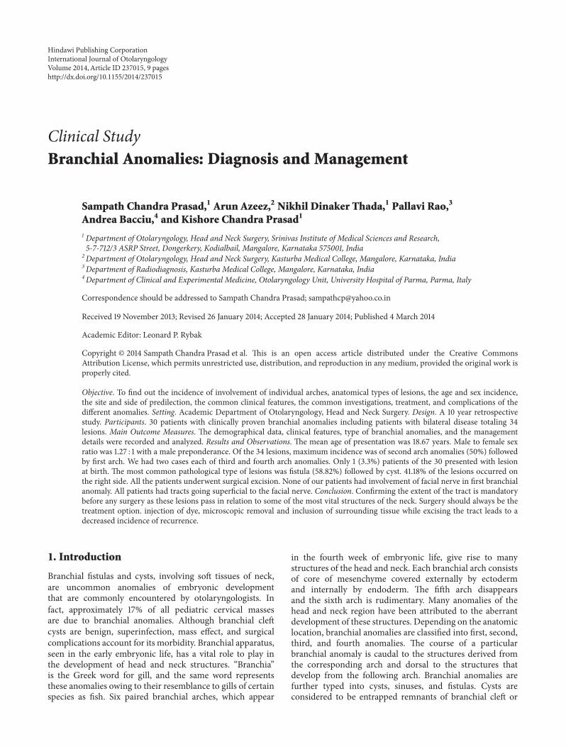
Clinical StudyBranchial Anomalies: Diagnosis and Management
Sampath Chandra Prasad,1 Arun Azeez,2 Nikhil Dinaker Thada,1 Pallavi Rao,3
Andrea Bacciu,4 and Kishore Chandra Prasad1
1 Department of Otolaryngology, Head and Neck Surgery, Srinivas Institute of Medical Sciences and Research,5-7-712/3 ASRP Street, Dongerkery, Kodialbail, Mangalore, Karnataka 575001, India
2Department of Otolaryngology, Head and Neck Surgery, Kasturba Medical College, Mangalore, Karnataka, India3 Department of Radiodiagnosis, Kasturba Medical College, Mangalore, Karnataka, India4Department of Clinical and Experimental Medicine, Otolaryngology Unit, University Hospital of Parma, Parma, Italy
Correspondence should be addressed to Sampath Chandra Prasad; [email protected]
Received 19 November 2013; Revised 26 January 2014; Accepted 28 January 2014; Published 4 March 2014
Academic Editor: Leonard P. Rybak
Copyright © 2014 Sampath Chandra Prasad et al. This is an open access article distributed under the Creative CommonsAttribution License, which permits unrestricted use, distribution, and reproduction in any medium, provided the original work isproperly cited.
Objective. To find out the incidence of involvement of individual arches, anatomical types of lesions, the age and sex incidence,the site and side of predilection, the common clinical features, the common investigations, treatment, and complications of thedifferent anomalies. Setting. Academic Department of Otolaryngology, Head and Neck Surgery. Design. A 10 year retrospectivestudy. Participants. 30 patients with clinically proven branchial anomalies including patients with bilateral disease totaling 34lesions. Main Outcome Measures. The demographical data, clinical features, type of branchial anomalies, and the managementdetails were recorded and analyzed. Results and Observations. The mean age of presentation was 18.67 years. Male to female sexratio was 1.27 : 1 with a male preponderance. Of the 34 lesions, maximum incidence was of second arch anomalies (50%) followedby first arch. We had two cases each of third and fourth arch anomalies. Only 1 (3.3%) patients of the 30 presented with lesionat birth. The most common pathological type of lesions was fistula (58.82%) followed by cyst. 41.18% of the lesions occurred onthe right side. All the patients underwent surgical excision. None of our patients had involvement of facial nerve in first branchialanomaly. All patients had tracts going superficial to the facial nerve. Conclusion. Confirming the extent of the tract is mandatorybefore any surgery as these lesions pass in relation to some of the most vital structures of the neck. Surgery should always be thetreatment option. injection of dye, microscopic removal and inclusion of surrounding tissue while excising the tract leads to adecreased incidence of recurrence.
1. Introduction
Branchial fistulas and cysts, involving soft tissues of neck,are uncommon anomalies of embryonic developmentthat are commonly encountered by otolaryngologists. Infact, approximately 17% of all pediatric cervical massesare due to branchial anomalies. Although branchial cleftcysts are benign, superinfection, mass effect, and surgicalcomplications account for its morbidity. Branchial apparatus,seen in the early embryonic life, has a vital role to play inthe development of head and neck structures. “Branchia”is the Greek word for gill, and the same word representsthese anomalies owing to their resemblance to gills of certainspecies as fish. Six paired branchial arches, which appear
in the fourth week of embryonic life, give rise to manystructures of the head and neck. Each branchial arch consistsof core of mesenchyme covered externally by ectodermand internally by endoderm. The fifth arch disappearsand the sixth arch is rudimentary. Many anomalies of thehead and neck region have been attributed to the aberrantdevelopment of these structures. Depending on the anatomiclocation, branchial anomalies are classified into first, second,third, and fourth anomalies. The course of a particularbranchial anomaly is caudal to the structures derived fromthe corresponding arch and dorsal to the structures thatdevelop from the following arch. Branchial anomalies arefurther typed into cysts, sinuses, and fistulas. Cysts areconsidered to be entrapped remnants of branchial cleft or
Hindawi Publishing CorporationInternational Journal of OtolaryngologyVolume 2014, Article ID 237015, 9 pageshttp://dx.doi.org/10.1155/2014/237015
2 International Journal of Otolaryngology
(a) (b)
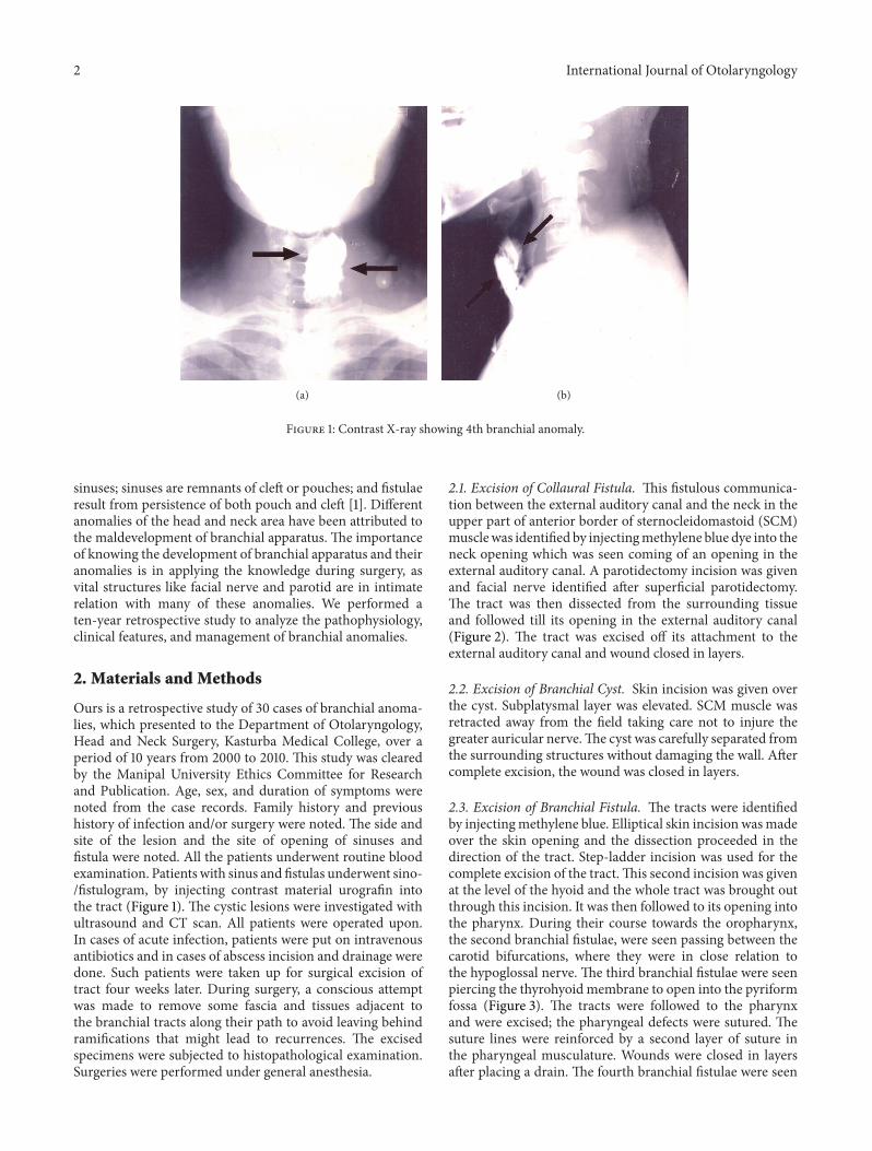
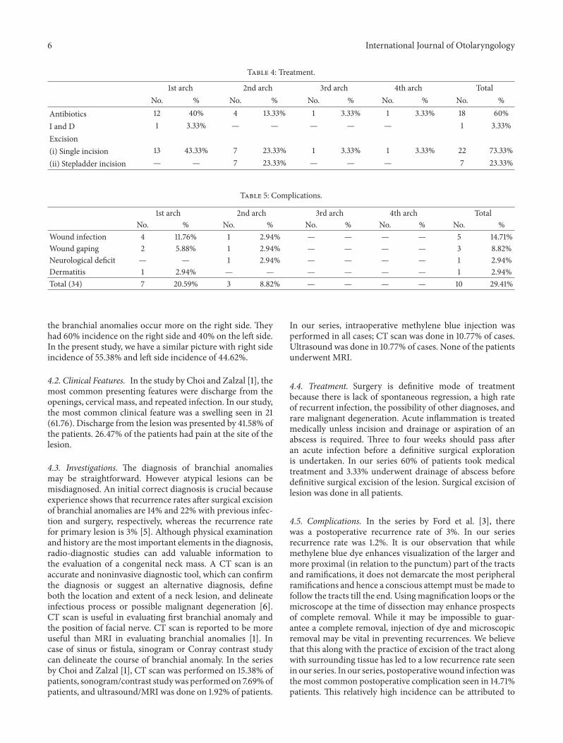
Figure 1: Contrast X-ray showing 4th branchial anomaly.
sinuses; sinuses are remnants of cleft or pouches; and fistulaeresult from persistence of both pouch and cleft [1]. Differentanomalies of the head and neck area have been attributed tothe maldevelopment of branchial apparatus. The importanceof knowing the development of branchial apparatus and theiranomalies is in applying the knowledge during surgery, asvital structures like facial nerve and parotid are in intimaterelation with many of these anomalies. We performed aten-year retrospective study to analyze the pathophysiology,clinical features, and management of branchial anomalies.
2. Materials and Methods
Ours is a retrospective study of 30 cases of branchial anoma-lies, which presented to the Department of Otolaryngology,Head and Neck Surgery, Kasturba Medical College, over aperiod of 10 years from 2000 to 2010. This study was clearedby the Manipal University Ethics Committee for Researchand Publication. Age, sex, and duration of symptoms werenoted from the case records. Family history and previoushistory of infection and/or surgery were noted. The side andsite of the lesion and the site of opening of sinuses andfistula were noted. All the patients underwent routine bloodexamination. Patients with sinus and fistulas underwent sino-/fistulogram, by injecting contrast material urografin intothe tract (Figure 1). The cystic lesions were investigated withultrasound and CT scan. All patients were operated upon.In cases of acute infection, patients were put on intravenousantibiotics and in cases of abscess incision and drainage weredone. Such patients were taken up for surgical excision oftract four weeks later. During surgery, a conscious attemptwas made to remove some fascia and tissues adjacent tothe branchial tracts along their path to avoid leaving behindramifications that might lead to recurrences. The excisedspecimens were subjected to histopathological examination.Surgeries were performed under general anesthesia.
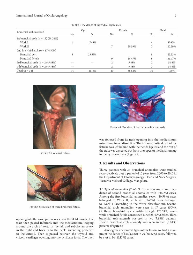
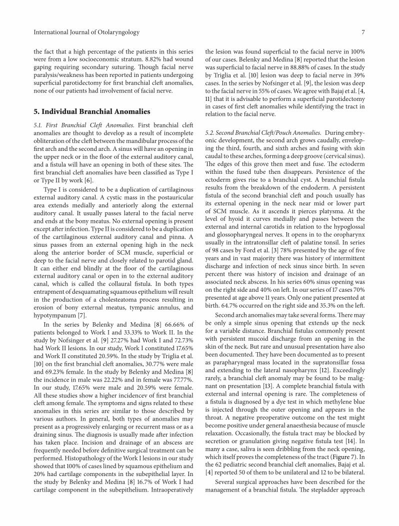
2.1. Excision of Collaural Fistula. This fistulous communica-tion between the external auditory canal and the neck in theupper part of anterior border of sternocleidomastoid (SCM)musclewas identified by injectingmethylene blue dye into theneck opening which was seen coming of an opening in theexternal auditory canal. A parotidectomy incision was givenand facial nerve identified after superficial parotidectomy.The tract was then dissected from the surrounding tissueand followed till its opening in the external auditory canal(Figure 2). The tract was excised off its attachment to theexternal auditory canal and wound closed in layers.
2.2. Excision of Branchial Cyst. Skin incision was given overthe cyst. Subplatysmal layer was elevated. SCM muscle wasretracted away from the field taking care not to injure thegreater auricular nerve.The cyst was carefully separated fromthe surrounding structures without damaging the wall. Aftercomplete excision, the wound was closed in layers.
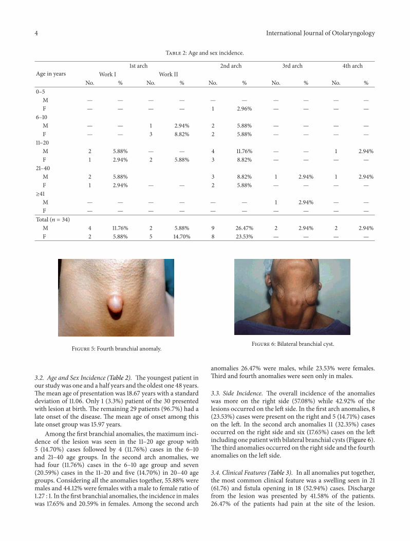
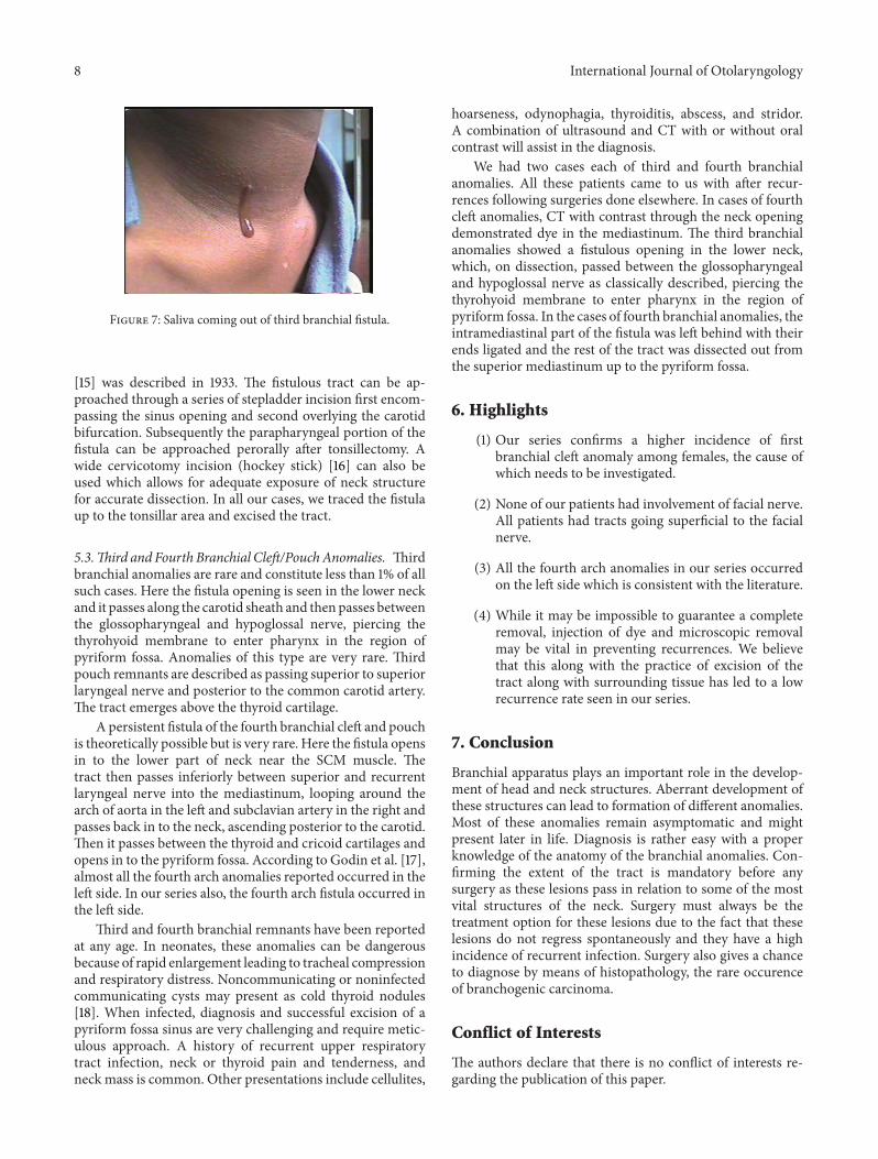
2.3. Excision of Branchial Fistula. The tracts were identifiedby injectingmethylene blue. Elliptical skin incision wasmadeover the skin opening and the dissection proceeded in thedirection of the tract. Step-ladder incision was used for thecomplete excision of the tract.This second incision was givenat the level of the hyoid and the whole tract was brought outthrough this incision. It was then followed to its opening intothe pharynx. During their course towards the oropharynx,the second branchial fistulae, were seen passing between thecarotid bifurcations, where they were in close relation tothe hypoglossal nerve. The third branchial fistulae were seenpiercing the thyrohyoid membrane to open into the pyriformfossa (Figure 3). The tracts were followed to the pharynxand were excised; the pharyngeal defects were sutured. Thesuture lines were reinforced by a second layer of suture inthe pharyngeal musculature. Wounds were closed in layersafter placing a drain. The fourth branchial fistulae were seen
International Journal of Otolaryngology 3
Table 1: Incidence of individual anomalies.
Branchial arch involved Cyst Fistula TotalNo. % No. % No. %
1st branchial arch (𝑛 = 13) (38.24%)Work I 6 17.65% 6 17.65%Work II 7 20.59% 7 20.59%
2nd branchial arch (𝑛 = 17) (50%)Branchial cyst 8 23.53% 8 23.53%Branchial fistula 9 26.47% 9 26.47%
3rd branchial arch (𝑛 = 2) (5.88%) — — 2 5.88% 2 5.88%4th branchial arch (𝑛 = 2) (5.88%) — 2 5.88% 2 5.88%Total (𝑛 = 34) 14 41.18% 20 58.82% 34 100%
Figure 2: Collaural fistula.
Figure 3: Excision of third branchial fistula.
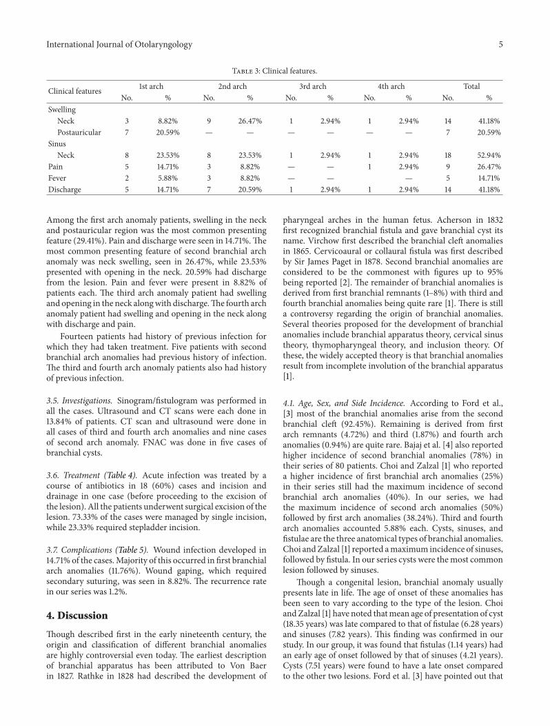
opening into the lower part of neck near the SCMmuscle.Thetract then passed inferiorly into the mediastinum, loopingaround the arch of aorta in the left and subclavian arteryin the right and back in to the neck, ascending posteriorto the carotid. Then it passed between the thyroid andcricoid cartilages opening into the pyriform fossa. The tract
Figure 4: Excision of fourth branchial anomaly.
was followed from its neck opening into the mediastinumusing blunt finger dissection.The intramediastinal part of thefistulae was left behind with their ends ligated and the rest ofthe tract was dissected out from the superiormediastinumupto the pyriform fossa (Figure 4).
3. Results and Observations
Thirty patients with 34 branchial anomalies were studiedretrospectively over a period of 10 years from 2000 to 2010 inthe Department of Otolaryngology, Head and Neck Surgery,Kasturba Medical College, Mangalore.
3.1. Type of Anomalies (Table 1). There was maximum inci-dence of second branchial anomalies with 17(50%) cases.Among the first branchial anomalies, seven (20.59%) casesbelonged to Work II, while six (17.65%) cases belongedto Work I (according to the Work classification). Secondbranchial arch anomalies were seen in 17 cases (50%).Of these, branchial cyst constituted eight (26.53%) caseswhile branchial fistula constituted nine (26.47%) cases.Thirdbranchial arch anomaly was seen in two (5.88%) patients.Fourth branchial arch anomaly was seen in two (5.88%)patients (Figure 5).
Among the anatomical types of the lesion, we had a max-imum incidence of fistula seen in 20 (58.82%) cases, followedby cyst in 14 (41.12%) cases.
4 International Journal of Otolaryngology
Table 2: Age and sex incidence.
Age in years1st arch 2nd arch 3rd arch 4th arch
Work I Work IINo. % No. % No. % No. % No. %
0–5M — — — — — — — — — —F — — — — 1 2.96% — — — —
6–10M — — 1 2.94% 2 5.88% — — — —F — — 3 8.82% 2 5.88% — — — —
11–20M 2 5.88% — — 4 11.76% — — 1 2.94%F 1 2.94% 2 5.88% 3 8.82% — — — —
21–40M 2 5.88% 3 8.82% 1 2.94% 1 2.94%F 1 2.94% — — 2 5.88% — — — —≥41
M — — — — — — 1 2.94% — —F — — — — — — — — — —
Total (𝑛 = 34)M 4 11.76% 2 5.88% 9 26.47% 2 2.94% 2 2.94%F 2 5.88% 5 14.70% 8 23.53% — — — —
Figure 5: Fourth branchial anomaly.
3.2. Age and Sex Incidence (Table 2). The youngest patient inour studywas one and a half years and the oldest one 48 years.Themean age of presentation was 18.67 years with a standarddeviation of 11.06. Only 1 (3.3%) patient of the 30 presentedwith lesion at birth. The remaining 29 patients (96.7%) had alate onset of the disease. The mean age of onset among thislate onset group was 15.97 years.
Among the first branchial anomalies, the maximum inci-dence of the lesion was seen in the 11–20 age group with5 (14.70%) cases followed by 4 (11.76%) cases in the 6–10and 21–40 age groups. In the second arch anomalies, wehad four (11.76%) cases in the 6–10 age group and seven(20.59%) cases in the 11–20 and five (14.70%) in 20–40 agegroups. Considering all the anomalies together, 55.88% weremales and 44.12% were females with a male to female ratio of1.27 : 1. In the first branchial anomalies, the incidence inmaleswas 17.65% and 20.59% in females. Among the second arch
Figure 6: Bilateral branchial cyst.
anomalies 26.47% were males, while 23.53% were females.Third and fourth anomalies were seen only in males.
3.3. Side Incidence. The overall incidence of the anomalieswas more on the right side (57.08%) while 42.92% of thelesions occurred on the left side. In the first arch anomalies, 8(23.53%) cases were present on the right and 5 (14.71%) caseson the left. In the second arch anomalies 11 (32.35%) casesoccurred on the right side and six (17.65%) cases on the leftincluding one patientwith bilateral branchial cysts (Figure 6).The third anomalies occurred on the right side and the fourthanomalies on the left side.
3.4. Clinical Features (Table 3). In all anomalies put together,the most common clinical feature was a swelling seen in 21(61.76) and fistula opening in 18 (52.94%) cases. Dischargefrom the lesion was presented by 41.58% of the patients.26.47% of the patients had pain at the site of the lesion.
International Journal of Otolaryngology 5
Table 3: Clinical features.
Clinical features 1st arch 2nd arch 3rd arch 4th arch TotalNo. % No. % No. % No. % No. %
SwellingNeck 3 8.82% 9 26.47% 1 2.94% 1 2.94% 14 41.18%Postauricular 7 20.59% — — — — — — 7 20.59%
SinusNeck 8 23.53% 8 23.53% 1 2.94% 1 2.94% 18 52.94%
Pain 5 14.71% 3 8.82% — — 1 2.94% 9 26.47%Fever 2 5.88% 3 8.82% — — — 5 14.71%Discharge 5 14.71% 7 20.59% 1 2.94% 1 2.94% 14 41.18%
Among the first arch anomaly patients, swelling in the neckand postauricular region was the most common presentingfeature (29.41%). Pain and discharge were seen in 14.71%.Themost common presenting feature of second branchial archanomaly was neck swelling, seen in 26.47%, while 23.53%presented with opening in the neck. 20.59% had dischargefrom the lesion. Pain and fever were present in 8.82% ofpatients each. The third arch anomaly patient had swellingand opening in the neck alongwith discharge.The fourth archanomaly patient had swelling and opening in the neck alongwith discharge and pain.
Fourteen patients had history of previous infection forwhich they had taken treatment. Five patients with secondbranchial arch anomalies had previous history of infection.The third and fourth arch anomaly patients also had historyof previous infection.
3.5. Investigations. Sinogram/fistulogram was performed inall the cases. Ultrasound and CT scans were each done in13.84% of patients. CT scan and ultrasound were done inall cases of third and fourth arch anomalies and nine casesof second arch anomaly. FNAC was done in five cases ofbranchial cysts.
3.6. Treatment (Table 4). Acute infection was treated by acourse of antibiotics in 18 (60%) cases and incision anddrainage in one case (before proceeding to the excision ofthe lesion). All the patients underwent surgical excision of thelesion. 73.33% of the cases were managed by single incision,while 23.33% required stepladder incision.
3.7. Complications (Table 5). Wound infection developed in14.71% of the cases.Majority of this occurred in first branchialarch anomalies (11.76%). Wound gaping, which requiredsecondary suturing, was seen in 8.82%. The recurrence ratein our series was 1.2%.
4. Discussion
Though described first in the early nineteenth century, theorigin and classification of different branchial anomaliesare highly controversial even today. The earliest descriptionof branchial apparatus has been attributed to Von Baerin 1827. Rathke in 1828 had described the development of
pharyngeal arches in the human fetus. Acherson in 1832first recognized branchial fistula and gave branchial cyst itsname. Virchow first described the branchial cleft anomaliesin 1865. Cervicoaural or collaural fistula was first describedby Sir James Paget in 1878. Second branchial anomalies areconsidered to be the commonest with figures up to 95%being reported [2]. The remainder of branchial anomalies isderived from first branchial remnants (1–8%) with third andfourth branchial anomalies being quite rare [1]. There is stilla controversy regarding the origin of branchial anomalies.Several theories proposed for the development of branchialanomalies include branchial apparatus theory, cervical sinustheory, thymopharyngeal theory, and inclusion theory. Ofthese, the widely accepted theory is that branchial anomaliesresult from incomplete involution of the branchial apparatus[1].
4.1. Age, Sex, and Side Incidence. According to Ford et al.,[3] most of the branchial anomalies arise from the secondbranchial cleft (92.45%). Remaining is derived from firstarch remnants (4.72%) and third (1.87%) and fourth archanomalies (0.94%) are quite rare. Bajaj et al. [4] also reportedhigher incidence of second branchial anomalies (78%) intheir series of 80 patients. Choi and Zalzal [1] who reporteda higher incidence of first branchial arch anomalies (25%)in their series still had the maximum incidence of secondbranchial arch anomalies (40%). In our series, we hadthe maximum incidence of second arch anomalies (50%)followed by first arch anomalies (38.24%). Third and fourtharch anomalies accounted 5.88% each. Cysts, sinuses, andfistulae are the three anatomical types of branchial anomalies.Choi andZalzal [1] reported amaximum incidence of sinuses,followed by fistula. In our series cysts were themost commonlesion followed by sinuses.
Though a congenital lesion, branchial anomaly usuallypresents late in life. The age of onset of these anomalies hasbeen seen to vary according to the type of the lesion. ChoiandZalzal [1] have noted thatmean age of presentation of cyst(18.35 years) was late compared to that of fistulae (6.28 years)and sinuses (7.82 years). This finding was confirmed in ourstudy. In our group, it was found that fistulas (1.14 years) hadan early age of onset followed by that of sinuses (4.21 years).Cysts (7.51 years) were found to have a late onset comparedto the other two lesions. Ford et al. [3] have pointed out that
6 International Journal of Otolaryngology
Table 4: Treatment.
1st arch 2nd arch 3rd arch 4th arch TotalNo. % No. % No. % No. % No. %
Antibiotics 12 40% 4 13.33% 1 3.33% 1 3.33% 18 60%I and D 1 3.33% — — — — — 1 3.33%Excision(i) Single incision 13 43.33% 7 23.33% 1 3.33% 1 3.33% 22 73.33%(ii) Stepladder incision — — 7 23.33% — — — 7 23.33%
Table 5: Complications.
1st arch 2nd arch 3rd arch 4th arch TotalNo. % No. % No. % No. % No. %
Wound infection 4 11.76% 1 2.94% — — — — 5 14.71%Wound gaping 2 5.88% 1 2.94% — — — — 3 8.82%Neurological deficit — — 1 2.94% — — — — 1 2.94%Dermatitis 1 2.94% — — — — — — 1 2.94%Total (34) 7 20.59% 3 8.82% — — — — 10 29.41%
the branchial anomalies occur more on the right side. Theyhad 60% incidence on the right side and 40% on the left side.In the present study, we have a similar picture with right sideincidence of 55.38% and left side incidence of 44.62%.
4.2. Clinical Features. In the study by Choi and Zalzal [1], themost common presenting features were discharge from theopenings, cervical mass, and repeated infection. In our study,the most common clinical feature was a swelling seen in 21(61.76). Discharge from the lesion was presented by 41.58% ofthe patients. 26.47% of the patients had pain at the site of thelesion.
4.3. Investigations. The diagnosis of branchial anomaliesmay be straightforward. However atypical lesions can bemisdiagnosed. An initial correct diagnosis is crucial becauseexperience shows that recurrence rates after surgical excisionof branchial anomalies are 14% and 22% with previous infec-tion and surgery, respectively, whereas the recurrence ratefor primary lesion is 3% [5]. Although physical examinationand history are themost important elements in the diagnosis,radio-diagnostic studies can add valuable information tothe evaluation of a congenital neck mass. A CT scan is anaccurate and noninvasive diagnostic tool, which can confirmthe diagnosis or suggest an alternative diagnosis, defineboth the location and extent of a neck lesion, and delineateinfectious process or possible malignant degeneration [6].CT scan is useful in evaluating first branchial anomaly andthe position of facial nerve. CT scan is reported to be moreuseful than MRI in evaluating branchial anomalies [1]. Incase of sinus or fistula, sinogram or Conray contrast studycan delineate the course of branchial anomaly. In the seriesby Choi and Zalzal [1], CT scan was performed on 15.38% ofpatients, sonogram/contrast studywas performed on 7.69%ofpatients, and ultrasound/MRI was done on 1.92% of patients.
In our series, intraoperative methylene blue injection wasperformed in all cases; CT scan was done in 10.77% of cases.Ultrasound was done in 10.77% of cases. None of the patientsunderwent MRI.
4.4. Treatment. Surgery is definitive mode of treatmentbecause there is lack of spontaneous regression, a high rateof recurrent infection, the possibility of other diagnoses, andrare malignant degeneration. Acute inflammation is treatedmedically unless incision and drainage or aspiration of anabscess is required. Three to four weeks should pass afteran acute infection before a definitive surgical explorationis undertaken. In our series 60% of patients took medicaltreatment and 3.33% underwent drainage of abscess beforedefinitive surgical excision of the lesion. Surgical excision oflesion was done in all patients.
4.5. Complications. In the series by Ford et al. [3], therewas a postoperative recurrence rate of 3%. In our seriesrecurrence rate was 1.2%. It is our observation that whilemethylene blue dye enhances visualization of the larger andmore proximal (in relation to the punctum) part of the tractsand ramifications, it does not demarcate the most peripheralramifications and hence a conscious attemptmust bemade tofollow the tracts till the end. Usingmagnification loops or themicroscope at the time of dissection may enhance prospectsof complete removal. While it may be impossible to guar-antee a complete removal, injection of dye and microscopicremoval may be vital in preventing recurrences. We believethat this along with the practice of excision of the tract alongwith surrounding tissue has led to a low recurrence rate seenin our series. In our series, postoperativewound infectionwasthe most common postoperative complication seen in 14.71%patients. This relatively high incidence can be attributed to
International Journal of Otolaryngology 7
the fact that a high percentage of the patients in this serieswere from a low socioeconomic stratum. 8.82% had woundgaping requiring secondary suturing. Though facial nerveparalysis/weakness has been reported in patients undergoingsuperficial parotidectomy for first branchial cleft anomalies,none of our patients had involvement of facial nerve.
5. Individual Branchial Anomalies
5.1. First Branchial Cleft Anomalies. First branchial cleftanomalies are thought to develop as a result of incompleteobliteration of the cleft between themandibular process of thefirst arch and the second arch. A sinus will have an opening inthe upper neck or in the floor of the external auditory canal,and a fistula will have an opening in both of these sites. Thefirst branchial cleft anomalies have been classified as Type Ior Type II by work [6].
Type I is considered to be a duplication of cartilaginousexternal auditory canal. A cystic mass in the postauriculararea extends medially and anteriorly along the externalauditory canal. It usually passes lateral to the facial nerveand ends at the bony meatus. No external opening is presentexcept after infection. Type II is considered to be a duplicationof the cartilaginous external auditory canal and pinna. Asinus passes from an external opening high in the neckalong the anterior border of SCM muscle, superficial ordeep to the facial nerve and closely related to parotid gland.It can either end blindly at the floor of the cartilaginousexternal auditory canal or open in to the external auditorycanal, which is called the collaural fistula. In both typesentrapment of desquamating squamous epitheliumwill resultin the production of a cholesteatoma process resulting inerosion of bony external meatus, tympanic annulus, andhypotympanum [7].
In the series by Belenky and Medina [8] 66.66% ofpatients belonged to Work I and 33.33% to Work II. In thestudy by Nofsinger et al. [9] 27.27% had Work I and 72.73%had Work II lesions. In our study, Work I constituted 17.65%and Work II constituted 20.59%. In the study by Triglia et al.[10] on the first branchial cleft anomalies, 30.77% were maleand 69.23% female. In the study by Belenky and Medina [8]the incidence in male was 22.22% and in female was 77.77%.In our study, 17.65% were male and 20.59% were female.All these studies show a higher incidencev of first branchialcleft among female. The symptoms and signs related to theseanomalies in this series are similar to those described byvarious authors. In general, both types of anomalies maypresent as a progressively enlarging or recurrent mass or as adraining sinus. The diagnosis is usually made after infectionhas taken place. Incision and drainage of an abscess arefrequently needed before definitive surgical treatment can beperformed. Histopathology of theWork I lesions in our studyshowed that 100% of cases lined by squamous epithelium and20% had cartilage components in the subepithelial layer. Inthe study by Belenky and Medina [8] 16.7% of Work I hadcartilage component in the subepithelium. Intraoperatively
the lesion was found superficial to the facial nerve in 100%of our cases. Belenky andMedina [8] reported that the lesionwas superficial to facial nerve in 88.88% of cases. In the studyby Triglia et al. [10] lesion was deep to facial nerve in 39%cases. In the series by Nofsinger et al. [9], the lesion was deepto the facial nerve in 55%of cases.We agreewith Bajaj et al. [4,11] that it is advisable to perform a superficial parotidectomyin cases of first cleft anomalies while identifying the tract inrelation to the facial nerve.
5.2. Second Branchial Cleft/Pouch Anomalies. During embry-onic development, the second arch grows caudally, envelop-ing the third, fourth, and sixth arches and fusing with skincaudal to these arches, forming a deep groove (cervical sinus).The edges of this grove then meet and fuse. The ectodermwithin the fused tube then disappears. Persistence of theectoderm gives rise to a branchial cyst. A branchial fistularesults from the breakdown of the endoderm. A persistentfistula of the second branchial cleft and pouch usually hasits external opening in the neck near mid or lower partof SCM muscle. As it ascends it pierces platysma. At thelevel of hyoid it curves medially and passes between theexternal and internal carotids in relation to the hypoglossaland glossopharyngeal nerves. It opens in to the oropharynxusually in the intratonsillar cleft of palatine tonsil. In seriesof 98 cases by Ford et al. [3] 78% presented by the age of fiveyears and in vast majority there was history of intermittentdischarge and infection of neck sinus since birth. In sevenpercent there was history of incision and drainage of anassociated neck abscess. In his series 60% sinus opening wason the right side and 40% on left. In our series of 17 cases 70%presented at age above 11 years. Only one patient presented atbirth. 64.7% occurred on the right side and 35.3% on the left.
Second arch anomaliesmay take several forms.Theremaybe only a simple sinus opening that extends up the neckfor a variable distance. Branchial fistulas commonly presentwith persistent mucoid discharge from an opening in theskin of the neck. But rare and unusual presentation have alsobeen documented.They have been documented as to presentas parapharyngeal mass located in the supratonsillar fossaand extending to the lateral nasopharynx [12]. Exceedinglyrarely, a branchial cleft anomaly may be found to be malig-nant on presentation [13]. A complete branchial fistula withexternal and internal opening is rare. The completeness ofa fistula is diagnosed by a dye test in which methylene blueis injected through the outer opening and appears in thethroat. A negative preoperative outcome on the test mightbecome positive under general anaesthesia because of musclerelaxation. Occasionally, the fistula tract may be blocked bysecretion or granulation giving negative fistula test [14]. Inmany a case, saliva is seen dribbling from the neck opening,which itself proves the completeness of the tract (Figure 7). Inthe 62 pediatric second branchial cleft anomalies, Bajaj et al.[4] reported 50 of them to be unilateral and 12 to be bilateral.
Several surgical approaches have been described for themanagement of a branchial fistula. The stepladder approach
8 International Journal of Otolaryngology
Figure 7: Saliva coming out of third branchial fistula.
[15] was described in 1933. The fistulous tract can be ap-proached through a series of stepladder incision first encom-passing the sinus opening and second overlying the carotidbifurcation. Subsequently the parapharyngeal portion of thefistula can be approached perorally after tonsillectomy. Awide cervicotomy incision (hockey stick) [16] can also beused which allows for adequate exposure of neck structurefor accurate dissection. In all our cases, we traced the fistulaup to the tonsillar area and excised the tract.
5.3.Third and Fourth Branchial Cleft/PouchAnomalies. Thirdbranchial anomalies are rare and constitute less than 1% of allsuch cases. Here the fistula opening is seen in the lower neckand it passes along the carotid sheath and thenpasses betweenthe glossopharyngeal and hypoglossal nerve, piercing thethyrohyoid membrane to enter pharynx in the region ofpyriform fossa. Anomalies of this type are very rare. Thirdpouch remnants are described as passing superior to superiorlaryngeal nerve and posterior to the common carotid artery.The tract emerges above the thyroid cartilage.
A persistent fistula of the fourth branchial cleft and pouchis theoretically possible but is very rare. Here the fistula opensin to the lower part of neck near the SCM muscle. Thetract then passes inferiorly between superior and recurrentlaryngeal nerve into the mediastinum, looping around thearch of aorta in the left and subclavian artery in the right andpasses back in to the neck, ascending posterior to the carotid.Then it passes between the thyroid and cricoid cartilages andopens in to the pyriform fossa. According to Godin et al. [17],almost all the fourth arch anomalies reported occurred in theleft side. In our series also, the fourth arch fistula occurred inthe left side.
Third and fourth branchial remnants have been reportedat any age. In neonates, these anomalies can be dangerousbecause of rapid enlargement leading to tracheal compressionand respiratory distress. Noncommunicating or noninfectedcommunicating cysts may present as cold thyroid nodules[18]. When infected, diagnosis and successful excision of apyriform fossa sinus are very challenging and require metic-ulous approach. A history of recurrent upper respiratorytract infection, neck or thyroid pain and tenderness, andneck mass is common. Other presentations include cellulites,
hoarseness, odynophagia, thyroiditis, abscess, and stridor.A combination of ultrasound and CT with or without oralcontrast will assist in the diagnosis.
We had two cases each of third and fourth branchialanomalies. All these patients came to us with after recur-rences following surgeries done elsewhere. In cases of fourthcleft anomalies, CT with contrast through the neck openingdemonstrated dye in the mediastinum. The third branchialanomalies showed a fistulous opening in the lower neck,which, on dissection, passed between the glossopharyngealand hypoglossal nerve as classically described, piercing thethyrohyoid membrane to enter pharynx in the region ofpyriform fossa. In the cases of fourth branchial anomalies, theintramediastinal part of the fistula was left behind with theirends ligated and the rest of the tract was dissected out fromthe superior mediastinum up to the pyriform fossa.
6. Highlights
(1) Our series confirms a higher incidence of firstbranchial cleft anomaly among females, the cause ofwhich needs to be investigated.
(2) None of our patients had involvement of facial nerve.All patients had tracts going superficial to the facialnerve.
(3) All the fourth arch anomalies in our series occurredon the left side which is consistent with the literature.
(4) While it may be impossible to guarantee a completeremoval, injection of dye and microscopic removalmay be vital in preventing recurrences. We believethat this along with the practice of excision of thetract along with surrounding tissue has led to a lowrecurrence rate seen in our series.
7. Conclusion
Branchial apparatus plays an important role in the develop-ment of head and neck structures. Aberrant development ofthese structures can lead to formation of different anomalies.Most of these anomalies remain asymptomatic and mightpresent later in life. Diagnosis is rather easy with a properknowledge of the anatomy of the branchial anomalies. Con-firming the extent of the tract is mandatory before anysurgery as these lesions pass in relation to some of the mostvital structures of the neck. Surgery must always be thetreatment option for these lesions due to the fact that theselesions do not regress spontaneously and they have a highincidence of recurrent infection. Surgery also gives a chanceto diagnose by means of histopathology, the rare occurenceof branchogenic carcinoma.
Conflict of Interests
The authors declare that there is no conflict of interests re-garding the publication of this paper.
International Journal of Otolaryngology 9
References
[1] S. S. Choi and G. H. Zalzal, “Branchial anomalies: a review of 52cases,” Laryngoscope, vol. 105, no. 9, part 1, pp. 909–913, 1995.
[2] J. F. Kenealy, A. J. Torsiglieri Jr., and L. W. Tom, “Branchial cleftanomalies: a five-year retrospective review,” Transactions of thePennsylvania Academy of Ophthalmology and Otolaryngology,vol. 42, pp. 1022–1025, 1990.
[3] G. R. Ford, A. Balakrishnan, J. N. G. Evans, and C. M. Bailey,“Branchial cleft and pouch anomalies,” Journal of Laryngologyand Otology, vol. 106, no. 2, pp. 137–143, 1992.
[4] Y. Bajaj, S. Ifeacho, D. Tweedie et al., “Branchial anomalies inchildren,” International Journal of Pediatric Otorhinolaryngol-ogy, vol. 75, no. 8, pp. 1020–1023, 2011.
[5] D. Reiter, “Third branchial cleft sinus: an unusual cause of neckabscess,” International Journal of Pediatric Otorhinolaryngology,vol. 4, no. 2, pp. 181–186, 1982.
[6] F. Coppens, P. Peene, and S. F. Lemahieu, “Diagnosis and differ-ential diagnosis of branchial cleft cysts by CT scan,” JournalBelge de Radiologie, vol. 73, no. 3, pp. 189–196, 1990.
[7] S. A. Hickey, G. A. Scott, and P. Traub, “Defects of the first bran-chial cleft,” Journal of Laryngology and Otology, vol. 108, no. 3,pp. 240–243, 1994.
[8] W. M. Belenky and J. E. Medina, “First branchial cleft anoma-lies,” Laryngoscope, vol. 90, no. 1, pp. 28–39, 1980.
[9] Y. C. Nofsinger, L. W. C. Tom, D. LaRossa, R. F. Wetmore, andS. D. Handler, “Periauricular cysts and sinuses,” Laryngoscope,vol. 107, no. 7, pp. 883–887, 1997.
[10] J. Triglia, R. Nicollas, V. Ducroz, P. J. Koltai, and E. Garabedian,“First branchial cleft anomalies: a study of 39 cases and a reviewof the literature,” Archives of Otolaryngology—Head and NeckSurgery, vol. 124, no. 3, pp. 291–295, 1998.
[11] Y. Bajaj, D. Tweedie, S. Ifeacho, R. Hewitt, and B. E. J. Hartley,“Surgical technique for excision of first branchial cleft anoma-lies: how we do it,” Clinical Otolaryngology, vol. 36, no. 4, pp.371–374, 2011.
[12] F. A. Papay, C. Kalucis, I. Eliachar, and H. M. Tucker, “Na-sopharyngeal presentation of secondbranchial cleft cyst,”Otola-ryngology—Head and Neck Surgery, vol. 110, no. 2, pp. 232–234,1994.
[13] A. K. Ohri, R. Makins, C. E. T. Smith, and P. W. Leopold,“Primary branchial cleft carcinoma—a case report,” Journal ofLaryngology and Otology, vol. 111, no. 1, pp. 80–82, 1997.
[14] A. H. Ang, K. P. Pang, and L. K. Tan, “Complete branchialfistula: case report and review of the literature,” Annals of Oto-logy, Rhinology and Laryngology, vol. 110, no. 11, pp. 1077–1079,2001.
[15] D. L. Mandell, “Head and neck anomalies related to the bran-chial apparatus,” Otolaryngologic Clinics of North America, vol.33, no. 6, pp. 1309–1332, 2000.
[16] F. C. Agaton-Bonilla and C. Gay-Escoda, “Diagnosis and treat-ment of branchial cleft cysts and fistulae. A retrospective studyof 183 patients,” International Journal of Oral and MaxillofacialSurgery, vol. 25, no. 6, pp. 449–452, 1996.
[17] M. S. Godin, D. B. Kearns, S. M. Pransky, A. B. Seid, and D. B.Wilson, “Fourth branchial pouch sinus: principles of diagnosisand management,” Laryngoscope, vol. 100, no. 2, part 1, pp. 174–178, 1990.
[18] M. Liberman, S. Kay, S. Emil et al., “Ten years of experiencewith third and fourth branchial remnants,” Journal of PediatricSurgery, vol. 37, no. 5, pp. 685–690, 2002.