Embed Size (px)
Citation preview
British Association of Veterinary Ophthalmologists
Spring Meeting Proceedings
Volume 11, Issue 16th April 2016Birmingham
•
•
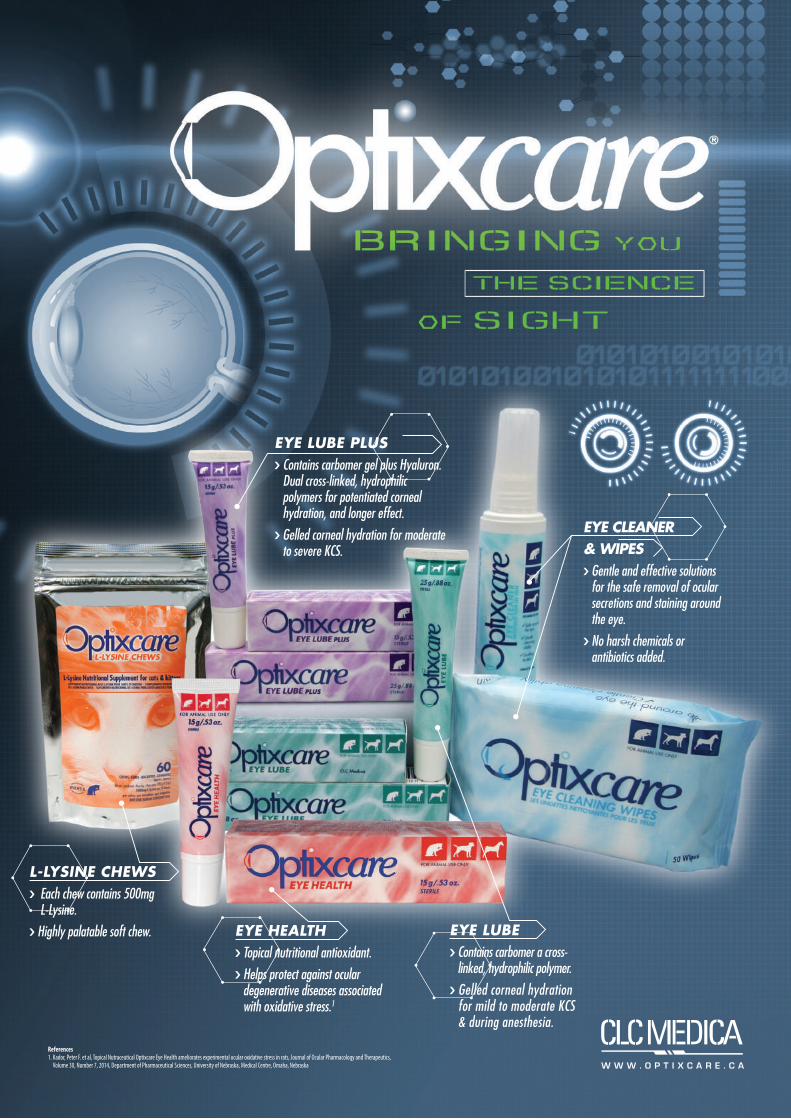
EYE CLEANER & WIPES Gentle and effective solutions
for the safe removal of ocular secretions and staining around the eye.
No harsh chemicals or antibiotics added.
EYE LUBE PLUS Contains carbomer gel plus Hyaluron.
Dual cross-linked, hydrophilic polymers for potentiated corneal hydration, and longer effect.
Gelled corneal hydration for moderate to severe KCS.
•
L-LYSINE CHEWS Each chew contains 500mg
L-Lysine. Highly palatable soft chew.
•
•
EYE LUBE Contains carbomer a cross-
linked, hydrophilic polymer. Gelled corneal hydration
for mild to moderate KCS & during anesthesia.
•
EYE HEALTH Topical nutritional antioxidant. Helps protect against ocular
degenerative diseases associated with oxidative stress.1
References1. Kador, Peter F. et al, Topical Nutraceutical Optixcare Eye Health ameliorates experimental ocular oxidative stress in rats, Journal of Ocular Pharmacology and Therapeutics,
Volume 30, Number 7, 2014, Department of Pharmaceutical Sciences, University of Nebraska, Medical Centre, Omaha, Nebraska W W W . O P T I X C A R E . C A
EYE CLEANER & WIPES Gentle and effective solutions for the safe removal of ocular secretions and staining around the eye. No harsh chemicals or antibiotics added.
EYE LUBE PLUS Contains carbomer gel plus Hyaluron. Dual cross-linked, hydrophilic polymers for potentiated corneal hydration, and longer effect.
Gelled corneal hydration for moderate to severe KCS.
L-LYSINE CHEWS Each chew contains 500mg L-Lysine.
Highly palatable soft chew. EYE LUBE Contains carbomer a cross- linked, hydrophilic polymer. Gelled corneal hydration for mild to moderate KCS & during anesthesia.
EYE HEALTH Topical nutritional antioxidant. Helps protect against ocular degenerative diseases associated with oxidative stress.1
References1. Kador, Peter F. et al, Topical Nutraceutical Optixcare Eye Health ameliorates experimental ocular oxidative stress in rats, Journal of Ocular Pharmacology and Therapeutics,
Volume 30, Number 7, 2014, Department of Pharmaceutical Sciences, University of Nebraska, Medical Centre, Omaha, Nebraska W W W . O P T I X C A R E . C A
1
Committee members • Tim Knott (Chairperson) • Ida Gilbert (Joint secretary) • Natasha Mitchell (Joint secretary) • Helen Appleboam (Hotel and
conference organiser) • Georgie Fricker (Exhibitor liaison) • Michael Ziglar (International liaison) • David Nutbrown-Hughes (Website and
audio-visual) • Rachael Grundon (Scientific
programme) • Christine Heinrich (Scientific
programme) • Alistair Oldfield (Scientific
programme) • Mike Rhodes (Editor) • Jenny Lambert (Clinical auditor) • Rob Lowe (Treasurer) Publisher British Association of Veterinary Ophthalmologists. Enquiries should be directed to Mike Rhodes, Willows Referral Service, Highlands Road, Shirley, Solihull, West Midlands, B90 4NH. Tel: 0121 712 7070. Email: [email protected] Membership information Full membership is open to veterinary surgeons with an interest in the field of ophthalmology. Associate membership is open to those with professional interest in the field of ophthalmology. Membership is for 12 months and starts on 1st April. Membership fees are £40 per year. Abstracts / lecture notes submission Please follow the guidelines below: • The first line should give the title. • The authors’ names should appear on
the next line – initials followed by last name.
• Provide the name of your practice / institution.
• Use single spacing. • Use 10 point Trebuchet MS font for all
text. • Photographs or diagrams are
encouraged, but do not place them within the main body text. Instead send each photograph or diagram as a
separate JPEG image with a title such as ‘Figure 1: The eye before surgery’. Up to four images may be included. If additional images are required, please discuss this in advance of the deadline.
• The abstract should be 500-1000 words.
• Please send in electronic format to [email protected]
Deadlines Deadlines will be published on the website but are usually the first day of the month, two months prior to the meeting. Abstracts will be reviewed and the author notified one week after the submission deadline with time allowed for alterations if required. Twelve minutes will be allocated for each presentation, with a further three minutes for discussion. All oral presentations must be made in English. Presentations must be compatible with Microsoft PowerPoint 2003. Copyright and Photocopying © British Association of Veterinary Ophthalmologists 2015. All rights reserved. No part of this publication may be produced, stored or transmitted in any form or means without the prior permission in writing from the copyright holder. Advertising Enquiries about advertising should be directed to the Editor. Copies need to be received one month prior to the meeting for inclusion in the proceedings. Disclaimer The Publisher, the British Association of Veterinary Ophthalmologists and the Editor cannot be held responsible for errors or any consequences arising from the use of material contained in this journal. The views and opinions expressed do not necessarily reflect those of the Publisher or Editor; neither does publication of advertisements constitute any endorsement by the Publisher or the Editor of the products advertised.

2
SPEAKER BIOGRAPHIES
David J. Maggs
BVSc Hons, Diplomate ACVO Professor, Comparative Ophthalmology
University of California Davis Davis CA 95616
Following graduation from the University of Melbourne in 1988, David spent 5 years in mixed practice throughout Australia, England, Scotland, and Wales. He then completed small animal and equine internships at Colorado State University, and a research fellowship and comparative ophthalmology residency at the University of Missouri. He joined the faculty at the University of California-Davis in 2000. He is the author of Slatter’s Fundamentals of Veterinary Ophthalmology, an editorial board member for the Journal of Feline Medicine and Surgery, Chair of the American Board of Veterinary Ophthalmology, President of the International Society of Veterinary Ophthalmology, and the 2012 WVC Small Animal Continuing Educator of the year. David’s major interests are ocular surface disease.
James Oliver BVSc CertVOphthal DipECVO MRCVS
RCVS and European Specialist in Veterinary Ophthalmology
Having graduated from the University of Bristol in 2002, James spent five years in general practice. James gained the RCVS Certificate in Veterinary Ophthalmology in 2007 and then undertook a three-year residency at Davies Veterinary Specialists. He became an ECVO diplomate in 2011 and is an RCVS recognised specialist in veterinary ophthalmology. James co-authored the Ocular Therapeutics chapter in the latest edition of the BSAVA Manual of Small Animal Ophthalmology and teaches this subject for the BSAVA Postgraduate Certificate. He recently co-authored the book Feline Ophthalmology – The Manual and is working towards a PhD in the genetics of canine primary glaucoma. James is Head of Ophthalmology at The Animal Health Trust.’
3
Dr Seth A Koch VMD Diplomate ACVO Ophthalmology
Veterinary education at University of Pennsylvania, 1965. Intern at the same institute, one year in internal medicine, N.I.H. post doctoral fellowship to study ophthalmology after the internal medicine year (in those days, there was no veterinary program residencies as such) mentors were Lon Rubin,VMD and Harold Scheie, MD, received a masters degree from the school of medicine (not the vet school) in comparative ophthalmology, thesis was on day blindness in Alaskan Malamutes. Started a private practice, one of the very first in the U.S. in 1970 in the Washington D.C. area. Taught as an adjunct associate professor at Pennsylvania every Monday and Tuesday for 25 plus years in ophthalmology clinics. Published 50+ papers in ophthalmology, and was recipient of AVMA practitioner research award, was a University of Pa alumni award recipient. Mentored a number of people in ophthalmology from both the U.S. and Europe (Bedford, Perrucio, Clerc, and others), Currently pretty well retired except for occasional consultations, an occasional request to come to a colleagues office and do a PDT, toxicology for a major drug company in the U.S., married, travel, fly fish in salt water, read, active in the boy scouts of America, have 9 grandkids among all the children, tell lots of jokes and laugh a lot, mostly at myself...
Mike Rhodes BVM&S CertVOphthal DipECVO
RCVS and European Specialist in Veterinary Ophthalmology
Mike graduated from Edinburgh University in 2004 and spent the next three and half years working in small animal practice in Peterborough and Suffolk. During this time he developed a keen interest in veterinary ophthalmology and completed the RCVS Certificate in Veterinary Ophthalmology in 2008. He then undertook a three-year ECVO residency programme at Willows Referral Service and obtained the European Diploma in Veterinary Ophthalmology in 2013. Mike has since stayed on at Willows becoming a recognised RCVS Specialist in Veterinary Ophthalmology in 2015. Mike’s clinical interests include the management of KCS and all aspects of ocular surgery, especially cataract removal.
JamesOliverBrAVOApril2016Antibacterial and antifungal agents in veterinary ophthalmology
Commensal ocular surface flora and pathogenic bacteria
For a more comprehensive review the reader is directed to the book chapter Clinical Microbiology
and Parasitology by Gould and Papsouliotis (In: Veterinary Ophthalmology, 5th Edition, Ed. Gelatt
KN, Gilger BC and Kern TJ).
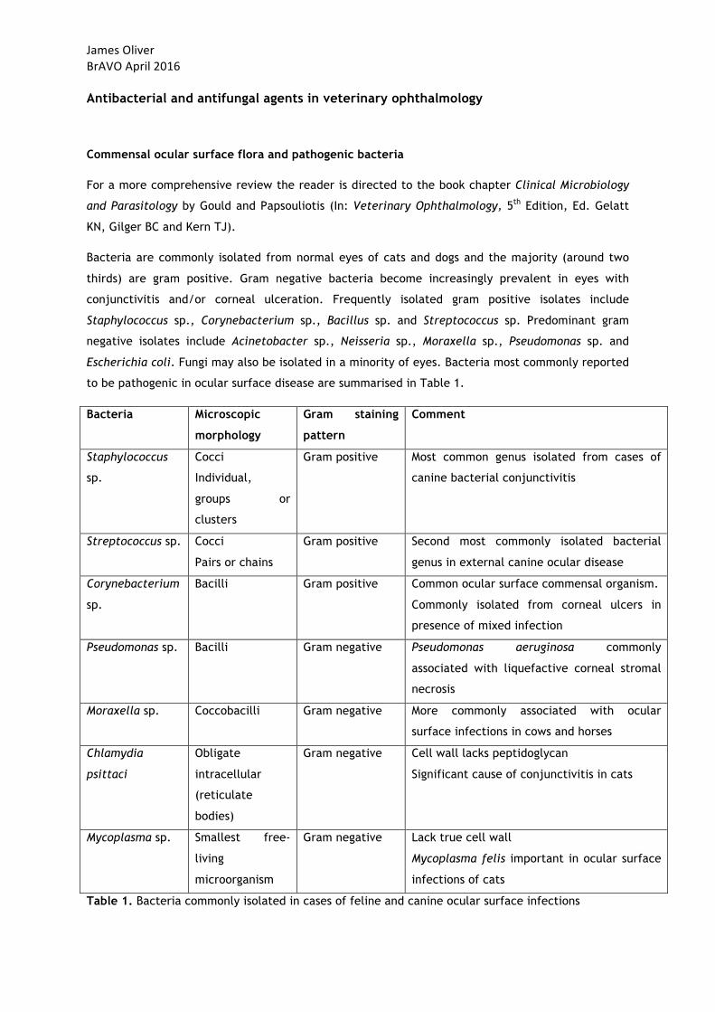
Bacteria are commonly isolated from normal eyes of cats and dogs and the majority (around two
thirds) are gram positive. Gram negative bacteria become increasingly prevalent in eyes with
conjunctivitis and/or corneal ulceration. Frequently isolated gram positive isolates include
Staphylococcus sp., Corynebacterium sp., Bacillus sp. and Streptococcus sp. Predominant gram
negative isolates include Acinetobacter sp., Neisseria sp., Moraxella sp., Pseudomonas sp. and
Escherichia coli. Fungi may also be isolated in a minority of eyes. Bacteria most commonly reported
to be pathogenic in ocular surface disease are summarised in Table 1.
Bacteria Microscopic
morphology
Gram staining
pattern
Comment
Staphylococcus
sp.
Cocci
Individual,
groups or
clusters
Gram positive Most common genus isolated from cases of
canine bacterial conjunctivitis
Streptococcus sp. Cocci
Pairs or chains
Gram positive Second most commonly isolated bacterial
genus in external canine ocular disease
Corynebacterium
sp.
Bacilli Gram positive Common ocular surface commensal organism.
Commonly isolated from corneal ulcers in
presence of mixed infection
Pseudomonas sp. Bacilli Gram negative Pseudomonas aeruginosa commonly
associated with liquefactive corneal stromal
necrosis
Moraxella sp. Coccobacilli Gram negative More commonly associated with ocular
surface infections in cows and horses
Chlamydia
psittaci
Obligate
intracellular
(reticulate
bodies)
Gram negative Cell wall lacks peptidoglycan
Significant cause of conjunctivitis in cats
Mycoplasma sp. Smallest free-
living
microorganism
Gram negative Lack true cell wall
Mycoplasma felis important in ocular surface
infections of cats
Table 1. Bacteria commonly isolated in cases of feline and canine ocular surface infections
JamesOliverBrAVOApril2016Antibacterial agents
Several principles should be followed in the selection of antibacterial therapy to maximise the
chance of successful management of ocular disease. Principles to consider include, but are not
limited to, the following:
• Availability of antibacterial agents
• Authorisation of products in small animals and implementation of the prescribing cascade
• Whether the agent is required for prophylaxis or treatment of infection
• Spectrum of antibacterial activity required (assisted by cytology and or culture &
susceptibility)
• Intended site of action of antibacterial agent (and ocular penetration characteristics of
agent)
• Owner compliance
• Potential for drug resistance
• Potential for adverse effects
Antibacterial agents are generally designed to exploit inherent differences between bacterial and
mammalian cells. They may exert their effects by a variety of means including: inhibition of
bacterial cell wall synthesis, alteration of bacterial cell membrane permeability, inhibition of
bacterial protein synthesis, inhibition of bacterial folic acid synthesis and interfering with bacterial
DNA synthesis and replication. Antibacterial agents may be classified as bacteriostatic or
bactericidal which largely relates to their mechanism of action. Some antibacterial agents may be
either bacteriostatic or bactericidal with their activity relating to the concentration of the agent at
the site of action. It is generally considered unwise to combine the use of bactericidal and
bacteriostatic agents owing to potential antagonism between the agents. This is of questionable
significance for agents used topically on the eye, however. It can also be argued that the use of
multiple classes of antibacterial agents on the eye increases the spectrum of activity and reduces
the chance of drug resistance. Such polypharmacy, however, should be used with some caution.
Accurate identification of the organism responsible for disease forms the ideal basis of antibacterial
agent selection. Culture and susceptibility testing is often impractical, however, and in house
cytological examination of Diff-Quik-stained or Gram-stained slides offers a quick and affordable
compromise.
Drugs that inhibit bacterial cell wall synthesis
Penicillins. Penicillins are bactericidal. They have a Β-lactam ring which binds to bacterial
transpeptidases which are required for formation of peptide cross-linkages between the
polysaccharide chains of peptidoglycan (a vital component of the bacterial cell wall). This results in
JamesOliverBrAVOApril2016incomplete cell wall synthesis and bacterial cell death. Penicillins G and V are susceptible to β-
lactamases and are rarely used in veterinary ophthalmology. Β-lactamase resistant penicillins
include methicillin, oxacillin and cloxacillin but alternative resistance pathways have been
developed by bacteria (‘methicillin-resistant’ strains). Amoxicillin, which is susceptible to β-
lactamase, becomes resistant when combined with clavulanic acid, extending its efficacy to Staph.
aureus and Staph. epidermidis. Amoxicillin/clavulanic acid remains the most commonly used
systemic antibacterial agent in veterinary medicine. In veterinary ophthalmology specifically, it is
used prophylactically following intraocular surgery and in the treatment of orbital and eyelid
infections. In cats with experimentally induce chlamydial conjunctivitis, treatment with
amoxicillin/clavulanic acid was as effective as doxycycline which is somewhat surprising as the cell
walls of Chlamydia spp. lack peptidoglycan (Sturgess et al. 2001). Penicillins should not be used in
individuals with known hypersensitivity to this class of antibacterial agents or to cephalosporins.
Cephalosporins. Cephalosporins have a very similar mechanism of action to penicillins although
have a different molecular structure. They may be susceptible to some β-lactamases produced by
some gram-negative bacteria but are generally resistant to those produced by Staph. aureus. They
are classified in generations which relate to their side-chain modifications which alter their
spectrum of activity. Cephalosporins are generally only available as systemic preparations
(intravenous and oral). In veterinary ophthalmology, there is rarely justification to use later
generation drugs. First generation cephalosporins include cephalexin and cefazolin. Cefazolin
(50mg/ml) can be used off-license to treat corneal ulcers infected with gram-positive organisms
although some streptococci have developed resistance. In dogs, IV cefazolin reaches a therapeutic
concentration in the anterior chamber and thus is a sensible choice during intraocular surgery. Oral
cephalexin is a good choice for staphylococcal eyelid infections. Second generation cephalosporins
include cefuroxime and cefoxitin and have increased gram-negative activity. Cefixime, cefoxatime
and ceftazidime are third generation and again have increased gram-negative activity. Ceftazidime
is used to treat endophthalmitis in humans via intravitreal injection. Cefepime is a fourth
generation cephalosporin and is very broad-spectrum having excellent activity against both gram-
negative and gram-positive organisms. Contraindications for use mainly relate to known
hypersensitivity to this class of agents and to pencillins. These drugs should also be used with
caution in animals with vitamin K deficiency and renal impairment.
Bacitracin. This bactericidal drug inhibits the movement of the precursor to bacterial
peptidoglycan (an essential bacterial cell wall component) across the bacterial cell membrane thus
inhibiting cell wall synthesis. It is mainly active against gram-positive species with little gram-
negative action. It is thus usually combined with other drugs such as neomycin and polymixin B to
provide a wider spectrum of activity. This ‘triple antibiotic’ is commonly used in the USA (and other
countries) both prophylactically (uncomplicated corneal ulcers) and therapeutically (non-specific
JamesOliverBrAVOApril2016ocular surface infections). Bacitracin has poor corneal penetration and thus is not useful for
intraocular infections. The main potential side effect of bacitracin is a local hypersensitivity
reaction.
Vancomycin. Vancomycin is bactericidal and inhibits the production of the mucopeptide portion of
peptidoglycan and thus cell wall synthesis. It has very good gram-positive activity including efficacy
against methicillin-resistant species. Its use is limited to patients with resistant infections or which
have known hypersensitivity to other antibiotics. Side effects of long-term parenteral use include
ototoxicity and nephrotoxicity.
Drugs that affect bacterial cell membranes
Owing to similarities in bacterial and mammalian cell membranes, the systemic use of these agents
would carry a high risk of toxicity. Thus these drugs are generally limited to topical use in the
treatment of ocular surface disease.
Polymixin B. This surfactant (detergent) interacts with the phospholipids of the cell membrane
which alters cell membrane permeability which ultimately leads to cell death. It has reasonable
gram-negative activity (including Pseudomonas spp.) and is used topically in combination with drugs
with gram-positive activity (e.g. bacitracin). Side effects include neurotoxicity and nephrotoxicity
(when used systemically) and local hypersensitivity reactions (when used topically).
Gramicidin. This bactericidal drug has mainly gram-positive activity. It is often combined, in place
of bacitracin, with drugs with gram-negative activity, such as neomycin and polymixin B, in topical
preparations.
Drugs that affect bacterial protein synthesis
These include aminoglycosides, tetracyclines, macrolides, lincosamides and chloramphenicol. They
alter bacterial protein synthesis by binding to either the 30S or 50S subunits of bacterial ribosomes.
Aminoglycosides. Aminoglycosides inhibit the 30S subunit of the bacterial ribosome and are
bactericidal. The most commonly used examples include neomycin, gentamicin, tobramycin,
kanamycin and amikacin. They have excellent gram-negative activity although neomycin is
generally inactive against Pseudomonas aeruginosa (an important pathogen of the ocular surface).
Their gram-positive activity is limited primarily to Staphylococcus aureus and they are inactive
against anaerobes. They have a synergistic/additive effect when used in conjunction with β-lactam
JamesOliverBrAVOApril2016antibacterial agents but must be given separately as they may be inactivated by these drugs.
Aminoglycosides are poorly absorbed when given orally and thus are used either topically or
intravenously. Neomycin is a common component of triple antibiotic topical preparations being
used either for prophylaxis or non-specific ocular surface infections. Its poor corneal penetration
renders it unsuitable for treatment of intraocular infections. Gentamicin may be applied topically
or be injected subconjunctivally to treat infectious keratitis particularly caused by Pseudomonas
aeruginosa. Subconjunctival injections may lead to therapeutic drug levels within the anterior
chamber but systemic absorption may lead to side effects. Licensed formulations exist for the dog,
cat and rabbit (Clinagel®, Tiacil®). Tobramycin has similar activity and indications to gentamicin.
This drug may be used as a fortified formulation for bacterial ulcers caused by P. aeruginosa strains
which are resistant to gentamicin. Kanamycin has been used topically to infectious bovine keratitis
caused by Moraxella bovis. Amikacin is not available as a topical product but can be compounded
as a topical preparation. It may also be injected subconjunctivally reaching an appreciable
intraocular concentration. Amikacin is less retinotoxic than other aminoglycosides thus may be
useful for endophthalmitis. All aminoglycosides should be used with caution in patients with renal
disease owing to their potential nephrotoxicity.
Tetracyclines. Tetracyclines interact with the 30S subunit of the bacterial ribosome and are
bacteriostatic. Tetracycline and oxytetracycline are examples of short-acting aminoglycosides and
doxycycline is long-acting. Good activity against Mycoplasma spp., Chlamydophila spp., Rickettsia
spp. and Moraxella spp. Resistance is common in Staphylococcus spp., Streptococcus spp. and
Pseudomonas aeruginosa. Doxycycline is used orally in cats with ocular disease caused by infections
with Chlamydophila felis and Mycoplasma spp. Tetracyclines are available in some countries as
topical preparations (not the UK). Apart from their use in ocular surface infections caused by the
aforementioned bacteria, they may also be used as treatment for ‘melting’ corneal ulcers owing to
their anticollagenase properties and may be useful in the management of spontaneous chronic
corneal epithelial defects (SCCEDs) (Chandler et al. 2010). Tetracyclines concentrate in the cornea
and lacrimal gland and may have beneficial ocular surface effects by a number of actions such as
chelation of cations, inhibition of gene expression, inhibition of α1-antitrypsin degradation and
inhibition of leucotaxis (Herring, 2007). Side effects include tooth enamel discoloration in kittens
and puppies and photosensitivity. Doxycycline has been associated with oesophageal stricture
formation and therefore oral administration should be followed by a water or food swallow.
Macrolides. Macrolides bind to the 50S subunit of the bacterial ribosome and are bacteriostatic.
Examples include erythromycin, clindamycin, azithromycin and clarithromycin. These drugs are
mainly used systemically. Azithromycin has been advocated to treat infections caused by Bartonella
henselae and Chlamydophila felis but appears to have lower efficacy compared to doxycycline.
JamesOliverBrAVOApril2016
Lincosamides. These have a similar mode of action to macrolides. Clindamycin remains the most
frequently used drug of this class in veterinary ophthalmology being prescribed in oral form to treat
infections suspected to be caused by Toxoplasma gondii as well as being used for anaerobic
infections.
Chloramphenicol. Chloramphenicol binds to the 50S subunit and is bacteriostatic. It has relative
broad-spectrum of activity being effective against gram-positive and gram-negative bacteria and
also having efficacy against Rickettsia, Chlamydophila, and Mycoplasma spp. Pseudomonas spp. are
resistant. It is not used systemically owing to serious side effect of bone marrow suppression. The
drug is used topically, being an excellent first line choice in the treatment of non-complicated
corneal ulcers and as prophylaxis following ocular surface and intraocular surgery owing to
excellent corneal penetration (due to the high lipophilicity of the drug). Chloramphenicol eye drops
should be stored under refrigerated conditions owing to the thermal instability of the drug at
warmer temperatures.
Fusidic acid. This drug prevents the turnover of elongation factor G from the bacterial ribosome
and is bacteriostatic. Fusidic acid is effective primarily against gram-positive bacteria such as
Staphylococcus spp. (including MRSA), Streptococcus spp. and Corynebacterium spp. In veterinary
ophthalmology, it is used topically being an excellent first-line choice for prophylaxis
(uncomplicated corneal ulcers) and ocular surface infections caused by gram-positive bacteria.
Fusidic acid may be used in conjunction with drugs with gram-negative activity (e.g. ofloxacin or
gentamicin) to widen spectrum of activity. It has good corneal penetration and a licensed
formulation exists for the dog, cat and rabbit (Isathal®).
Drugs that alter folic acid synthesis
Examples include sulphonamides and trimethoprim and these drugs are generally considered to be
bacteriostatic. Sulphonamides and trimethoprim are often combined as they have synergistic
effects as they inhibit different steps in the pathway of folate synthesis. They have relatively good
gram-positive but variable gram-negative activity. They have poor intraocular penetration when
given systemically. Numerous potential side effects including hepatotoxicity, nephrotoxicity and
blood dyscrasias. Systemic use of TMS also carries the very real risk of keratoconjunctivitis sicca
owing to a direct toxic effect on lacrimal acinar cells.
JamesOliverBrAVOApril2016
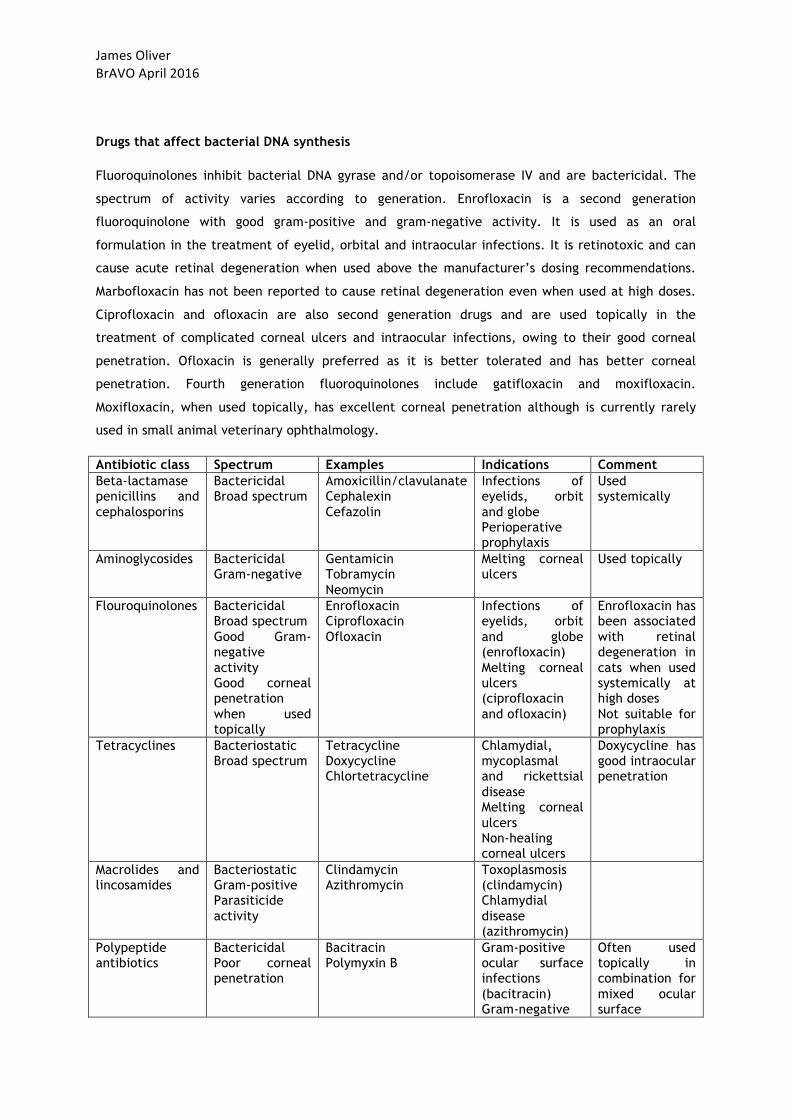
Drugs that affect bacterial DNA synthesis
Fluoroquinolones inhibit bacterial DNA gyrase and/or topoisomerase IV and are bactericidal. The
spectrum of activity varies according to generation. Enrofloxacin is a second generation
fluoroquinolone with good gram-positive and gram-negative activity. It is used as an oral
formulation in the treatment of eyelid, orbital and intraocular infections. It is retinotoxic and can
cause acute retinal degeneration when used above the manufacturer’s dosing recommendations.
Marbofloxacin has not been reported to cause retinal degeneration even when used at high doses.
Ciprofloxacin and ofloxacin are also second generation drugs and are used topically in the
treatment of complicated corneal ulcers and intraocular infections, owing to their good corneal
penetration. Ofloxacin is generally preferred as it is better tolerated and has better corneal
penetration. Fourth generation fluoroquinolones include gatifloxacin and moxifloxacin.
Moxifloxacin, when used topically, has excellent corneal penetration although is currently rarely
used in small animal veterinary ophthalmology.
Antibiotic class Spectrum Examples Indications Comment Beta-lactamase penicillins and cephalosporins
Bactericidal Broad spectrum
Amoxicillin/clavulanate Cephalexin Cefazolin
Infections of eyelids, orbit and globe Perioperative prophylaxis
Used systemically
Aminoglycosides Bactericidal Gram-negative
Gentamicin Tobramycin Neomycin
Melting corneal ulcers
Used topically
Flouroquinolones Bactericidal Broad spectrum Good Gram-negative activity Good corneal penetration when used topically
Enrofloxacin Ciprofloxacin Ofloxacin
Infections of eyelids, orbit and globe (enrofloxacin) Melting corneal ulcers (ciprofloxacin and ofloxacin)
Enrofloxacin has been associated with retinal degeneration in cats when used systemically at high doses Not suitable for prophylaxis
Tetracyclines Bacteriostatic Broad spectrum
Tetracycline Doxycycline Chlortetracycline
Chlamydial, mycoplasmal and rickettsial disease Melting corneal ulcers Non-healing corneal ulcers
Doxycycline has good intraocular penetration
Macrolides and lincosamides
Bacteriostatic Gram-positive Parasiticide activity
Clindamycin Azithromycin
Toxoplasmosis (clindamycin) Chlamydial disease (azithromycin)
Polypeptide antibiotics
Bactericidal Poor corneal penetration
Bacitracin Polymyxin B
Gram-positive ocular surface infections (bacitracin) Gram-negative
Often used topically in combination for mixed ocular surface
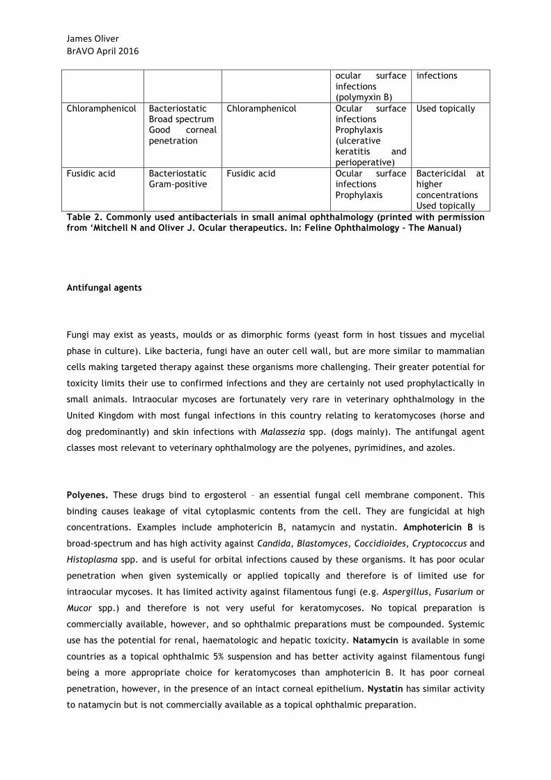
JamesOliverBrAVOApril2016
ocular surface infections (polymyxin B)
infections
Chloramphenicol Bacteriostatic Broad spectrum Good corneal penetration
Chloramphenicol Ocular surface infections Prophylaxis (ulcerative keratitis and perioperative)
Used topically
Fusidic acid Bacteriostatic Gram-positive
Fusidic acid Ocular surface infections Prophylaxis
Bactericidal at higher concentrations Used topically
Table 2. Commonly used antibacterials in small animal ophthalmology (printed with permission from ‘Mitchell N and Oliver J. Ocular therapeutics. In: Feline Ophthalmology – The Manual)
Antifungal agents
Fungi may exist as yeasts, moulds or as dimorphic forms (yeast form in host tissues and mycelial
phase in culture). Like bacteria, fungi have an outer cell wall, but are more similar to mammalian
cells making targeted therapy against these organisms more challenging. Their greater potential for
toxicity limits their use to confirmed infections and they are certainly not used prophylactically in
small animals. Intraocular mycoses are fortunately very rare in veterinary ophthalmology in the
United Kingdom with most fungal infections in this country relating to keratomycoses (horse and
dog predominantly) and skin infections with Malassezia spp. (dogs mainly). The antifungal agent
classes most relevant to veterinary ophthalmology are the polyenes, pyrimidines, and azoles.
Polyenes. These drugs bind to ergosterol – an essential fungal cell membrane component. This
binding causes leakage of vital cytoplasmic contents from the cell. They are fungicidal at high
concentrations. Examples include amphotericin B, natamycin and nystatin. Amphotericin B is
broad-spectrum and has high activity against Candida, Blastomyces, Coccidioides, Cryptococcus and
Histoplasma spp. and is useful for orbital infections caused by these organisms. It has poor ocular
penetration when given systemically or applied topically and therefore is of limited use for
intraocular mycoses. It has limited activity against filamentous fungi (e.g. Aspergillus, Fusarium or
Mucor spp.) and therefore is not very useful for keratomycoses. No topical preparation is
commercially available, however, and so ophthalmic preparations must be compounded. Systemic
use has the potential for renal, haematologic and hepatic toxicity. Natamycin is available in some
countries as a topical ophthalmic 5% suspension and has better activity against filamentous fungi
being a more appropriate choice for keratomycoses than amphotericin B. It has poor corneal
penetration, however, in the presence of an intact corneal epithelium. Nystatin has similar activity
to natamycin but is not commercially available as a topical ophthalmic preparation.
JamesOliverBrAVOApril2016Pyrimidines. These drugs block thymidine, and therefore DNA and RNA synthesis, and are
fungistatic. Flucytosine-C is the only medically relevant drug of this class and its activity is mainly
against yeasts. Resistance is fairly high and it is rarely used in veterinary ophthalmology.
Azoles. Azoles inhibit ergosterol synthesis by inhibiting the cytochrome P450 enzyme pathway
resulting in inhibition of fungal growth and increasing cell membrane permeability. Owing to the
potential effects on mammalian cytochrome P450 when given systemically, they should be used
with caution in individuals being treated with drugs that are metabolised via this pathway. Azoles
are further classified as imidazoles or triazoles on the basis of their parent ring structure.
Imidazoles. Imidazoles include ketoconazole and miconazole. Ketoconazole has good efficacy
against Candida spp. but has reduced efficacy against filamentous fungi compared to other azoles.
It is also relatively slow in onset of action and its systemic use has a greater potential for
hepatotoxicity. Can be compounded as a 1% topical solution and demonstrates good corneal
penetration. Miconazole is broad-spectrum with better activity against filamentous fungi. It can be
compounded as a 1% ophthalmic suspension and 2% dermatological creams are commercially
available. It is well tolerated and has good corneal penetration.
Triazoles. Include fluconazole, itraconazole and voriconazole. Fluconazole has good activity
against yeasts including Candida and Cryptococcus but limited efficacy against moulds such as
Aspergillus and Fusarium. It is well tolerated when given systemically and appears to have good
intraocular penetration. Itraconazole has a broader spectrum of activity than fluconazole
particularly against filamentous fungi. Intraocular penetration is poor when given systemically,
however. Can be compounded as a 1% ointment. Voriconazole has a very broad spectrum of
activity and has good intraocular penetration when given systemically. The IV solution can be used
topically in the treatment of keratomycosis in dogs and horses and is well tolerated.
References and further reading
1. Chandler H, Gemensky-Metzler A, Bras I (2010) In vivo effects of adjunctive tetracycline
treatment on refractory corneal ulcers in dogs. Journal of American Veterinary Medical
Association; 237:387-386
2. Gould D and Papasouliotis K (2013) Clinical microbiology and parasitology. In:
Veterinary Ophthalmology, 5th edn, ed. Gelatt KN, Gilger BC and Kern TJ, pp 300-350.
Blackwell Publishing, Iowa
3. Herring IP (2007) Clinical pharmacology and therapeutics In: Veterinary Ophthalmology,
4th edn, ed. KN Gelatt, pp 332-354. Blackwell Publishing, Iowa.
4. Mitchell N and Oliver J. Ocular therapeutics. In: Feline Ophthalmology – The Manual,
pp31-40. Servet ISBN 978-84-16315-11-6

JamesOliverBrAVOApril2016
5. Oliver J and Smith K (2014) Ophthalmic drugs. In: BSAVA Manual of Canine and Feline
Ophthalmology, 3rd edn. Ed. Gould D and McLellan G, pp97-110. British Small Animal
Veterinary Association, Gloucester.
6. Pennisi MG, Hartmann K, Lloret A et al. (2013) Cryptococcosis in cats: ABCD guidelines
on prevention and management. Journal of Feline Medicine and Surgery; 15:611-618.
7. Sturgess CP, Gruffydd-Jones TJ, Harbour DA et al. (2001) Controlled study of the
efficacy of clavulanic acid-potentiated amoxicillin in the treatment of Chlamydia
psittaci in cats. Veterinary Record; 149:73–76.
FHV-1 – LATEST THOUGHTS ON PATHOGENESIS AND DIAGNOSIS
David J. Maggs
Some Clinically Relevant Virology
Feline herpesvirus is a ubiquitous virus that varies very little worldwide with respect to its clinical virulence. And yet, we see a huge range of clinical signs in cats infected with this virus. There are probably a large number of reasons for this; however principle among these is likely the host’s response to this virus (not the virus itself). FHV-1-naïve kittens infected in the first few weeks of life against a backdrop waning maternal immunity almost inevitably get severe upper respiratory and ocular disease with high morbidity but rare mortality. By contrast, adult cats can undergo viral reactivation with viral shedding and can infect in-contact cats; all without demonstrating clinical signs themselves. These two scenarios represent just the two extremes of infection. Within your clinic, you see cats with a huge diversity of clinical signs in between. For this reason, I like to consider clinical signs associated with FHV-1 under one of three broad categories: primary infection, recrudescent infections, and FHV-1-associated syndromes. Layered on top of this is the mechanism by which the virus causes injury and disease. Herpetic disease in humans (and to a growing extent in cats) can be categorized as resulting from 1 of 3 pathophysiologic mechanisms. Note that these are particularly useful to the clinician, not just the virologist!) because they can be used to guide treatment:
1. Cytolytic disease, where cell rupture occurs as a direct result of viral replication. In this form of disease, virus can be cultured from diseased tissue and antiviral drugs are recommended whereas immunomodulatory therapy is not.
2. Immunopathologic disease, where the host’s reaction to viral antigens or altered auto-antigens is believed to be the major cause of disease. In this disease subset, virus is less reliably isolated, ulceration is less common, antiviral drugs are typically ineffective when used alone, and concurrent immunomodulatory therapy is often required.
3. Metaherpetic disease, which develops as a result of structural tissue damage as a
result of cytolytic and/or immunopathologic disease. Traditional antiviral or immunomodulatory therapies alone or together are ineffective, and therapy specific to the anatomic or physiologic disruption is required. Of particular relevance to cats is a specific metaherpetic syndrome in which virally-induced damage to the trigeminal nerve axons and their ganglion is believed to reduce corneal sensation and reflex tearing, thereby inducing an unusual form of dry eye in cats.
Clinical Presentations
PRIMARY HERPETIC DISEASE
Primary ocular FHV-1 infection is characterized by blepharospasm, conjunctival hyperaemia, serous ocular discharge that becomes purulent by day 5-7 of infection, mild to moderate conjunctival swelling, and often conjunctival ulcers. Corneal involvement is not reliable; however some cats develop corneal ulcers which are transiently dendritic at the
very earliest phase only. These dendrites quickly coalesce to become geographic ulcers. The ocular signs are seen in association with typical signs of upper respiratory infection. These signs are caused almost exclusively by cytolytic disease, and antiviral drugs are therefore helpful (although not always indicated). The uncomplicated clinical course is typically 10-14 days; however it is critical to realize that almost all cats become latently infected within ganglia for life. Reactivation from latency is likely in at least 50% of cats, sometimes with viral shedding.
RECRUDESCENT FHV-1 SYNDROMES
Despite the frequency with which latently infected cats undergo viral reactivation at the ganglia and viral shedding at peripheral epithelial sites, recrudescent disease occurs in a minority of these. Further, disease severity and tissue involvement can range very widely between individuals and even between episodes in the same cat. Recrudescent conjunctivitis is usually milder than in acute infections, but can become chronic and “smouldering”. Although recrudescent conjunctivitis is usually nonulcerative, substantial conjunctival thickening and hyperaemia can occur secondary to inflammatory cell infiltration. Corneal disease may involve the corneal epithelium or stroma, and may be ulcerative (due to cytolytic disease) as in primary infections. Corneal stromal disease is typically immunopathological (i.e., immune-mediated, but not necessarily autoimmune) in origin and includes stromal neovascularization, oedema, stromal cell infiltration, and ultimately fibrosis usually under an intact epithelium. Consensus has not been reached regarding the antigens responsible for the subepithelial immunological response within cornea and/or conjunctiva. Some believe the process is driven by viral antigens, while others are suspicious that altered self-antigens are the focus of the immunological response.
FHV-1-ASSOCIATED DISEASE SYNDROMES
The following diseases have been associated with detection of FHV-1 in affected tissues; however the causative role of the virus in each syndrome has been variably proven.
Symblepharon. There is little question that symblepharon can be a sequela to severe primary FHV-1 infection. It is commonly seen in young animals, and presumably occurs as a result of widespread ulceration with exposure of the conjunctival substantia propria and sometimes also the corneal stroma. FHV-1 is almost certainly the predominant cause of symblepharon formation in cats and other infectious agents are unlikely to cause symblepharon formation.
Corneal sequestration. Experimentally, FHV-1 inoculation (in cats receiving corticosteroids) can result in corneal sequestration. However, the prevalence of detectable FHV-1 in samples collected from cats with sequestra has varied widely in the clinical setting and the link between FHV-1 and sequestra has not been shown to be causative. It seems likely that sequestration is a non-specific response to stromal exposure or damage and that FHV-1 is just one possible cause of this disease. This is borne out in a study by Nasisse et al who reported identification of FHV-1 DNA in 86 of 156 (55%) of sequestra analysed (compared with only 6% of clinically normal corneas). A lower prevalence of FHV-1 DNA was found in corneas of Persian and Himalayan cats with sequestration, suggesting that other non-viral causes of sequestration are more likely to be operative in these breeds.
Eosinophilic keratitis. Prior clinical studies have suggested a link between FHV-1 infection and eosinophilic keratitis. In one study, PCR testing of corneal scrapings from cats with cytology-confirmed eosinophilic keratitis has revealed 76% (45/59) of cases to be FHV-1 positive. However, PCR performed on tears collected onto a STT was negative in 10 cats with cytologically proven eosinophilic keratitis. As with corneal sequestra, the role of the virus in the initiation or exacerbation of this disease has not been determined; however anecdotally some patients with this syndrome improve with antiviral therapy alone.
Dermatitis. Periodically, FHV-1 has been identified as a cause of dermatological lesions, particularly those surrounding the eyes and involving nasal skin of domestic and wild felidae. This is not surprising when one considers the marked epithelial tropism of this virus and the reliability with which herpes simplex virus (HSV-1) causes dermal lesions. We have recently examined the diagnostic utility of FHV-1 PCR for this disease. FHV-1 DNA was detected in all 9 biopsy specimens from 5 cats with herpetic dermatitis but in 1 of 17 biopsy specimens from the 14 cats with nonherpetic dermatitis, and was not detected in any of the 21 biopsy specimens from the 8 cats without dermatitis. This is in sharp contrast to the use of this technique in ocular tissues where the extent of viral shedding in normal animals dramatically reduces the sensitivity of a positive test in affected animals. When results of histologic examination were used as the gold standard in this study of cats with dermatitis, sensitivity and specificity of the PCR assay were 100% and 95%, respectively. We concluded that FHV-1 DNA can be detected in the skin of cats with herpetic dermatitis, that the virus may play a causative role in the disease, and that this PCR assay may be useful in confirming a diagnosis of herpetic dermatitis.
Uveitis. HSV-1 is a well-documented cause of uveitis in humans. Given the shared biological behaviour of these 2 alphaherpesviruses, we examined the role of FHV-1 in feline idiopathic uveitis. The PCR assay used demonstrated FHV-1 DNA in the aqueous humor of 12/86 cats, all but one of which had uveitis. The same study also used ELISA to examine FHV-1-specific antibody concentrations in aqueous humor and serum. While seropositivity did not vary among cats, intraocular antibody production, as determined by a Goldman-Witmer coefficient (C-value) > 1, was detected only in cats with uveitis. Additionally, a C-value > 8, which is frequently quoted as a more clinically useful indicator of intraocular antibody production, was found only in cats with idiopathic uveitis. A subsequent investigation also demonstrated FHV-1 DNA could be detected in the aqueous humor of cats and more often in the blood of cats with uveitis than those without uveitis. Taken together, these data suggest that intraocular FHV-1 infection occurs and that, at least in some cats, stimulates a specific local intraocular antibody response. Because the trigeminal nerve supplies the uveal tract, it is possible that virus may reactivate spontaneously or via induction and arrive in the uvea (and aqueous humor) via the “round trip theory”, as for surface ocular disease. Viral pathogenic mechanisms just like those reported in surface disease (i.e., virally mediated cytolysis and immunopathological responses directed at auto or viral antigens) are both plausible causes of herpetic uveitis. However, proving a casual association remains difficult.
A Diagnostic Approach to Cats with Keratoconjunctivitis
One of my least favourite questions is “What is the best laboratory test for cats with corneal or conjunctival disease?”. In reality there is not one. Explaining this position requires an understanding of an essential fact about feline herpesvirus (FHV-1) - clinically normal cats (and lots of them) can shed FHV-1 at their ocular surface. Because PCR is more
sensitive than IFA or VI, this assay exacerbates this problem. In fact, in some humane shelter-based populations, about half of all normal cats are shedding FHV-1 DNA as determined by PCR. Therefore, in some circumstances, the number of false positive test results we can expect is extraordinarily high and we may be better to flip a coin than to run that PCR assay! Given the predictably high rate of false positive (particularly with serology and PCR) and negative test results (particularly with VI and IFA), I no longer conduct laboratory tests for FHV-1 or Chlamydia felis (previously Chlamydophila felis and before that Chlamydia psittaci) in individual cats with keratoconjunctivitis. Rather, I resort to good old fashioned clinical acumen. My diagnostic “tests” now are (i) the history and clinical exam findings followed by (ii) response to therapy. This requires acceptance of a couple of critical facts: first I have to be willing to be wrong when making an educated guess regarding the aetiological diagnosis and, second, I have to use the absolute best therapeutic trial and demand excellent owner compliance in executing that trial.
USING CLINICAL SIGNS AS YOUR DIAGNOSTIC GUIDE
Using clinical signs of surface ocular disease as a “diagnostic assay” requires a philosophical approach that I liken to adding pebbles to one of two sides of an old-fashioned scale or balance. I start with the paradigm that feline keratoconjunctivitis is infectious till proven otherwise and that by far and away the most commonly implicated infectious organisms are FHV-1 and C. felis or possibly Mycoplasma spp. [Note that since my next “diagnostic test” is response to therapy and since Chlamydia and Mycoplasma spp. respond similarly to doxycycline (my preferred therapy), I am not particularly interested in separating them from each other as causes]. I then consider the clinical signs outlined in this table. Using each clinical sign as a discerning feature I aim to place one of my “diagnostic pebbles” on the herpetic or chlamydial sides of the balance, thereby making a clinical judgment at the end of the examination as to which of these 2 organisms is more likely to be the cause of the disease seen.
Clinical Signs FHV-1 C. felis/Mycoplasma Conjunctival hyperaemia +++ + Chemosis + +++ Ulceration (conj/cornea) +/- - Keratitis +/- - Dendrites Pathognomonic - Respiratory signs/malaise ++ +/- Note that some of the signs are caused by both agents and that it is, therefore, a weighted assessment. This introduces a notable element of subjectivity into the assessment. I unashamedly tell clients this and explain that I still believe that this is better than wasting their money on a laboratory test. I also take this opportunity to introduce the concept that the clients themselves will form the critical next step in the diagnostic process – “response to therapy”. We will discuss this more fully in the next session.
FHV-1 TREATMENT OPTIONS –
ARE YOU UP TO DATE WITH THE TRUTHS AND MYTHS?
David J. Maggs
Introductory Philosophy & Treatment for Chlamydia
If we are to use response to therapy as a “diagnostic test” (see previous session), then we must choose the optimum therapeutic approach possible for each cat, and impress on the client the importance of compliance and accurate feedback on progress (or regression). If, at the end of the clinical assessment, I believe that Chlamydia felis or Mycoplasma spp. are the most likely pathogens, then I recommend a 3-week course of orally administered doxycycline at 5-10 mg/kg once daily. Based on excellent placebo-controlled comparative trials in experimentally inoculated cats, I do not use azithromycin or topical tetracycline-containing antibiotics. Where reasonable, I treat all in-contact cats too as “silent shedders” in the household are likely. I typically do not use a topical antibiotic and never use a topical corticosteroid or NSAID. Because there is evidence that chronic conjunctivitis of multiple causes can lead to a vicious cycle of goblet cell deficiency and because topical administration of a mucinomimetic ophthalmic solution can ameliorate this cycle, I will also dispense an ophthalmic solution of (preferably non-preserved) hyaluronate for administration at least 4 times daily if the owners are able. Unlike doxycycline, this needs only be administered to those cats in the household which are demonstrating ocular clinical signs. This is discussed further in the session on feline tear film disorders and at the end of this session. The rest of this presentation describes my approach to response to therapy as a “diagnostic test” when I suspect herpetic ocular surface disease.
Herpetic Therapy
As a general rule I tend to think of herpetic therapy in 3 broad categories: R&D antiviral therapies, supportive care, and “other”. Here we will emphasize the agents developed by research and development (R&D) pharmaceutical companies as well as some of the more popular other therapies.
R&D ANTIVIRAL DRUGS
Although a large variety of R&D antiviral agents exists for oral or topical treatment of cats infected with feline herpesvirus type 1 (FHV-1), some general comments regarding these agents are possible: • No antiviral agent has been developed for FHV-1; although many have been tested for efficacy against this virus. Agents highly effective against closely-related human herpesviruses are not necessarily or predictably effective against FHV-1 and all should be tested in vitro before they are administered to cats. • No antiviral agent has been developed for cats; although some have been tested for safety in this species. Agents with a reasonable safety profile for topical application in humans tend to be safe in cats; however, systemically administered agents tolerated by humans are not always or predictably non-toxic when administered to cats and all require safety and efficacy testing in vivo. • Many antiviral agents require host metabolism before achieving their active form. These agents are not reliably or predictably metabolized by cats and pharmacokinetic studies in cats are required.
• Antiviral agents tend to be more toxic than do antibacterial agents since viruses are obligate intracellular organisms and co-opt or have close analogues of the host’s cellular “machinery”. This limits many antiviral agents to topical (ophthalmic) rather than systemic use. • All antiviral agents currently used for cats infected with FHV-1 are virostatic. Therefore, they typically require frequent administration to be effective. The following antiviral agents have been studied to varying degrees for their efficacy against FHV-1, their pharmacokinetics in cats, and/or their safety and efficacy in treating cats infected with FHV-1. Trifluridine is too toxic to be administered systemically, but topically administered trifluridine is considered one of the most effective drugs for treating HSV-1 keratitis. This is in part due to its superior corneal epithelial penetration. It is also one of the more potent antiviral drugs for FHV-1. It is formulated as a 1% ophthalmic solution that should be applied to the affected eye 5-6 times daily. Unfortunately, it is often not well tolerated by cats, presumably due to a stinging reaction reported in humans. In a retrospective case series of cats with ocular disease attributed to FHV-1, 1% trifluridine solution was used every 4-8 hours with improvement in 1 cat and no improvement or worsening in 2 cats. Idoxuridine is a nonspecific inhibitor of DNA synthesis, affecting any process requiring thymidine. Therefore, host cells are similarly affected, systemic therapy is not possible, and corneal toxicity can occur. It has been used as a 0.1% ophthalmic solution or 0.5% ophthalmic ointment. This drug is reasonably well tolerated by most cats and seems efficacious in some. In a retrospective case series of cats with ocular disease attributed to FHV-1, 0.1% idoxuridine solution was used every 4-6 hours with improvement or resolution of clinical signs in 3 cats and no improvement or worsening in 4 cats. It should be applied to the affected eye 5-6 times daily. Vidarabine interferes with DNA polymerase and, like idoxuridine, is non-selective in its effect and so is associated with notable host toxicity if administered systemically. Because it affects a viral replication step different from that targeted by idoxuridine, vidarabine may be effective in patients whose disease seems resistant to idoxuridine. As a 3% ophthalmic ointment, vidarabine often appears to be better tolerated than many of the antiviral solutions including idoxuridine. In a retrospective case series of cats with ocular disease attributed to FHV-1, 3% vidarabine ointment was used every 4 to 6 hours with improvement noted in 1 cat and no improvement or worsening noted in 2 cats. Like idoxuridine, it should be applied to the affected eye 5-6 times daily. Cidofovir is commercially available only in injectable form in the USA but has been studied when compounded as a 0.5% solution in methylcellulose artificial tears and applied topically twice daily to cats experimentally infected with FHV-1. Its use in these cats was associated with reduced viral shedding and less severe clinical disease. Its efficacy at only twice daily (despite being virostatic) is believed to be due to the long tissue half-lives of the metabolites of this drug. There are occasional reports of its experimental topical use in humans being associated with stenosis of the nasolacrimal drainage system components and, as yet, it is not commercially available as an ophthalmic agent in humans. Therefore, although the in vitro and short-term in vivo efficacy of cidofovir against FHV-1 is proven, cats should be monitored for nasolacrimal cicatrization. Cidofovir 0.5% retained efficacy
when compounded in normal saline and refrigerated (4 ºC) or frozen (-20 or -80 ºC) in plastic or glass for up to 6 months. However, safety data including change in pH, tonicity, etc., and risk of contamination were not evaluated. Acyclovir has relatively low antiviral potency against FHV-1, poor bioavailability, and is potentially toxic when systemically administered to cats. Oral administration of 50 mg/kg acyclovir to cats was associated with peak plasma levels of only approximately one third those required for this virus. Common signs of toxicity are referable to bone marrow suppression. However, acyclovir is also available as a 3% ophthalmic ointment in some countries. In one study in which a 0.5% ointment was used 5 times daily, the median time to resolution of clinical signs was 10 days. Cats treated only 3 times daily took approximately twice as long to resolve and did so only once therapy was increased to 5 times daily. Taken together, these data suggest that frequent topical application of acyclovir may produce concentrations at the corneal surface that do exceed the reported concentration required for this virus but are not associated with toxicity. There are also in vitro data suggesting that interferon exerts a synergistic effect with acyclovir that could permit an approximately 8-fold reduction in acyclovir dose. In vivo investigation and validation of these data are needed. Valacyclovir is a prodrug of acyclovir that, in humans and cats, is more efficiently absorbed from the gastrointestinal tract compared with acyclovir and is converted to acyclovir by a hepatic hydrolase. Plasma concentrations of acyclovir that surpass the IC50 for FHV-1 can be achieved after oral administration of this drug to cats. However, in cats experimentally infected with FHV-1, valacyclovir induced fatal hepatic and renal necrosis, along with bone marrow suppression, and did not reduce viral shedding or clinical disease severity. This likely resulted because high plasma concentrations of acyclovir were achieved - reinforcing the toxicity of acyclovir in cats. Despite its superior pharmacokinetics, valacyclovir should never be used in cats. Ganciclovir is at least 10-fold more effective against FHV-1 in vitro than is acyclovir. It is available for oral, intravenous, and intravitreal use in humans, where it is associated with greater toxicity than acyclovir. It is also available as a 0.15% ophthalmic gel. Although the in vitro efficacy of ganciclovir against FHV-1 and anecdotal reports of its topical administration to cats in Europe are very promising, to the author’s knowledge, neither the safety nor pharmacokinetics of ganciclovir in any form (or of its prodrug – valganciclovir) has been reported in cats. Penciclovir has a similar mechanism of action to acyclovir and potent antiviral activity against a number of human herpesviruses. It is highly effective against FHV-1 in vitro and in vivo. In a rabbit model of human HSV-1 keratitis, a 3% penciclovir ointment administered once, twice or four times daily decreased epithelial keratitis severity. Thus, a topical ophthalmic penciclovir ointment may be effective in cats with FHV-1 keratitis and/or conjunctivitis, but, to the author’s knowledge, there are no commercial or compounded preparations available for ophthalmic use. Penciclovir is available as a 1% dermatologic cream for humans, but that should not be applied to the eye. Famciclovir is a highly bioavailable prodrug of penciclovir; however metabolism of famciclovir to penciclovir in humans is complex; requiring di-deacetylation to BRL42359, in the blood, liver, or small intestine, and subsequent oxidation to penciclovir by aldehyde
oxidase in the liver. Neither famciclovir nor BRL42359 has any in vitro antiviral activity against FHV-1, therefore complete metabolism to penciclovir is required. However, hepatic aldehyde oxidase activity in cats is about 2% of that seen in humans and lower than in any other species reported to date. Not surprisingly, therefore, famciclovir and penciclovir pharmacokinetics in the cat are extremely complex and nonlinear (i.e., doubling of famciclovir dose does not lead to doubling of plasma penciclovir concentration) due to saturation of the hepatic oxidase. As a result, very high plasma concentrations of BRL42359 accumulate in the cat. Fortunately, this compound demonstrates very little cytotoxicity in vitro. Despite an increasing bank of information regarding the pharmacokinetics of famciclovir and penciclovir in tears and plasma of normal cats, definitive dose recommendations are still not possible. In addition to the complexity of the drug’s pharmacokinetics, this is in part because recommendation of an appropriate famciclovir dose requires: • Knowledge of whether penciclovir concentrations in plasma, tears, or the infected tissues themselves are most relevant • Selection of an appropriate target penciclovir concentration based on in vitro IC50s (which range at least 10-fold from 304 to 3500 ng/mL). • Knowledge of whether the targeted IC50 should be exceeded by the trough or the peak penciclovir concentrations, and for how long. Together, these uncertainties have led to much controversy about the optimum famciclovir dose in cats, with reported doses ranging from 8 mg/kg SID to 140 mg/kg TID. The following data are provided to inform dose selection. In the only masked, placebo-controlled efficacy trial to date, experimentally infected cats receiving 90 mg/kg famciclovir TID had significantly reduced clinical signs, serum globulin concentrations, histologic evidence of conjunctivitis, viral shedding, and serum FHV-1 titres, as well as increased goblet cell density relative to control cats. No important adverse clinical, hematologic or biochemical changes were associated with famciclovir administration. A subsequent study revealed that client-owned cats receiving 40 mg/kg TID had tear penciclovir concentrations likely to be effective against FHV-1 (using a target IC50 of 304 ng/mL) for at least 3 hours after each dose (i.e., for ≥ 9 hours/day). In the most comprehensive pharmacokinetic study to date, healthy cats were administered famciclovir at 30, 40 or 90 mg/kg BID or TID, and plasma and tear famciclovir, BRL42359, and penciclovir concentrations were measured. This resulted in the recommendation that cats should receive 90 mg famciclovir/kg twice daily because this regimen achieved comparable plasma and tear penciclovir concentrations to those achieved with 90 mg/kg TID, whereas the lower doses tested did not result in adequate tear penciclovir concentrations, even when administered TID. Perhaps most revealing so far, are data from a retrospective study comparing outcomes when famciclovir was administered TID to client-owned cats with presumed herpetic disease at approximately 40 (n = 33 cats) or 90 mg/kg (n = 26 cats). Median duration of therapy required for clinical improvement was significantly longer in cats administered 40 versus 90 mg/kg. Furthermore, cats in the 90 mg/kg group showed significantly greater and faster improvement than did cats in the 40 mg/kg group. Adverse events (most commonly gastrointestinal) potentially attributable to famciclovir were reported in 17% of cats receiving 40 or 90 mg famciclovir/kg TID, but the prevalence was not different between the 2 dose groups. The reduction in treatment duration with the higher
famciclovir dose was estimated to decrease overall client costs due to a reduction in total famciclovir administered (and potentially the number of recheck examinations required). These data, suggest that administration of 90 mg/kg TID is clinically and cost effective. Meanwhile, pharmacokinetic data from a separate study suggest that tear and plasma penciclovir concentrations are similar whether cats receive 90 mg famciclovir /kg 2 or 3 times daily. Therefore, taken together, data from these 2 studies suggest that 90 mg famciclovir/kg twice daily is likely to be effective in treating cats with herpetic disease. Assessing all in vivo tolerance data for famciclovir, this drug appears to be markedly safer than acyclovir and valacyclovir - the only other systemic antiviral drugs to be orally administered to cats. However, patients administered famciclovir should be closely monitored, and assessment of a complete blood count, serum biochemistry panel, and urinalysis should be considered in cats with known concurrent disease or cats expected to receive famciclovir for long periods. As in humans, reduction in dose frequency should be considered in cats with renal insufficiency.
LYSINE
Lysine is perhaps the best studied and yet maybe one of the more controversial of all of the other compounds with putative efficacy against FHV-1 in cats. As with the R&D antiviral drugs, initial interest arose from in vitro data and clinical trials in humans. Lysine’s antiviral effect is believed to arise because arginine is an essential amino acid for FHV-1 and HSV-1 replication, and assumes that lysine antagonizes arginine availability to or utilization by these viruses during protein synthesis. This was hypothesized to affect protein synthesis of the virus more than the host because viral proteins had a higher arginine-to-lysine content than did human (and feline) proteins; however recent analysis suggests that the difference in feline versus FHV-1 protein amino acid content is minimal. Markedly elevated lysine concentrations in combination with notably low arginine concentrations suppress HSV-1 and FHV-1 replication in vitro. However, this was not borne out with more physiologic amino acid concentrations. In vivo data in cats are also contradictory. Oral administration of 500 mg L-lysine every 12 hours beginning 6 hours prior to inoculation with FHV-1 was associated with less severe conjunctivitis but similar viral shedding to cats receiving placebo. In cats latently infected by experimental inoculation but without clinical signs, oral administration of 400 mg L-lysine once daily reduced viral shedding relative to placebo-treated cats. Despite significant elevations in plasma lysine concentration, no change in plasma arginine concentration was observed in either study. Mild, reversible gastrointestinal disturbance potentially attributable to lysine administration was noted in some cats. In the only study to assess bolus administration of lysine in naturally infected cats, 144 shelter-housed cats received 250 mg (kittens) or 500 mg (adult cats) lysine once daily for their entire shelter stay; outcomes were compared with an untreated control group. No significant treatment effect was detected for any parameter. Safety and efficacy of dietary lysine supplementation have also been assessed. No ill effects were seen in cats fed diets supplemented to up to 8.6% (dry matter) lysine. In 2 subsequent efficacy trials, cats in environments where FHV-1 was enzootic were fed a diet supplemented to 5.1% lysine while control cats received a basal ration (approximately 1% lysine). In both studies, disease was more severe and viral shedding was increased in cats fed the supplemented ration relative to those fed the basal diet. This may be partially
explained by the observation that cats decreased their food (and therefore lysine) intake coincident with peak disease and viral presence. In summary, there is considerable variability among these studies, especially with respect to methodology, study population, and dose and method of lysine administration. However, taken together, they suggest that lysine is safe when orally administered to cats and, provided that it is administered as a bolus, may reduce viral shedding in latently infected cats and clinical signs in cats undergoing primary exposure to the virus. However, the stress of bolus administration in shelter situations may well negate its effects and data do not support dietary supplementation. Unfortunately, no clinical trials have been conducted on the group in which this drug is commonly used – client-owned cats with recurrent herpetic disease.
THE INTERFERONS
Interferons (IFNs) are cytokines with diverse immunological and antiviral functions and which may be divided into 4 groups (α, β, γ, and ω) and numerous subtypes. Viral infection stimulates cells to secrete IFNs into the extracellular space where they limit viral spread to adjacent cells without being virucidal. This knowledge should be used to set reasonable expectations of how therapeutically efficacious IFNs may be, and to decide in which patients and at what stages of disease they might be expected to be most effective. I am aware of only 2 experimental inoculation studies. In the first, 5 SPF cats were pre-treated with 10,000 IU of recombinant feline IFNω OU q 12 hours and 2,000 IU administered PO q 24 hours for 2 days prior to viral inoculation; IFN therapy was not continued after inoculation. No beneficial effects were shown. In the second study, twice daily subcutaneous administration of 108 IU/kg IFNα on two consecutive days prior to inoculation did lead to lower cumulative clinical scores for treated cats. In clinical trials, there are reports of IFN administration to 37 client-owned and 13 shelter-housed cats testing negative for FeLV and FIV, 24 shelter housed cats testing negative for FeLV ± FIV, and 16 shelter-housed cats testing positive for FeLV, FIV or both. These cats were of widely ranging ages, and showed signs of acute, unrecorded, or chronic unresponsive, spontaneously-occurring upper respiratory disease. They were treated with recombinant human IFNα at 10,000 U/kg subcutaneously once daily for 14 days, three 5-day cycles of once-daily subcutaneous injections of 1 million U/kg recombinant feline IFNω on Days 0, 14, and 60, 1 drop of 1 million U/ml recombinant feline IFNω or human IFNα OU twice daily for 14 days, or 2.5 million units of recombinant IFNω injected subcutaneously once on Day 0 followed by 0.5 million units applied every 8 hours for 21 days in each nostril and conjunctival sac (1 drop each) and the oral cavity (the remainder). Only 2 of the studies were placebo-controlled; neither showed a significant treatment effect. Taken together, the data to date are not strongly supportive of interferon use in the management of herpetic disease in cats.
TOPICAL HYALURONATE
I use the following pieces of evidence to support topical application of (preferably non-preserved) hyaluronate in almost every case of herpetic surface ocular disease I treat:

1. There is evidence that herpetic disease chronically and dramatically ablates conjunctival goblet cells beyond the time point when slit lamp examination suggests return to normality.
2. There is increasing evidence that topical application of hyaluronate to the ocular surface permits goblet cell regeneration and leads to clinical improvement.
3. Experimental evidence reveals that famciclovir administration improves but does not normalize goblet cell density in treated cats.
4. I am unaware of any evidence that non-preserved hyaluronate harms the ocular surface.
Management of Superficial Chronic Corneal Epithelial Defects (SCCEDs) in dogs with Multiple Punctate Keratotomy with a Third-Eyelid Flap.
E. C. Jeanes and K. J. Fraser.
K. J. Fraser Veterinary Surgery, 37 Caldecott Road, Abingdon, Oxfordshire, OX14 5EZ, UK.
Objective: To evaluate the effectiveness of multiple punctate keratotomy with a third eyelid flap (MPK TELF) in the treatment of Superficial Chronic Corneal Epithelial Defects (SCCEDs) in dogs.
Animals studied: 139 cases, consisting of 134 eyes of 116 dogs.
Procedures: A retrospective review was made of the clinical notes between November 2010 and December 2015. Cases diagnosed with SCCEDs which were treated with MPK TELF were included in the study. Cases with concurrent ocular conditions and cases that were lost to follow up were excluded from the study. Successful healing was defined as a visual eye with no fluorescein uptake on the cornea. Data were analysed for the age, sex, breed, time required to heal, repeat operations required, complications attributed to the MPK TELF procedure and additional surgical procedures required to achieve healing.
Method: The non-adherent corneal epithelium was debrided using a cotton bud. A disposable 25g 5/8”needle was used with the bevel facing upwards, held at approximately 45 - 60o to the corneal surface, to cover the corneal surface with approximately 100 shallow prick marks. The nictitans membrane was then pulled up over the cornea and attached to the dorsal bulbar conjunctiva using 5-0 silk suture (Mersilk, Ethicon). The central suture was passed through the bulbar conjunctiva from the fornix to the limbus, then under the T-shaped cartilage of the nictitans membrane taking care not to penetrate through the posterior surface of the nictitans membrane, then back through the bulbar conjunctiva from the limbus to the fornix before being securely tied. A supporting simple interrupted suture was placed on either side of the central suture, again securing the dorsal bulbar conjunctiva to the nictitans membrane. 3 weeks later, the third eyelid was released under topical anaesthesia and fluorescein used to assess the corneal healing.
Results: 97.84% (136/139) of cases healed following MPK TELF. The mean age at surgery was 8 years and 10 months. 60 dogs were female (of which 48 were neutered) and 56 dogs were male (of which 36 were neutered). 22 of the 116 dogs in the study had more than one incidence of the disease. In 17 of these 22 dogs the second SCCED occurred on the contralateral eye to the one first affected. In animals where the second SCCED occurred on the ipsilateral eye (n=4), the interval between the cases ranged from 1 month to 2 years. The Boxer was the most frequently affected breed (52/116 dogs), followed by the Labrador and the Staffordshire Bull Terrier (both 10/116 dogs). The mean time to heal with MPK TELF was 23.77 days. 5.04% of these cases (7/139) required a second MPK TELF. No cases required a third MPK TELF. Recorded complications included abrasions to the cornea from the suture material (11/139), dehiscence of the third eyelid flap (11/139), scarring significant enough to require topical steroid treatment (4/139), deepening of the ulcer to become a descemetocele (1/139), inflammation of the adnexa (1/139), suture wounds in the third eyelid (1/139), stromal lipid accumulation (1/139) and purulent discharge (1/139). The most serious complication was the development of a descemetocele in one dog (a Shih Tzu), this case required a conjunctival graft after the third eyelid flap was released. Suture abrasions usually healed without complication within a week, only 1 suture abrasion lead to the MPK TELF being repeated. Dehiscence of the third eyelid flap only required a repeat of the MPK TELF in one case. Of the three eyes where MPK TELF alone did not achieve success, one healed successfully following debridement under local anaesthesia and cyanoacrylate application, one required a conjunctival graft, and one required a superficial keratectomy before healing.
Discussion: The success rates for MPK TELF are comparable with other published techniques for the treatment of SCCED lesions2-4,6,7. MPK is used in humans with recurrent corneal erosion syndrome1, and has been found to be safe and effective. We hypothesise that the third eyelid flap acts in the same way as a bandage contact lens, by protecting the growing epithelium and helping the epithelium to adhere to the stroma. Bandage contact lenses have been shown to improve patient comfort and reduce the healing time required3,7. However, bandage contact lenses can be easily lost5. The disadvantages of a third eyelid flap are that it prevents inspection of the eye to monitor healing, and because it prevents the patient from seeing with the affected eye for the duration of the treatment. No problems were recorded due to the lack of sight in the affected eye. In this study, third eyelid flaps were released after 3 weeks, allowing plenty of time for the cornea to heal. However it would be possible to release the third eyelid flap earlier if examination of the eye was necessary and replace it if healing had not taken place. Our data show that the risk of serious complications following use of a third eyelid flap in an uncomplicated SCCED lesion is low.
Conclusion: MPK TELF is an effective and safe procedure for treatment of SCCEDs in dogs.
1. N. Avni Zaubermann, P. Artomsombudh, U. Elbaz, Y. Goldich, D.S. Rootman, C.C. Chan. Anterior stromal puncture for the treatment of recurrent corneal erosion syndrome: patient clinical features and outsomes. American Journal of Ophthalmology 2014, 157(2): 273-279.
2. A.A. Gosling, A.L. Labelle, C.B. Breaux. Management of spontaneous chronic corneal epithelial defects (SCCEDs) in dogs with diamond burr debridement and placement of a bandage contact lens. Veterinary Ophthalmology 2013, 16(2):83-88.
3. P. Grinninger, A.M.J. Verbrugger, I.M.G. Kraijer-Huver, S.C. Djajadiningrat-Laanen, E. Teske, M.H.Boevé. Use of bandage contact lenses for treatment of spontaneous chronic corneal epithelial defects in dogs. Journal of Small Animal Practice 2015, 56(7): 446 – 449.
4. E.C. Ledbetter, R.J. Munger, R.D. Ring, J.M.Scarlett. Efficacy of two chondroitin sulfate ophthalmic solutions in the therapy of spontaneous chronic corneal epithelial defects and ulcerative keratitis associated with bullous keratopathy in dogs. Veterinary Ophthalmology 2006, 9(2):77-87.
5. G.M. Schmidt, G.L. Blanchard, W.F. Keller. The use of hydrophilic contact lenses in corneal diseases of the dog and cat: a preliminary report. Journal of Small Animal Practice 1977, 18: 773-777.
6. R.G. Stanley, C. Hardman, B.W. Johnson. Results of grid keratotomy, superficial keratectomy and debridement for the management of persistent corneal erosions in 92 dogs. Veterinary Ophthalmology 1998, 1:233 – 238.
7. P.J. Wooff, J.C. Norman. Effect of corneal contact lens wear on healing time and comfort post LGK for treatment of SCCEDs in boxers. Veterinary Ophthalmology 2015, 18(5): 364 – 370.
CANINE CORNEAL SURFACE DISEASE - BEYOND THE STT
David J. Maggs
Introductory Philosophy
Prior to and especially since the introduction of cyclosporine as a treatment for canine dry eye, I think we have had a strong tendency to consider keratoconjunctivitis sicca (KCS) as a simple deficiency of aqueous tear production. To be sure, this is justified (and reinforced on a daily basis in our clinics) because the vast majority of our canine patients respond so remarkably to topical administration of cyclosporine. Therefore, we sometimes need to remind ourselves that the nasolacrimal system consists of complex secretory, distributional, and drainage components all of which must act in superb harmony to effectively protect the corneal and conjunctival surfaces. In some ways, the fact that 0.2% cyclosporine ointment (Optimmune®) is so effective in the majority of KCS patients has made treating tear film disease truly fascinating. I enjoy the challenge of thinking about and better managing those less common cases that are unresponsive or only partially responsive to immunomodulatory stimulation of aqueous tear production. A complete but brief review of nasolacrimal anatomy and physiology is a necessary first step.
A Clinician’s Approach to the Lacrimal Unit
SECRETION
• Orbital and third eyelid lacrimal glands that produce the aqueous component of tears
• Tarsal (or meibomian) glands, which are modified sebaceous glands that secrete an oily fluid similar to sebum and responsible for reducing evaporation of the aqueous component of the tears.
• Conjunctival goblet cells, especially those in the ventral fornix, that produce mucin, which improves retention of the aqueous tears by the hydrophobic corneal epithelium.
DISTRIBUTION AND LOSS
The composite preocular tear film produced by lacrimal, tarsal and conjunctival glands is critical for corneal, conjunctival, and general ocular health. It is distributed by normal blinking and movements of the third eyelid and globe before pooling in a space referred to as the "lacrimal lake" between the anterior faces of the cornea and third eyelid and the posterior margin of the lower eyelid. A percentage of tears determined in large part by tearfilm stability (and therefore composition), ocular surface topography, and blink rate (determined in part by skull shape, corneal sensitivity, emotional state) then evaporates or is lost over the eyelid margins. Excess tears drain via upper or lower nasolacrimal puncta into the nasolacrimal system.
Qualitative vs. Quantitative Tear Film Deficiencies
Classically, qualitative deficiencies describe biochemical alteration of a component of the tear film, while quantitative deficiencies describe decreased volume of a tear component. Because the aqueous component of tears comprises the major volume of the tear film, qualitative tear film disturbance and KCS are sometimes considered synonymous. The majority of canine KCS is believed to be due to an idiopathic, but immune mediated dacryoadenitis involving the lacrimal glands. These cases are most likely to respond to therapy with topical cyclosporine A (CsA). This treatment is so reliably successful in immune-mediated dacryoadenitis, that I wonder if failure to consider other causes of KCS is possibly the most common cause of poor response to this standard therapy.
USING THE “DAMNIT” LIST TO DIRECT EXAMINATION AND TESTING
As the internists have taught us, a logical approach to apparently idiopathic or disease or cases unresponsive to “best guess” therapy is very wise. I think this is particularly true for canine dry eye patients unresponsive to topical administration of CsA. Here’s a few causes I consider (for completeness, I have included feline as well as canine causes): Developmental KCS (acinar hypoplasia) is reasonably common in Yorkshire terriers and other toy breeds and is often associated with absolute sicca (STT = 0). Curiously, this can be unilateral. As might be expected, this form of KCS is unlikely to respond to topical CsA and is one of the more challenging forms to treat, usually requiring parotid duct transposition. Autoimmune disease with mononuclear cell infiltration and fibrosis of the lacrimal gland is the most common aetiology for KCS in dogs. The stimulus for this disease is unknown, however the observation that it occurs more commonly in some breeds suggests that a familial predisposition may exist. Commonly-affected breeds are West Highland White Terriers, Bulldogs, and Cocker Spaniels. These patients seem the most likely to respond to CsA. Metabolic causes of KCS are limited. Although some studies suggest an association between KCS and certain endocrine diseases such as diabetes and hypothyroidism, this is not universally proven. Regardless, concurrent treatment of any endocrinopathy and topical application of CsA would appear wise and may improve prognosis. Neoplasia of the lacrimal glands is rare; however glandular dysfunction can be seen in association with any form of orbital disease, particularly cellulitis or space-occupying masses. Exophthalmos or strabismus with reduced globe retropulsion should arouse suspicion of such a cause and prompt orbital imaging. Hypovitaminosis A has been associated with nutritional KCS; however this is most common in food animals. Infectious aetiologies of reduced aqueous production include distemper virus in dogs and feline herpesvirus (FHV-1) in cats. In these diseases, signs of KCS are usually overshadowed by more overt ocular or systemic lesions. However, assessment of tear production and supplementation of the tear film when necessary should be a routine part of management of these diseases. Unlike other causes of KCS, tear production usually resumes if the primary infectious aetiology is resolved. Perhaps of more relevance is the way in which infectious diseases may affect tear quality through destruction or dysfunction of the conjunctival goblet cells and meibomian glands. For example, conjunctivitis of any cause,
is often associated with reduction in goblet cell density, an unstable tear film and worsening conjunctival (and sometimes corneal) disease – thus setting up a “vicious cycle”. Likewise, bacterial blepharoconjunctivitis or orbital cellulitis may also extend to the tarsal and orbital lacrimal glands respectively. Surgical removal of the third eyelid gland following third eyelid gland prolapse (or cherry eye) can be an iatrogenic cause of KCS. Traumatic disruption of the lacrimal gland, its blood supply, or innervation (CN V or VII) is a known cause of KCS. Trauma may be anatomically distant from the gland if the nerve or vascular supply is involved. Possibly one of the most common causes of neurologic KCS is injury to the facial nerve, particularly in association with middle ear disease. Neurogenic reduction or failure of blinking due to facial nerve dysfunction and/or dysfunction of the sensory fibres of the trigeminal nerve can exacerbate KCS in these cases. Concurrent desiccation and crusting of the ipsilateral nostril (xeromycteria) strongly suggests neurogenic dysfunction. The most commonly incriminated toxic causes of KCS are sulphur drugs and atropine; however etodolac (Etogesic®) appears to be associated with a rapid onset of usually absolute sicca poorly responsive to cessation of the drug and/or administration of cyclosporine. General anaesthesia and sedation can also cause a temporary depression of STT values. Regardless of cause, the pathogenesis and end result of deficient aqueous production is multifactorial. Surface dehydration, tear hypertonicity, hypoxia and necrosis of surface tissues, accumulation of exudates, and secondary infections are important mechanisms.
Diagnostic Approach
CLINICAL SIGNS
Clinical signs will always be the initial alert that tear film dysfunction should be considered. The classic signs of aqueous tear film deficiency are familiar, with the hallmark clinical sign being accumulation of tenacious, adherent mucopurulent discharge over the corneal surface, conjunctiva, and eyelids. The mechanisms underlying this are likely over-production of mucins to compensate for aqueous deficiency as well as reduced hydration and flushing of those mucins secreted. It should come as no surprise to us therefore that analogous mechanisms are at play in mucin or lipid deficiency. For example, reductions in the quantity or quality of ocular surface mucins should be expected to cause a compensatory increase in aqueous production (and likely a less visible increase in lipid production) as well as a tear film that is less well “anchored” to the ocular surface. In other words one of the signs of qualitative tear film deficiency may well be… epiphora!! Somewhat irrespective of which tear component is deficient, the secondary corneal changes are relatively non-specific and reflect the chronic, irritating nature of the disease. These include a lacklustre corneal surface (especially with aqueous deficiency), superficial corneal vascularization and pigmentation, and sometimes (if the onset of dry eye is acute) corneal ulceration. Ocular discomfort and conjunctival thickening due to squamous metaplasia are also common.
I am always careful to thoroughly assess all visible conjunctival regions (especially the deep fornicial regions) using both diffuse and a slit beam. In particular, I look for evidence of thickening/cellular infiltrate, chemosis, hyperaemia, follicles, papillary conjunctivitis, or excessive folding. I also pay particular attention to the meibomian gland profiles visible through the palpebral conjunctiva and any glandular secretions naturally occurring or forcibly expressed from the gland orifices. Look particularly for secretions that are more difficult to express, more opaque than translucent, thicker or “waxier” than normal, or those that form a small inspissated “bubble” from the orifice (so-called “choked” meibomian glands)
DIAGNOSTIC TESTING
The “workhorse” of dry eye testing in veterinary medicine is of course is Schirmer’s tear test type 1 (STT-1). In dogs, I consider STT values less than 15 mm/minute in conjunction with consistent clinical signs diagnostic. However, it is important to recall that the STT-1 result merely reflects the volume of tear film in the lacrimal lake plus the volume of reflex tears stimulated to be produced and released by the STT strip gently abrading the cornea. It is interesting to ponder, therefore, the effects of lid conformation, emotional state, corneal sensitivity, placement of the STT strip (medially, centrally or laterally in the ventral conjunctival fornix), lacrimal gland function, and patency of the lacrimal gland ductules. I am confident that a patient cold have normal tear production but a sufficiently dysfunctional tear delivery system (due to conjunctivitis-induced compression of the lacrimal gland ductules) to (likely reversibly) reduce his STT-1 result. I do not routinely use STT-2 (STT following topical anaesthesia) or STT-3 (STT following or during a noxious stimuli) in canine patients but am beginning to appreciate their value in cats. Despite the utility of the STT, there are many other potentially underused tests that are of value – particularly in those patients who are unresponsive to topically applied CsA. I like to use an assessment of blink rate and effectiveness. It amazes me – especially in many brachycephalic breeds how poorly and infrequently they blink. Unless they have a remarkably increased tearfilm stability to compensate for this, one must assume that they have greater evaporative losses than dolichocephalic dogs. Perhaps these are patients who would benefit from a medial canthoplasty. Our best clinical test of tear film stability in vivo appears to be the tear film breakup time (TFBUT). Although patient compliance sometimes makes this test difficult, I believe that it provides highly valuable information in select patients. As we learn more about this test, we would do well to pay attention to what the physicians have known for some time about performing this test very consistently especially with regards timing relative to the rest of the exam, amount of fluorescein applied. I think that the specially prepared Dry Eye Test (DET) strips by Amcon Labs (www.dryeyetest.com) are worth considering. The normal range has not been established using sufficiently large population of normal dogs of various skull shapes, but most manuscripts to date report mean ± SD values of around 20 ± 5 seconds. In patients where the clinical exam suggests it may be informative, I consider culture and sensitivity and cytology of expressed mebum and/or an eyelid (meibomian gland) and/or conjunctival biopsy. If I am interested primarily in the conjunctiva (and especially the goblet cells) I simply do a snip biopsy of the fornicial conjunctiva under topical anaesthesia. In patients where I am more interested in the entire qualitative tear film unit I do a full-thickness punch biopsy from dermis to conjunctiva through an affected area of

the eyelid. If there is marginal disease, I consider a wedge biopsy. In all cases, I work with our in-house ocular pathologist to ensure goblet cell density (GCD) is reported. These are typically calculated (and reported) as a percentage of the non-goblet conjunctival epithelial cells. Like TFBUT, the number of normal dogs which have been sampled and assessed in a uniform manner is insufficient to permit the statement yet of a true reference range, and there is much variation in GCD according to site sampled; however the GCD of the palpebral/forniceal sites (which are the most readily sampled) are typically reported to be around 20-30%. The periodic acid Schiff (PAS) staining technique can greatly facilitate counts. An often overlooked but critical component of the exam of some dry-eye patients is assessment of corneal sensitivity (or corneal touch threshold – CTT) using the Cochet-Bonnet aesthesiometer. If we recall the critical role of the trigeminal nerve in sensing ocular surface dryness, reflex and basal tearing, reflex and basal blinking, and carriage of the parasympathetic fibres of lacrimation as well as trophic factors for the ocular surface, it is difficult to underrate the importance of normal function of this nerve to the lacrimal unit. It is involved in the afferent and efferent arm of tear production and delivery, and in tear distribution and retention via normal lid position and blinking. In all cases, the ocular surface should be stained with vital dyes. It is critical to recall that these stain the corneal epithelium (rose bengal or lissamine green) or subepithelial collage n (fluorescein) of both conjunctiva and cornea and the entire visible ocular surface should be examined following stain application. Recalling the DAMNIT list facilitates an efficient but directed examination of the body systems and signs sometimes associated with those less common causes of KCS are essential. I include a thorough history directed at the known causes, followed by examination for associated systemic diseases, a thorough assessment of cranial nerve function, especially palpebral and corneal reflexes, and careful evaluation of upper, lower, and third eyelids. This must include assessment of their position in relationship to the cornea, and appearance of eyelid margins, cilia, and the meibomian glands and orifices. Globe retropulsion and jaw opening, “slipping” the oral mucous membranes, and assessment of the nares for dryness is also essential – sometimes in association with an otic exam. Culture and sensitivity, along with cytology is unnecessary as microbial overgrowth is secondary and typically responds as soon as tear production is improved.
Treatment
MY FIVE MAIN TREATMENT GOALS:
1. Always diagnose and treat the underlying cause if possible. (This is especially important in patients unresponsive to CsA)
2. Minimize further tear loss and maximize tear distribution
3. Stimulate of tear production (CsA irrespective of cause)
4. Supplement the tear film in a manner that considers which of the components is inadequate
5. Treat or prevent secondary infection
Underlying causes
Thorough attention to the “DAMNIT” list, a careful assessment of history and clinical signs, and appropriate diagnostic testing will facilitate recognition of any underlying cause, expedite appropriate treatment, and improve prognosis for full return of secretory function.
Minimization of tear loss and maximization of tear distribution
Minimization of tear loss and maximization of tear distribution relies on a thorough assessment of lid anatomy and function. Many dogs with only marginal tear production can be made more comfortable with correction of mild ectropion or entropion, removal of distichia, and/or reduction of palpebral fissure size.
Stimulation of normal tear production remains the main goal of medical therapy
Tear replacement products are no substitute for improved production of endogenous tears with their multitude of immunologic and nutritive factors, and appropriate pH and osmolarity. Cyclosporine remains the most effective drug for this purpose in my opinion. In addition to its ability to reduce immune-mediated infiltration of the lacrimal gland, this compound has a direct lacrimogenic function, and it promotes mucin production from conjunctival goblet cells. Its direct lacrimogenic function appears to rely on frequent application, while immunosuppression and remodelling of glandular tissue presumably require more chronic use. Therefore, in most cases this drug should be instituted twice daily and the patient rechecked in approximately 2 weeks. It is important that the client be instructed to apply CsA as scheduled right up until the time of recheck examination. Omitting the morning treatment because the dog was going to be examined later that day may cause an artificial depression in STT values. Clients should also be advised that initial response to therapy is best judged by change in STT values, mucoid discharge, and ocular comfort, rather than decrease in pigmentation or corneal vascularization. Improvement in these corneal changes occurs at a similar rate to that which they occurred – slowly. Tapering of dose frequency or product concentration is typically not possible and should be based on clinical and measured (STT) responses. Failure to respond to 0.2% CsA BID is a reason to trial a higher concentration such as 1% or 2%. In my experience, increased frequency beyond BID does not have a satisfactory effect. Information regarding tacrolimus is encouraging. This drug acts by a similar mechanism to CsA but is more potent and operates via a different cellular receptor. Reports confirm that it is effective in some cases that are unresponsive to CsA. It is compounded in various ways by many pharmacies. To date I am aware of data for a 0.02% aqueous and a 0.03% suspension in olive oil only. Although its safety and efficacy as an ophthalmic drug in dogs have been preliminarily tested, an FDA alert in the USA suggests that topical application of this drug as a dermatologic preparation in humans, especially children, may be associated with development of lymphoma and squamous cell carcinoma. The FDA currently recommends that tacrolimus be used only when other drugs have failed or not been tolerated, and then with caution. I follow this guideline for our veterinary patients too. Consider recommending that clients wear gloves when handling this product and that children do not administer the drug to their pets.
Some advocate use of topical corticosteroids to further reduce dacryoadenitis. This has some rationale but requires caution in an eye that is already more prone to ulceration. Addition of a topical, penetrating steroid such as dexamethasone or prednisolone after initial treatment with CsA has successfully promoted some tear production and improved corneal health may be justified.
Cholinergic agents such as pilocarpine may be used to provide parasympathetic stimulation of the lacrimal gland. This alternative mechanism might be expected to be more physiologic and therefore likely to succeed in cases of neurogenic KCS than more common cases of immune-mediated dacryoadenitis. Topical use of this drug is very irritating, produces a noticeable uveitis, and may not provide adequate drug concentrations at neurologic synapses. This has led to the suggestion that oral dosing on an empirical but individualized basis is necessary. This requires that the dose be titrated to just below systemic toxicity in each animal. Signs of toxicity include vomiting, diarrhoea, and bradycardia. Ophthalmic pilocarpine is used orally usually via a doctored food bolus. One dosage recommendation (credit Dr. Randy Scagliotti) is that 1% pilocarpine is used for dogs < 4 kg, 2% for dogs weighing 4-20 kg, and 4% pilocarpine for dogs > 20 kg. The initial dose is one drop PO twice daily for three days. This dose is increased by one drop every three days until the earliest signs of toxicity (usually vomiting or anorexia without diarrhoea) are observed. The drug is discontinued for 24 hours or until GI signs abate and then re-instituted at the highest dose which did not produce signs of toxicity. Because of the different mechanism by which CsA acts and because of its additional desirable effects, the two drugs are expected to be synergistic. There is a case report supporting the addition of a topical sympathomimetic eye drop to this regimen, and my personal experience supports this. I use 2.5% phenylephrine. While this seems counter-intuitive at first, it appears that there are smooth muscle fibres associated with the lacrimal glands that act via contraction to express tears over the eyes. Thus the initial use of pilocarpine to stimulate tear production followed by the addition of phenylephrine to stimulate tear secretion has been advocated by some. We have tried this with remarkable results in a small number of dogs.
Artificial tears
Supplementation of tears has traditionally been provided in one of three forms: aqueous (“artificial tear”) solutions, more viscous polymers or methylcellulose solutions, and ointments in a petrolatum base. However, no product currently available adequately replaces all of the functions served by tears. As such, application of tear supplements can have a dilutional effect on those tears being naturally produced. In addition, any product (and especially the preservatives most contain) can cause surface irritation. Finally, tear supplement solutions may require extremely frequent application to be effective. These factors have made this a problematic area in veterinary medicine. Commercial introduction of hyaluronan tear replacement products has provided an important adjunctive therapy for most dogs with KCS. These products have mucinomimetic properties and some are available in a preservative-free formulation. They are extremely well tolerated in dogs and cats. I typically use hyaluronans early in the treatment schedule while CsA is being introduced but often continue them even if adequate tear production returns.
Secondary infection
Secondary infection is common when tear quality or quantity declines. This is best treated with a well-tolerated, reasonably broad-spectrum antibiotic with the major goal being control of normal Gram-positive flora overgrowth. Triple antibiotic (neomycin-polymyxin-bacitracin) ophthalmic ointment is an excellent choice. This can be discontinued as soon as STT values improve and mucopurulent discharge declines since chronic topical antibiotic therapy is contraindicated for maximal ocular surface health.
Parotid duct transposition
It is my opinion that parotid duct transposition (PDT) is associated with significant complications in some patients and does not obviate the need for ongoing medical management. Therefore, medical management is the preferred method of treatment and should always be attempted first. I reserve PDT for those cases in which a thorough clinical examination has failed to reveal a cause and which have not responded to protracted and multiple medical therapies – typically patients with congenital glandular aplasia/hypoplasia.

FELINE TEAR FILM DISEASES – INTERACTIVE MANAGEMENT OF CHALLENGING CASE SCENARIOS
David J. Maggs
Introductory Philosophy
Dry eye disease (DED) in dogs typically is due to aqueous deficiency, relatively well understood, easily diagnosed, and responds well and relatively predictably to therapy. By contrast, tear film dysfunction and DED in cats are less commonly recognized, remain very poorly understood, are associated with more subtle clinical signs, and seem poorly responsive to therapies typically effective in dogs. For all of these reasons I prefer not to call DED in cats “keratoconjunctivitis sicca” or “KCS”, since in dogs this term has developed – entirely appropriately – strong connotations of an aqueous tear film deficiency consequent to immune-mediated destruction of the lacrimal glands. As you know from my “sister” presentation on DED in dogs, I am trying to become better at thinking more broadly than that about KCS in dogs. I am trying to do this even more-so in cats where I suspect that immune-mediated destruction of the lacrimal glands is actually very uncommon. Rather, I think that DED in cats is much more likely to result from qualitative dysfunction of the meibomian or goblet cell secretions, or herpetic destruction of normal trigeminal nerve function. It is important that I credit Dr. Lionel Sebbag – an intuitive resident with whom I work – for leading the way and stimulating the development and testing of these hypotheses.
What’s Different about DED in Cats?
QUALITATIVE TEAR FILM DYSFUNCTION
Although keratitis and conjunctivitis are common in cats, tear film integrity has been only superficially assessed as a potential originating or exacerbating cause of such syndromes, and methods of diagnosing tear film deficiency in cats remain rudimentary. There is increasing evidence, however, that qualitative tear film deficiency is an important cofactor or potential cause of some of the most common and frustrating ocular diseases of cats, such as keratoconjunctivitis, chronic nonhealing superficial (‘indolent”) corneal ulcers, corneal sequestra, and herpetic disease. For example, cats infected with feline herpesvirus have marked and protracted reduction in conjunctival goblet cell density (GCD) and associated reduction in tear film stability, but no change in their Schirmer tear test (STT) values. These changes persist beyond the time when cats appear to have clinically recovered. Meanwhile, cats with indolent ulcers or conjunctivitis show notable improvement when treated with an artificial tear designed to replace the mucinous layer of the tears. This suggests that clinically important alterations in tear film are easily overlooked unless specifically tested for, but that simple treatment strategies may exist in some cases. Data from these studies, supported by experimental evidence in other species, and our clinical impression suggest that there is a “vicious cycle” of debilitation in which conjunctivitis causes reduction in GCD with associated reduced mucin quality/quantity, which, in turn, causes decreased tear film stability and, thus, worsens conjunctivitis, and so on. Topical application of a mucinomimetic eye drop such as hyaluronate appears to partially or completely break this cycle.
FELINE METAHERPETIC TEAR FILM DYSFUNTION
Feline herpesvirus type 1 (FHV-1) is a leading cause of a variety of feline ocular, upper respiratory, and dermatologic diseases that closely mimic those in humans infected with herpes simplex virus type 1 (HSV-1). During primary herpetic infection, a proportion of viral particles ascend sensory nerve axons of the trigeminal nerve and establish lifelong latency in the trigeminal ganglia (TG). Here, they typically remain quiescent but can sporadically reactivate and travel back down the same axons to peripheral epithelial tissues sometimes causing acute or chronic recrudescent rhinosinusitis, keratoconjunctivitis, and/or dermatitis. This “round-trip” theory unites FHV-1 and HSV-1 as closely-related alphaherpesviruses, which are ubiquitous in their natural host populations, share similar biological behaviour, cause painful clinical syndromes sometimes resulting in blindness, and are variably responsive to antiviral compounds. In both species, viral exposure is widespread with seroprevalence of up to 97%, and herpetic disease must be managed, not cured, due to lifelong latency. Herpetic disease in humans (and to a growing extent in cats) has been categorized as resulting from 1 of 3 pathophysiologic mechanisms which are particularly useful because they can be used to guide treatment:
1. Cytolytic disease, where cell rupture occurs as a direct result of viral replication. In this form of disease, virus can be cultured from diseased tissue and antiviral drugs are recommended whereas immunomodulatory therapy is not.
2. Immunopathologic disease, where the host’s reaction to viral antigens or altered auto-antigens is believed to be the major cause of disease. In this disease subset, virus is less reliably isolated, ulceration is less common, antiviral drugs are typically ineffective when used alone, and concurrent immunomodulatory therapy is often required.
3. Metaherpetic disease, which develops as a result of structural tissue damage as a
result of cytolytic and/or immunopathologic disease. Traditional antiviral or immunomodulatory therapies alone or together are ineffective, and therapy specific to the anatomic or physiologic disruption is required.
Of particular relevance to DED is a specific metaherpetic syndrome in which virally-induced damage to the trigeminal nerve axons and their ganglion is believed to reduce corneal sensation and, thus, reduce reflex tearing. This causes an extremely debilitating form of DED in humans with marked corneal changes and visual disturbance. Biopsy of these critical neural tissues of affected humans is not possible. However, in mice experimentally infected with HSV-1, astrocyte loss and demyelination at the trigeminal root entry zone (TREZ) were notable features. We have recognized feline patients in our clinical practice which we believe have metaherpetic DED closely paralleling that reported in dogs and humans. Like affected humans, these cats were naturally infected with herpesvirus and had very low baseline tear production which was unresponsive to typical DED therapies. However, tear production was markedly increased by noxious olfactory stimulation. We believe that, like humans, some cats naturally infected with herpesvirus
have DED secondary to viral injury of nerves critical for tear stimulation, production, and release.
Diagnostic Testing of the Tear Film in Cats
The essentials of tear film testing are identical in cats and in dogs and so the reader is referred to the “sister” presentation to this one on canine KCS. In particular, (and as in dogs) nothing can replace a thorough and directed assessment of the ocular surface and tear film, with special attention to each of the elements involved in the secretion, distribution, retention, and drainage of tears. The following discussion highlights how the performance or interpretation of some diagnostic tests may differ in cats.
DIAGNOSING QUALITATIVE TEAR FILM DYSFUNCTION IN CATS
It was only 2015 that normative values in cats of some tear tests in routine use in dogs and humans were published. Meanwhile, what we believe to be the first normative goblet cell density (GCD) data in cats are currently in-press. Table 1 provides our normative tear test values for cats as median and 95% central range data. In addition, test-retest reliability (within the same cat on a different day) is estimated using 95% limits of agreement (LOA) and intraclass correlation coefficients (ICC).
Tear Test Median 95% Central
Range 95% Limits of Agreement
Intraclass Correlation Coefficients
STT-1 (mm/min)
18 9-34 -11 to +11 0.44
PRTT (mm/15 sec)
29 15-37 -10 to +13 0.19
TFBUT (sec)
12.4 9.1-17.7 -6.2 to +5.9 0.2
Osmolarity (mOsm/L)
322 297-364 -39 to +57 0.19
Meibometry (MU)
32 11-114 -52 to +66 0.51
This study utilized 3 tests not described in the canine presentation. For the phenol red thread test (PRTT), commercial threads 75 mm long with a bend approximately 3 mm from 1 end were used (Zone-Quick, FCI Ophthalmics Inc, Pembroke, Mass). The lower eyelid was gently everted, and the bent portion of the thread was placed into the lateral third of the ventral conjunctival fornix with forceps for 15 seconds. The wetted length of the thread (as indicated by colour change from yellow to red) was measured in millimetres from the end of the thread, not the bend. Secondly, tear film osmolarity was measured with a proprietary osmometer (TearLab system, OcuSense Inc, San Diego, Calif.) that used single-use test cards containing microchannels to collect tear fluid. These were read with a portable reader that measured tear film osmolarity using electrical impedance. Tear samples were collected by passive capillary action from the inferior tear meniscus near the lateral canthus, without everting the lower eyelid. Finally, meibometry was performed with a commercial meibometer (MB 560, Courage-Khazaka Electronic GmbH, Cologne, Germany) using samples of meibomian gland secretions collected from the
central lower eyelid. The meibometry tape loop was held in place until a translucent lipid line was visible (typically a few seconds); care was taken to avoid contacting the lacrimal lake with the strip. Since methodology likely introduces much variation in tear film breakup time (TFBUT) data in particular, an extra description of how it was performed in this study is also valuable. A modified fluorescein-impregnated paper strip [Dry Eye Test (DET) strips by Amcon Labs (www.dryeyetest.com)] was used to deliver fluorescein to the ocular surface. Prior to use, a single drop of eyewash was applied to the fluorescein strip. The strip was then gently shaken until the entire fluorescein strip was moistened and a small droplet of fluorescein was formed at the tip of the strip. The strip then was touched to the dorsolateral bulbar conjunctiva so that the flat side of the strip briefly made contact with the conjunctival surface. After 3 manually controlled blinks, the eyelids were gently held open and the dorsolateral corneal surface was observed with 16X magnification with light passed through the cobalt blue filter of a slit-lamp biomicroscope. The TFBUT was measured as the time from eyelid opening to the first signs of tear film breakup, evident as the appearance of 1 or more dark spots within the fluorescent green tear film. A stopwatch was used to determine 1 measurement/eye, recorded to the nearest tenth of a second. Perhaps one of the most clinically important findings from the study was the poor test-retest repeatability for all of the diagnostic assays assessed, despite our ability to keep constant many more variables than would typically be possible in the clinic. In general, the higher the ICC and the narrower the LOA, the more reliable the measurement, with ICC > 0.75 reported to indicate good reliability. The ICCs for the 5 tear film tests performed in this study were all below this preferred value. The relatively wide 95% LOA confirm low tear test repeatability in the feline population tested. Let’s use the 95% LOA for the STT-1 from the present study (–11 to +11 mm/min) as an example. Assuming that a value of 20 mm/min is obtained for the STT-1 in a healthy cat, 95% of repeated measurements in the same cat would be predicted by our data to lie between 9 and 31 mm/min (i.e., 20-11 to 20+11). Thus, any value within this range would be considered measurement error and not represent a physiologic, therapeutic, or pathologic change in STT-1 measurements caused by a change in a cat’s condition. Whether the same LOAs apply in diseased animals is not known; however, such information is critical to interpreting test results when they are used to monitor disease progression or response to treatment.
DIAGNOSING FELINE METAHERPETIC TEAR FILM DYSFUNTION IN CATS
This is a new and developing area for us. In cats with suspected metaherpetic DED, in addition to a very thorough clinical exam, we conduct a bank of standard tear film tests including measurement of spontaneous blink rate, STT-1, STT-2, TFBUT, corneal touch threshold (CTT), and sometimes conjunctival biopsy with GCD calculation. But the most revealing assay is a stimulated STT (similar in intent to the originally-described STT-3). The goal is to stimulate reflex tearing (while a STT strip is in place in the conjunctival fornix) by stimulation of a nerve other than the trigeminal nerve (which we hypothesize is ineffective in this role in affected cats). We currently do this by placing a cotton ball

soaked in alcohol in front of but not touching the cat’s nose during performance of an otherwise standard STT-1. Cats with a functional gland and capable therefore of tear production and secretion (but lacking the normal trigeminal reflex to initiate or promote such tearing) have dramatic increases in their STT result in response to olfactory stimulation.
� PDT introduction M Rhodes � 6/4/16
� 1
PDT introduction….
Mike Rhodes BVM&S CertVOphthal DipECVO MRCVS RCVS and European Specialist in Veterinary Ophthalmology
April 2016
VOTING SLIDES
� PDT introduction M Rhodes � 6/4/16
� 2
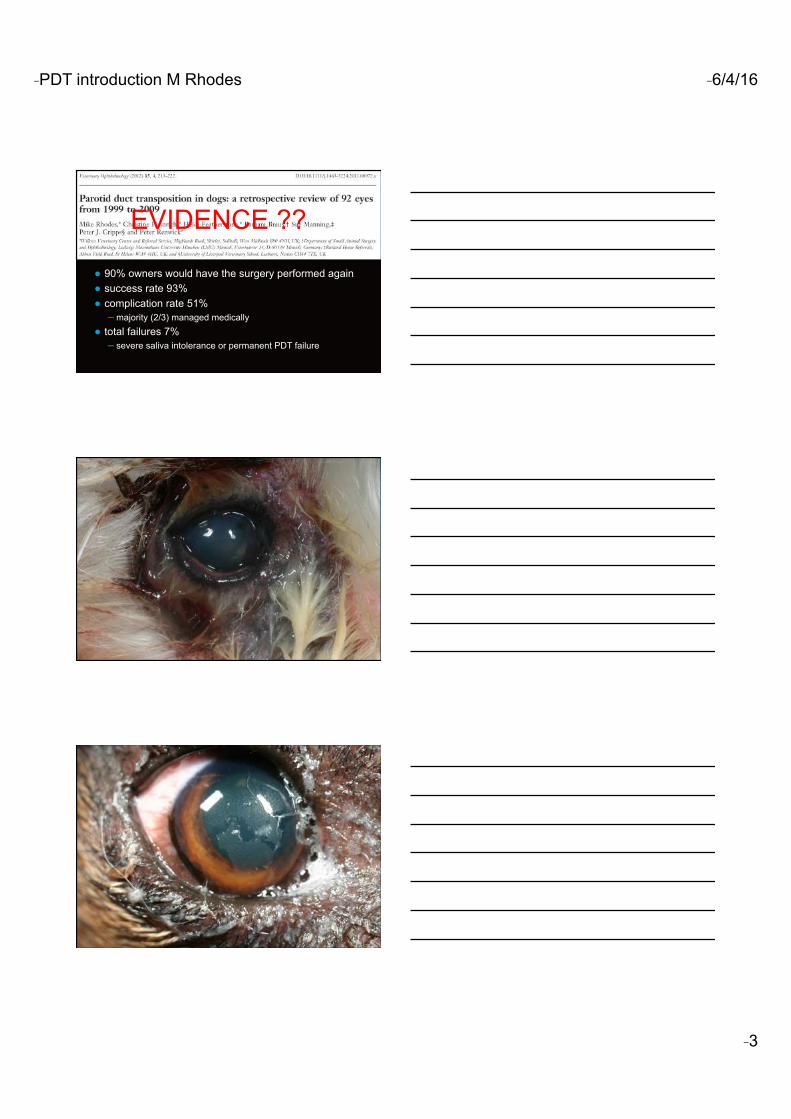
� PDT introduction M Rhodes � 6/4/16
� 3
● 90% owners would have the surgery performed again ● success rate 93% ● complication rate 51%
– majority (2/3) managed medically ● total failures 7%
– severe saliva intolerance or permanent PDT failure
EVIDENCE ??
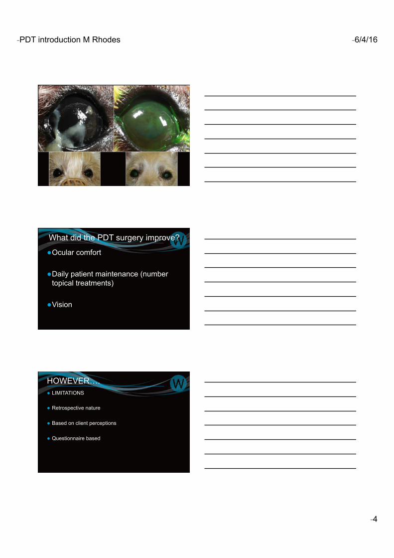
� PDT introduction M Rhodes � 6/4/16
� 4
pre PDT 4 weeks post PDT
What did the PDT surgery improve?
● Ocular comfort
● Daily patient maintenance (number topical treatments)
● Vision
HOWEVER…. ● LIMITATIONS
● Retrospective nature
● Based on client perceptions
● Questionnaire based

� PDT introduction M Rhodes � 6/4/16
� 5
Not in the study…. ● How diet affects saliva content ? ● Sodium hexametaphosphate ● Future study…
CHARACTERISTIC PAROTID SECRETION LACRIMAL SECRETION
pH 5.2-8.4 5.3-7.8
Osmolarity Physiologic Physiologic
Lysozyme Present Present
Translucency Clear Clear
Total solids (%) 1.8 1.6
Ash 1.05 0.81
From Gelatt, K.N. (1991) Canine lacrimal and nasolacrimal systems. In: Gelatt, K.N., ed., Veterinary Ophthalmology. Philadelphia: Lea & Febinger, 283.
Dr. Seth Koch VMD Diplomate ACVO Ophthalmology
Over to the expert…

PDT: A review Dr S Koch 6/4/16
1
PAROTID DUCT
TRANSPOSITION:
A Review
Seth A. Koch, VMD, MMSc, ACVO
“You gave us back our life.”
Statement from client that had been
treating medically for three years,
with no change.
“There is no need for
surgery.”
Statement from an academic
ophthalmologist who sees a client
once then sends back to referring
veterinarian.
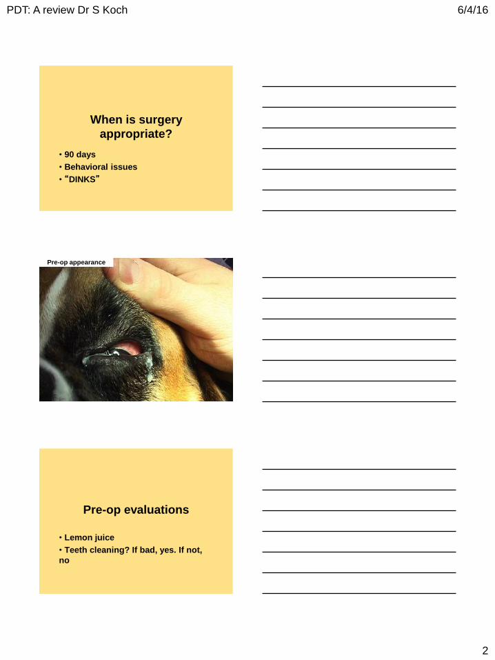
PDT: A review Dr S Koch 6/4/16
2
When is surgery
appropriate?
• 90 days
• Behavioral issues
• “DINKS”
Pre-op appearance
Pre-op evaluations
• Lemon juice
• Teeth cleaning? If bad, yes. If not,
no
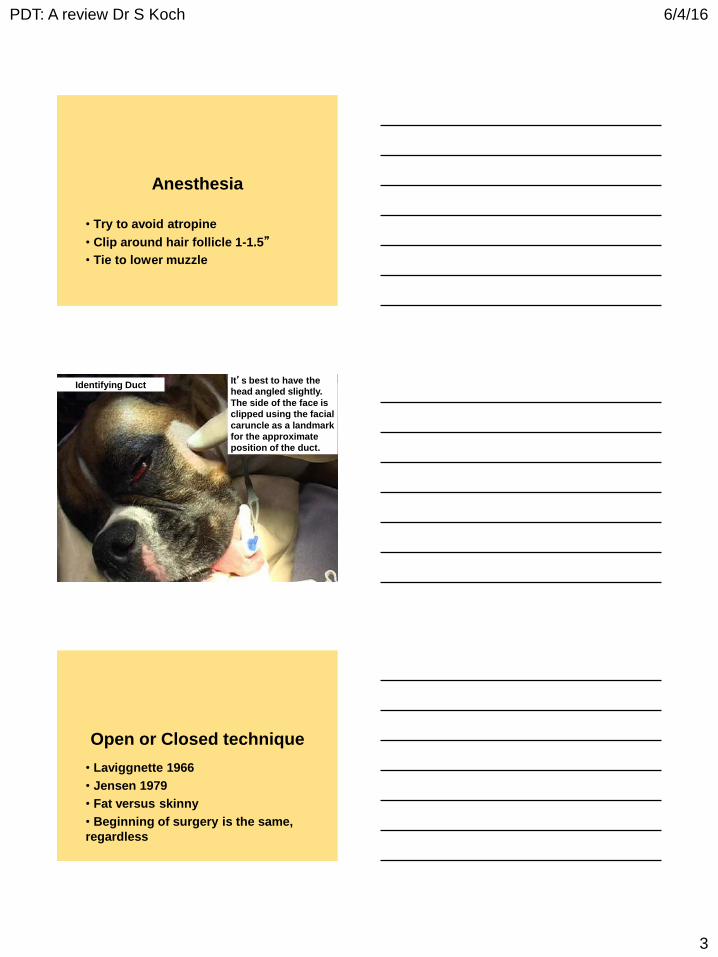
PDT: A review Dr S Koch 6/4/16
3
Anesthesia
• Try to avoid atropine
• Clip around hair follicle 1-1.5”
• Tie to lower muzzle
Identifying DuctIt’s best to have the head angled slightly.
The side of the face is
clipped using the facial
caruncle as a landmark
for the approximate
position of the duct.
Open or Closed technique
• Laviggnette 1966
• Jensen 1979
• Fat versus skinny
• Beginning of surgery is the same,
regardless
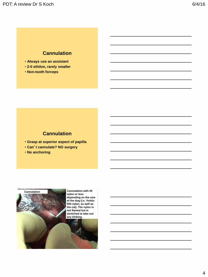
PDT: A review Dr S Koch 6/4/16
4
Cannulation
• Always use an assistant
• 2-0 ethilon, rarely smaller
• Non-tooth forceps
Cannulation
• Grasp at superior aspect of papilla
• Can’t cannulate? NO surgery
• No anchoring
Cannulation Cannulation with 00
nylon or less
depending on the size
of the dog (i.e. Yorkie:
000 nylon, as well as
the cat). The nylon is
not flamed but is
stretched to take out
any kinking.
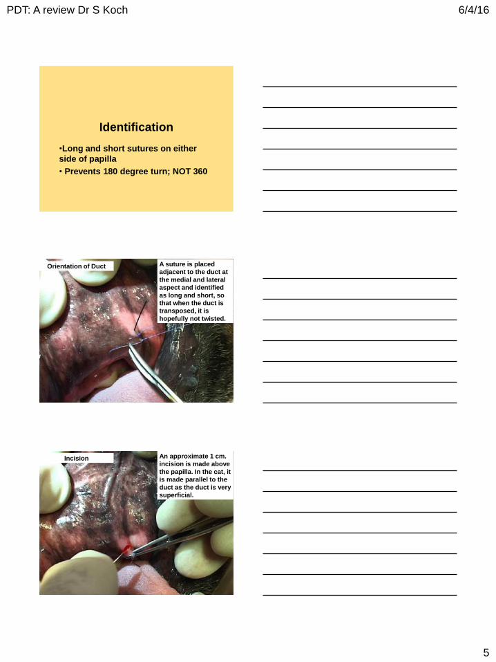
PDT: A review Dr S Koch 6/4/16
5
Identification
•Long and short sutures on either
side of papilla
• Prevents 180 degree turn; NOT 360
Orientation of Duct A suture is placed
adjacent to the duct at
the medial and lateral
aspect and identified
as long and short, so
that when the duct is
transposed, it is
hopefully not twisted.
Incision An approximate 1 cm.
incision is made above
the papilla. In the cat, it
is made parallel to the
duct as the duct is very
superficial.
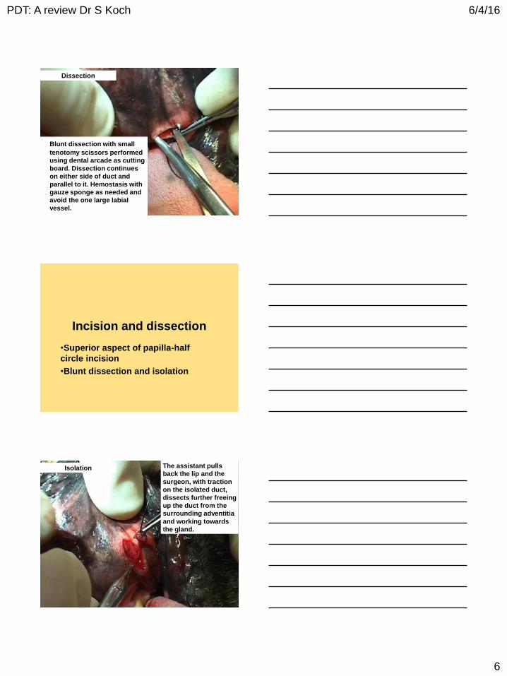
PDT: A review Dr S Koch 6/4/16
6
Dissection
Blunt dissection with small
tenotomy scissors performed
using dental arcade as cutting
board. Dissection continues
on either side of duct and
parallel to it. Hemostasis with
gauze sponge as needed and
avoid the one large labial
vessel.
Incision and dissection
•Superior aspect of papilla-half
circle incision
•Blunt dissection and isolation
Isolation The assistant pulls
back the lip and the
surgeon, with traction
on the isolated duct,
dissects further freeing
up the duct from the
surrounding adventitia
and working towards
the gland.
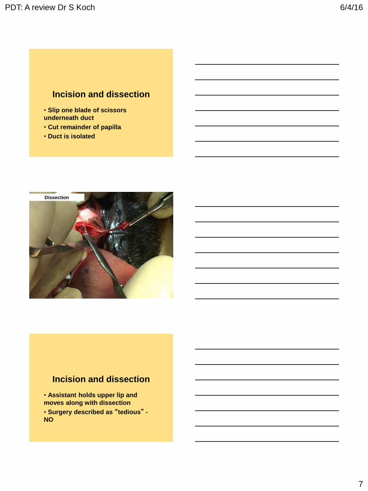
PDT: A review Dr S Koch 6/4/16
7
Incision and dissection
• Slip one blade of scissors
underneath duct
• Cut remainder of papilla
• Duct is isolated
Dissection
Incision and dissection
• Assistant holds upper lip and
moves along with dissection
• Surgery described as “tedious” -
NO
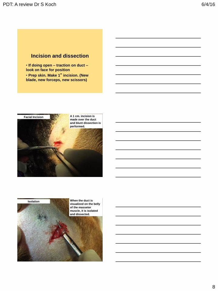
PDT: A review Dr S Koch 6/4/16
8
Incision and dissection
• If doing open – traction on duct –
look on face for position
• Prep skin. Make 1” incision. (New
blade, new forceps, new scissors)
Facial Incision A 1 cm. incision is
made over the duct
and blunt dissection is
performed.
Isolation When the duct is
visualized on the belly
of the masseter
muscle, it is isolated
and dissected.
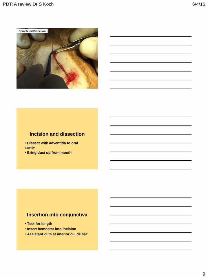
PDT: A review Dr S Koch 6/4/16
9
Completed Disection
Incision and dissection
• Dissect with adventitia to oral
cavity
• Bring duct up from mouth
Insertion into conjunctiva
• Test for length
• Insert hemostat into incision
• Assistant cuts at inferior cul de sac
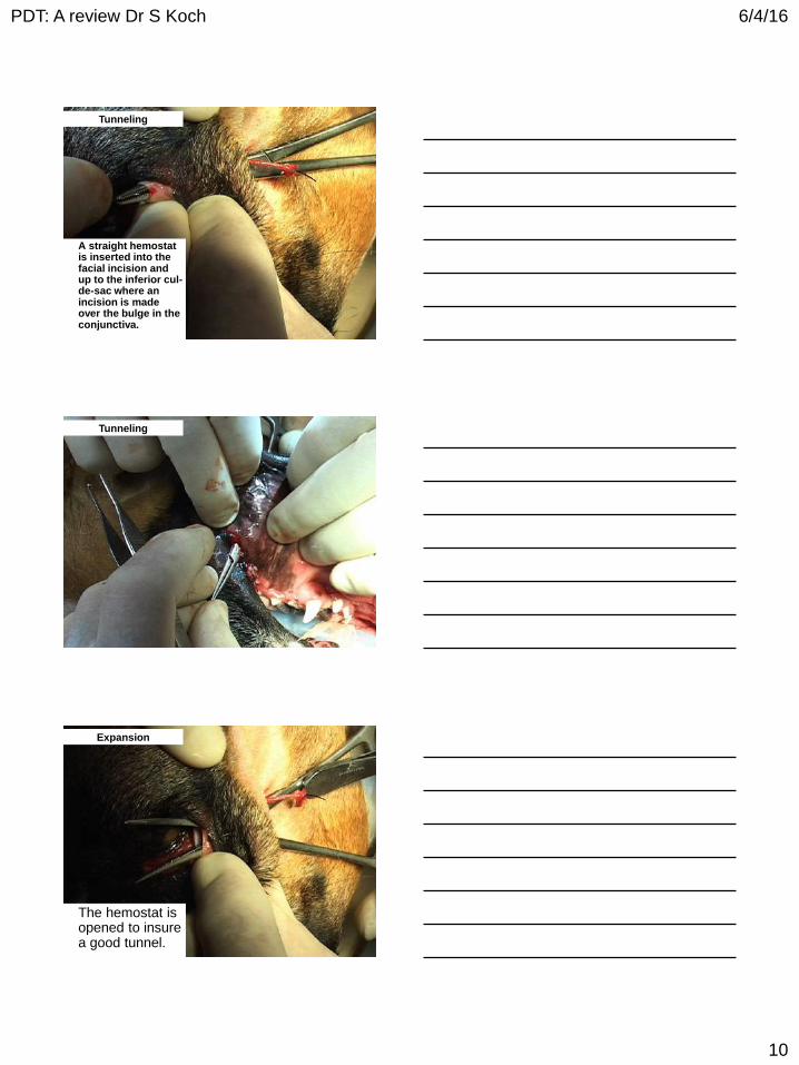
PDT: A review Dr S Koch 6/4/16
10
Tunneling
A straight hemostat is inserted into the facial incision and up to the inferior cul-de-sac where an incision is made over the bulge in the conjunctiva.
Tunneling
Expansion
The hemostat is opened to insure a good tunnel.
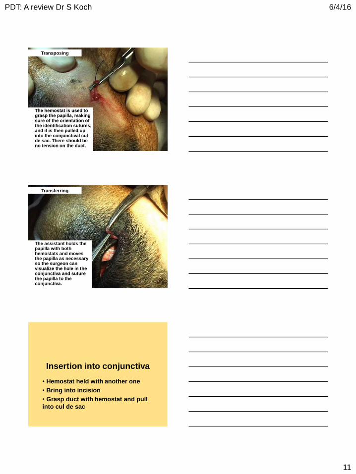
PDT: A review Dr S Koch 6/4/16
11
Transposing
The hemostat is used to grasp the papilla, making sure of the orientation of the identification sutures, and it is then pulled up into the conjunctival cul de sac. There should be no tension on the duct.
Transferring
The assistant holds the papilla with both hemostats and moves the papilla as necessary so the surgeon can visualize the hole in the conjunctiva and suture the papilla to the conjunctiva.
Insertion into conjunctiva
• Hemostat held with another one
• Bring into incision
• Grasp duct with hemostat and pull
into cul de sac
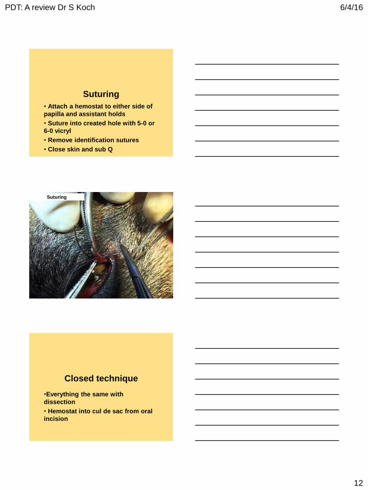
PDT: A review Dr S Koch 6/4/16
12
Suturing
• Attach a hemostat to either side of
papilla and assistant holds
• Suture into created hole with 5-0 or
6-0 vicryl
• Remove identification sutures
• Close skin and sub Q
Suturing
Closed technique
•Everything the same with
dissection
• Hemostat into cul de sac from oral
incision

PDT: A review Dr S Koch 6/4/16
13
Closed technique
• More swelling of face with closed
technique
• No oral sutures for either
technique
Post operative
• Rarely need a collar
• Oral and topical antibiotics
• Cold packs if swelling
Instructions to owner
• Frequent small feedings
• In A.M. feed and cleanse

PDT: A review Dr S Koch 6/4/16
14
Complications and
side effects• Stricture:
•Not enough dissection
•Too much tension
•Or who knows?
Complications and
side effects• Too much flow:
•Ligature after cannulation
•Tie down to ligature
Complications and
side effects• Tartar:
•EDTA useless
•Buttermilk, distilled water - anecdotal

PDT: A review Dr S Koch 6/4/16
15
To order video:
Seth A Koch, VMD, MMSc, ACVO
1420 Locust Street, 31K
Philadelphia, PA 19102
Clinicopathological features of uveal peripheral nerve sheath tumours in cats. JD Bujan, GC Shaw, RR Dubielzig, LBC Teixeira Comparative Ocular Pathology Laboratory of Wisconsin (COPLOW) University of Wisconsin-Madison. Purpose: To describe the clinical and histopathological features of 6 cases of uveal peripheral nerve sheath tumours (PNST) cats. Methods: 6 cases diagnosed as uveal PNST were identified in the Comparative Ocular Pathology Laboratory of Wisconsin (COPLOW) database from 2005 to 2015. Clinical presentation, signalment, follow up, histopathologic and immunohistochemical characteristics were recorded. Enucleated eyes were formalin-fixed, paraffin-embedded, stained with H&E, alcian blue/PAS and immunohistochemically for S-100, glial fibrillary acid protein (GFAP) and melan-A. Results: The median age of animals was 11.5 years (range 8-15). There were 4 spayed female and 2 neutered males. No breed predisposition was found. The left eye was affected in five cases and right eye in one. All cats had yellow-coloured irises. Except for one cat that presented thrombocytopenia the animals presented in good general health. The main ophthalmic findings were glaucoma (5/6), presence of an intraocular mass (3/6), uveitis (2/6) and buphthalmia (2/6). The main reasons for enucleation were glaucoma (3/6) and presence of a mass (3/6). The time between first clinical signs and enucleation ranged between 3 to 12 weeks. Grossly, 3/6 eyes had a white firm mass in the iris, ciliary body, and choroid that variably extended into the sclera and cornea (Figure 1). Histologically the neoplastic cells infiltrated and variably expanded and distorted the iris and sometimes the ciliary body and usually extended into the the choroid. There was scleral and extra-scleral invasion in 4 of 6 cases and in one case the tumour extended into the peripheral cornea. In 1 of 6 cases there was involvement of uveal, scleral and episcleral nerves (Figure 2). Neoplastic cells were arranged in streams and bundles and sometimes formed dense aggregates of elongated nuclei palisading around a densely eosinophilic stromal matrix characteristic of an Antoni-A type tissue pattern (6/6) with classic Verocay bodies (5/6) and less cellular areas with cells embedded in a loose matrix characteristic of Antoni-B type tissue pattern (3/6). Cellular pleomorphism ranged from severe (1/6), moderate (3/6) and mild (2/6) and 3/6 had areas of necrosis. Mitotic index (MI) ranged from 1-21 mitotic figures per 10 high power fields and 2/6 tumours presented MI > 4. Based on the MI, cellular pleomorphism and tissue necrosis 2/6 tumour were diagnosed as malignant peripheral nerve sheath tumours. Immunohistochemical staining revealed that all tumours (6/6) were positive for GFAP and negative for melan-A and 3/3 cases stained with S-100 were positive. Conclusion: A wide range of tumours in cats can present as nonpigmented anterior uveal masses, including lymphoma, amelanotic melanoma, feline ocular post-traumatic sarcoma, ciliary body adenoma and carcinomas and metastatic tumours.4-9 PNSTs are neoplasms that arise from Schwann cells, perineurial cells, and intraneural fibroblasts and can be subdivided based on morphologic features and benignancy versus malignancy into schwannoma neurofibroma and malignant PNST.10 In cats these tumours have been reported in multiple cutaneous sites, associated with major nerves and one case was reported in the uvea.1,10 The tumours in the present series revealed histologic features and immunohistochemical reaction (GFAP and S100 positivity with melan-A negativity11,12) that are characteristic of PNSTs. There is a close resemblance between the present tumours and the Schwannoma (spindle cell tumour) of blue eyed dogs.3 In conclusion, uveal PNSTs in cats are relatively rare tumours that affect adult animals without apparent breed or sex predilection. The presence of Antoni type A and B tissue patterns along with positive immunohistochemical staining for GFAP and S-100 and negative staining for melan-A are diagnostic features of these tumours. Although rare, PNSTs should be considered as a differential diagnosis of iridal masses in cats. To the authors’ knowledge this is the largest series of such intraocular tumours in cats.
REFERENCES 1. Evans, P. M., Lynch, G. L. and Dubielzig, R. R. (2010), Anterior uveal spindle cell
tumor in a cat. Veterinary Ophthalmology, 13: 387–390. 2. Palm SL, Furcht LT. Production of laminin and fibronectin by Schwannoma cells: cell-
protein interactions in vitro and protein localization in peripheral nerve in vivo. J Cell Biol. 1983 May 1; 96(5): 1218–1226.
3. Zarfoss MK, Klauss G, Newkirk K et al. Uveal spindle cell tumor of blue-eyed dogs: an immunohistochemical study. Veterinary Pathology 2007; 44: 276–284.
4. Labelle P, Holmberg BJ. Ocular myxoid leiomyosarcoma in a cat. Veterinary Ophthalmology 2010; 13: 58–62.
5. Groskopf BS, Dubielzig RR, Beaumont SL. Orbital extraskeletal osteosarcoma enucleation in a cat: a case report. Veterinary Ophthalmology 2010; 13: 179–183.
6. 6. Harris BP, Dubielzig RR. Atypical primary ocular melanoma in cats. Veterinary Ophthalmology 1999; 2: 121–124.
7. Dubielzig RR. Ocular neoplasia in small animals. The Veterinary Clinics of North America 1990; 20: 837–848.
8. Wippold FJ 2nd, Lubner M, Perrin RJ et al. Neuropathology for the neuroradiologist: Antoni A and Antoni B tissue patterns. American Journal of Neuroradiology 2007; 28: 1633–1638.
9. Hoffman A, Blocker T, Dubielzig R et al. Feline periocular peripheral nerve sheath tumor: a case series. Veterinary Ophthalmology 2005; 8: 153–158.
10. Schulman FY1, Facemire PR, Fanburg-Smith JC. Feline peripheral nerve sheath tumors: histologic, immunohistochemical, and clinicopathologic correlation (59tumors in 53 cats). 2009 Nov;46(6):1166-80.
11. Weiss SW, Langloss JM, Enzinger FM. Value of S-100 protein in the diagnosis of soft tissue tumors with particular reference to benign and malignant Schwann cell tumors. Laboratory Investigation 1983; 49: 299–308.
12. Orchard G, Wilson Jones E. Immunocytochemistry in the diagnosis of malignantmelanoma.BritishJournalofBiomedicalScience1994;51:44–56.
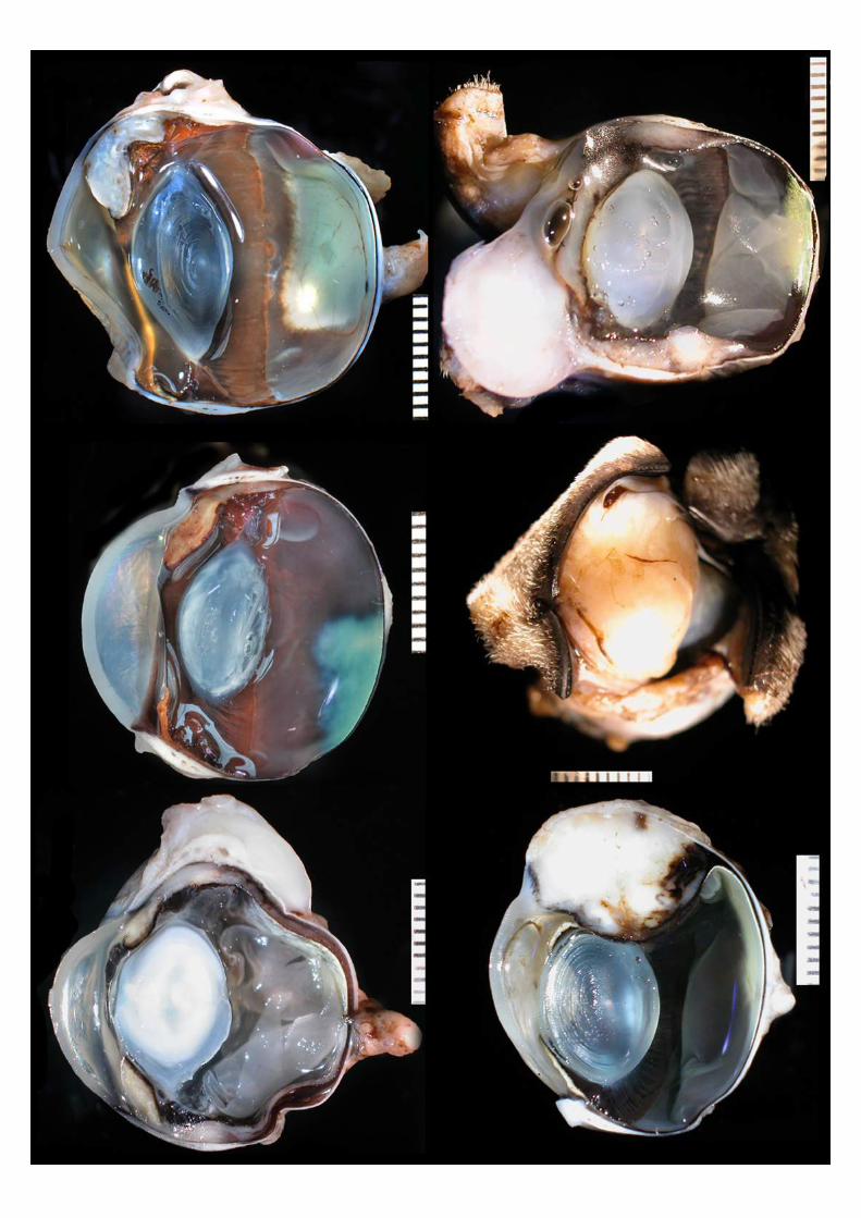

- When eyelid lesions are not what they seem….. M Rhodes
- 6/4/16
- 1
When eyelid lesions are not what they seem….
Mike Rhodes BVM&S CertVOphthal DipECVO MRCVS
RCVS and European Specialist in Veterinary Ophthalmology
April 2016
VOTING SLIDE
CASE 1
- When eyelid lesions are not what they seem….. M Rhodes
- 6/4/16
- 2
Bobby Greene
Canine meibomian gland adenomas and epitheliomas
● Meibomian gland adenoma
– mostly made up of fully differentiated meibomian gland tissue ● holocrine secretory cells ● keratinizing ducts
– smaller, more superficial, more likely exophytic
● Meibomian gland epithelioma
– mostly undifferentiated basal cells
– rare sebaceous or squamous differentiation
– larger, more likely pigmented and likely to be deeper, in lid margin
- When eyelid lesions are not what they seem….. M Rhodes
- 6/4/16
- 3
histo – meibomian gland adenoma
courtesy of E Scurrell
meibomian gland epithelioma
courtesy of E Scurrell
- When eyelid lesions are not what they seem….. M Rhodes
- 6/4/16
- 4
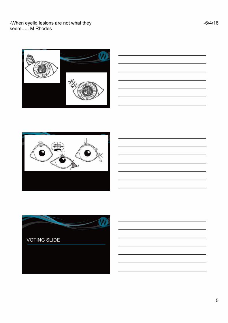
- When eyelid lesions are not what they seem….. M Rhodes
- 6/4/16
- 5
VOTING SLIDE
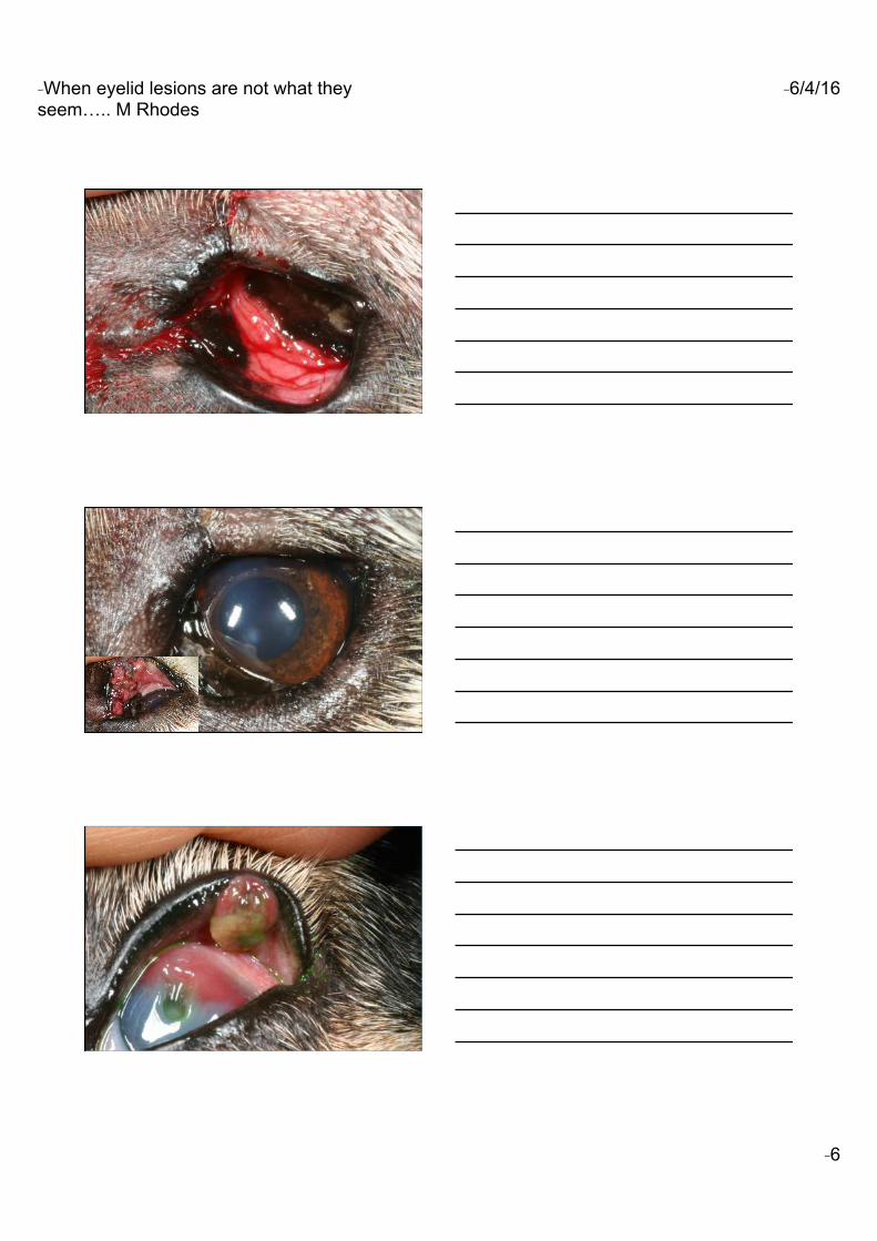
- When eyelid lesions are not what they seem….. M Rhodes
- 6/4/16
- 6
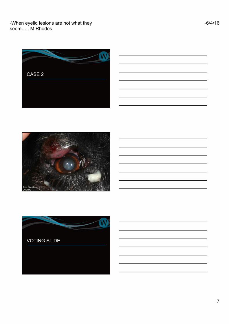
- When eyelid lesions are not what they seem….. M Rhodes
- 6/4/16
- 7
CASE 2
Tess Goulding 04/07/12
VOTING SLIDE
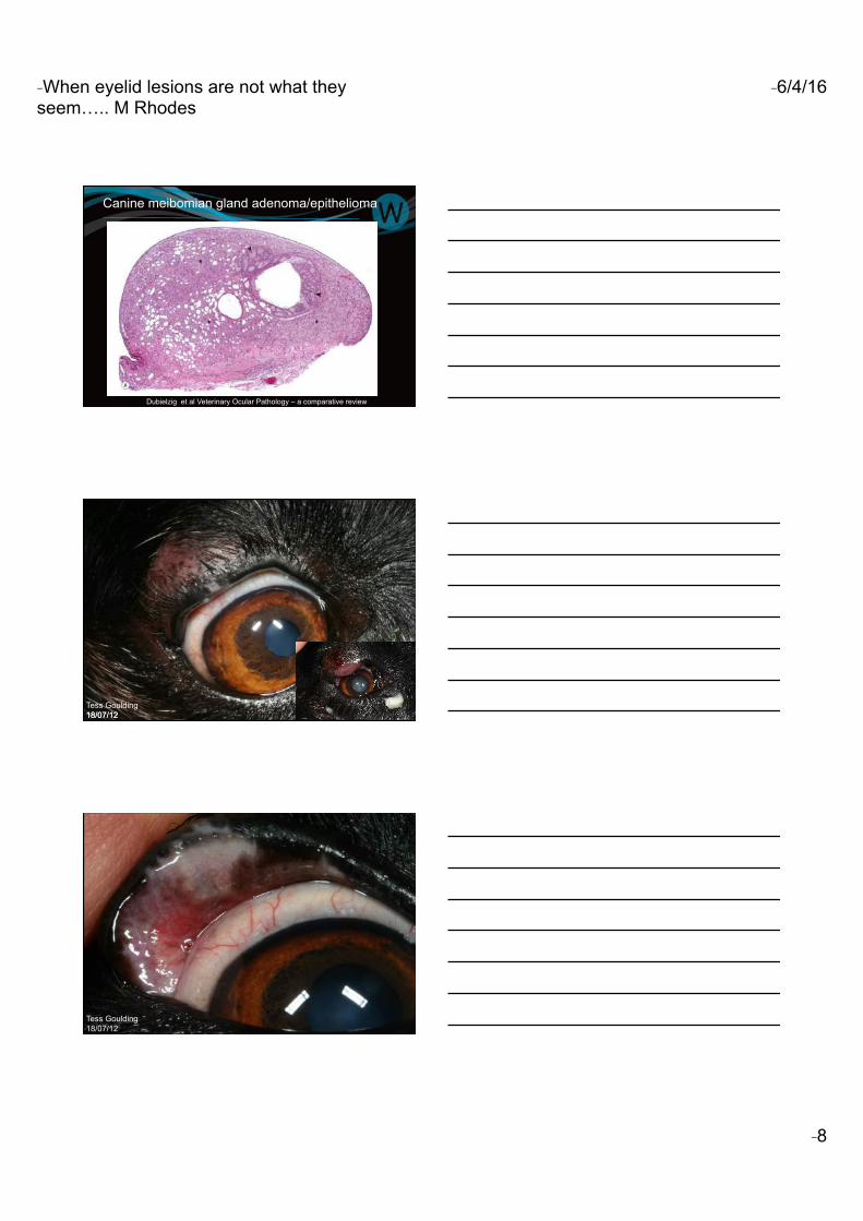
- When eyelid lesions are not what they seem….. M Rhodes
- 6/4/16
- 8
Canine meibomian gland adenoma/epithelioma
Dubielzig et al Veterinary Ocular Pathology – a comparative review
18/07/12 Tess Goulding 18/07/12
Tess Goulding 18/07/12
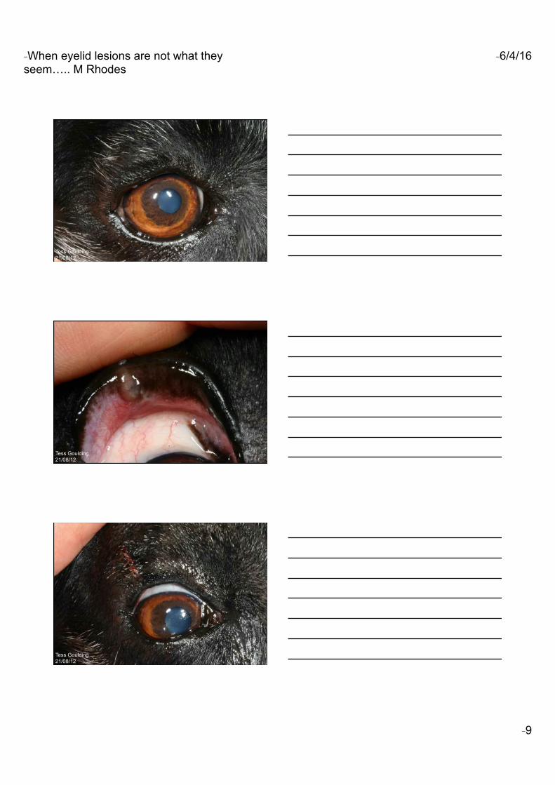
- When eyelid lesions are not what they seem….. M Rhodes
- 6/4/16
- 9
Tess Goulding 21/08/12
Tess Goulding 21/08/12
Tess Goulding 21/08/12
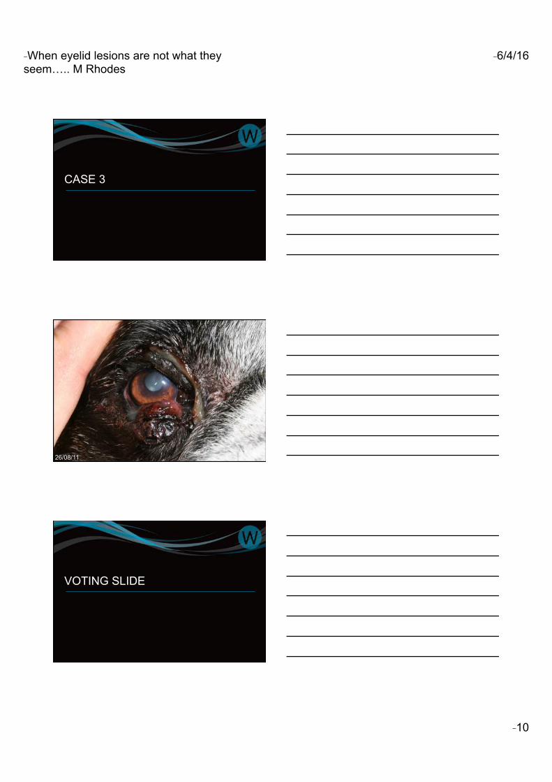
- When eyelid lesions are not what they seem….. M Rhodes
- 6/4/16
- 10
CASE 3
26/08/11
VOTING SLIDE
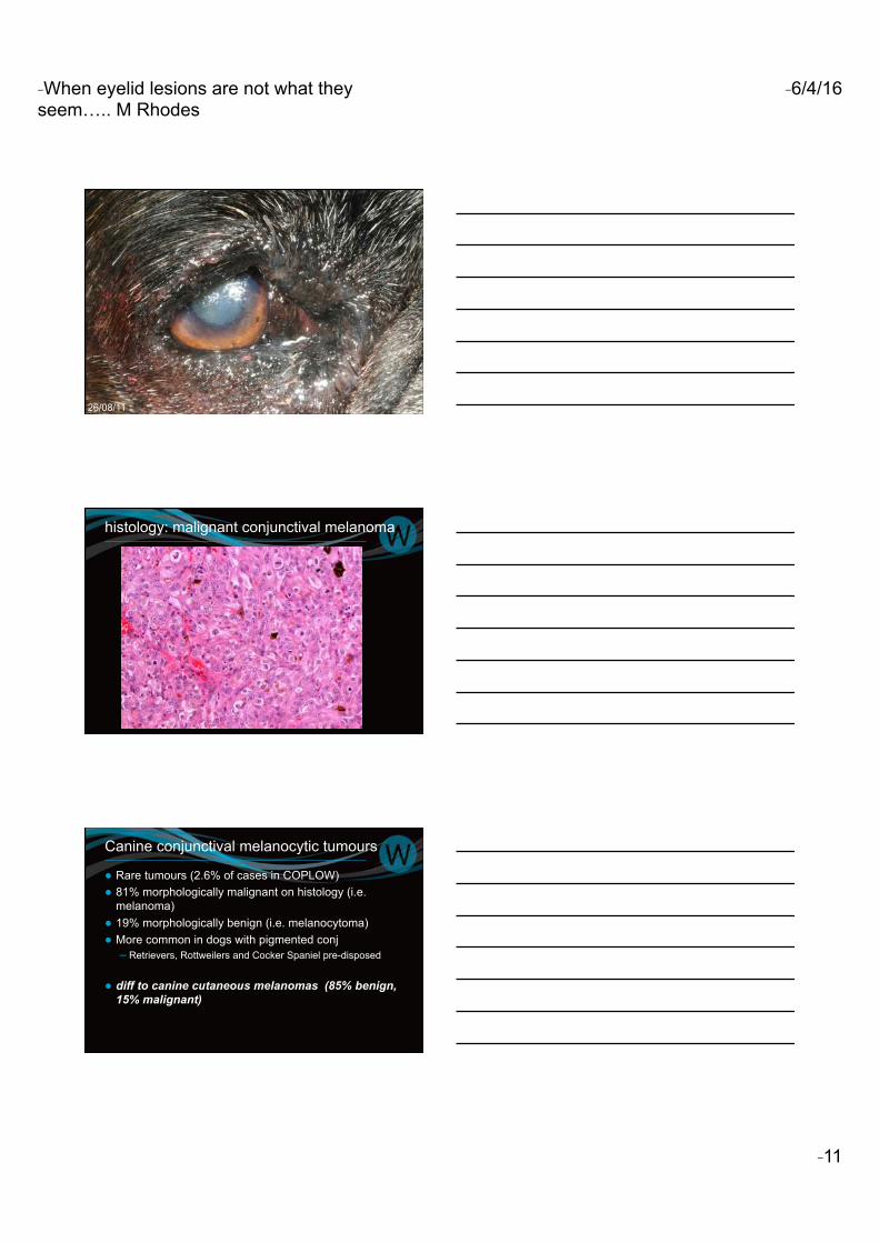
- When eyelid lesions are not what they seem….. M Rhodes
- 6/4/16
- 11
26/08/11
histology: malignant conjunctival melanoma
Canine conjunctival melanocytic tumours
● Rare tumours (2.6% of cases in COPLOW) ● 81% morphologically malignant on histology (i.e.
melanoma) ● 19% morphologically benign (i.e. melanocytoma) ● More common in dogs with pigmented conj
– Retrievers, Rottweilers and Cocker Spaniel pre-disposed
● diff to canine cutaneous melanomas (85% benign, 15% malignant)
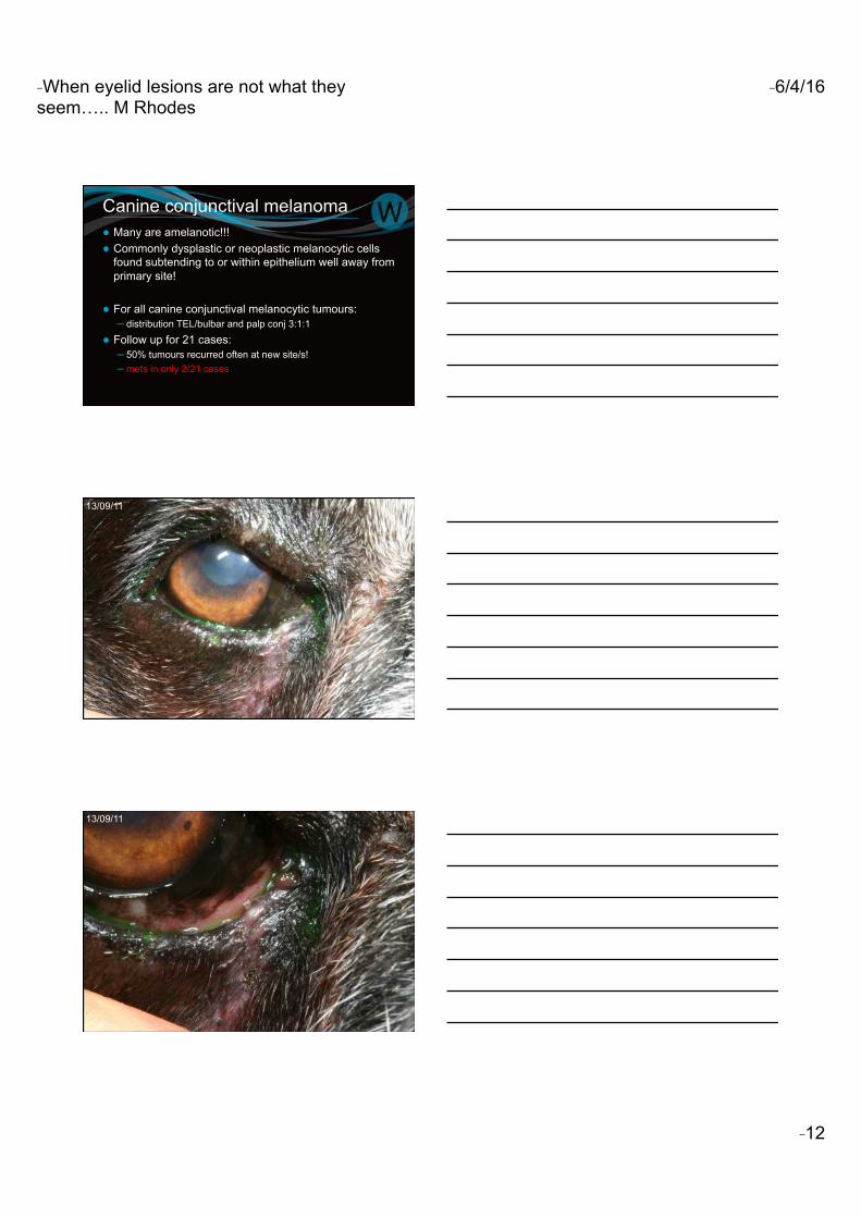
- When eyelid lesions are not what they seem….. M Rhodes
- 6/4/16
- 12
Canine conjunctival melanoma ● Many are amelanotic!!! ● Commonly dysplastic or neoplastic melanocytic cells
found subtending to or within epithelium well away from primary site!
● For all canine conjunctival melanocytic tumours: – distribution TEL/bulbar and palp conj 3:1:1
● Follow up for 21 cases: – 50% tumours recurred often at new site/s! – mets in only 2/21 cases
13/09/11
13/09/11
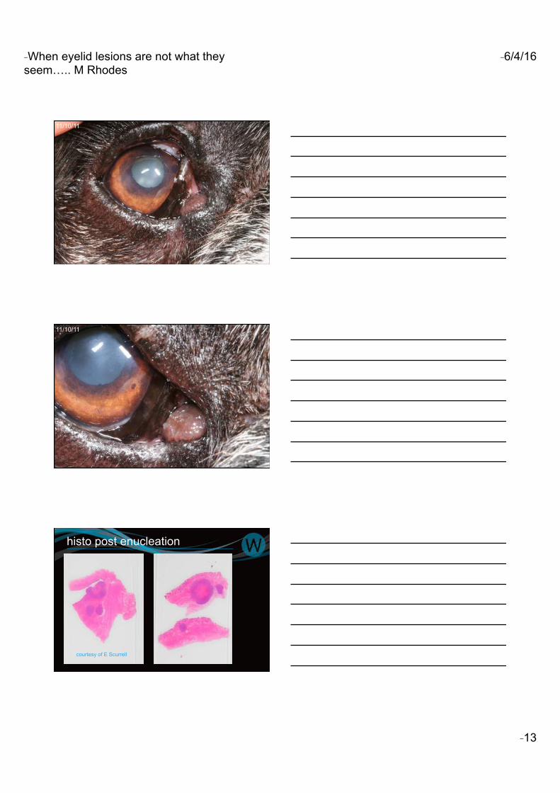
- When eyelid lesions are not what they seem….. M Rhodes
- 6/4/16
- 13
11/10/11
11/10/11
histo post enucleation
courtesy of E Scurrell
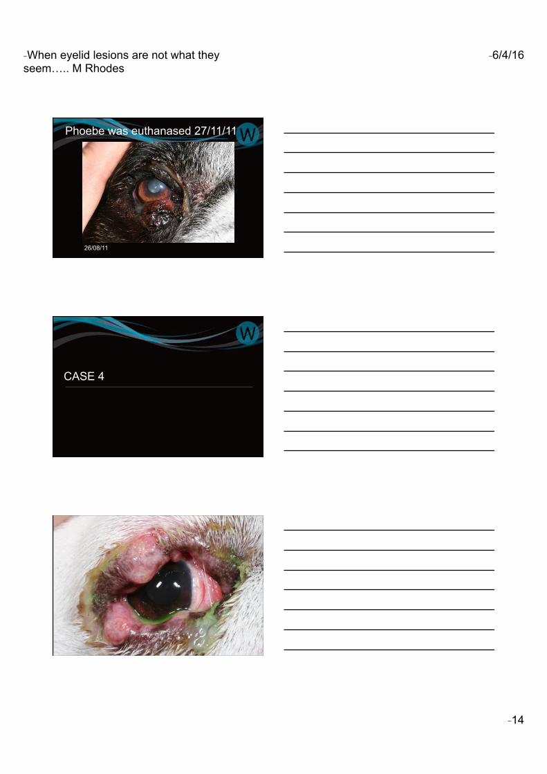
- When eyelid lesions are not what they seem….. M Rhodes
- 6/4/16
- 14
26/08/11
Phoebe was euthanased 27/11/11
CASE 4
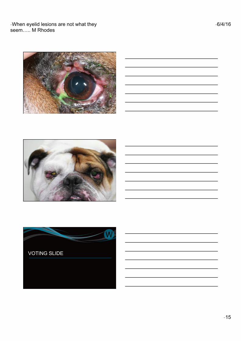
- When eyelid lesions are not what they seem….. M Rhodes
- 6/4/16
- 15
VOTING SLIDE
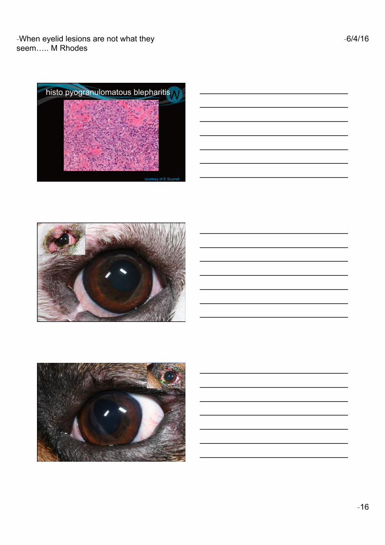
- When eyelid lesions are not what they seem….. M Rhodes
- 6/4/16
- 16
histo pyogranulomatous blepharitis
courtesy of E Scurrell
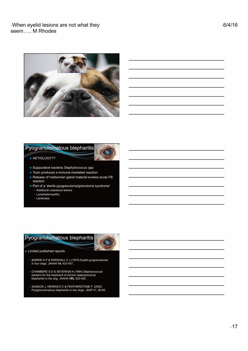
- When eyelid lesions are not what they seem….. M Rhodes
- 6/4/16
- 17
Pyogranulomatous blepharitis
● AETIOLOGY??
● Suppurative bacteria Staphylococcus spp. ● Toxin produces a immune-mediated reaction ● Release of meibomian gland material evokes acute FB
reaction ● Part of a ‘sterile pyogranuloma/granuloma syndrome’
– Additional cutaneous lesions – Lymphadenopathy – Lameness
Pyogranulomatous blepharitis
● Limited published reports
– BARRIE K P & PARSHALL C J (1979) Eyelid pyogranulomas in four dogs. JAAHA 14, 433-437.
– CHAMBERE S D & SEVERIGN A (1984) Staphylococcal bacterin for the treatment of chronic staphylococcal blepharitis in the dog. JAAHA 185, 422-425.
– SANSON J, HEINRICH C & FEATHERSTONE F. (2000) Pyogranulomatous blepharitis in two dogs. JSAP 41, 80-83.
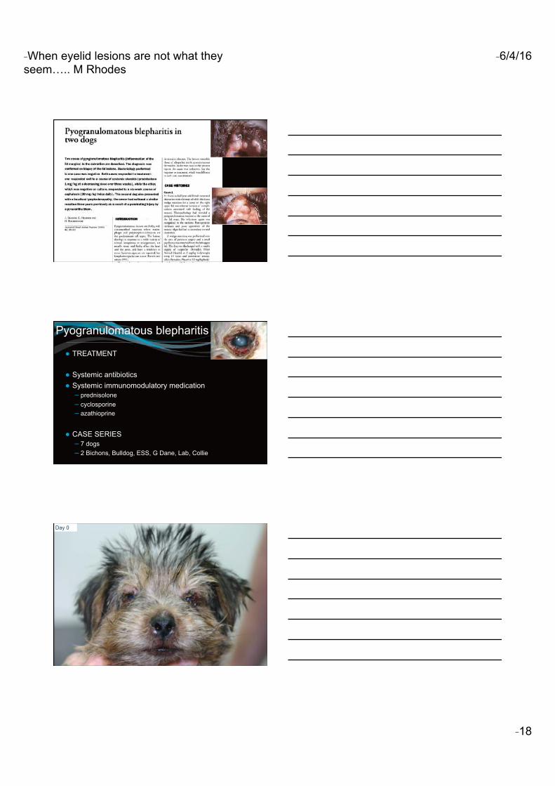
- When eyelid lesions are not what they seem….. M Rhodes
- 6/4/16
- 18
Pyogranulomatous blepharitis
● TREATMENT
● Systemic antibiotics ● Systemic immunomodulatory medication
– prednisolone – cyclosporine – azathioprine
● CASE SERIES – 7 dogs – 2 Bichons, Bulldog, ESS, G Dane, Lab, Collie
Day 0
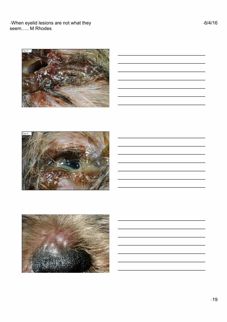
- When eyelid lesions are not what they seem….. M Rhodes
- 6/4/16
- 19
Day 0
Day 0
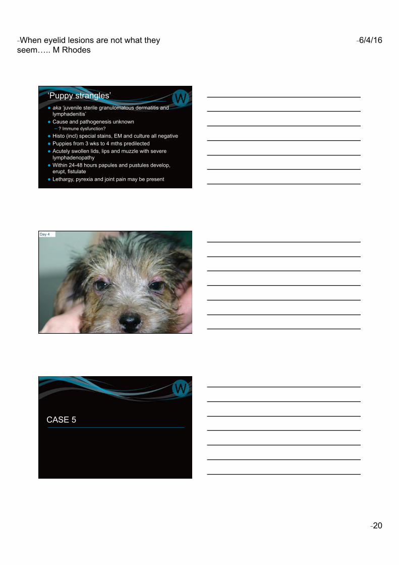
- When eyelid lesions are not what they seem….. M Rhodes
- 6/4/16
- 20
‘Puppy strangles’ ● aka ‘juvenile sterile granulomatous dermatitis and
lymphadenitis’ ● Cause and pathogenesis unknown
– ? Immune dysfunction?
● Histo (incl) special stains, EM and culture all negative ● Puppies from 3 wks to 4 mths predilected ● Acutely swollen lids, lips and muzzle with severe
lymphadenopathy ● Within 24-48 hours papules and pustules develop,
erupt, fistulate ● Lethargy, pyrexia and joint pain may be present
Day 4
CASE 5
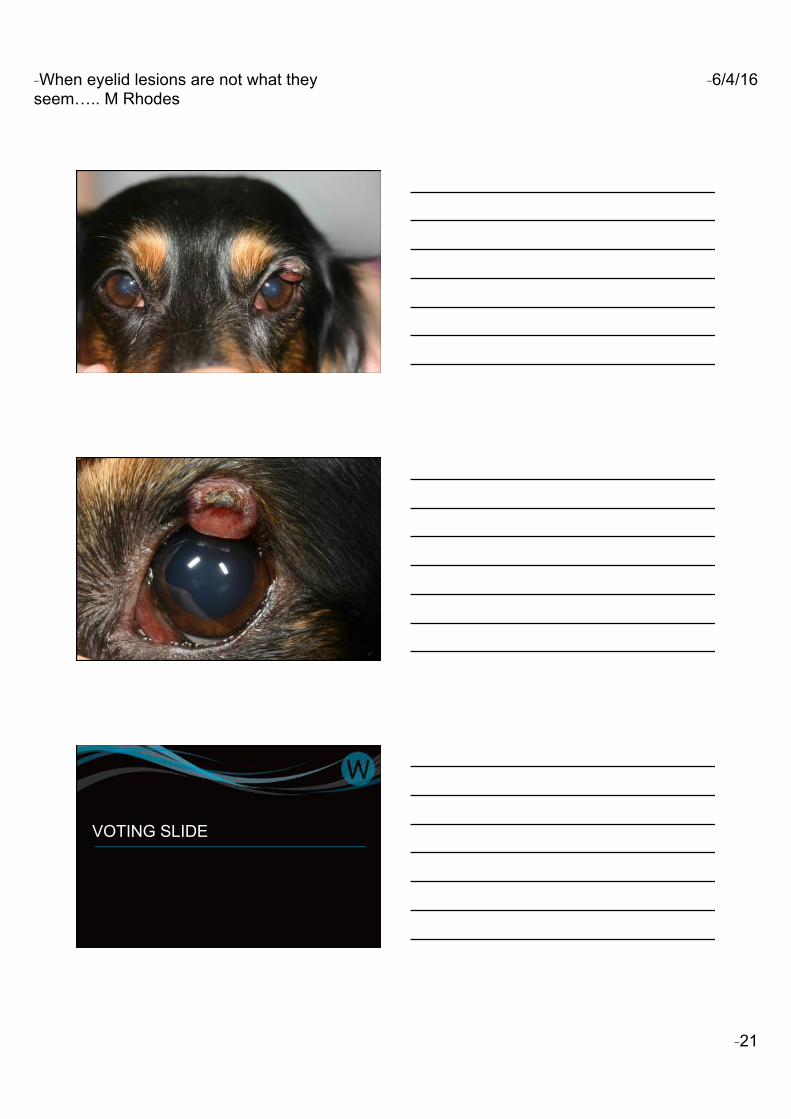
- When eyelid lesions are not what they seem….. M Rhodes
- 6/4/16
- 21
VOTING SLIDE
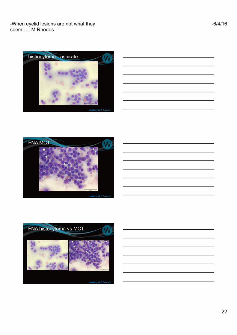
- When eyelid lesions are not what they seem….. M Rhodes
- 6/4/16
- 22
histiocytoma - aspirate
courtesy of E Scurrell
FNA MCT
courtesy of E Scurrell
FNA histocytoma vs MCT
courtesy of E Scurrell
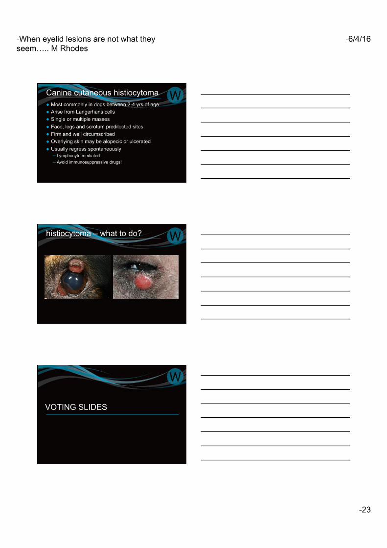
- When eyelid lesions are not what they seem….. M Rhodes
- 6/4/16
- 23
Canine cutaneous histiocytoma ● Most commonly in dogs between 2-4 yrs of age ● Arise from Langerhans cells ● Single or multiple masses ● Face, legs and scrotum predilected sites ● Firm and well circumscribed ● Overlying skin may be alopecic or ulcerated ● Usually regress spontaneously
– Lymphocyte mediated – Avoid immunosuppressive drugs!
histiocytoma – what to do?
VOTING SLIDES
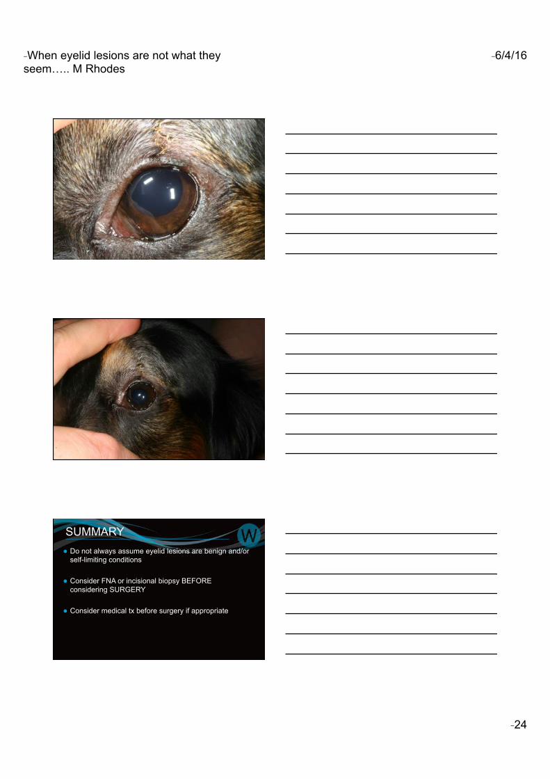
- When eyelid lesions are not what they seem….. M Rhodes
- 6/4/16
- 24
SUMMARY ● Do not always assume eyelid lesions are benign and/or
self-limiting conditions
● Consider FNA or incisional biopsy BEFORE considering SURGERY
● Consider medical tx before surgery if appropriate
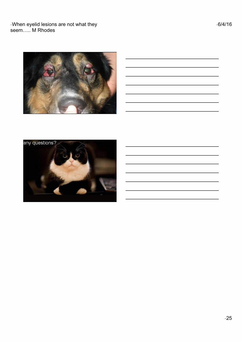
- When eyelid lesions are not what they seem….. M Rhodes
- 6/4/16
- 25
Corneo-scleral repair in five dogs using an autogenous auricular cartilage graft G A Lewin, C J Dixon, Veterinary Vision, Penrith, UK Abstract Five dogs presented with a melanoma arising at or immediately caudal to the dorsomedial or dorsolateral limbus of the globe. Examination of the affected eyes, including ultrasonography and gonioscopy, identified no intra-ocular extension of the lesions. The position, appearance and behaviour of the mass in each case was characteristic of a limbal melanoma. Excisional biopsy by full thickness corneal and scleral resection, including a margin of non-pigmented tissue, was carried out. An auricular cartilage graft was harvested from the patient’s pinna via a skin incision on the medial aspect of the pinna and was trimmed to correspond to the size of the corneo-scleral defect. The graft was sutured in position with a double continuous suture of 8/0 Vicryl, and whenever possible was overlain with bulbar conjunctiva. Post-operative medication comprised routine topical antibiosis and mydriasis and oral non-steroidal anti-inflammatory medication The skin at the donor site was closed routinely. The excised tissue was submitted for histopoathological examination and surgical margins appeared to have been achieved in all cases In all cases healing was uneventful and over a period of weeks the auricular graft became confluent with the adjacent tissue. Seroma formation was encountered in 2 cases at the donor site but resolved uneventfully. Cases have been followed up for up to 30 months, No complications have been encountered, there have been no signs of recurrence of the limbal melanoma in any of the cases and all eyes have remained visual. From the results of this series auricular cartilage appears to be a suitable material for repairing full thickness corneoscleral defects, and the quantity of graft material that the pinna provides enabled curative excision of the limbal melanoma to be undertaken in all of these cases.
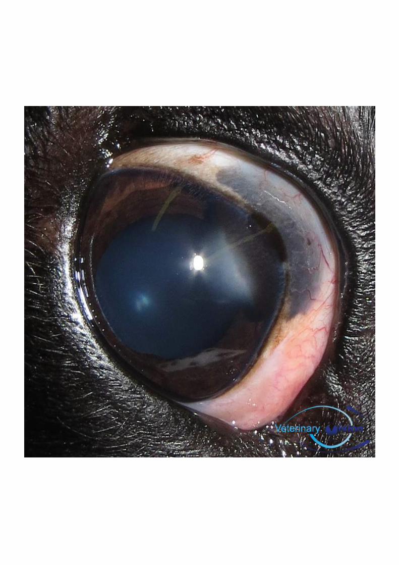
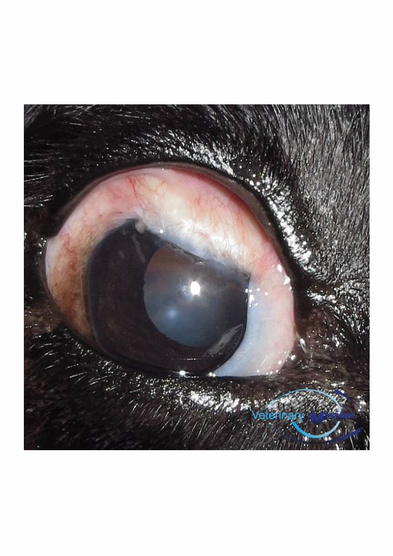
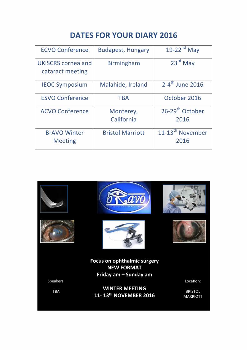
DATESFORYOURDIARY2016ECVOConference Budapest,Hungary 19-22ndMay
UKISCRScorneaandcataractmeeting
Birmingham 23rdMay
IEOCSymposium Malahide,Ireland 2-4thJune2016
ESVOConference TBA October2016
ACVOConference Monterey,California
26-29thOctober2016
BrAVOWinterMeeting
BristolMarriott 11-13thNovember2016
Focus&on&ophthalmic&surgery&NEW&FORMAT&
Friday&am&–&Sunday&am&&
WINTER&MEETING&&11A&13th&NOVEMBER&2016&
&&
Speakers:))
TBA)
Loca0on:))
BRISTOL)MARRIOTT)

NOTES

NOTES

NOTES

BRAVO COMMITTEE MEMBERS 2015
Chairperson – Tim KnottTim Graduated with an honours degree in Anatomical Science from Bristol University in 1991, followed by his Veterinary degree in 1995. Tim holds the RCVS certificate in Veterinary Ophthalmology and runs Rowe Referrals, Bristol. Hi is interested in all aspects of mixed practice but has special interests in ophthalmology, exotic animal medicine and surgery and fish disease. As chairperson, Tim overseas the running of the committee and chairs our meetings.
Joint Secretary – Ida GilbertIda graduated from Bristol Veterinary School in 1995. Following two years in mixed practice she decided to commit to her interests in ophthalmology and moved to Eastcott Veterinary hospital in Swindon, where she gained her RCVS Certificate in Veterinary Ophthalmology in 2001. She enjoys all areas of Veterinary Ophthalmology and still works and lives in rural Wiltshire. As secretary, Ida is involved with enrolment of new members as well as dealing with general enquiries and helping herding the Committee members!
Joint Secretary – Natasha MitchellNatasha graduated from University College Dublin in 1998 with a degree in Veterinary Medicine. She obtained the Royal College of Veterinary Surgeons’ (RCVS) Certificate in Veterinary Ophthalmology in 2004. She later joined the Eye Veterinary Clinic in Herefordshire where she completed an alternative residency programme for the RCVS Diploma in Veterinary Ophthalmology, which she obtained in 2011. She is a Veterinary Council of Ireland recognised specialist in Veterinary Ophthalmology. Natasha runs a referral veterinary ophthalmology service, Eye Vet, in Limerick, Ireland. She assists Ida in her very busy role as secretary, enrolling new members and answering general enquiries.
Hotel and conference organiser – Helen Appelboam Helen qualified from Royal Veterinary College, London in 2001. Originally from Hampshire but keen to see the world, veterinary work has taken her to South Africa and New Zealand where she gained experience in small animal and equine practice. Her interest in ophthalmology grew after seeing practice with specialist veterinary ophthalmologists in these countries. On returning to the UK, she spent 4 years developing her skills in a small animal and eye referral practice in Bristol and studying for the RCVS Veterinary Ophthalmology Certificate, which she achieved in 2011. She joined Optivet Referrals in 2012. Helen’s job is to research the venues for our conferences each year and oversee the running of the conference itself.
Exhibition liaison – Georgie Fricker Georgie graduated from Liverpool in 2005. She worked in small animal practice but had a keen interest in ophthalmology, resulting in her joining Robert Lowe at Optivet Referrals in 2008. Georgie gained her RCVS Certificate in Veterinary Ophthalmology in 2011 before moving to Davies Veterinary Specialists in March 2012 where she is undertaking an ECVO residency. Georgie is in charge of communicating with our sponsors.
International liaison (new role) – Michael Ziglar Michael completed his Bachelor of Science degree in 1975 at the University of Guelph. He went on to complete his DVM in 1979 at the Ontario Veterinary College, University of Guelph.From 1979 to 1982 he worked as an associate veterinarian in two different Alberta small-animal clinics. He then returned to Ontario in 1982 and opened Bronte Road Animal Hospital in 1984. In 1993 Michael completed examinations and received the RCVS Certificate in Veterinary Ophthalmology. Michael has been involved with both undergraduate teaching and delivering numerous continuing education presentations across North America. He has also been past President of both the American Society of Veterinary Ophthalmology and Canadian Association of Veterinary Ophthalmology where he is also an Honorary Life Member.This new role of international liaison is to try and promote BrAVO abroad and encourage international contacts.
Website and audio-visual – David Nutbrown-Hughes After graduating from Bristol in 1995, David had worked in practices in Worcestershire, Somerset and West Sussex where he built on his interest in ophthalmology, gaining the RCVS Certificate in Veterinary Ophthalmology in 2004. Since June 2012 David has joined the ophthalmology team at Rowe Referrals, Bristol. David runs the BrAVO website and is also involved with setting up the audio-visual facilities at each meeting.
Scientific programme organiser – Rachael GrundonRachael graduated from Cambridge and spent many years in mixed general practice before specialising in ophthalmology. She undertook her residency in Melbourne gaining the ANZCVS Fellowship in 2014. She is now working at the Eye Vet Clinic in Herefordshire while completing an ECVO residency.
Scientific programme organiser – Alistair OldfieldHaving graduated in 1997 from Bristol University, Alistair joined Woodcroft Veterinary Group in June 2002 after working nearly five years at the PDSA clinic in Manchester. Having attained his RCVS Certificate in Veterinary Ophthalmology, Alistair sees referral ophthalmology cases as well as continuing his work in general small animal practice. Alongside James and Mike, Alistair co-ordinates the scientific programme for each meeting and liaises with speakers.
Scientific programme organiser – Christine HeinrichChristine graduated from Munich Veterinary School in 1994 and immediately afterwards moved to the UK, where she undertook both an Internship and a Residency in Ophthalmology at the Animal Health Trust in Newmarket. Since 2000, Christine has been in private ophthalmic referral practice in the UK and she is both a diplomate of the Royal College of Veterinary Surgeons (Ophthalmology) and of the European College of Veterinary Ophthalmologists. In 2015, Christine took over the Eye Veterinary Clinic in Leominster, a dedicated ophthalmic referral clinic for all species. Despite a busy clinical work-load, Christine has continued to enjoy the teaching of Veterinary Ophthalmology both to pre-and post-graduate veterinary surgeons, including lecturing nationally and internationally and the mentoring of residents under the ECVO residency program.
Editor – Mike Rhodes Mike graduated from Edinburgh University in 2004 and spent the next three and half years working in small animal practice in Peterborough and Suffolk. During this time he developed a keen interest in veterinary ophthalmology and completed the RCVS Certificate in Veterinary Ophthalmology in 2008. He then undertook a three-year ECVO residency programme at Willows Referral Service and obtained the European Diploma in Veterinary Ophthalmology in 2013. Mike currently resides at Willows Referral Service. Mike’s job is to prepare the meeting proceedings as well as to assist Christine, Rachael and Alistair in putting together the scientific programme.
Clinical Auditor – Jenny LambertJenny works at Bath Referrals and obtained the RCVS Certificate in Veterinary Ophthalmology in 2003. As clinical auditor, Jenny aims to create an ongoing cycle of continuous improvement, by collecting data and comparing current practice with evidence of good practice.
PROGRAMME 08:30-09:15 Registration/coffee 09:15-10:00 Rational use of antimicrobials – case based approaches (James Oliver) 10:00-10:45 FHV1 - latest thoughts on pathogenesis and diagnosis (David Maggs) 10:45-11:15 - Coffee and exhibition 11:15-12:00 FHV1 - treatment options - are you up to date with the truths and
myths? (David Maggs) 12:00-12:15 Abstract 1: Management of Superficial Chronic Corneal Epithelial Defects (SCCEDs) in dogs with Multiple Punctate Keratotomy with a Third-Eyelid Flap (Emily Jeanes) 12:15-13:00 Canine corneal surface disease - beyond the STT (David Maggs) 13:00-14:15 Lunch and exhibition 14:15-14:45 Feline tear film diseases interactive management of challenging case
scenarios (David Maggs) 14:45-15:45 To PDT or not to PDT - that is the question (Seth Koch, Mike Rhodes,
David Maggs, James Oliver) 15:45-15:50 Using an evidence-based approach in your practice (Marnie Brennan) 15:50-16:15 Coffee and exhibition 16:15-16:30 Abstract 2: Clinicopathological features of uveal peripheral nerve sheath
tumours in cats (Jesus Bujan) 16:30 -17:15 When eyelid lesions are not what they seem? (Mike Rhodes) �17:15-17:30 Abstract 3: Corneo-scleral repair in five dogs using an autogenous
auricular cartilage graft 17:30 Close
SPONSORS Please remember to visit our sponsors’ stands during the breaks
Company Telephone Website CLC Medica, Aventix Animal Health +1 905 332 4744 www.optixcare.ca Eickemeyer Ltd. 020 8891 2007 www.eickemeyer.co.uk Vision Matrix Ltd. 01423 705058 www.visionmatrix.co.uk Veterinary Speciality Products 01948 780624 www.sjhales.co.uk Visionary Surgical Solutions Ltd. 01246 252810 www.visionarysurgicalsolutions.com NuVision 0115 7840120 www.nu-vision.co.uk