Embed Size (px)
Citation preview
Thallium-201-Chloride Lung Imaging for Bronchogenic Carcinoma
Wei-Jen Shih, Sylvia Magoun, Vickie Stipp, Kelly Gross, Sara Brandenburg, Becky Wierzbinski, U. Yun Ryo, and Marcus Dillon
Veterans Administration and University of Kentucky Medical Centers, Lexington. Kentucky
To evaluate the clinical utility ofthallium-201-f'" Tl) chloride lung imaging for patients with suspected lung tumor or recurrent tumor before and/or after thoracic surgery, 33 men (aged 50 to 79; mean 62) with recurrent or suspected carcinoma of the lung underwent 201 Tl-chloride planar lung imaging. Planar lung images (anterior, posterior, and lateral views) were obtained 15 min after i.v. injection of2-4 mCi oP01 Tl-chloride. Thallium-201 lung images were compared with concurrent computed tomography of the chest and correlated to the pathologic results from bronchial washing, bronchoscopic biopsy, lobectomy and/or pneumonectomy.1n 23 of33 patients, planar images were compatible with carcinoma manifesting focal areas ofuptau. Ten of the 33 patients had diffuse lung uptau or focal area of lung uptau, while six patients had diffuse or focal uptau of the lung in a nonmalignant condition which interfered with interpretation. Six benign lesions included one in chronic inflammation, one in pneumonia, one in granulomatous inflammation, one in squamous metaplasia, and two in nonmalignancy. Three of the six patients' lung images showed focal areas of uptau and lung images of three others demonstrated diffuse lung uptau. Diffuse lung uptau in malignant lesions(s) of four patients interfered with scan interpretation. Four of these six patients with nonmalignant conditions and two of four patients with diffuse uptau in malignant lesion(s) had a history of smoking and/or obstructive lung disease, two had undergone recent thoracotomy and one postirradiation. These results suggest 201 Tl-chloride locali:.ed in the benign lesions of the lung and/or diffuse lung uptau may interfere with the interpretation of 201 TI-chloride lung images.
Thallous ion is a biochemical analog of potassium and has the same pathway as potassium across cell membranes into myocardial cells (J). The exact mechanism of thallium-201 e0 'Tl) uptake in tumors is still unclear. Experimental results suggest that 201 Tl avidity in neoplasms depends on increased vascularity, cellularity, and changes in the cell membrane (1,2).
Since 201 Tl is taken up by viable tumor cells and infectious foci usually do not show high avidity for thallium ( 1 ,2), 201 Tl-
For reprints contact: Wei-Jen Shih. MD, Dept. of Veterans Affairs, Univer· sity of Kentucky Medical Centers, Lexington. KY 40511.
VOLUME 19, NUMBER 2, JUNE 1991
chloride has been used in the detection of tumor and tumor recurrence in brain tumors, lymphomas, thyroid tumors, and hepatomas (3,6). To evaluate the clinical utility of 201 Tlchloride lung imaging for patients with suspected lung tumor before and/or after thoracic surgery, we performed 20 'Tllung imaging in 33 consecutive patients with suspected or known cancer.
MATERIALS AND METHODS
Thirty-three men aged 50 to 79 (mean 62) with suspected or recurrent carcinoma of the lung were included in the study. Malignancy had been diagnosed by cytology of bronchial washing and/or histopathology of bronchoscopic biopsy, lobectomy, or pneumonectomy. Thallium-201 planar lung images, including anterior, posterior, and lateral views, were obtained 10-15 min after i.v. injection of2-4 mCi of 201 Tlchloride. Each image contained 500,000 counts. Thallium-201 lung images were compared with concurrent computed tomography (CT) of the chest and correlated with surgical pathologic findings. Surgical-specimen images were also obtained if pneumonectomy or lobectomy had been performed within 48 hr of the lung imaging.
Single-photon emission computed tomography (SPECT) imaging was obtained when planar images were negative. A gamma camera (Orbiter, Siemens, Des Plaines, IL) equipped with a high-resolution collimator was interfaced with a computer (PDP-11/34, Digital Equipment Corp., Marlboro, MA). The patient was placed in a supine position. The detector focusing on the chest was rotated every 3° for a total of 360°, and image data were collected for 20 sec at each stop. A 64 x 64 matrix was used for data acquisition, and the transaxial images were reconstructed by the use of Shepp and Logan filters. Coronal and sagittal images were assembled from the transaxial images.
RESULTS
Of the 33 patients, 3 underwent pneumonectomy, II lobectomy, 9lung biopsy, and 10 bronchial washing. The results of final diagnosis consisted of 27 with malignant tumors and 6 with nonmalignant conditions. Twenty-five of the patients had bronchogenic carcinoma (Fig. I). two patients had met-
83
astatic lung carcinoma (Fig. 2). The six patients with nonmalignant conditions had one squamous cell metaplasia, one pneumonia, one granulomatous inflammation (Fig. 3), one chronic inflammation, and two were negative for malignancy
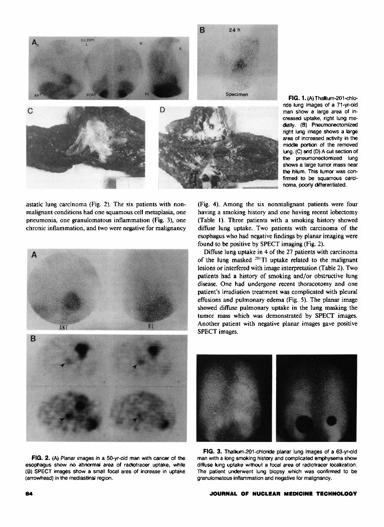
FIG. 2. (A) Planar images in a 50-yr-old man with cancer of the esophagus show no abnormal area of radiotracer uptake, while (B) SPECT images show a small focal area of increase in uptake (arrowhead) in the mediastinal region.
84
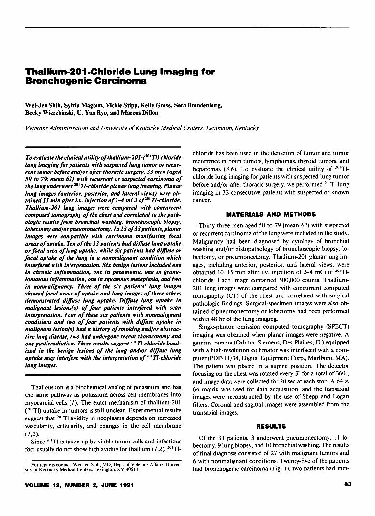
FIG. 1. (A) Thallium-201-chloride lung images of a 71-yr-old man show a large area of increased uptake, right lung medially. (B) Pneumonectomized right lung image shows a large area of increased activity in the middle portion of the removed lung. (C) and (D) A cut section of the pneumonectomized lung shows a large tumor mass near the hilum. This tumor was confirmed to be squamous carcinoma, poorly differentiated.
(Fig. 4). Among the six nonmalignant patients were four having a smoking history and one having recent lobectomy (Table l ). Three patients with a smoking history showed diffuse lung uptake. Two patients with carcinoma of the esophagus who had negative findings by planar imaging were found to be positive by SPECT imaging (Fig. 2).
Diffuse lung uptake in 4 of the 27 patients with carcinoma of the lung masked 201 TI uptake related to the malignant lesions or interfered with image interpretation (Table 2). Two patients had a history of smoking and/or obstructive lung disease. One had undergone recent thoracotomy and one patient's irradiation treatment was complicated with pleural effusions and pulmonary edema (Fig. 5). The planar image showed diffuse pulmonary uptake in the lung masking the tumor mass which was demonstrated by SPECT images. Another patient with negative planar images gave positive SPECT images.
FIG. 3. Thallium-201-chloride planar lung images of a 63-yr-old man with a long smoking history and complicated emphysema show diffuse lung uptake without a focal area of radiotracer localization. The patient underwent lung biopsy which was confirmed to be granulomatous inflammation and negative for malignancy.
JOURNAL OF NUCLEAR MEDICINE TECHNOLOGY
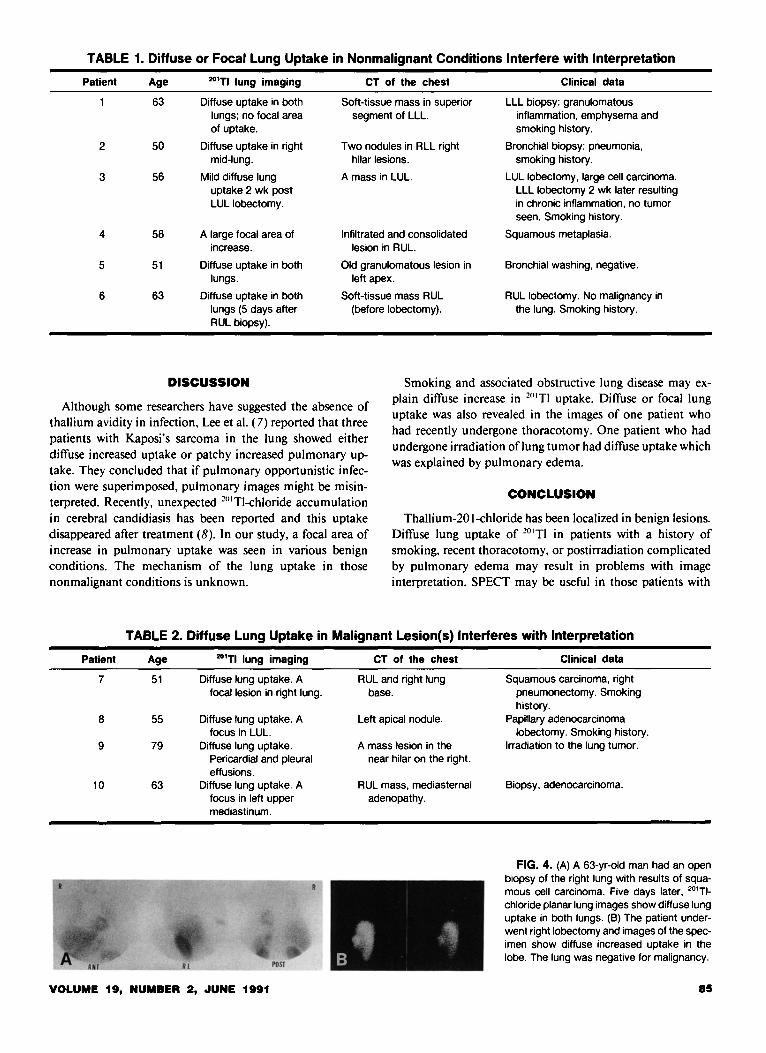
TABLE 1. Diffuse or Focal Lung Uptake in Nonmalignant Conditions Interfere with Interpretation
Patient Age 201TI lung imaging CT of the chest Clinical data
63 Diffuse uptake in both Soft-tissue mass in superior LLL biopsy: granulomatous lungs; no focal area segment of LLL. inflammation, emphysema and of uptake. smoking history.
2 50 Diffuse uptake in right Two nodules in RLL right Bronchial biopsy: pneumonia, mid-lung. hilar lesions. smoking history.
3 56 Mild diffuse lung A mass in LUL. LUL lobectomy, large cell carcinoma. uptake 2 wk post LLL lobectomy 2 wk later resulting LUL lobectomy. in chronic inflammation, no tumor
seen. Smoking history.
4 58 A large focal area of Infiltrated and consolidated Squamous metaplasia. increase. lesion in RUL.
5 51 Diffuse uptake in both Old granulomatous lesion in Bronchial washing, negative. lungs. left apex.
6 63 Diffuse uptake in both Soft-tissue mass RUL RUL lobectomy. No malignancy in lungs (5 days after (before lobectomy). the lung. Smoking history. RUL biopsy).
DISCUSSION
Although some researchers have suggested the absence of thallium avidity in infection, Lee et al. ( 7) reported that three patients with Kaposi's sarcoma in the lung showed either diffuse increased uptake or patchy increased pulmonary uptake. They concluded that if pulmonary opportunistic infection were superimposed, pulmonary images might be misinterpreted. Recently, unexpected 201 TI-chloride accumulation in cerebral candidiasis has been reported and this uptake disappeared after treatment (8). In our study, a focal area of increase in pulmonary uptake was seen in various benign conditions. The mechanism of the lung uptake in those nonmalignant conditions is unknown.
Smoking and associated obstructive lung disease may explain diffuse increase in 201 TI uptake. Diffuse or focal lung uptake was also revealed in the images of one patient who had recently undergone thoracotomy. One patient who had undergone irradiation oflung tumor had diffuse uptake which was explained by pulmonary edema.
CONCLUSION
Thallium-201-chloride has been localized in benign lesions. Diffuse lung uptake of 201 Tl in patients with a history of smoking, recent thoracotomy, or postirradiation complicated by pulmonary edema may result in problems with image interpretation. SPECT may be useful in those patients with
TABLE 2. Diffuse Lung Uptake in Malignant Lesion(s) Interferes with Interpretation
Patient Age 201TI lung imaging
7 51 Diffuse lung uptake. A focal lesion in right lung.
8 55 Diffuse lung uptake. A focus in LUL.
9 79 Diffuse lung uptake. Pericardia! and pleural effusions.
10 63 Diffuse lung uptake. A focus in left upper mediastinum.
VOLUME 19, NUMBER 2, JUNE 1991
CT of the chest
RUL and right lung base.
Left apical nodule.
A mass lesion in the near hilar on the right.
RUL mass, mediasternal adenopathy.
Clinical data
Squamous carcinoma, right pneumonectomy. Smoking history.
Papillary adenocarcinoma lobectomy. Smoking history.
Irradiation to the lung tumor.
Biopsy, adenocarcinoma.
FIG. 4. (A) A 63-yr-old man had an open biopsy of the right lung with results of squamous cell carcinoma. Five days later, 20'TIchloride planar lung images show diffuse lung uptake in both lungs. (B) The patient underwent right lobectomy and images of the specimen show diffuse increased uptake in the lobe. The lung was negative for malignancy.
85
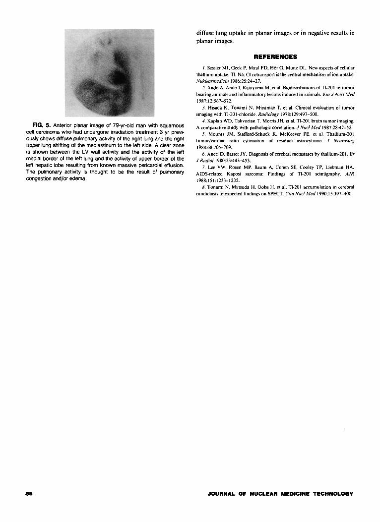
FIG. 5. Anterior planar image of 79-yr-old man with squamous cell carcinoma who had undergone irradiation treatment 3 yr previously shows diffuse pulmonary activity of the right lung and the right upper lung shifting of the mediastinum to the left side. A clear zone is shown between the LV wall activity and the activity of the left medial border of the left lung and the activity of upper border of the left hepatic lobe resulting from known massive pericardia! effusion. The pulmonary activity is thought to be the result of pulmonary congestion andfor edema.
86
diffuse lung uptake in planar images or in negative results in planar images.
REFERENCES
/. Sessler MJ. Geck P. Maul FD. Hor G. Munz DL. New aspects of cellular thallium uptake: Tl. Na. Cl cot ran sport is the central mechanism of ion uptake: Nuk/earmedi=in 1986:25:24-27.
2. Ando A. Ando I. Katayama M. et al. Biodistributions ofTI-201 in tumor bearing animals and inflammatory lesions induced in animals. Eur J Nuc/ Med 1987:12:567-572.
J. Hisada K. Tonami N. Miyamae T. et al. Clinical evaluation of tumor imaging with Tl-201-chloride. Radiolog,l' 1978:129:497-500.
4. Kaplan WD. Takvorian T. Morris JH, et al. Tl-201 brain tumor imaging: A comparative study with pathologic correlation. J Nucl Med 1987:28:47-52.
5. Mountz JM. Stafford-Schuck K. McKeever PE. et al. Thallium-201 tumor/cardiac ratio estimation of residual astrocytoma. J Neurosurg 1988:68:705-709.
6. Ancri D. Basset JY. Diagnosis of cerebral metastases by thallium-20 I. Br
J Radio/ 1980:53:443-453. 7. Lee VW. Rosen MP. Baum A, Cohen SE, Cooley TP, Liebman HA.
AIDS-related Kaposi sarcoma: Findings of Tl-201 scintigraphy. AJR
1988:151:1233-1235. 8. Tonami N. Matsuda H. Ooba H. et al. Tl-201 accumulation in cerebral
candidiasis unexpected findings on SPECT. Clin Nuc/ Med 1990:15:397-400.
JOURNAL OF NUCLEAR MEDICINE TECHNOLOGY


![Transbronchial Needle Aspiration Staging of Bronchogenic ...downloads.hindawi.com/journals/dte/1996/237680.pdfChest, 80,48-50. [18] Transbronchialneedle bronchogenic carcinoma, In:](https://img.pdfslide.net/doc/110x75/5fef28f6c0cad34ae7313439/transbronchial-needle-aspiration-staging-of-bronchogenic-chest-8048-50-18.jpg)