Embed Size (px)
Citation preview
Polish Journal of Microbiology2018, Vol. 67, No 2, 151–161DOI: 10.21307/pjm-2018-029
MINIREVIEW
* Corresponding author: P. Głowacka, Biological Threats Identification and Countermeasure Center of the General Karol Kaczkowski Military Institute of Hygiene and Epidemiology, Puławy, Poland; e-mail: [email protected]
Introduction
Brucella is a genus of bacteria belonging to the phy-lum Proteobacteria, class Alphaproteobacteria, order Rhizobiales, family Brucellaceae. Alphaproteobacteria is a very diverse group as to this class belongs both, the pathogens associated with plants: Agrobacterium spp., Sinorhizobium spp., Mesorhizobium spp. and the patho-gens, which cause dangerous infections of animals, e.g. Ricketsia spp., Bartonella spp., Brucella spp. and many others (Dwight and Bowman, 2011).
Brucella genus is responsible for brucellosis, a severe febrile disease. Brucellosis is a worldwide problem, causing abortion and infertility in domestic and wild animals (Lapaque et al., 2005). Infection factors are aerobic, small, Gram-negative rods. Brucella, a genus discovered in 1887 by David Bruce, contains the follow-ing species: Brucella suis, Brucella ovis, Brucella abortus, Brucella canis, Brucella melitensis, Brucella neotomae, Brucella ceti, Brucella pinnipedialis, Brucella microti, Brucella inopinata, Brucella papionis, Brucella vulpis and other strains without standing in nomenclature, that include environmental samples (Galińska and Zagór-
ski, 2013; Whatmore et al., 2014; Scholz et al., 2016). Some species contain biovars, for example: B. suis have five biovars, B. melitensis contain three and B. abortus – nine biovars (Mizak et al., 2014). Most of these species infect mainly specific hosts. B. abortus causes disease in cattle and infections usually lead to abortion; whereas B. suis is responsible for brucellosis in pigs, resulting in reproductive problems. Sheep are hosts for B. melitensis; infection causes impaired fertility. B. ovis is an etiologi-cal factor in sterility of rams (Megid et al., 2010). Cur-rently, about 500 000 cases of human brucellosis have been reported worldwide annually (Byndloss and Tsolis, 2016). Brucellosis is an endemic zoonosis with infection predominantly occurring in Middle East, Mediterra-nean rim (Portugal, Spain, Greece), Asia, Africa, South and Central America where the intake of dairy products is high, and protection of animal health is insufficient (Rubach et al., 2013). There are single cases reported in Poland, however connected with occupational exposure or with traveling to Mediterranean countries (Galińska and Zagórski, 2013). B. abortus and B. suis are isolated not only from livestock but also from different wild-life species (bears, buffalo, bison, caribu, camelids, elk,
Brucella – Virulence Factors, Pathogenesis and Treatment
PATRYCJA GŁOWACKA1*, DOROTA ŻAKOWSKA1, KATARZYNA NAYLOR2,MARCIN NIEMCEWICZ1 and AGATA BIELAWSKA-DRÓZD1
1 Biological Threats Identification and Countermeasure Center of the General Karol KaczkowskiMilitary Institute of Hygiene and Epidemiology, Puławy, Poland
2 Lublin Medical University, Department of Didactics and Medical Simulation, Lublin, Poland
Submitted 17 October 2017, revised 21 December 2017, accepted 28 February 2018
A b s t r a c t
Brucellae are Gram-negative, small rods infecting mammals and capable of causing disease called brucellosis. The infection results in abortion and sterility in domestic animals (sheeps, pigs, rams etc). Especially dangerous for humans are: Brucella melitensis, Brucella suis, Brucella abortus, and Brucella canis that trigger unspecific symptoms (flu-like manifestation). Brucella rods are introduced via host cells, by inhalation, skin abrasions, ingestion or mucosal membranes. The most important feature of Brucella is the ability to survive and multiply within both phagocytic and non-phagocytic cells. Brucella does not produce classical virulence factors: exotoxin, cytolisins, exoenzymes, plasmids, fimbria, and drug resistant forms. Major virulence factors are: lipopolysaccharide (LPS), T4SS secretion system and BvrR/BvrS system, which allow interaction with host cell surface, formation of an early, late BCV (Brucella Containing Vacuole) and interaction with endoplasmic reticulum (ER) when the bacteria multiply. The treatment of brucellosis is based on two-drug therapy, the most common combinations of antibiotics are: doxycycline with rifampicin or fluoroquinolones with rifampicin. Currently, also other methods are used to disrupt Brucella intracellular replication (tauroursodeoxycholic acid or ginseng saponin fraction A).
Key words: Brucella, endoplasmic reticulum, macrophage, replication, virulence factors
Głowacka P. et al. 2152
ferrets, deer, foxes, rodents, rabbits, wolves) and marine mammals (dolphins, dugongs, manatees, otters, sea porpoise) (Coelho et al., 2015). B. melitensis is rarely encountered in wildlife, nonetheless individual cases have been reported in ibex and in chamois in Alps. B. ovis and B. canis have not been detected in wildlife in Europe up to date. B. pinnipedialis and B. ceti cause the most common infections in marine mammals. Birds are resistant to Brucella infection, whereas fish seem to be susceptible to B. pinnipedialis and B. ceti infections (Godfroid et al., 2013). Infection is transmitted through close contact and during a common pasture.
Brucellosis is transferred from animals to humans, frequently human to human transmission occurs (Osman et al., 2016). Especially dangerous for humans are: B. melitensis, B. suis, and B. abortus, B. canis. Brucel-losis in human presents with symptoms like influenza: undulating fever, depression, weight loss, hepatomegaly, and splenomegaly (Bingöl et al., 1999). Mainly human cases are connected to occupational risk or consumption of unprocessed dairy products (Boschiroli et al., 2001; de Figureido et al., 2015). Brucella rods can enter via host cells by inhalation, ingestion, skin abrasion, or mucosal membranes (Franco et al., 2007). After penetration into host, rods multiply in lymph nodes; afterward, they pen-etrate other organs (Galińska and Zagórski, 2013). Brucella, can modify immune response in host cells; it has an affinity to the cells of specific tissues, e.g. placental trophoblast in fetal lung, pregnant females or reproduc-tive system (de Figureido et al., 2015). Brucellosis causes enlargement of lymph nodes, liver and spleen (Perkins et al., 2010). Pathogenicity of Brucella is dependent on their ability to multiply and survive within macrophages (Sangari and Agűero, 1996; Christopher et al., 2010).
Characteristics of Brucella
Species of the genus Brucella belong to small cocco-bacilli, measuring about 0,6–1,5 µm (Alton and Forsyth, 1996). They occur in single forms; rarely they create pairs or chains (Mizak et al., 2014). Brucella are non-spore forming and non-motile Gram-negative coco-bacilli (GNCB) (Alton and Forsyth, 1996). Brucella is an intracellular pathogen, during an infection it sur-vives and multiplies in macrophages; the bacteria adapt to the acidic pH, low levels of oxygen, and low levels of nutrients (Kőhler et al., 2002).
Lipopolisaccharide (LPS) is an essential element of structure building in each Gram-negative bacterial cell. Brucella is a genus that creates two forms of LPS. The smooth forms present complete LPS in the outer membrane, the rooth phenotype does not contain poli-saccharide O-chain (Lapaque et al., 2005; Seleem et al., 2008). These infectious agents are able to produce cyto-chrome oxidase, catalase, and most of them are able to
hydrolyze urea (Iowa State University, 2009). Brucella does not produce classical pathogenic factors, such as: exotoxin, cytolisins, exoensymes, exoproteins, capsules, plasmids, fimbria, and drug resistant forms (Seleem et al., 2008; Baldi and Giambartolomei, 2013; Tan et al., 2015). Bacterial cells are able to survive for a prolonged time in water, aborted fetus, soil, dairy products, meat, dung, and dust (Gwida et al., 2010). For isolation of Brucella spp. the enrichment and selective media such as Thayer-Martin’s medium or Farrell’s medium are commonly used. The colonies mature after four to six days of incubation at temperature of 37°C. They can also grow at 28°C, but poorly and slowly. Moreover, these bacteria can grow in both aerobic atmosphere and in 10% CO2; while, their growth is enhanced without addi-tional CO2 on a serum dextrose agar (Iowa State Univer-sity, 2009; Whatmore et al., 2014; Gupte and Kaur, 2015).
A wide range of bacterial detection methods is available. The predominatingly used culture media are: Bacto Tryptose (Difco), Triptcase soy (BBL), Tryp-tone soya (Oxoid), Triptic soy (Gibco). For culture of blood or body fluid a biphase medium called Castaneda should be used. Castaneda consists of two phases: liq-uid and solid closed in bottle. Liquid medium contains 1–2% of sodium citrate. Sample (5–10 ml) is added to the medium and incubated in 37°C in perpendiculary standing bottle in 10% carbon dioxide atmosphere (Gupte and Kaur, 2015). Serological tests are used to detect infection by examination of a specific antibodies level in serum. In the first week of Brucella infection the titres of IgM are dominant, but in the second week IgG class is prevalent. After four weeks, both types of antibodies reach a peak; durable, high titres of IgG can evidence failure in treatment (Al Dahouk et al., 2013). Serum Agglutination Test (SAT) and Enzyme linked immunosorbent assay (ELISA) are the most common serological tests used for diagnosis of brucellosis. SAT is based on a survey of agglutination titer of different serum dilution against Brucella cell suspension (Alshaa-lan et al., 2014). ELISA depends on detection of anti-bodies against the antigen – smooth LPS in serum (Gerasu and Kassa, 2016). The most effective methods for detection of brucellosis are molecular techniques (classical PCR, real-time PCR). The PCR method applies various pairs of primers to amplify different fragments of the genome. The examples of genes used to identification of Brucella spp. are: BCSP 31 (primers: B4/B5), sequence of 16S rRNA (primers: F4/R2), omp2 gene (primers: JPF/JPR) (Baddour and Alkhalifa, 2008).
Virulence factors
Lipopolisaccharide. LPS is an essential virulence factor of Brucella. LPS consists of lipid A, oligosaccha-ride core and O-antigen in Gram-negative bacteria.
Brucella – virulence factors and pathogenesis2 153
Lipopolisaccharide is different and non-classical in Brucella as compared to other Gram-negative bacte-ria, for example E. coli (Cardosos et al., 2006; Chris-topher et al., 2010). Lipopolisaccharide from Brucella strains is less toxic and less active than the classical LPS isolated from E. coli. Classical LPS causes a high pyrogenicity, while non-classical LPS shows low pyro-genicity, being a weak inducer of tumor necrosis factor (Christopher et al., 2010). Three features distinguish-ing it from other Gram-negatives characterize lipid A found in B. abortus: i) the fundamental component is diaminoglucose instead glucosamine, ii) longer acyl groups, and iii) lipid A is connected to the core by amide bonds, instead ester and amide bounds (Lapaque et al., 2005). In strains with smooth colonies, the smooth LPS, (S-LPS) contains: i) lipid A, that consists of two types of aminoglycose, and fatty acid besides β-hydroxymiristic acid, ii) core comprises mannose, glucose, quinovosamine, and iii) O-chains are compo-sed of 4-formamido-4,6-dideoxymannose. The struc - ture of the R-LPS in strains with rough colonies is similar to the S-LPS, except for O-chains, which are reduced or absent (Corbel, 1997). B. suis has S-LPS. The O-chain connects with lipid rafts on the macrophage surface and the bacteria enter the cell. Brucella strains with R-LPS, for example B. ovis or B. canis do not con-nect with lipid rafts and rapidly connect with lysosomes (Lapaque et al., 2005). The strains with S-LPS are able to restrain host cell apoptosis by the interaction of the O-chain with TNF-α (tumor necrosis factor). Thus, dead cells do not release specific factors, therefore they do not activate the immune system and Brucellae are able to avoid host immune surveillance (Fernandez- -Prada et al., 2003).
Type IV secretion system (T4SS). T4SS is a multi-protein complex and participates in secretion of bacte-rial macromolecues (Cascales and Christie, 2003). This system is typified by virB operon encoding 12 proteins (11 860 bp) and exhibits in Brucella spp. with a high degree of similarity to T4SSs found in rhizobia, for example in phytopatogenic Agrobacterium tumefaciens (O’Callaghan et al., 1999). Expression of the virB operon is regulated by the regulator of quorum-sensing – VjbR (Seleem et al., 2008). The wild strains of Brucella are able to multiply only in the endoplasmic reticulum. VirB mutants of Brucella spp. are unable to multiply within the endoplasmic reticulum, it can result from the incapability to reach the ER, or multiply within (Delure et al., 2001). In macrophages, rods of Brucella spp. are localized in Brucella-containing vacuole (BCV); this organelle interacts with the ER and is responsible for formation of specialized brucellae-multiplication com-partment (Kőhler et al., 2002). The acquisition of endo-plasmic reticulum membrane depends on a functional virB secretion system – T4SS (Celli et al., 2003).
Superoxide dismutase and catalase. Macrophages with Brucella produce reactive oxygen intermediates (ROIs), this is a primary mechanism of destruction of the bacteria ingested, and it also prevents their intracel-lular replication (Gee et al., 2005; Seleem et al., 2008). The following ROIs: O2– (superoxide), H2O2 (hydrogen peroxide), OH– (hydroxyl radical) are very detrimen-tal for cell structure. The production of enzymes is the main line of defense, counteracting reactive oxy-gen intermediates. These enzymes include superoxide dismutase (SOD) and catalase (Gee et al., 2005). SOD (metalloenzyme) is encoded by sod sequence. An enzyme contains iron, magnesium, or zinc and cop-per at its active site (Benov and Fridovich, 1994). SOD is responsible for dismutation of O2– (superoxide) to H2O2 (hydrogen peroxide) and O2 (oxygen) – transfer from one molecule to another (2O2–+2H+ →H2O2 + O2) (Gopal and Elumalai, 2017). Some species possess two types of SOD (B. abortus, B. melitensis, B. suis). The first is cytoplasmic – a Mn cofactor – SodA. SodA neutral-izes endogenously generated O2– – product of aerobic metabolism. The second one, SodC is periplasmic Cu, Zn-SOD, an enzyme responsible for neutralizing exo-genously generated O2– and protection from the res-piratory burst within macrophages (Beck et al., 1990, Seleem et al., 2008, Martin et al., 2012).
Catalase decomposes hydrogen peroxide into oxygen and water. Catalase activity is limited to the periplasmic space, where together with Cu-Zn SOD leave external sources of ROI unchanged (Kim et al., 2000). Catalase is not necessary virulence factor, the other enzymes can compensate lack of this enzyme in catalase mutants, e.g. alkyl hydroperoxide reductase or enzymes involved in DNA repair mechanisms (Seleem et al., 2008). Catalase is encoded by a sequence similar to katE gene of Escherichia coli. B. abortus, B. melitensis and B. suis catalase production is regulated by an increased external level of H2O2 (Gee et al., 2004).
Cyclic β-1-2-glucans (CβG). Brucella CβG belongs to II OPGs (Osmoregulated periplasmic glucans) fam-ily (Bohin, 2000). B. abortus CβG impacts intracellu-lar trafficking by acting on lipid rafts on macrophage surface. These glucans participate in control of the phagosome-lysosome fusion. Mutants are destroyed in phagolysosome and they are not able to multiply. Even more, mutants treated by CβG are able to control vacuole maturation and lysosome fusion, so they can reach to the ER and replicate there (Arellano-Reynoso et al., 2005).
Urease. In Brucella there are non-identical urease operons in two separate genomes. Urease is a metal-loenzyme, that decomposes urea to carbonic acid and to ammonium form, and it results in pH increase. This feature enables its survival in acid environment (Seleem et al., 2008). In I chromosome, there are two
Głowacka P. et al. 2154
urea-operons: ure1 and ure2, separated by 1 Mb of DNA. Ure1 and ure2 encode structural genes: ureA, ureB, ureC and accessory genes: ureD, ureE, ureF, ureG (Mobley et al., 1995). That urease may protect Brucella during passage through the digestive tract (stomach), when the bacteria access their host through the oral route (Bandara et al., 2007). Urease is produced by all bacteria belonging to the genus Brucella but B. ovis (Sangari et al., 2007).
Cytochrome oxidase. Cytochrome oxidase is an enzyme facilitating Brucella’s survival inside the macro-phages, where oxygen availability is limited. There are two operons in genome encoding two types of high oxygen-affinity oxidases: cytochrome cbb3-type and cytochrome bd (ubiquinol oxidases) oxidases. Cyto-chrome cbb3 oxidase is expressed in vitro and allows for colonization of anoxic tissues (maximal action in microaerobiosis). Cytochrome bd oxidase is expressed during intracellular multiplication and enables adjust-ment to the replicative niche (Loiser-Meyer et al., 2005), by restraining the creation of oxidative free radi-cals and detoxification of compartment inside the cell (Endley et al., 2001).
Alkyl hydroperoxide reductase (AhpC, AhpD). These enzymes attempt protection against oxygen radi-cal and reactive nitrogen (Chen et al., 1998). AhpC and ahpD are organized in an operon under one promoter control. AhpC mutants are more sensitive to peroxide killing and are vulnerable to spontaneous mutagenesis (DelVecchio et al., 2002; Seleem et al., 2008).
Nitric oxide reductase (NorD). Reduction of nitrate to dinitrogen gas is an essential process for bac-teria in case of oxygen deficiency inside the cell; this process allows for respiration of nitrate (Stevanin et al., 2005). The infected macrophages produce nitric oxide (NO), and Brucella can use it for own purposes. Brucella NorD consists of four types of reductases: Nir – nitrite reductase, Nar – nitrate reductase, Nor – nitric oxide reductase and Nos – nitrous oxide reductase, called the nitrification island. Possibility concerning produc-tions of this enzymes helps to protect Brucella against low-oxygen conditions inside macrophages (Seleem et al., 2008).
Brucella virulence factor A (BvfA). Periplasmic protein that occurs only in genus Brucella; there are no homologous sequences in Gen Bank. The bvfA expres-sion is induced in macrophages, through phagosome acidification. Presumably this protein is involved in forming the replication intracellular niche. BvfA func-tion is not precisely identified (Lavigne et al., 2005).
Base excision repair (BER). XthA gene encodes exonuclease III, which takes part in the base excision repair of DNA. Two different sequences of xthA occur in the Brucella genome: xthA1 and xthA2. XthA1 mutants exhibit increased sensitivity to reactive oxygen
species (ROS), so this enzyme is responsible for protec-tion against oxidative destruction (Seleem et al., 2008).
BvrR/BvrS system. The analysis of Brucella genomic library has confirmed an occurrence of two open reading frames: bvrR and bvrS. The bvrR encodes BvrR proteins (237 amino acid) and bvrS encodes BvrS (601 amino acid). There are two potential promoters (–10 and 35 seq. located 50 bp upstream ORF of bvrR), and ribosome-binding sequence (9 bp upstream of the first codon) (Sola-Landa et al., 1998). BvrR exhibits resem-blance to response regulators proteins, as N-terminal domain is composed of highly conserved aminoacids: aspartic (pos: 14, 15, 58) and lysine (pos: 107). C-termi-nal domain showed high similarity sequence to OmpR family; therefore, this protein can be included as part of this family (Mizuno and Tanaka, 1997; Martínez-Nūñez et al., 2010). The protein is composed of three highly conserved domains: N-terminal sensing, periplasmic domain together with transmembrane component, cyto-plasmic domain with distinctive histidine residue and C-terminal ATP-binding domain (Viadas et al., 2010). BvrS includes four highly conserved regions on C-ter-minal domain: H, N, D/F, and G. This feature causes BvrS homologous to sensor proteins of the histidine pro-tein kinase family (Stock et al., 1995). BvrS is located in the cell membrane (Martínez-Nūñez et al., 2010). Brucella BvrR/BvrS are the best characterized compo-nents of the virulence system; mutants are incapable of invasion, prevention phagosome-lysosome fusion and intracellular replication. BvrR/BvrS system is a regula-tor of expression of multiple genes (Viadas et al., 2010). These proteins affect the transcription of the membrane proteins: Omp3b (Omp22) or Omp3a (Omp25a) and have the influence on other non-protein membrane molecules and hence on functional and structural mem-brane homeostasis (Manterola et al., 2007). BvrR/bvrS mutants show structural changes in LPS, but O-chains seem to be undisturbed. These mutants are incapable of activation of GTPase (Cdc42) before entry into the cell, so they persist extracellularly and in consequence they do not infect the cell (Guzmán-Verri et al., 2001). BvrR/BvrS is also responsible for limited lysosome fusion and intracellular trafficking (López-Goñi et al., 2002).
Brucella BvrR/BvrS regulatory system action acti-vate sensor domain of the BvrS protein by environmen-tal signals through kinase activity. Additionally, BvrS causes phosphorylation and activation of BvrR protein. BvrR activates transcription of omp3a, omp3b and other genes responsible for lipid A structure and perhaps core of LPS. In consequence, bvrS/bvrR mutants are more sensitive to cationic peptides and display increased permeability for surfactants (López-Goñi et al., 2002; Seleem et al., 2008). The influence of Omp3a and Omp3b on virulence remains unexplained in details (Manterola et al., 2007).
Brucella – virulence factors and pathogenesis2 155
It has been proven that BvrR/BvrS two-component system regulate the expression of virB by positive stimulation of vjbR transcription. VjbR transcriptional factor interacts with virB promoter (Martínez-Nūñez et al., 2002).
Role of virulence factors in chronic persistence. Evading an immunological response to Brucella anti-gens depends on LPS structure. The appearance of elongated fatty acid on the lipid A (Brucella-C28 com-pared to others Enterobacteriaceae C12 – C16) leads to poor activation of TLR4 (Tool-like receptor 4) (de Figueiredo et al., 2015; Byndloss and Tsolis, 2016). The other feature of Brucella spp. LPS is core oligosaccharide glycosylation pattern that prevents a connection of the bacteria with TLR4 (co-receptor MD-2) (Byndloss and Tsolis, 2016). Toll-like recep-tors, the transmembrane proteins, act as PRRs (Pattern recognition receptors) and initiate the innate immune responses. TLRs are responsible for the recognition of components of microorganism (Uemattsu et al., 2008). The TLR protein is composed of two domains: extracellular domain that is rich in leucine repeats and it is responsible for recognition of microbial components, and cytoplasmic domain – TIR, involved in signal transmission, activation of intermediate proteins and finally an activation of NF-ĸβ and cytokines (Radhakrishnan et al., 2009). TLR5 detects flagellin, but flagellin in Brucella spp. can avoid inter-action with TLR5 as it lacks the domain recognized by TLR. (Andersen-Nissen et al., 2005; Kim, 2015). Brucella encoded TcpB/BtpA protein that acts as a fol-lowing virulence factor (TcpB – B. melitensis, Btp1/BtpA – B. abortus). Three pathogenic microorgan-ism can produce similar proteins: Salmonella spp., E. coli and Brucella (Radhakrishnan et al., 2009). These proteins contain TIR domain and show similarity to TIRAP (MAL) – a TLR adaptor protein. TcpB promotes a degradation of TIRAP and disrupts TLR4 signalling that result in inhibition of proinflammatory cytokines production and dendritic cell maturation (Newman et al., 2006; Oliveira et al., 2008; Radhakrishnan et al., 2009; Sengupta et al., 2010; Byndloss and Tsolis, 2016). TIRAP triggers recruitment of MyD88 and hence mediates TLR4 and TLR2 signalling (Kagan and Medzhitov, 2006). It seems to be likely that TcpB is able to interact with Death Domain of MyD88 and affect the signalling pathway (Chaudhary et al., 2012). Another group of researchers has proven that Brucella encodes another TIR domain-containing protein, called BtpB (present in all Brucella strains). This protein reacts with MyD88, inhibits TLR signalling, and disrupts activation of dendritic cells BtpB, restraining TLR2, TLR4, TLR5 and TLR9 signalling; and together with BtpA affects DC maturation and inflects host inflammatory responses (Salcedo et al., 2013).
B. abortus has proline racemase and thus is able to produce anti-inflammatory cytokine IL-10. This cyto-kine modulates macrophage activity during early phase of infection and leads to persistence and long-term survival of microorganism inside host cells (Byndloss and Tsolis, 2016).
Role of virulence factors in reproductive dis-ease. The investigations performed on bovine placen-tal explants have proven that in early infection with B. aborus the suppression of proinflammatory cytokines occured. This process is dependent on BtpB and T4SS proteins (use of mutants of the virB and btpB genes results in the reverse effect by enhancement of the pro-inflammatory cytokines production) (Mol et al., 2014). In later phases, 12 h after infection with B. abortus, stimulation of proinflammatory cytokines and CXC chemokines production – CXCL6 (GCP-2) and CXCL8 (interleukin 8) takes place. CXCL6 and IL-8 known as neutrophil chemoattractants cause neutrophil influx and have been reported to cause necrotizing placentis after infecting a pregnant cow (Carvalho Neta et al., 2008).
Pathogenesis
Invasion to cell. The Brucella strains survive and multiply within both phagocytic and non-phagocytic cells. The main targets for this bacterium are mac-rophages, dendritic cells and trophoblast cells. How-ever, Brucella can also multiply within other cells, for example epithelioid cell (HeLa) or murine fibroblast (NIH3T3) (Pizarro-Cerdá et al., 2000; Celli, 2006; Xavier et al., 2010). Brucella translocates across the mucosal epithelial cells layer, where the professional phagocytes (macrophages and DC cells) engulf the bacteria. Brucella survives within non-phagocytic cells up to 72 hours after infection, overcomes the epithe-lial barrier and then penetrates the phagocytic cells. Approximately 10% of these bacteria survive this initial phase. In macrophages the pathogen avoids the host immune response; therefore, it can multiply and spread to other tissues using cellular tropism. The Brucella strains penetrate the host cells through a zipper-like mechanism (Gorvel and Moreno, 2002; de Figureido et al., 2015). Bacteria can spread in a host through the lymph nodes and then translocate to the preferred tis-sues in reproductive tract (Kim, 2015). There, Brucella induces acute or chronic infection of reproductive tract that leads to abortion or severe reproductive tract dis-eases. (He, 2012).
The non-opsonized Brucella organisms are inter-nalized through lectin or fibronectin receptors but opsonized by complement and Fc receptors. The opsonized bacteria are more prone to be destroyed within macrophages than non-opsonized ones. The
Głowacka P. et al. 2156
pathogen binds to receptors containing of sulfated residues and sialic acid on surfaces of epithelial cells (de Figureido et al., 2015).
Penetration into the epithelial cell requires actin polymerization (Kim, 2015). The adhesion of B. abortus to the cell surface leads to activation of GTPases of Rho subfamily, e.g. Rho, Rac, and Cdc42 (Guzmán-Verri et al., 2001). These proteins are involved in cytoskel-etal regulation and have an impact on parasitic bacte-rial internalization. Cdc42 is the only GTPase activated directly by B. abortus during the contact with a non-phagocytic cell. It seems that other GTP-ases (Rho or Rac) are activated indirectly, because their inhibition impede invasion into host cells (Waterman-Storer et al., 1999). Other protein acting as second messengers, i.e. cGMP, PIP3-kinase, MAP-kinase and tyrosine kinase are involved in adhesion of bacteria to the host cell sur-face (Guzmán-Verri et al., 2001).
Adhesion to macrophage surface is also associated with small GTPases activation (Guzmán-Verri et al., 2001) and F-actin polymerization (transient and rapid F-actin accumulation). In the early stages of adhesion Annexin I is also involved, a protein that is impli-cated in membrane fusion (Kusumawati et al., 2000). Bacterial internalization occurs also by the lipid rafts – microdomains that occur in the macrophage cell membrane. These structure contribute to intracellu-
lar trafficking of Brucella (Fig.1) (Xavier et al., 2010). Non-opsonized Brucella strains internalize in human monocytes and murine macrophages by lipid rafts. This process requires activation of TLR4 and PI3-kinase. However, this process in human dendritic cells is only partly dependent on lipid rafts (von Bargen et al., 2012). Brucella strains with lack of O-polysaccharides in LPS (R-LPS) do not penetrate eukaryotic cells by lipid rafts, and thus are exterminated by macrophages (Porte et al., 2003; Gomez et al., 2013). Lipid rafts are rich in cholesterol, GPI (glycosylphosphatidylinositol) and GM1 (gangliosides) (Brown and London, 1998). Lipid rafts-associated proteins: GPI and GM1 as well as cho-lesterol inosculate with Brucella-contain macropino-somes and facilitate internalization with macrophage (Naroeni and Porte, 2002).
Brucella can be recognized by TLRs, but owing to modifications its interaction with TLRs is 10-fold lesser than for Enterobacteria. Hence, the activation of NF-κβ and production of inflammatory cytokines is weaker (de Figureido et al., 2015).
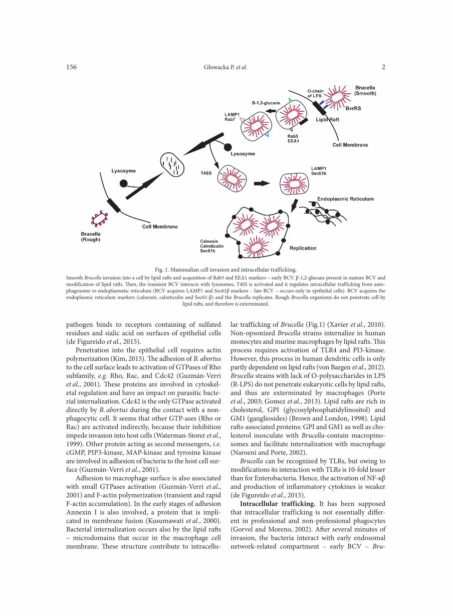
Intracellular trafficking. It has been supposed that intracellular trafficking is not essentially differ-ent in professional and non-professional phagocytes (Gorvel and Moreno, 2002). After several minutes of invasion, the bacteria interact with early endosomal network-related compartment – early BCV – Bru
Fig. 1. Mammalian cell invasion and intracellular trafficking.Smooth Brucella invasion into a cell by lipid rafts and acquisition of Rab5 and EEA1 markers – early BCV. β-1,2-glucans present in mature BCV and modification of lipid rafts. Then, the transient BCV interacts with lysosomes, T4SS is activated and it regulates intracellular trafficking from auto-phagosome to endoplasmatic reticulum (BCV acquires LAMP1 and Sec61β markers – late BCV – occurs only in epithelial cells). BCV acquires the endoplasmic reticulum markers (calnexin, calreticulin and Sec61-β) and the Brucella replicates. Rough Brucella organisms do not penetrate cell by
lipid rafts, and therefore is exterminated.
Brucella – virulence factors and pathogenesis2 157
cella Containing Vacuole. This compartment is char-acterized by Rab5 (GTP-binding protein) and EEA1 (early endosomal antigen 1) markers (Pizarro-Cerdá et al., 1998; Pizarro-Cerdá et al., 2000). β-1,2-glucans are necessary for the regulation of BCV maturation in macrophages as well as in epithelial cells. Additional function of β-1,2-glucan is the modification of rich in cholesterol lipid rafts, which are located on BCV membrane surface (Arellano-Reynoso et al., 2005). The interaction with early endocytic network last about 10 minutes (Pizarro-Cerdá et al., 1998). At this stage, acidification of BCV takes place leading to changes on the bacterial genes expression and enabling intracellu-lar survival (Carvalho Neta et al., 2010). BCV does not interact with late endosome and it avoids fusion with lysosomes (by β-glucans and LPS occurrence) (Gorvel and Moreno, 2002; Celli, 2006). However, early BCV is transformed to intermediate BCV that is LAMP1 and Rab-7 marked (late endosomal/lysosomal markers), indicating that interactions with late endosomal com-partments and lysosomes become necessary. What is more, in this step BCV acquires also Rab-interacting lysosomal protein (RILP) that is Rab-7 effector (Starr et al., 2008; von Bargen et al., 2012; Gomez et al., 2013). The interaction between BCV and late endosomes/lys-osomes is transitional and controlled. This event allows for acidification of BCV and expression of acidic-con-tingent bacterial factors, e.g., virB; simultaneously, the cathepsin D action does not take place (Boschiroli et al., 2001; Starr et al., 2008; von Bargen et al., 2012). Brucella type IV secretion (T4SS encoded by virB operon) is responsible for regulation of intracellular trafficking from autophagosome to endoplasmatic reticulum (ER) (Fig.1) (Gorvel and Moreno, 2002).
About 1 hour after internalization, Brucella organ-isms are located within multimembranous autopha-gosome with LAMP1 and Sec61β (calreculin). This structure is also called as a late BCV and occurs only in epithelial cells (Pizarro-Cerdá et al., 1998). LAMP1 function is not accurately described; however, it presum-ably participates in the pathogen intracellular survival (Gorvel and Moreno, 2002). The final step of Brucella intercellular trafficking is an acquisition of the markers characteristic of the endoplasmic reticulum: calnexin, calreticulin and Sec61β, although in this step BCV lose the LAMP-1 (Pizarro-Cerdá et al., 1998). However, this protein is constantly present in large vacuole only in human monocytes, in which opsonized Brucella mul-tiply (Bellaire et al., 2005). ER is the only compartment that is suitable for Brucella replication (Pizarro-Cerdá et al., 1998). The mechanism of BCV-ER connection remains unclear. In this process there are involved small GTPases Rab2, GAPDH – glyceraldehyde-3-phosphate dehydrogenase, the COPI complex (Coat Protein Com-plex I) and protein kinase C (PKCI). COPI and PKCI
control vesicular trafficking to the ER from Golgi. GAPDH/COPI/Rab2/PKCI complex is responsible for Brucella replication within the ER (Fugier et al., 2009).
Treatment of brucellosis
Currently, there are no effective vaccines for human, although several Brucella vaccines are accessible for livestock. Live, attenuated vaccines bereft of viru-lence factors, still present residual virulence (e.g., Live B. abortus vaccine strain 19, Live B. abortus vaccine strain RB51, Live B. melitensis vaccine strain Rev-1). Subunit vaccines are proven to be relatively safe and they raise less concerns compared to live vaccines. They do not cause infection, as they present purified proteins or DNA to stimulate immune response. Researchers are still working on the improvement in livestock vaccines and their application in preventing human infections (Yang et al., 2013).
To implement a successful treatment against brucel-losis, antibiotics penetrating into macrophages as well as active in acidic environment are essential (Ranjbar, 2015). Brucellosis is a disease, that rarely leads to death and responds well to diverse therapeutic strategies (Solís et al., 2015). However, single-antibiotics therapy is inadequate in brucellosis, as it leads to relapse of dis-ease (Pappas et al., 2006). Similarly, the therapy with single agent like: oxytetracycline, rifampin or doxycy-cline, causes high rate of relapses (9–25%) and prolon-gation of therapy does not provide satisfying effects. The treatment with trimethoprim-sulfamethoxazole or ciprofloxacin results in relapse in 30% and 83% of cases, respectively (Ranjbar, 2015). Treatment should prevent relapse of disease, further complications (arthritis, spondylitis, sacroilitis etc.) and enable quick relief of symptoms. The combination of two antibiotics in the-rapy of infections caused by Brucella is more effective than monotherapy. The WHO in 1986 recommended doxycycline with rifampicin for six weeks, replaced with tetracycline in combination with streptomycin. Cur-rently, the combinations of other antibiotics or chemo-therapeutics in therapy of brucellosis are used, such as fluoroquinolones or co-trimoxazole with rifampicin, doxycycline-streptomycin and doxycycline-rifampicin (Skalsky et al., 2008). During the treatment of brucel-losis with streptomycin and doxycycline (SD), a failure of treatment and relapse rates at 7.4% and 4.8%, respec-tively were noted. Almost similar results of therapy were observed during therapy with doxycycline and rifampin (DR) or streptomycin together with tetracycline (ST); however, their relapse rates were higher than in SD treatment. Another dual therapies of brucellosis are known, for example doxycycline and gentamicin (DG) with the average failure rate of 5.2% and the relapse rate
Głowacka P. et al. 2158
of 5.9%, or cotrimoxazole and rifampicin (RCTM) used in children brucellosis with the failure of treatment and relapse rates at 0–16.4% and 3.1–10%, respectively. The treatment with ciprofloxacin or ofloxacin with doxy-cycline, cotrimoxazole, rifampicin brought about the relapse rate between 3.2 to 26% (average 11.4%) and the failure rate between 3.2% to 26% (12.2%) (Alavi and Alavi, 2013). There were three clinical trials that used triple-drug therapy with doxycycline, rifampicin and aminoglycoside. There is no clear evidence on the superiority of triple-drug therapy when compared to the two-drug therapy. Nevertheless, it seems that triple-drug therapy is more effective in preventing relapses, but less successful in short-term treatment than two-drug therapy (Solís et al., 2015). The research by Alavi and Alavi (2013) suggested triple therapy for eight weeks in complicated cases (with spondylitis, or arthritis) due to lower treatment failure rates than two-drug therapy. Doxycycline and aminoglycoside therapy is recommended in uncomplicated chronic, or acute cases and in complicated cases without endocarditis, spondylitis, arthritis. In uncomplicated cases, strepto-mycin and doxycycline, or gentamicin are also advo-cated (Alavi and Alavi, 2013).
Smith and colleagues (2013) found a new strategy to treat brucellosis. The connection of BCV with the ER requires remodeling of endoplasmatic reticulum, which is necessary in modification of the ER structure during host stress response, which is called the Unfolded Pro-tein Response (UPR). The disruption of UPR, through tauroursodeoxycholic acid drug can inhibit Brucella replication. UPR can be a novel target in the of brucel-losis (Smith et al., 2013).
There are studies concerning the influence of gin-seng saponin fraction A (RGSF-A) for combating bru-cellosis. Ginseng is a valued plant in Asia, considered a panacea to variety range of diseases. Arayan et al. (2015) examined the influence of RGSF-A for eradica-tion of bacterial infection in RAW 264.7 cells. In this study, the bacterial internalization and adhesion were reduced in the cells treated when compared to the control cells without treatment. RGSF-A takes part in downregulation of MAPKs (mitogen-activated protein kinases) and hence, limits polymerization of F-actin and inhibits bacterial penetration into the cells. RGSF-A influences also intracellular trafficking of B. abortus and favors interaction of B. abortus-containing phagosomes (BCPs) with LAMP-1. LAMP-1 is trans-membrane protein, that is responsible for the fusion of lysosomes with phagosomes, enabling the connec-tion of BCPs with lysosome and elimination of bacte-ria (Arayan et al., 2015; Reyes et al., 2016; Huy et al., 2017). Huy et al. (2017) have proven that ginsenoside Rg3 – panaxadiol saponin components of RGSF-A have been the major factor controlling brucellosis.
Undoubtedly, there are also other promising herbal plants e.g. Teucrium polium, Scophularia deserti, Alhagi, Eucalyptus, garlic and roots of barberry that contain bioactive ingredients (flavones, flavonoids, anthocya-nins and tanins) that can be effecive in preventing or even combat brucellosis (Naghadi et al., 2016).
Conclusions
Brucella is an intracellular pathogen, especially dangerous for domestic animals, which causes mas-sive infections and thus significant economic losses. Moreover, people who work with infected animals comprise a risk group, e.g., farmers, veterinarians, or laboratorians and they are most endangered of being exposed to the pathogen. Brucellosis in human causes non-specific symptoms, therefore no plausible estima-tion can be managed to detect the number of infected people. Brucella is an inquisitive etiological agent, as does not produce classical virulence factors. The pro-cess of infection is a complex one, and there are many unexplained issues associated with it. Therefore, further studies of infection mechanisms are required.
Literature
Alavi S.M. and L. Alavi. 2013. Treatment of brucellosis: a systematic review of studies in recent twenty years. Caspian. J. Intern. Med. 4(2): 636–641. Al Dahouk S., L.D. Sprague and H. Neubauer. 2013. New devel-opments in the diagnostic procedures for zoonotic brucellosis in humans. Rev. Sci. Tech. 32(1): 177–188.Alshaalan M.A., S.A. Alalola, M.A. Almuneef, E.A. Albanyan, H.H. Balkhy, D.A. AlShahrani and S. AlJohani. 2014. Brucellosis in children: Prevention, diagnosis and management guidelines for general pediatricians endorsed by the Saudi Pediatric Infectious Dis-eases Society (SPIDS). Int. J. Ped. Adolesc. Med. 1(1): 40–46.Alton G.G. and J.R.L. Forsyth. 1996. Brucella, pp. 514–521. In: Baron S. (ed.). Medical Microbiology. University of Texas Medical Branch at Galveston, Galveston.Andersen-Nissen E., K.D. Smith, K.L. Strobe, S.L. Barrett, B.T. Cookson, S.M. Logan and A. Aderem. 2005. Evasion of Toll-like receptor 5 by flagellated bacteria. PNAS. 102(26): 9247–9252. Arayan S.T., H.L. Simborio, A.W. Reyes, H.T. Hop, W. Min and H.J. Lee. 2015. The effects of red ginseng saponin fraction-A (RGSF-A) on phagocytosis and intracellular signaling in Brucella abortus infected RAW 264.7 cells. FEMS Microbiol. Lett. 362(11). doi:10.1093/femsle/fnv070.Arellano-Reynoso B., S. Lapaque, S. Salcedo, G. Briones, A.E. Ciocchini and R. Ugadle. 2005. Cyclic-1,2-glucan is a Brucella virulence factor required for intracellular survival. Nat. Immunol. 6(6): 618–625.Baddour M.M. and D.H. Alkhalifa. 2008. Evaluation of three poly-merase chain reaction techniques for detection of Brucella DNA in peripheral human blood. Can. J. Microbiol. 54(5): 352–357.Baldi P.C. and G.H. Giambartolomei. 2013. Pathogenesis and pathobiology of zoonotic brucellosis in humans. Rev. Sci. Tech. Off. Int. Epiz. 32(1): 117–125.
Brucella – virulence factors and pathogenesis2 159
Bandara A.B., N. Sriranganathan, G.G. Schurig and S.M. Boyle. 2007. Carboxyl-terminal protease regulates Brucella suis morphol-ogy in culture and persistence in macrophages and mice. J. Bacteriol. 187(16): 5767–5775.von Bargen K., J.P. Gorvel and S.P. Salcedo. 2012. Internal affairs: investigating the Brucella intracellular lifestyle. FEMS Microbiol. Rev. 36(3): 533–562.Beck B.L., L. Tabatabi and J.E. Mayfield. 1990. A protein isolated from Brucella abortus is a Cu-Zn superoxide dismutase. Biochem. 29(2): 372–376. Bellaire B.H., R.M. Roop II and J.A. Cardelli. 2005. Opsonized virulent Brucella abortus replicates within nonacidic, endoplasmic reticulum-negative, LAMP-1-positive phagosomes in human mono-cytes. Infect. Immun. 73(6): 3702–3713.Benov L.T. and I. Fridovich. 1994. Escherichia coli Expresses a Cop-per- and Zinc-containing Superoxide Dismutase. J. Biol. Chem. 269(41): 25310–25314.Bingöl A., N. Yücemen and O. Meço. 1999. Medically treated intraspinal “Brucella” granuloma. Surg. Neur. 52(6): 570–576.Bohin J.P. 2000. Osmoregulated periplasmic glucans in Proteo-bacteria. FEMS Misrobiol. Lett. 186(1): 11–19.Boschiroli M.L., V. Foulongne and D. O’Callaghan. 2011. Brucel-losis: a worldwide zoonosis. Curr. Opin. Microbiol. 4: 58–64.Brown D.A. and E. London. 1998. Functions of lipid rafts in bio-logical membranes. Annu. Rev. Cell. Dev. Biol. 14: 111–136.Byndloss M.X. and R.M. Tsolis. 2016. Brucella spp. virulence fac-tors and immunity. Annu. Rev. Anim. Biosci. 4: 111–127.O’Callaghan D., C. Cazevieille, A. Allardet-Servent, M.L. Boschi-roli, G. Bourg, V. Foulongne, P. Frutos, Y. Kulakov and M. Ramuz. 1999. A homologue of the Agrobacterium tumefaciens VirB and Bordetella pertussis Ptl type IV secretion systems is essential for intra-cellular survival of Brucella suis. Mol. Microbiol. 33(6): 1210–1220. Cardosos P.G., G.C. Macedo, V. Azevedo and S.C. Oliveira. 2006. Brucella spp. noncanonical LPS: structure, biosynthesis, and interaction with host immune system. Microb. Cell Fact. 5: 13. doi:10.1186/1475-2859-5-13.Carvalho Neta A.V., A.P.R. Stynen, T.A. Paixăo, K.L. Miranda, F.L. Silva, C.M. Roux, R.M. Tsolis, R.E. Everts, H.A. Lewin, L.G. Adams and others. 2008. Modulation of bovine trophoblastic innate immune response by Brucella abortus. Infect. Immun.76(5): 1897–1907.Carvalho Neta A.V., J.P. Mol, M.N. Xavier, T.A. Paixão, A.P. Lage and R.L. Santos. 2010. Pathogenesis of bovine brucellosis. Vet. J. 184(2): 146–155. Cascales E. and P.J. Christie. 2003. The versatile bacterial type IV secretion system. Nat. Rev. Microbiol. 1(2): 137–149.Celli J., C. de Chastelier, D.M. Franchini, J. Pizzaro-Cerda, E. Moreno and J.P. Gorvel. 2003. Brucella evades macrophage kill-ing via VirB-dependent sustained interactions with the endoplasmic reticulum. J. Exp. Med. 198(4): 545–556. Celli J. 2006. Surviving inside a macrophage: the many ways of Bru-cella. Res. Microbiol. 157: 93–98.Chaudhary A., K. Ganguly, S. Cabantous, G.S. Waldo, S.N. Micheva- Viteva, K. Nag, W.S. Hlavacek and C.S. Tung. 2012. The Brucella TIR-like protein TcpB interacts with the death domain of MyD88. Biochem. Biophys. Res. Commun. 417(1): 299–304.Chen L., Q.W. Xie and C. Nathan. 1998. Alkyl hydroperoxide reductase subunit C (AhpC) protects bacterial and human cells against reactive nitrogen intermediates. Mol. Cell. 1(6): 795–805. Christopher S., B.L. Umapathy and K.L. Ravikumar. 2010. Brucel-losis: Review on the recent trends in pathogenicity and laboratory diagnosis. J. Lab. Physicians. 2(2): 55–60. Coelho A.C., J.G. Díez and A.M. Coelho. 2015. Risk Factors for Brucella spp. in Domestic and Wild Animals, pp. 2–31. In: Baddour M (ed.). Updates on Brucellosis. InTech.
Corbel M.J. 1997. Brucellosis: an overview. Emerg. Infect. Dis. 3(2): 213–221.Delure R.M., M. Martinez-Lorenzo, P. Lestrate, I. Danese, V. Bie- larz, P. Mertens, X. De Bolle, A. Tibor, J.P. Gorvel and J.J. Letes-son. 2001. Identification of Brucella spp. genes involved in intracel-lular trafficking. Cell Microbiol. 3(7): 487–497. Del Vecchio V.G., V. Kapatral, R.J. Redkar, G. Patra, C. Mujer, T. Los, N. Ivanova, I. Anderson, A. Bhattacharyya, A. Lykidis and others. 2002. The genome sequence of the facultative intracel-lular pathogen Brucella melitensis. Proc. Natl. Acad. Sci. USA 99(1): 443–448.Dwight D. and M.S. Bowman. 2011. Introduction to the Alpha-proteobacteria: Wolbachia and Bartonella, Rickettsia, Brucella, Ehrlichia and Anaplasma. Top. Companion Anim. Med. 26(4): 173–177.Endley S., D. McMurray and T.A. Ficht. 2001. Interruption of the cydB locus in Brucella abortus attenuates intracellular survival and virulence in the mouse model of infection. J. Bacteriol. 183(8): 2454–2462.Fernandez-Prada C.M., E.B. Zelazowska, M. Nikolich, T. Hadfield, R.M. Roop II, G.L. Robertson and D.L. Hoover. 2003. Interac-tions between Brucella melitensis and human phagocytes: Bacterial surface O-polysaccharide inhibits phagocytosis, bacterial kill-ing, and subsequent host cell apoptosis. Infect. Immunity. 71(4): 2110–2119.de Figueiredo P., T.A. Ficht, A. Rice-Ficht, C.A. Rossetti and L.G. Adams. 2015. Pathogenesis and immunobiology of brucel-losis review of Brucellae host interactions. Am. J. Pathol. 185(6): 1505–1517.Franco M.P., M. Mulder, R.H. Gilman and H.L. Smits. 2007. Human brucellosis. Lancet. Infect. Dis. 7(12): 775–786.Fugier E., S.P. Salcedo, C. de Chastellier, M. Pophillat, A. Muller, V. Arce-Gorvel, P. Fourquet and J.P. Gorvel. 2009. The glycer-aldehyde-3-phosphate dehydrogenase and the small GTPase Rab 2 are crucial for Brucella replication. PLoS Path. 5(6):e1000487. doi:10.1371/journal.ppat.1000487.Galińska E.M. and J. Zagórski. 2013. Brucellosis in humans – etio logy, diagnostics, clinical forms. Ann. Agric. Environ. Med. 20(2): 233–238.Gee J.M., M.E. Kovach, V.K. Grippe, S. Hagius, J.V. Walker, P.H. Elzer and others. 2004. Role of catalase in the virulence of Bru-cella melitensis in pregnant goats. Vet. Microbiol. 102(1–2): 111–115.Gee J.M., M.E. Kovach, V.K. Grippe, S. Hagius, J.V. Walker, P.H. Elzer and R.M. Roop 2nd. 2005. The Brucella abortus Cu, Zn superoxide dismutase is required for optimal resistance to oxidative killing by murine macrophages and wild-type virulence in experi-mentally infected mice. Infect. Immun. 73(5): 2873–2880.Gerasu M.A. and G.M. Kassa. 2016. A review on diagnostic meth-ods of brucellosis. Vet. Sci. Tech. 7:3. doi:10.4172/2157-7579.1000323.Godfroid J., B. Garin-Bastuji, C. Saegerman and J.M. Blasco. 2013. Brucellosis in terrestrial wildlife. Rev. Sci. Tech. 32(1): 27–42.Gomez G., L.G. Adams, A. Rice-Ficht and T.A. Ficht. 2013. Host-Brucella interactions and the Brucella genome as tools for subunit antigen discovery and immunization against brucellosis. Front. Cell. Infect. Microbiol. 3:17. doi:10.3389/fcimb.2013.00017.Gopal R.K. and S. Elumalai. 2017. Industrial production of Super-oxide Dismutase (SOD): A mini review. J. Probe. Health. 5:3. doi:10.4172/2329-8901.1000179.Gorvel J. and E. Moreno. 2002. Brucella intracellular life: from inva-sion to intracellular replication. Vet. Microbiol. 90(1–4): 281–297.Gupte S. and T. Kaur. 2015. Diagnosis of Human Brucellosis. J. Top. Dis. 4:1. doi:10.4185/2329-891X.1000185.Guzmán-Verri C., E. Chaves-Olarte, C. von Eichel-Streiber, I. López-Goñi, M. Thelestam, S. Arvidson, J.P. Gorvel and E. Moreno. 2001. GTPases of the Rho subfamily are required for Brucella abortus internalization in nonprofessional phagocytes. J. Biol. Chem. 276(48): 44435–44443.
Głowacka P. et al. 2160
Gwida M., S. Al Dahouk, F. Melzer, U. Rősler, H. Neubauer and H. Tomaso. 2010. Brucellosis – regionally emerging zoonotic dis-ease? Croat. Med. J.51(4): 289–295. He Y. 2012. Analyses of Brucella pathogenesis, host immunity, and vaccine targets using systems biology and bioinformatics. Front. Cell. Infect. Microbiol. 2:2. doi:10.3389/fcimb.2012.00002.Huy T.X., A.W. Reyes, H.T. Hop, L.T. Arayan, W. Min and H.J. Lee. 2017. Intracellular trafficking modulation by ginseno-side Rg3 Inhibits Brucella abortus uptake and intracellular survival within RAW 264.7 cells. J. Microbiol. Biotechnol. 27(3): 616–623.Iowa State University. The Centerfor Food Security & Public Health. 2009. Ovine and Caprine Brucellosis: Brucella melitensis. http://www.cfsph.iastate.edu/Factsheets/pdfs/brucellosis_melitensis.pdf. Kagan J.C. and R. Medzhitov. 2006. Phosphoinositide-mediated adaptor recruitment controls toll-like receptor signaling. Cell. 125(5): 943–955.Kim S. 2015. The interaction between Brucella and the host cell in phagocytosis, pp. 45–60. In: Baddour M. (ed.). Updates on Brucellosis. InTech, Jinju.Kőhler S., V. Foulongne, S. Ouahrani-Bettache, G. Bourg, J. Teys-sler, M. Ramuz and J.P. Liautard. 2002. The analysis of the intra-macrophagic virulome of Brucella suis deciphers the environment encountered by the pathogen inside the macrophage host cell. Proc. Natl. Acad. Sci. USA. 99(24): 15711–15716. Kusumawati A., C. Cazevieille, F. Porte , S. Bettache, J.P. Liautard and J. Widada. 2000. Early events and implication of F-actin and annexin I associated structures in the phagocytic uptake of Brucella suis by the J-774A.1 murine cell line and human monocytes. Microb. Pathog. 28(6): 343–352. Lapaque N., I. Moriyon, E. Moreno and J.P. Gorvel. 2005. Brucella lipopolysaccharide acts as a virulence factor. Curr Opin Microbiol. 8(1): 60–66. Lavigne J.P., G. Petey, F.J. Sangari, G. Bourg, M. Ramuz, D. O’Cal- la ghan, S. Michaux-Charachon. 2005. Identification of a new viru-lence factor, BvfA, in Brucella suis. Infect. Immun. 73(9): 5524–5529.Loiser-Meyer S., M.P.J. de Bagüés, S. Köhler, J.P. Liautard and V. Jubier-Maurin. 2005. Differential use of the two high-oxygen-affinity terminal oxidases of Brucella suis for in vitro and intramac-rophagic multiplication. Infect. Immun. 73(11): 7768–7771. López-Goñi I., C. Guzmán-Verri, L. Manterola, A. Sola-Landa, I. Moriyón, E. Moreno. 2002. Regulation of Brucella virulence by the two – component system BvrR/BvrS. Vet. Microbiol. 90(1–4): 329–339.Manterola L., C. Guzmán-Verri, E. Chaves-Olarte, E. Barquero-Calvo, M.J. de Miguel, I. Moriyón, M.J. Grilló, I. López-Goñi and E. Moreno. 2007. BvrR/BvrS-Controlled Outer Membrane Proteins Omp3a and Omp3b are not essential for Brucella abortus virulence. Infect. Immun. 75(10): 4867–4874.Martin D.W., J.E. Baumgartner, J.M. Gee, E.S. Anderson and R.M. Roop II. 2012. SodA is a major metabolic antioxidant in Bru-cella abortus 2308 that plays a significant, but limited, role in the virulence of this strain in the mouse model. Microbiology. 158(Pt7): 1767–1774.Martínez-Nūñez C., P. Altamirano-Silva, F. Alvarado-Guillén, E. Moreno, C. Guzmán-Verri and E. Chaves-Olarte. 2010. The Two-Component System BvrR/BvrS regulates the expression of the type IV secretion system VirB in Brucella abortus. J. Bacteriol. 192(21): 5603–5608.Megid J., L.A. Mathias and C.A. Robles. 2010. Clinical manifesta-tions of brucellosis in domestic animals and humans. Op. Vet. Sci. J. 4: 119–126.Mizak L., R. Gryko, S. Parasion and M. Kwiatek. 2014. Brucellosis – a worldwide zoonosis (in Polish). Życ. Wet. 89(1): 35–40.Mizuno T. and I. Tanaka. 1997. Structure of the DNA-binding domain of the OmpR family of response regulators. Mol. Microbiol. 24(3): 665–667.
Mol J.P.S., E.A. Costa, A.F. Carvalho, Y-H. Sun, R.M. Tsolis, T.A. Paixăo and R.L. Santos. 1995. Molecular biology of microbial ureases. Microbiol. Rev. 59(3): 451–480. Mol J.P.S., E.A. Costa, A.F. Carvalho, Y-H. Sun, R.M. Tsolis, T.A. Paixão and others. 2014. Early transcriptional responses of bovine chorioallantoic membrane explants to wild type, ΔvirB2 or ΔbtpB Brucella abortus infection. PLoS One. 9(9): e108606. doi:10.1371/journal.pone.0108606.Naghadi N., H. Assanzad-Azar and A. Delpisheh. 2016. The most important medicinal plants for treatment of brucellosis. J. Prev. Epi. 1(2): e20. Naroeni A. and F. Porte. 2002. Role of Cholesterol and the Gan-glioside GM 1 in entry and short-term survival of Brucella suis in murine macrophages. Infect. Immun. 70(3): 1640–1644.Newman R.M., P. Salunkhe, A. Godzik and J.C. Reed. 2006. Iden-tification and characterization of a novel bacterial virulence factor that shares homology with mammalian toll/interleukin-1 receptor family proteins. Infect. Immun. 74(1): 594–601.Oliveira S.C., F.S. de Oliviera, G.C. Macedo, L.A. de Almeida and N.B. Carvalho. 2008. The role of innate immune receptors in the control of Brucella abortus infection: Toll-like receptors and beyond. Microbes Infect. 10(9): 1005–1009.Osman A.Y., F.F. Jesse, A. Abdul Kadir and A.A. Saharee. 2016. The epidemiology and immunopathophysiology of brucellosis in small ruminant. PJSRR. 2(1): 11–21.Pappas G., P. Papadimitriou, N. Akritidis, L. Christou and E. Tsia-nos. 2006. The new global map of human brucellosis. Lancet. Infect. Dis. 6(2): 91–99. Perkins S.D., S.J. Smither and H.S. Atkins. 2010. Towards a Brucella vaccine for humans. FEMS Microbiol. Rev. 34: 379–394.Pizarro-Cerdá J., E. Moreno and J.P. Gorvel. 2000. Invasion and intracellular trafficking of Brucella abortus in nonphagocytic cells. Microbes Infect. 2(7): 829–835. Pizarro-Cerdá J., S. Méresse, R.G. Parton, G. van der Goot, A. Sola-Landa, I. López-Goñi, E. Moreno and J.P. Gorvel. 1998. Brucella abortus transits through the autophagic pathway and rep-licates in the endoplasmic reticulum of nonprofessional phagocytes. Infect. Immun. 66(12): 5711–5724. Porte F., A. Naroeni, S.Ouahrani-Bettache and J.P. Liautard. 2003. Role of the Brucella suis lipopolysaccharide O antigen in phago-somal genesis and in inhibition of phagosome-lysosome fusion in murine macrophages. Infect. Immun. 71(3): 1481–1490. Radhakrishnan G.K., Q. Yu , J.S. Harms and G.A. Splitter. 2009. Brucella TIR domain-containing protein mimics properties of the toll-like receptor adaptor protein TIRAP. J. Biol. Chem. 284(15): 9892–9898.Ranjbar M. 2015. Treatment of brucellosis, pp. 171-184. In: Bad-dour M. (ed.). Updates on Brucellosis. InTech, Teheran.Reyes A.W., H.L. Simborio, H.T. Hop, L.T. Arayan, W.G. Min, H.J. Lee, M.H. Rhee, H.H. Chang and S. Kim. 2016. Inhibitory effect of red ginseng acidic polysaccharide from Korean red gin-seng on phagocytic activity and intracellular replication of Brucella abortus in RAW 264.7 cells. J. Vet. Sci. 17(3): 315–321.Rubach M.P., J.E. Halliday, S. Cleaveland and J.A. Crump. 2013. Brucellosis in low-income and middle-income countries. Curr. Opin. Infect. Dis. 26(5): 404–412.Salcedo S.P., M.I. Marchesini, C. Degos, M. Terwagne, K. Von Bar-gen, H. Lepidi, C.K. Hermann, T.L. Santos Lacerda, P.R. Imbert, P. Pierre and others. 2013. BtpB, a novel Brucella TIR-containing effector protein with immune modulatory functions. Front. Cell. Infect. Microbiol. 3:28. doi:10.3389/fcimb.2013.00028.Sangari F.J. and J. Agűero. 1996. Molecular basis of Brucella patho-genicity: an update. Microbiologia. 12(2): 207–218. Sangari F.J., A. Seoane, M.C. Rodriguez, J. Agüero and J.M. Gar-cia Lobo. 2007. characterization of the urease operon of Brucella
Brucella – virulence factors and pathogenesis2 161
abortus and assessment of its role in virulence of the bacterium. Infect. Immun. 75(2): 774–780.Scholz H.C., S. Revilla-Fernández, S. Al Dahouk, J.A. Hammerl, M.S. Zygmunt, A. Cloeckaert, M. Koylass, A.M. Whatmore, J. Blom, G. Vergnaut and others. 2016. Brucella vulpis sp. nov., isolated from mandibular lymph nodes of red foxes (Vulpes vulpes). Int. J. Syst. Evol. Microbiol. 66(5): 2090–2098.Seleem M.N., S.M. Boyle and N. Sriranganathan. 2008. Brucella: A pathogen without classic virulence genes. Vet. Microbiol. 129(1–2): 1–14.Sengupta D., A. Koblansky, J. Gaines, T. Brown, A.P. West and D. Zhang. 2010. Subversion of innate immune responses by Brucella through the targeted degradation of the TLR signaling adapter, MAL. J. Immunol. 184(2): 956–964.Skalsky K., D. Yahav, J. Bishara, S. Pitlik, L. Leibovici and M. Paul. 2008. Treatment of human brucellosis: systematic review and meta-analysis of randomised controlled trials. BMJ. 336(7646): 701–704.Smith J.A., M. Khan, D.D. Magnani, J.S. Harms, M. Durward, G.K. Radhakrishnan. 2013. Brucella induces an unfolded pro-tein response via TcpB that supports intracellular replication in macrophages. PLoS Path. 9(12): e1003785. doi:10.1371/journal.ppat.1003785.Sola-Landa A., J. Pizarro-Cerdá, M.J. Grilló, E. Moreno, I. Mori-yón and J.M. Blasco. 1998. A two-component regulatory system playing a critical role in plant pathogens and endosymbionts is pre-sent in Brucella abortus and controls cell invasion and virulence. Mol. Microbiol. 29(1): 125–138.Solís J., G. del Pozo and J. Solera. 2015. Treatment of human brucel-losis-review of evidence from clinical trials, pp. 186–189. In: Baddour M. (eds). Updates on Brucellosis. InTech, Villarrobledo, Albacete.Starr T., T.W. Ng, T.D. Wehrly, L.A. Knodler and J. Celli. 2008. Brucella intracellular replication requires trafficking through the late endosomal/lysosomal compartment. Traffic. 9(5): 678–694.Stevanin T.M., J.W.B. Moir and R.C. Read. 2005. Nitric oxide detoxification systems enhance survival of Neisseria meningitidis in human macrophages and in nasopharyngeal mucosa. Infect. Immun. 73(6): 3322–3329.
Stock J.B., M.G. Surette, M. Levit and P. Park. 1995. Two-com-ponent siganl transduction systems: structure-function relation- ships and mechanism of catalysis, pp. 25–51. In: Hoch J.A. and T.J. Silhavy (eds). Two Component Signal Transduction. ASM Press, Washington, D.C.Tan K.K., Y.C.Tan, L.Y. Chang, K.W. Lee, S.S. Nore, W.Y. Yee, M.N.M. Isa, F.L. Jafar, C.C. Hoh and S. AbuBakar. 2015. Full genome SNP-based phylogenetic analysis reveals the origin and global spread of Brucella melitensis. BMC Genomics. 16(1):93. doi:10.1186/s12864-015-1294-x.Uematsu S. and S. Akira. 2008. Toll-Like Receptors (TLRs) and their ligands. Handb. Exp. Pharmacol. (183): 1–20.Waterman-Storer C.M., R.A. Worthylake, B.P. Liu, K. Burridge and E.D. Salmon. 1999. Microtubule growth activates Rac1 to pro-mote lamellipodial protrusion in fibroblasts. Nat. Cell. Biol. 1(1): 45–50.Whatmore A.M., N. Davison, A. Cloeckaert, S. Al Dahouk, M.S. Zygmunt, S.D. Brew, L.L. Perrett, M.S. Koylass, G. Verg-naud, C. Quance and others. 2014. Brucella papionis sp. nov., iso-lated from baboons (Papio spp.). Int. J. Syst. Evol. Microbiol. 64(Pt12): 4120–4128. Viadas C., M.C. Rodríguez, F.J. Sangari, J.P. Gorvel, J.M. Garcıía-Lobo and I. López-Goñi. 2010. Transcriptome Analysis of the Brucella abortus BvrR/BvrS TwoComponent Regulatory System. PLoS ONE. 5(4): e10216. doi:10.1371/journal.pone.0010216.Xavier M.N., T.A. Paixao, A.B. den Hartigh, R.M. Tsolis and R.L. Santos. 2010. Pathogenesis of Brucella spp. Op. Vet. Sci. J. 4: 109–118. Yang X., J.A. Skyberg, L. Cao, B. Clapp, T. Thornburg and D.W. Pascual. 2013. Progress in Brucella vaccine development. Front. Biol. 8(1): 60–77.
This article is published in Open Access model and licensed under a Creative Commons CC BY-NC-ND 4.0, licence available at: https://creativecommons.org/licenses/by-nc-nd/4.0/