Embed Size (px)
Citation preview
Brucella Periplasmic Protein EipB Is a Molecular Determinantof Cell Envelope Integrity and Virulence
Julien Herrou,a* Jonathan W. Willett,a Aretha Fiebig,a Daniel M. Czyz,b Jason X. Cheng,c Eveline Ultee,d Ariane Briegel,d
Lance Bigelow,e Gyorgy Babnigg,e Youngchang Kim,e Sean Crossona
aDepartment of Biochemistry and Molecular Biology, University of Chicago, Chicago, Illinois, USAbDepartment of Microbiology and Cell Science, University of Florida, Gainesville, Florida, USAcDepartment of Pathology, The University of Chicago, Chicago, Illinois, USAdDepartment of Biology, Universiteit Leiden, Leiden, NetherlandseBiosciences Division, Argonne National Laboratory, Argonne, Illinois, USA
ABSTRACT The Gram-negative cell envelope is a remarkable structure with corecomponents that include an inner membrane, an outer membrane, and a pepti-doglycan layer in the periplasmic space between. Multiple molecular systems func-tion to maintain integrity of this essential barrier between the interior of the celland its surrounding environment. We show that a conserved DUF1849 family pro-tein, EipB, is secreted to the periplasmic space of Brucella species, a monophyleticgroup of intracellular pathogens. In the periplasm, EipB folds into an unusual 14-stranded �-spiral structure that resembles the LolA and LolB lipoprotein delivery sys-tem, though the overall fold of EipB is distinct from LolA/LolB. Deletion of eipB re-sults in defects in Brucella cell envelope integrity in vitro and in maintenance ofspleen colonization in a mouse model of Brucella abortus infection. Transposon dis-ruption of ttpA, which encodes a periplasmic protein containing tetratricopeptide re-peats, is synthetically lethal with eipB deletion. ttpA is a reported virulence determi-nant in Brucella, and our studies of ttpA deletion and overexpression strains provideevidence that this gene also contributes to cell envelope function. We conclude thateipB and ttpA function in the Brucella periplasmic space to maintain cell envelope in-tegrity, which facilitates survival in a mammalian host.
IMPORTANCE Brucella species cause brucellosis, a global zoonosis. A gene encodinga conserved DUF1849-family protein, which we have named EipB, is present in allsequenced Brucella and several other genera in the class Alphaproteobacteria. Themanuscript provides the first functional and structural characterization of a DUF1849protein. We show that EipB is secreted to the periplasm where it forms a spiral-shaped antiparallel � protein that is a determinant of cell envelope integrity in vitroand virulence in an animal model of disease. eipB genetically interacts with ttpA,which also encodes a periplasmic protein. We propose that EipB and TtpA functionas part of a system required for cell envelope homeostasis in select Alphaproteobac-teria.
KEYWORDS Alphaproteobacteria, Brucella, DUF1849, PF08904, TPR, stress response
Brucella spp. are the causative agents of brucellosis, which afflicts wildlife andlivestock on a global scale and can occur in humans through contact with infected
animals or animal products (1, 2). These intracellular pathogens are members of theclass Alphaproteobacteria, a group of Gram-negative species that exhibit tremendousdiversity in metabolic capacity, cell morphology, and ecological niches (3). In theirmammalian hosts, Brucella cells must contend with the host immune system (4) andadapt to stresses, including oxidative assault from immune cells, acidic pH in the
Citation Herrou J, Willett JW, Fiebig A, CzyzDM, Cheng JX, Ultee E, Briegel A, Bigelow L,Babnigg G, Kim Y, Crosson S. 2019. Brucellaperiplasmic protein EipB is a moleculardeterminant of cell envelope integrity andvirulence. J Bacteriol 201:e00134-19. https://doi.org/10.1128/JB.00134-19.
Editor Thomas J. Silhavy, Princeton University
Copyright © 2019 American Society forMicrobiology. All Rights Reserved.
Address correspondence to Sean Crosson,[email protected].
* Present address: Julien Herrou, Laboratoire deChimie Bactérienne, UMR7283, Institut deMicrobiologie de la Méditerranée, CNRS,Marseille, France.
J.H. and J.W.W. contributed equally to thiswork.
For a commentary on this article, see https://doi.org/10.1128/JB.00216-19.
Received 14 February 2019Accepted 25 March 2019
Accepted manuscript posted online 1 April2019Published
RESEARCH ARTICLE
crossm
June 2019 Volume 201 Issue 12 e00134-19 jb.asm.org 1Journal of Bacteriology
22 May 2019
on July 19, 2020 by guesthttp://jb.asm
.org/D
ownloaded from
phagosomal compartment, and nutrient shifts during intracellular trafficking (5). Mo-lecular components of the cell envelope play a key role in the ability of Brucella spp. tosurvive these stresses and to replicate in the intracellular niche (6, 7). As part of asystematic experimental survey of conserved Alphaproteobacterial protein domains ofunknown function (DUFs), we recently described envelope integrity protein A (EipA).This periplasmic protein confers resistance to cell envelope stressors and determinesBrucella abortus virulence in a mouse model of infection (8). In this study, we report afunctional and structural analysis of envelope integrity protein B (EipB), a member ofthe uncharacterized gene family DUF1849.
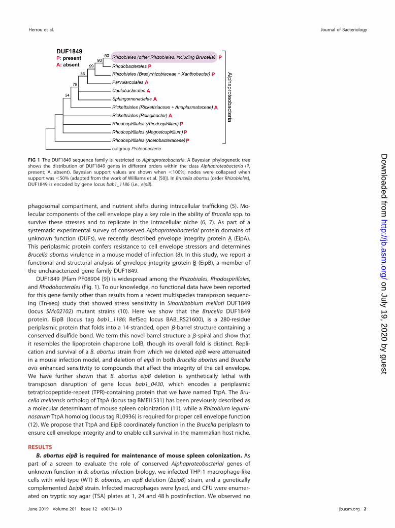
DUF1849 (Pfam PF08904 [9]) is widespread among the Rhizobiales, Rhodospirillales,and Rhodobacterales (Fig. 1). To our knowledge, no functional data have been reportedfor this gene family other than results from a recent multispecies transposon sequenc-ing (Tn-seq) study that showed stress sensitivity in Sinorhizobium meliloti DUF1849(locus SMc02102) mutant strains (10). Here we show that the Brucella DUF1849protein, EipB (locus tag bab1_1186; RefSeq locus BAB_RS21600), is a 280-residueperiplasmic protein that folds into a 14-stranded, open �-barrel structure containing aconserved disulfide bond. We term this novel barrel structure a �-spiral and show thatit resembles the lipoprotein chaperone LolB, though its overall fold is distinct. Repli-cation and survival of a B. abortus strain from which we deleted eipB were attenuatedin a mouse infection model, and deletion of eipB in both Brucella abortus and Brucellaovis enhanced sensitivity to compounds that affect the integrity of the cell envelope.We have further shown that B. abortus eipB deletion is synthetically lethal withtransposon disruption of gene locus bab1_0430, which encodes a periplasmictetratricopeptide-repeat (TPR)-containing protein that we have named TtpA. The Bru-cella melitensis ortholog of TtpA (locus tag BMEI1531) has been previously described asa molecular determinant of mouse spleen colonization (11), while a Rhizobium legumi-nosarum TtpA homolog (locus tag RL0936) is required for proper cell envelope function(12). We propose that TtpA and EipB coordinately function in the Brucella periplasm toensure cell envelope integrity and to enable cell survival in the mammalian host niche.
RESULTSB. abortus eipB is required for maintenance of mouse spleen colonization. As
part of a screen to evaluate the role of conserved Alphaproteobacterial genes ofunknown function in B. abortus infection biology, we infected THP-1 macrophage-likecells with wild-type (WT) B. abortus, an eipB deletion (ΔeipB) strain, and a geneticallycomplemented ΔeipB strain. Infected macrophages were lysed, and CFU were enumer-ated on tryptic soy agar (TSA) plates at 1, 24 and 48 h postinfection. We observed no
FIG 1 The DUF1849 sequence family is restricted to Alphaproteobacteria. A Bayesian phylogenetic treeshows the distribution of DUF1849 genes in different orders within the class Alphaproteobacteria (P,present; A, absent). Bayesian support values are shown when �100%; nodes were collapsed whensupport was �50% (adapted from the work of Williams et al. [50]). In Brucella abortus (order Rhizobiales),DUF1849 is encoded by gene locus bab1_1186 (i.e., eipB).
Herrou et al. Journal of Bacteriology
June 2019 Volume 201 Issue 12 e00134-19 jb.asm.org 2
on July 19, 2020 by guesthttp://jb.asm
.org/D
ownloaded from
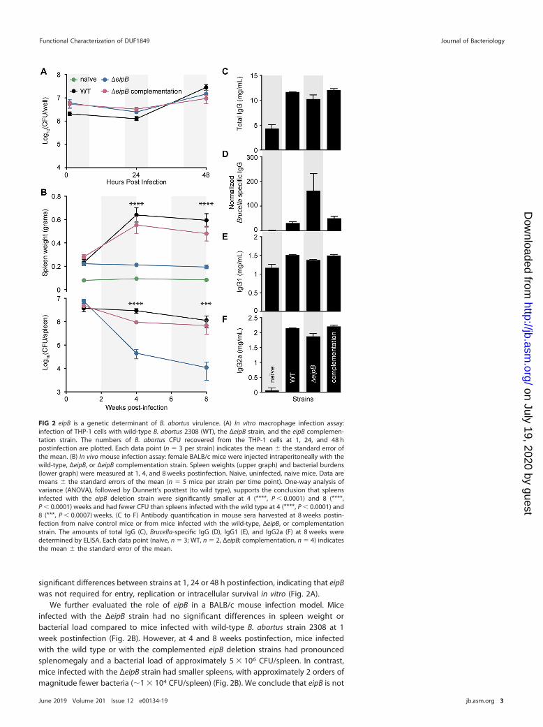
significant differences between strains at 1, 24 or 48 h postinfection, indicating that eipBwas not required for entry, replication or intracellular survival in vitro (Fig. 2A).
We further evaluated the role of eipB in a BALB/c mouse infection model. Miceinfected with the ΔeipB strain had no significant differences in spleen weight orbacterial load compared to mice infected with wild-type B. abortus strain 2308 at 1week postinfection (Fig. 2B). However, at 4 and 8 weeks postinfection, mice infectedwith the wild type or with the complemented eipB deletion strains had pronouncedsplenomegaly and a bacterial load of approximately 5 � 106 CFU/spleen. In contrast,mice infected with the ΔeipB strain had smaller spleens, with approximately 2 orders ofmagnitude fewer bacteria (�1 � 104 CFU/spleen) (Fig. 2B). We conclude that eipB is not
FIG 2 eipB is a genetic determinant of B. abortus virulence. (A) In vitro macrophage infection assay:infection of THP-1 cells with wild-type B. abortus 2308 (WT), the ΔeipB strain, and the eipB complemen-tation strain. The numbers of B. abortus CFU recovered from the THP-1 cells at 1, 24, and 48 hpostinfection are plotted. Each data point (n � 3 per strain) indicates the mean � the standard error ofthe mean. (B) In vivo mouse infection assay: female BALB/c mice were injected intraperitoneally with thewild-type, ΔeipB, or ΔeipB complementation strain. Spleen weights (upper graph) and bacterial burdens(lower graph) were measured at 1, 4, and 8 weeks postinfection. Naïve, uninfected, naive mice. Data aremeans � the standard errors of the mean (n � 5 mice per strain per time point). One-way analysis ofvariance (ANOVA), followed by Dunnett’s posttest (to wild type), supports the conclusion that spleensinfected with the eipB deletion strain were significantly smaller at 4 (****, P � 0.0001) and 8 (****,P � 0.0001) weeks and had fewer CFU than spleens infected with the wild type at 4 (****, P � 0.0001) and8 (***, P � 0.0007) weeks. (C to F) Antibody quantification in mouse sera harvested at 8 weeks postin-fection from naive control mice or from mice infected with the wild-type, ΔeipB, or complementationstrain. The amounts of total IgG (C), Brucella-specific IgG (D), IgG1 (E), and IgG2a (F) at 8 weeks weredetermined by ELISA. Each data point (naive, n � 3; WT, n � 2, ΔeipB; complementation, n � 4) indicatesthe mean � the standard error of the mean.
Functional Characterization of DUF1849 Journal of Bacteriology
June 2019 Volume 201 Issue 12 e00134-19 jb.asm.org 3
on July 19, 2020 by guesthttp://jb.asm
.org/D
ownloaded from
required for initial spleen colonization but is necessary for full virulence and persistencein the spleen over an 8-week time course.
To assess the pathology of mice infected with wild-type and ΔeipB strains, weharvested spleens at 8 weeks postinfection and fixed, mounted, and subjected thesamples to hematoxylin and eosin (H&E) staining (see Fig. S1 in the supplementalmaterial). Compared to naive (uninfected) mice (Fig. S1A), we observed higher ex-tramedullary hematopoiesis, histiocytic proliferation, granulomas, and the presence ofBrucella immunoreactivities in spleens of mice infected with wild-type B. abortus 2308and the genetically complemented mutant strain (Fig. S1B and D). Both the wild typeand the complemented strain caused spleen inflammation with a reduced white to redpulp ratio as a result of lymphoid follicle disruption and red pulp expansion, whichtypically correlates with infiltration of inflammatory cells; these spleens also hadincreased marginal zones (Fig. S1B and D). As expected from the CFU enumeration data,mice infected with the ΔeipB strain had reduced pathological features: there wasminimal change in the white/red pulp ratio and a minimal increase in marginal zones(Fig. S1C). There was no evidence of extramedullary hematopoiesis in mice infectedwith the ΔeipB strain, though histiocytic proliferation was mildly increased. Granulomasand Brucella immunoreactivities were rare in the ΔeipB strain (Fig. S1C). These resultsare consistent with a model in which eipB is required for full B. abortus virulence ina mouse model of infection. A summary of spleen pathology scores is presented inTable S1.
We further measured antibody responses in mice infected with ΔeipB and wild-typestrains. The serum levels of total IgG, Brucella-specific IgG, subclass IgG1, and subclassIgG2a were measured by enzyme-linked immunosorbent assays (ELISA) (Fig. 2C to F).Antibody subclasses IgG2a and IgG1 were measured as markers of T helper 1 (Th1)- andTh2-specific immune responses, respectively. At 8 weeks postinfection, the total serumIgG level was higher in all infected mice relative to the uninfected control (Fig. 2C). Thelevel of Brucella-specific IgG was approximately 5 times higher in ΔeipB strain-infectedmice than in mice infected with wild type or the complemented mutant strain (Fig. 2D).Uninfected mice and mice infected with wild-type, ΔeipB, and ΔeipB-complementedstrains showed no significant differences in IgG1 levels after 8 weeks (Fig. 2E). Allinfected mice had highly increased levels of IgG2a at 8 weeks postinfection relative tonaive mice, though there was no difference between B. abortus strains (Fig. 2F). Weconclude that ΔeipB strain infection results in the production of more B. abortus-specificantibodies than does infection with the wild type. Subclasses IgG1 and IgG2a do notapparently account for the higher levels of these specific antibodies. Large induction ofIgG2a by all B. abortus strains is consistent with the known ability of B. abortus topromote a strong Th1 response (13, 14). However, the ΔeipB strain does not induce amore robust Th1 response than does the wild type based on our IgG2a measurements.We did not test whether antibodies contribute to clearance of the ΔeipB strain.Enhanced Brucella-specific antibody production may simply be a consequence ofantigen release triggered by host clearance of the ΔeipB strain by other immunemechanisms.
The �eipB strain is sensitive to cell envelope stressors. To test whether reducedvirulence of the ΔeipB strain correlates with an increased sensitivity to stress in vitro, weevaluated B. abortus ΔeipB strain growth on TSA plates supplemented with known cellmembrane/envelope stressors, including EDTA, ampicillin, and deoxycholate. The ΔeipBstrain had 1.5 to 3 orders of magnitude fewer CFU compared to the wild type whentiters were determined on TSA plates containing these compounds. All phenotypeswere complemented by restoring the �eipB locus to the wild type (Fig. 3A). Together,these data provide evidence that eipB contributes to the resistance to compounds thatcompromise the integrity of the B. abortus cell membrane/envelope.
Although ΔeipB CFU were reduced relative to the wild type on agar plates contain-ing all three envelope stressors that we assayed, we observed no apparent defects inΔeipB cell morphology by light microscopy or cryo-electron microscopy when the strain
Herrou et al. Journal of Bacteriology
June 2019 Volume 201 Issue 12 e00134-19 jb.asm.org 4
on July 19, 2020 by guesthttp://jb.asm
.org/D
ownloaded from
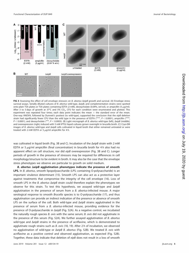
was cultivated in liquid broth (Fig. 3B and C). Incubation of the ΔeipB strain with 2 mMEDTA or 5 �g/ml ampicillin (final concentration) in brucella broth for 4 h also had noapparent effect on cell structure, nor did eipB overexpression (Fig. 3B and C). Longerperiods of growth in the presence of stressors may be required for differences in cellmorphology/structure to be evident in broth. It may also be the case that the envelopestress phenotypes we observe are particular to growth on solid medium.
B. abortus �eipB agglutination phenotypes indicate the presence of smoothLPS. In B. abortus, smooth lipopolysaccharide (LPS; containing O-polysaccharide) is animportant virulence determinant (15). Smooth LPS can also act as a protective layeragainst treatments that compromise the integrity of the cell envelope (16). Loss ofsmooth LPS in the B. abortus ΔeipB strain could therefore explain the phenotypes weobserve for this strain. To test this hypothesis, we assayed wild-type and ΔeipBagglutination in the presence of serum from a B. abortus-infected mouse. A majorserological response to smooth Brucella species is to O-polysaccharide (17), and thusagglutination can provide an indirect indication of the presence or absence of smoothLPS on the surface of the cell. Both wild-type and ΔeipB strains agglutinated in thepresence of serum from a B. abortus-infected mouse, providing evidence for thepresence of O-polysaccharide in ΔeipB (Fig. S2A). As a negative control, we incubatedthe naturally rough species B. ovis with the same serum; B. ovis did not agglutinate inthe presence of this serum (Fig. S2A). We further assayed agglutination of B. abortuswild-type and ΔeipB strains in the presence of acriflavine, which is demonstrated toagglutinate rough strains such as B. ovis (18, 19). After 2 h of incubation, we observedno agglutination of wild-type or ΔeipB B. abortus (Fig. S2B). We treated B. ovis withacriflavine as a positive control and observed agglutination, as expected (Fig. S2B).Together, these data indicate that deletion of eipB does not result in a loss of smooth
FIG 3 Assessing the effect of cell envelope stressors on B. abortus ΔeipB growth and survival. (A) Envelope stresssurvival assays. Serially diluted cultures of B. abortus wild-type, ΔeipB, and complementation strains were spottedonto plain TSA plates or TSA plates containing EDTA (2 mM), deoxycholate (0.04%, wt/vol), or ampicillin (5 �g/ml).After 3 to 5 days of growth at 37°C and 5% CO2, CFU for each condition were enumerated and plotted. Thisexperiment was repeated four times; each data point indicates the mean � the standard error of the mean.One-way ANOVA, followed by Dunnett’s posttest (to wild-type), supported the conclusion that the eipB deletionstrain had significantly fewer CFU than the wild type in the presence of EDTA (****, P � 0.0001), ampicillin (****,P � 0.0001), and deoxycholate (***, P � 0.0003). (B) Light micrograph of B. abortus wild-type (left), ΔeipB (middle),and overexpression (right; induced with 5 mM IPTG) liquid cultures grown overnight in brucella broth. (C) Cryo-EMimages of B. abortus wild-type and ΔeipB cells cultivated in liquid broth that either remained untreated or weretreated with 2 mM EDTA or 5 �g/ml ampicillin for 4 h.
Functional Characterization of DUF1849 Journal of Bacteriology
June 2019 Volume 201 Issue 12 e00134-19 jb.asm.org 5
on July 19, 2020 by guesthttp://jb.asm
.org/D
ownloaded from
LPS. However, we cannot rule out the possibility that the chemical structure ofO-polysaccharide is altered in the ΔeipB strain.
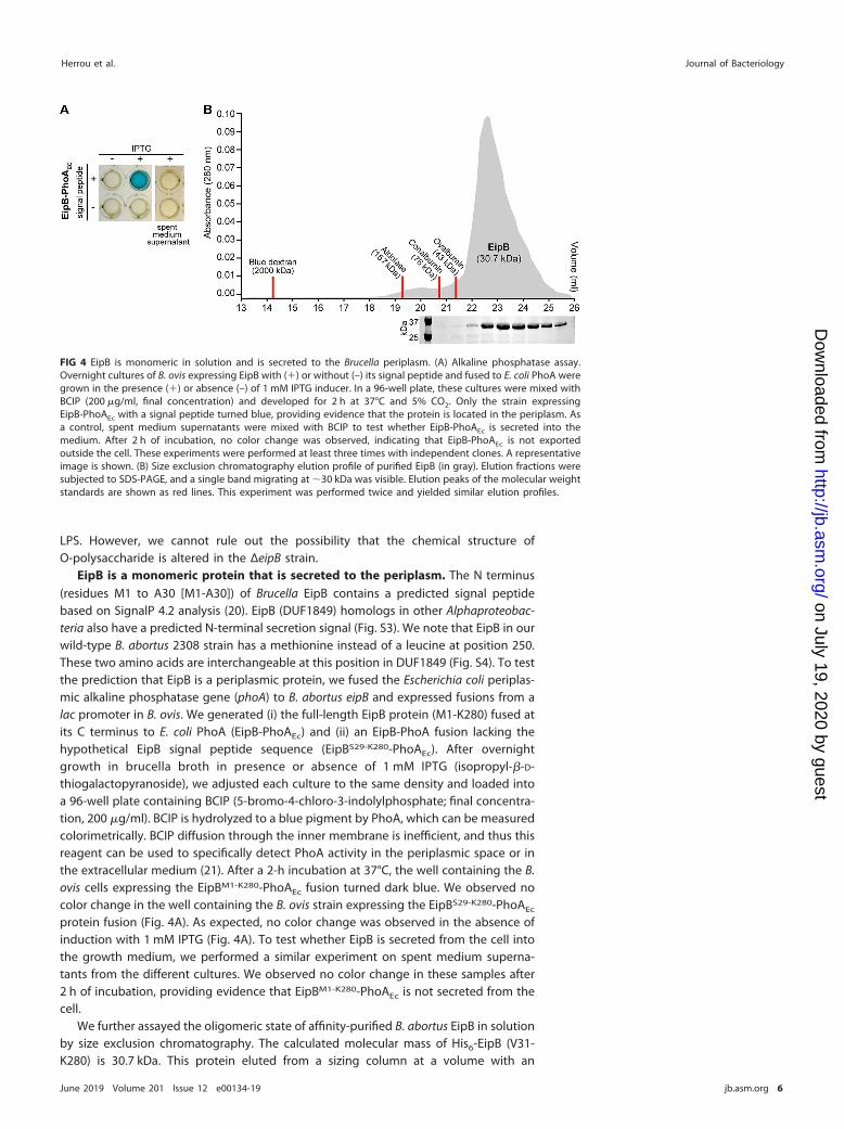
EipB is a monomeric protein that is secreted to the periplasm. The N terminus(residues M1 to A30 [M1-A30]) of Brucella EipB contains a predicted signal peptidebased on SignalP 4.2 analysis (20). EipB (DUF1849) homologs in other Alphaproteobac-teria also have a predicted N-terminal secretion signal (Fig. S3). We note that EipB in ourwild-type B. abortus 2308 strain has a methionine instead of a leucine at position 250.These two amino acids are interchangeable at this position in DUF1849 (Fig. S4). To testthe prediction that EipB is a periplasmic protein, we fused the Escherichia coli periplas-mic alkaline phosphatase gene (phoA) to B. abortus eipB and expressed fusions from alac promoter in B. ovis. We generated (i) the full-length EipB protein (M1-K280) fused atits C terminus to E. coli PhoA (EipB-PhoAEc) and (ii) an EipB-PhoA fusion lacking thehypothetical EipB signal peptide sequence (EipBS29-K280-PhoAEc). After overnightgrowth in brucella broth in presence or absence of 1 mM IPTG (isopropyl-�-D-thiogalactopyranoside), we adjusted each culture to the same density and loaded intoa 96-well plate containing BCIP (5-bromo-4-chloro-3-indolylphosphate; final concentra-tion, 200 �g/ml). BCIP is hydrolyzed to a blue pigment by PhoA, which can be measuredcolorimetrically. BCIP diffusion through the inner membrane is inefficient, and thus thisreagent can be used to specifically detect PhoA activity in the periplasmic space or inthe extracellular medium (21). After a 2-h incubation at 37°C, the well containing the B.ovis cells expressing the EipBM1-K280-PhoAEc fusion turned dark blue. We observed nocolor change in the well containing the B. ovis strain expressing the EipBS29-K280-PhoAEc
protein fusion (Fig. 4A). As expected, no color change was observed in the absence ofinduction with 1 mM IPTG (Fig. 4A). To test whether EipB is secreted from the cell intothe growth medium, we performed a similar experiment on spent medium superna-tants from the different cultures. We observed no color change in these samples after2 h of incubation, providing evidence that EipBM1-K280-PhoAEc is not secreted from thecell.
We further assayed the oligomeric state of affinity-purified B. abortus EipB in solutionby size exclusion chromatography. The calculated molecular mass of His6-EipB (V31-K280) is 30.7 kDa. This protein eluted from a sizing column at a volume with an
FIG 4 EipB is monomeric in solution and is secreted to the Brucella periplasm. (A) Alkaline phosphatase assay.Overnight cultures of B. ovis expressing EipB with (�) or without (–) its signal peptide and fused to E. coli PhoA weregrown in the presence (�) or absence (–) of 1 mM IPTG inducer. In a 96-well plate, these cultures were mixed withBCIP (200 �g/ml, final concentration) and developed for 2 h at 37°C and 5% CO2. Only the strain expressingEipB-PhoAEc with a signal peptide turned blue, providing evidence that the protein is located in the periplasm. Asa control, spent medium supernatants were mixed with BCIP to test whether EipB-PhoAEc is secreted into themedium. After 2 h of incubation, no color change was observed, indicating that EipB-PhoAEc is not exportedoutside the cell. These experiments were performed at least three times with independent clones. A representativeimage is shown. (B) Size exclusion chromatography elution profile of purified EipB (in gray). Elution fractions weresubjected to SDS-PAGE, and a single band migrating at �30 kDa was visible. Elution peaks of the molecular weightstandards are shown as red lines. This experiment was performed twice and yielded similar elution profiles.
Herrou et al. Journal of Bacteriology
June 2019 Volume 201 Issue 12 e00134-19 jb.asm.org 6
on July 19, 2020 by guesthttp://jb.asm
.org/D
ownloaded from
apparent molecular mass of �23 kDa, which is consistent with a monomer (Fig. 4B).There was no evidence of larger oligomers determined by size exclusion chromatog-raphy. From these data, we conclude that EipB is a monomeric periplasmic protein.
EipB folds into a spiral-like �-sheet that resembles PA1994, LolA, and LolB. Wepostulated that the three-dimensional structure of EipB may provide molecular-levelinsight into its function in the cell. As such, we solved an X-ray crystal structure of B.abortus EipB (residues A30 to K280; PDB ID 6NTR). EipB lacking its signal peptide formedtriclinic crystals (a � 47.4 Å, b � 69.2 Å, c � 83.2 Å, � � 90.1°, � � 90.0°, � � 78.7°) thatdiffracted to 2.1-Å resolution; we refined this structure to Rwork� 0.195 and Rfree�
0.245. Crystallographic data and refinement statistics are summarized in Table S2. FourEipB molecules (chains A to D) are present in the crystallographic asymmetric unit.
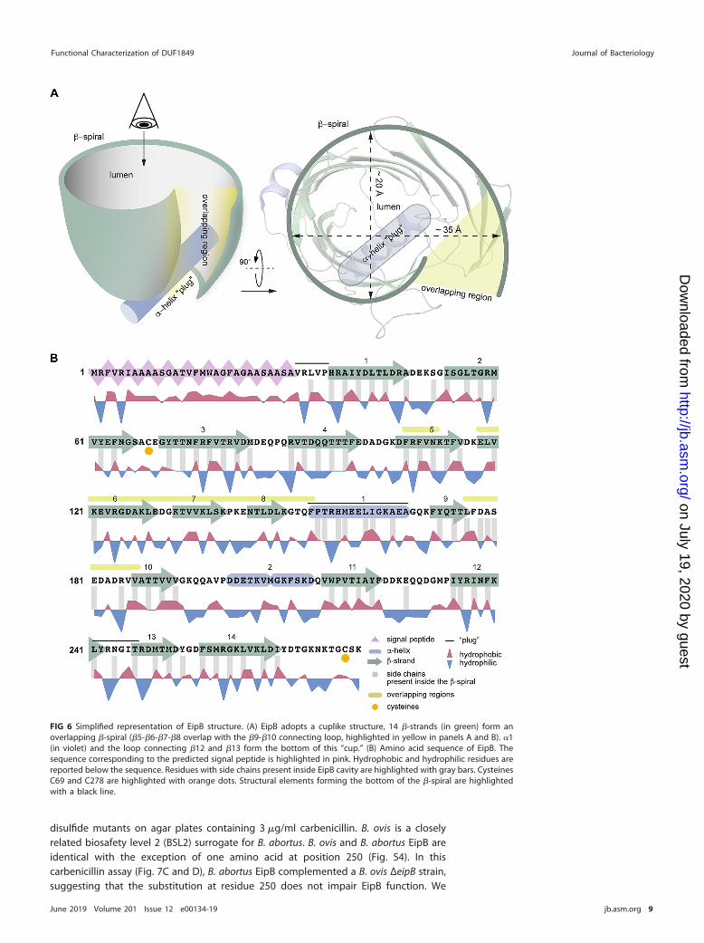
Each EipB monomer consists of 14 antiparallel �-strands (�1 to �14) forming an oval,spiral-like �-sheet (minor axis diameter, �25 Å; major axis diameter, �35 Å). Tworegions of this �-spiral, involving �5, �6, �7, and �8, and the hairpin loop connecting�9 and �10 overlap (Fig. 5). Interactions between these two overlapping portions ofstructure are mostly hydrophobic, though polar contacts are also found in theseregions (Fig. 5 and 6). One side of the spiral is occluded by the N terminus, a loopconnecting �-strands 12 and 13, and �-helix 1, which form the bottom of this “cup”shaped protein (Fig. 5 and 6A). The external surface of EipB is positively and negativelycharged and also presents small hydrophobic patches (Fig. S5); one helix, �2, is kinkedand positioned at the surface of the cylindrical �-spiral (Fig. 5). The lumen of EipB issolvent accessible and is partially filled with the side chains of hydrophobic or acidicresidues. Hydrophobic residues represent �66% of the residues present inside the EipBcavity (Fig. 5 and 6B). The size of this cavity suggests that EipB, in this conformation, canaccommodate small molecules or ligands in its lumen.
We searched the EipB structure against the protein structure database using Dali(22) but failed to identify clear structural homologs. Pseudomonas aeruginosa PA1994(PDB ID 2H1T) (23) was the closest structural match to EipB (root mean squaredeviation, �3.5; Z-score, �11) (Fig. S6A). Despite very low sequence identity (�8%),PA1994 has noticeable structural similarities to EipB: it adopts a spiral-like �-foldinvolving 15 �-strands, which is occluded at one end with a long �-helix. Unlike EipB,PA1994 lacks a signal peptide and is predicted to be a cytoplasmic protein. Structuralparallels between PA1994 and the periplasmic lipoprotein chaperones LolA/LolB havebeen noted and a role for PA1994 in glycolipid metabolism has been postulated (23),though this prediction remains untested. Like PA1994, EipB has structural similarities toLolA and LolB, in particular the antiparallel and curved �-sheet scaffold that engulfs acentral �-helical plug (Fig. S6B). Whether Brucella EipB or DUF1849 proteins moregenerally function in trafficking lipoproteins or other molecules in the periplasmremains to be tested.
EipB has a conserved disulfide bond. We identified two cysteines in EipB, C69 andC278, which are the two most conserved residues in the DUF1849 sequence family (Fig.S3 and S4). C69 is solvent exposed in Brucella EipB and positioned in a loop connecting�2 and �3. C278 is present at the C terminus of the protein, which immediately follows�14. �14 interacts with �13 and �1 and is spatially proximal to �2 and �3 (Fig. 7A).Given the proximity of these two cysteines in the EipB structure, we hypothesized thatC69 and C278 form an internal disulfide bond. However, electron density for the10 C-terminal residues (containing C278) is not well resolved in the EipB crystal struc-ture, and a disulfide bond is not evident, likely because the protein was dialyzed againsta buffer containing 2 mM 1,4-dithiothreitol (DTT) prior to crystallization.
To biochemically test whether these two cysteines form a disulfide bond, wepurified B. abortus EipB under nonreducing conditions and mixed the protein withsodium dodecyl sulfate (SDS) gel loading dye with or without 1 mM DTT. We observedtwo bands that migrated differently in the 30-kDa region when the protein wasresolved by 12% SDS-PAGE. EipB without DTT migrated farther than the DTT-treatedprotein, suggesting the presence of a disulfide bond (Fig. 7B). We performed this same
Functional Characterization of DUF1849 Journal of Bacteriology
June 2019 Volume 201 Issue 12 e00134-19 jb.asm.org 7
on July 19, 2020 by guesthttp://jb.asm
.org/D
ownloaded from

experiment with three different EipB cysteine mutant proteins in which C69, C278, orboth were mutated to serine. In the absence of DTT, EipBC69S and EipBC278S migratedat an apparent molecular weight of �60 kDa, corresponding to a dimeric EipB inter-acting through a S-S bond. After DTT treatment, these mutant proteins migrated thesame as the reduced wild-type protein (Fig. 7B). As expected, the double cysteinemutant (EipBC69S�C278S) did not form an apparent dimer and was unaffected by DTT(Fig. 7B). From these data, we conclude that an internal disulfide bond can formbetween C69 and C278 in EipB and is likely present in vivo, since EipB resides in theoxidizing environment of the periplasm.
To test whether this disulfide bond affects EipB function, we measured CFU of aBrucella ovis ΔeipB (Δbov_1121) strain expressing wild-type B. abortus EipB or cysteine
FIG 5 EipB adopts a �-spiral fold. (A, left) X-ray structure of EipB. EipB consist of 14 �-strands (in green) and 2 �-helices(in violet). The N terminus (A30) and the C terminus (D270) are reported on this structure. (Right) Simplified representationof EipB. The color code is the same as for the left panel. (B) Different orientations of EipB structure. The color code is thesame as for panel A.
Herrou et al. Journal of Bacteriology
June 2019 Volume 201 Issue 12 e00134-19 jb.asm.org 8
on July 19, 2020 by guesthttp://jb.asm
.org/D
ownloaded from
disulfide mutants on agar plates containing 3 �g/ml carbenicillin. B. ovis is a closelyrelated biosafety level 2 (BSL2) surrogate for B. abortus. B. ovis and B. abortus EipB areidentical with the exception of one amino acid at position 250 (Fig. S4). In thiscarbenicillin assay (Fig. 7C and D), B. abortus EipB complemented a B. ovis ΔeipB strain,suggesting that the substitution at residue 250 does not impair EipB function. We
FIG 6 Simplified representation of EipB structure. (A) EipB adopts a cuplike structure, 14 �-strands (in green) form anoverlapping �-spiral (�5-�6-�7-�8 overlap with the �9-�10 connecting loop, highlighted in yellow in panels A and B). �1(in violet) and the loop connecting �12 and �13 form the bottom of this “cup.” (B) Amino acid sequence of EipB. Thesequence corresponding to the predicted signal peptide is highlighted in pink. Hydrophobic and hydrophilic residues arereported below the sequence. Residues with side chains present inside EipB cavity are highlighted with gray bars. CysteinesC69 and C278 are highlighted with orange dots. Structural elements forming the bottom of the �-spiral are highlightedwith a black line.
Functional Characterization of DUF1849 Journal of Bacteriology
June 2019 Volume 201 Issue 12 e00134-19 jb.asm.org 9
on July 19, 2020 by guesthttp://jb.asm
.org/D
ownloaded from

FIG 7 EipB has an internal disulfide bond. (A) Cysteines C69 and C278 are spatially proximal in the EipB structureand form a disulfide bond. C278 is present at the EipB C terminus that follows �14, and C69 is present in a loopconnecting �2 and �3. (B) His-tagged wild-type EipB and EipB cysteine mutant proteins (C69S, C278S, andC69S�C278S) were purified and mixed with a protein loading buffer with (�) or without () 1 mM DTT. Proteinsamples were resolved by SDS–12% PAGE. This experiment was performed three times. The picture of arepresentative gel is presented. (C) Growth on SBA plates containing 3 �g/ml of carbenicillin with (�) or without(–) 2 mM IPTG of a serially diluted (10-fold dilution) B. ovis ΔeipB strain ectopically expressing wild-type EipB(Plac-eipB), C69S mutant (Plac-eipBC69S), C278S mutant (Plac-eipBC278S), or C69S�C278S mutant (Plac-eipBC69S�C278S). B.ovis wild-type (WT) and ΔeipB carrying the pSRK empty vector (EV) were used as a control. The days of growth at37°C and 5% CO2 are reported for each plate. A representative picture of the different plates is presented. (D)Enumerated CFU after growth on SBA plates containing 3 �g/ml of carbenicillin with (�) or without (–) 2 mM IPTGof serially diluted (10-fold dilution) B. ovis ΔeipB strains expressing different versions of eipB from a plasmid (wildtype and cysteine mutants; see panel C legend). EV strains and SBA plates with no carbenicillin, with or withoutIPTG, were used as controls. This experiment was independently performed twice, with two different clones eachtime, and all plate assays were done in triplicate. Each data point indicates the mean � the standard error of themean. One-way ANOVA, followed by Dunnett’s posttest (to the wild type), supports the conclusion that eipB-
(Continued on next page)
Herrou et al. Journal of Bacteriology
June 2019 Volume 201 Issue 12 e00134-19 jb.asm.org 10
on July 19, 2020 by guesthttp://jb.asm
.org/D
ownloaded from
placed four different versions of eipB under the control of a lac promoter (Plac):Plac-eipBWT, Plac-eipBC69S, Plac-eipBC278S, and Plac-eipBC69S�C278S; the empty vector (EV)was used as a control. After 5 to 6 days of growth on Schaedler blood agar (SBA) platescontaining 3 �g/ml of carbenicillin and no IPTG, we observed poor growth at only thelowest dilution for wild-type and ΔeipB strains carrying the empty vector control (alsosee Fig. S7A for an example of growth on 2 �g/ml carbenicillin plates). Correspondingcolonies for the strains carrying the different Plac-eipB overexpression plasmids weremore abundant though very small in the absence of IPTG induction. However, the strainharboring the wild-type eipB plasmid systematically grew at a 1-log-higher dilutionthan the cysteine mutant strains, indicating that the presence of the disulfide bond ineipB contributes to carbenicillin resistance on solid medium (Fig. 7C and D; see also Fig.S7A). These results indicate some level of leaky expression from the multicopy Plac-eipBplasmids. When induced with IPTG, overexpression of the different EipB variantsenhanced growth in all strains (Fig. 7C and D). As expected, strains grown on controlplates without carbenicillin had no growth defect, with or without IPTG induction (Fig.7D). The morphology of B. ovis �eipB strains expressing the different variants of eipBappeared normal by phase-contrast microscopy (see Fig. S7B). These results provideevidence that EipB is necessary for full carbenicillin resistance in B. ovis and thatcysteines 69 and 278 contribute to EipB function in vivo.
To evaluate the effect of these two cysteines on EipB stability in vitro, we measuredthe thermal stability of purified wild-type B. abortus EipB (EipBWT) and double cysteinemutant (EipBC69S�C278S) in presence or absence of 2 mM DTT. EipBWT melted at �46°Cin the absence of DTT and at �41.5°C in the presence of DTT. EipBC69S�C278S melted at�42.3°C in the presence or absence of DTT (see Fig. S8). We conclude that an internaldisulfide bond stabilizes EipB structure in vitro. Reduced stability of EipB lacking itsconserved disulfide bond may contribute to the 1-log relative growth defect of ΔeipBstrains expressing EipB cysteine mutants on SBA carbenicillin plates (Fig. 7C and D).
eipB deletion is synthetically lethal with bab1_0430 (ttpA) disruption andsynthetically sick with disruption of multiple genes with cell envelope functions.To further characterize how eipB functions in the Brucella cell, we aimed to identifytransposon (Tn) insertion mutations that are synthetically lethal with eipB deletion in B.abortus (see Tables S3 and S4). In other words, we sought to discover genes that aredispensable in a wild-type genetic background but that cannot be disrupted in a �eipBbackground. By sequencing a Tn-Himar insertion library generated in B. abortus �eipB(NCBI Sequence Read Archive accession number SRR8322167) and a Tn-Himar librarygenerated in wild-type B. abortus (NCBI Sequence Read Archive accession numberSRR7943723), we uncovered evidence that disruption of bab1_0430 (RefSeq locusBAB_RS17965) is synthetically lethal with eipB deletion. Specifically, reproducible readscorresponding to insertions in the central 10 to 90% of bab1_0430 were not evident in�eipB but were present in wild-type (Fig. 8A). bab1_0430 encodes a 621-residuetetratricopeptide repeat-containing (TPR) protein with a predicted signal peptide andsignal peptidase site at its N terminus. This protein was previously detected by massspectrometry analyses of B. abortus extracts and was described as a cell envelope-associated protein (24) or periplasmic protein (25). Here, we refer to this gene as ttpA(tetratricopeptide repeat protein A gene) based on its similarity to Rhizobium legumi-nosarum ttpA (12).
Genes involved in LPS O-antigen synthesis and previously described as syntheticlethal with eipA (bab1_1612) deletion in B. abortus (8) were synthetic sick with eipBdeletion (Fig. 8A), as were genes involved in peptidoglycan synthesis: mltA (bab1_2076,lytic murein transglycosylase A) and bab1_0607 (glycosyl transferase/penicillin-binding
FIG 7 Legend (Continued)dependent protection against the cell wall antibiotic, carbenicillin, is significantly diminished when disulfide-forming residues C69 (**, P � 0.005) and C278 (**, P � 0.003) are individually or both (*, P � 0.01) mutated to serine.This is effect is evident with leaky eipB expression from Plac but diminished when the expression of wild-type andmutant eipB alleles is induced by IPTG.
Functional Characterization of DUF1849 Journal of Bacteriology
June 2019 Volume 201 Issue 12 e00134-19 jb.asm.org 11
on July 19, 2020 by guesthttp://jb.asm
.org/D
ownloaded from
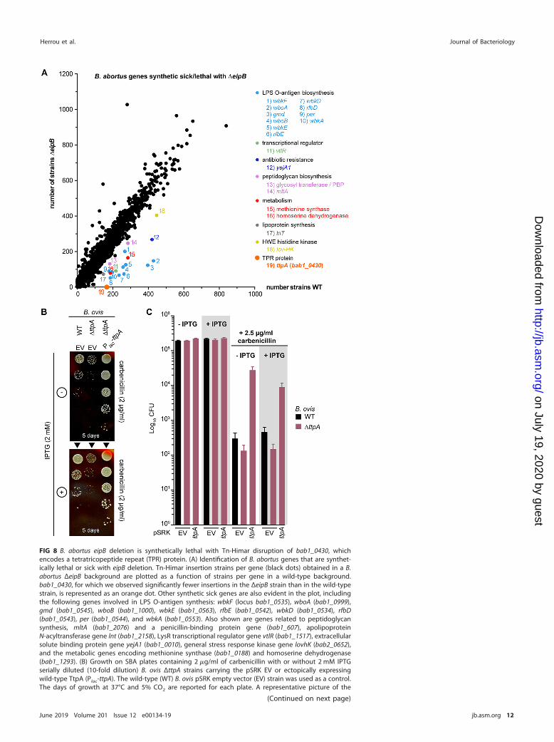
FIG 8 B. abortus eipB deletion is synthetically lethal with Tn-Himar disruption of bab1_0430, whichencodes a tetratricopeptide repeat (TPR) protein. (A) Identification of B. abortus genes that are synthet-ically lethal or sick with eipB deletion. Tn-Himar insertion strains per gene (black dots) obtained in a B.abortus ΔeipB background are plotted as a function of strains per gene in a wild-type background.bab1_0430, for which we observed significantly fewer insertions in the ΔeipB strain than in the wild-typestrain, is represented as an orange dot. Other synthetic sick genes are also evident in the plot, includingthe following genes involved in LPS O-antigen synthesis: wbkF (locus bab1_0535), wboA (bab1_0999),gmd (bab1_0545), wboB (bab1_1000), wbkE (bab1_0563), rfbE (bab1_0542), wbkD (bab1_0534), rfbD(bab1_0543), per (bab1_0544), and wbkA (bab1_0553). Also shown are genes related to peptidoglycansynthesis, mltA (bab1_2076) and a penicillin-binding protein gene (bab1_607), apolipoproteinN-acyltransferase gene lnt (bab1_2158), LysR transcriptional regulator gene vtlR (bab1_1517), extracellularsolute binding protein gene yejA1 (bab1_0010), general stress response kinase gene lovhK (bab2_0652),and the metabolic genes encoding methionine synthase (bab1_0188) and homoserine dehydrogenase(bab1_1293). (B) Growth on SBA plates containing 2 �g/ml of carbenicillin with or without 2 mM IPTGserially diluted (10-fold dilution) B. ovis ΔttpA strains carrying the pSRK EV or ectopically expressingwild-type TtpA (Plac-ttpA). The wild-type (WT) B. ovis pSRK empty vector (EV) strain was used as a control.The days of growth at 37°C and 5% CO2 are reported for each plate. A representative picture of the
(Continued on next page)
Herrou et al. Journal of Bacteriology
June 2019 Volume 201 Issue 12 e00134-19 jb.asm.org 12
on July 19, 2020 by guesthttp://jb.asm
.org/D
ownloaded from
protein 1A) (26) (Fig. 8A). There were reduced transposon insertions in solute bindingprotein gene yejA1 (bab1_0010) (Fig. 8A), which is involved in B. melitensis resistance topolymyxin (27). lnt (bab1_2158) and vtlR (bab1_1517) were also synthetic sick with theΔeipB strain. lnt is an apolipoprotein N-acyltransferase gene involved in lipoproteinsynthesis (28); vtlR encodes a LysR transcriptional regulator required for full B. abortusvirulence (29) (Fig. 8A). Finally, the general stress sensor kinase gene lovHK (bab2_0652)(30), bab1_1293 (homoserine dehydrogenase), and bab1_0188 (methionine synthase)had fewer Tn insertions in the ΔeipB background relative to the wild type (Fig. 8A).
ttpA contributes to carbenicillin resistance. As ttpA disruption is synthetic lethalwith eipB deletion, we postulated that these two genes have complementary functionsor are involved in a common physiological process (i.e., envelope integrity). Thus, tocharacterize ttpA and the nature of its connection to eipB, we deleted ttpA in B. ovis andevaluated its sensitivity to carbenicillin. All efforts to delete B. ovis ttpA (locus tagbov_0411) using a classic crossover recombination and sacB counterselection approachwere unsuccessful, though hundreds of clones were screened. Efforts to delete thechromosomal copy by expressing a copy of ttpA from a plasmid also failed. This resultis surprising considering that transposon insertions in B. abortus ttpA (NCBI SequenceRead Archive accession number SRR7943723) and B. ovis ttpA (NCBI Sequence ReadArchive accession number SRR7943724) are tolerated in wild-type backgrounds (8). Asan alternative approach to study the function of this gene, we inactivated ttpA using asingle crossover recombination strategy. The resulting strain expressed a truncatedversion of TtpA containing the first 205 amino acids (including the signal peptide),immediately followed by 22 amino acids from the suicide plasmid. The correspondingB. ovis strain (ΔttpA) was then transformed with a plasmid-borne IPTG-inducible copy ofttpA (pSRK-ttpA) or with an empty plasmid vector (EV). We evaluated the sensitivity ofthese strains to carbenicillin by plating a dilution series on SBA plates containing 2 or2.5 �g/ml carbenicillin, with or without IPTG inducer (Fig. 8B and C). Compared to thewild-type strain with an empty vector, the B. ovis ΔttpA strain with an empty vector had�0.5-log-unit-reduced CFU on carbenicillin SBA. The corresponding colonies of the B.ovis ΔttpA strain were noticeably smaller than the wild-type colonies. Genetic comple-mentation of the ΔttpA strain with pSRK-ttpA restored growth on carbenicillin plates. B.ovis ΔttpA/pSRK-ttpA had �1.5-log-units more colonies than did the wild type in thepresence of carbenicillin, with or without IPTG induction. Thus, leaky expression of ttpAfrom the lac promoter on pSRK-ttpA is apparently sufficient to protect this strain fromcarbenicillin on solid medium. The morphology of the B. ovis �ttpA strains appearednormal by phase-contrast microscopy at �630 magnification (Fig. S9).
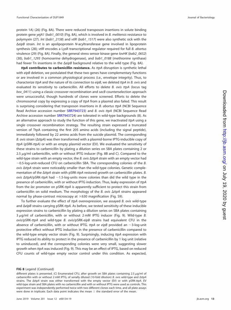
To further evaluate the effect of ttpA overexpression, we assayed B. ovis wild-typeand ΔeipB strains carrying pSRK-ttpA. As before, we tested sensitivity of these inducibleexpression strains to carbenicillin by plating a dilution series on SBA plates containing3 �g/ml of carbenicillin, with or without 2 mM IPTG inducer (Fig. 9). Wild-type B.ovis/pSRK-ttpA and wild-type B. ovis/pSRK-eipB strains had equivalent CFU in theabsence of carbenicillin, with or without IPTG. ttpA or eipB provided an �3-log-unitprotective effect without IPTG induction in the presence of carbenicillin compared tothe wild-type empty vector strain (Fig. 9). Surprisingly, inducing ttpA expression withIPTG reduced its ability to protect in the presence of carbenicillin by 1 log unit (relativeto uninduced), and the corresponding colonies were very small, suggesting slowergrowth when ttpA was induced (Fig. 9). This may be an effect of IPTG, based on reducedCFU counts of wild-type empty vector control under this condition. As expected,
FIG 8 Legend (Continued)different plates is presented. (C) Enumerated CFU, after growth on SBA plates containing 2.5 �g/ml ofcarbenicillin with or without 2 mM IPTG, of serially diluted (10-fold dilution) B. ovis wild-type and ΔttpAstrains. The ΔttpA strain was either transformed with the empty vector (EV) or with pSRK-ttpA. EVwild-type strain and SBA plates with no carbenicillin and with or without IPTG were used as controls. Thisexperiment was independently performed twice with two different clones each time, and all plate assayswere done in triplicate. Each data point indicates the mean � the standard error of the mean.
Functional Characterization of DUF1849 Journal of Bacteriology
June 2019 Volume 201 Issue 12 e00134-19 jb.asm.org 13
on July 19, 2020 by guesthttp://jb.asm
.org/D
ownloaded from
induced expression of eipB from Plac-eipB rescued the carbenicillin viability defect of�eipB. However, induced expression of ttpA from Plac-ttpA was not sufficient to rescuethe �eipB carbenicillin phenotype (Fig. 9). As before, we observed highly reduced CFUfor B. ovis wild-type or ΔeipB control strains carrying the pSRK empty vector (EV), whenchallenged with 3 �g/ml of carbenicillin. The morphology of wild-type or �eipB B. ovisstrains overexpressing ttpA appeared normal by phase-contrast microscopy at �630magnification (Fig. S10).
The observed genetic interaction between eipB and ttpA, the fact that both singlemutants have envelope phenotypes, and the fact that both gene products are secretedto the periplasm raised the possibility that EipB and TtpA physically interact. We testedinteraction between EipB and TtpA proteins using bacterial two-hybrid and biochem-ical pulldown assays. We further evaluated whether a possible EipB-TtpA interaction isinfluenced by the presence or absence of the EipB internal disulfide bond using abiochemical pulldown. For our bacterial two-hybrid assay, EipBV31-K280 was fused to theT25 adenylate cyclase fragment, and TtpAK31-D621 was fused to the T18 or T18Cadenylate cyclase fragments. For the pulldown assay, MBP-tagged TtpA (K31-D621) andHis-tagged EipB (V31-K280; wild type and the different cysteine mutants) were copu-rified in the presence or absence of DTT. We found no evidence for direct interactionbetween EipB and TtpA, suggesting that the function of these two proteins in Brucellaenvelope stress adaptation is not achieved through direct interaction (Fig. S11).
DISCUSSION
Bacterial genome sequencing efforts over the past 2 decades have revealed thou-sands of protein domains of unknown function (DUFs). The DUF1849 sequence familyis prevalent in the orders Rhizobiales, Rhodobacterales, and Rhodospirillales. To date, thefunction of DUF1849 has remained undefined. We have shown that a DUF1849 gene in
FIG 9 Overexpression of TtpA protects against carbenicillin treatment; protection requires EipB. (A) Growth on SBAplates containing 3 �g/ml of carbenicillin with or without 2 mM IPTG of serially diluted (10-fold dilution) B. oviswild-type (WT) and ΔeipB strains expressing wild-type EipB (Plac-eipB) or TtpA (Plac-ttpA). B. ovis strains carrying thepSRK empty vector were used as a control. The days of growth at 37°C and 5% CO2 are reported for each plate.A representative picture of the different plates is presented. (B) Enumerated CFU after growth on SBA platescontaining 3 �g/ml of carbenicillin with or without 2 mM IPTG of serially diluted (10-fold dilution) B. ovis wild-typeand ΔeipB strains ectopically expressing eipB or ttpA. EV strains and SBA plates with no carbenicillin and with orwithout IPTG were used as controls. This experiment was independently performed twice with two different cloneseach time, and all plate assays were done in triplicate. Each data point indicates the mean � the standard errorof the mean.
Herrou et al. Journal of Bacteriology
June 2019 Volume 201 Issue 12 e00134-19 jb.asm.org 14
on July 19, 2020 by guesthttp://jb.asm
.org/D
ownloaded from
Brucella spp., which we have named eipB, encodes a 14-stranded �-spiral protein thatis secreted to the periplasm. eipB is required for maintenance of B. abortus spleencolonization in a mouse model of infection (Fig. 2), and eipB deletion in B. abortus andin B. ovis results in sensitivity to treatments that compromise the integrity of the cellenvelope in vitro (Fig. 3). The envelope stress sensitivity of the B. abortus ΔeipB mutantlikely contributes to its reduced virulence in a mouse. We further demonstrate that EipBcontains a conserved disulfide bond that contributes to protein stability and functionin vitro; the importance of this conserved disulfide to EipB function in vivo remains tobe determined (Fig. 6 and 7; see also Fig. S3 and S4 in the supplemental material).
A lipoprotein connection? An X-ray crystal structure of EipB shows that thisperiplasmic protein adopts an unusual �-spiral fold that shares structural similarity(DALI Z-score, 11.0) with a functionally uncharacterized P. aeruginosa protein, PA1994,despite low sequence identity (Fig. S6). It was previously noted (23) that PA1994 hasstructural features that resemble the lipoprotein carrier and chaperone proteins LolAand LolB, which have a central role in lipoprotein localization in select Gram-negativebacteria (31). Like LolA, LolB, and PA1994, Brucella EipB forms a curved hydrophobic�-sheet that is wrapped around an �-helix (Fig. S6B). Homologs of LolA are present inBrucella and other Alphaproteobacteria, but homologs of LolB are missing (28). Giventhe EipB structure, its periplasmic localization, and the phenotypes of a ΔeipB deletionstrain, it is tempting to speculate that EipB (DUF1849) has a LolB-like function in theBrucella cell. However, it seems unlikely that LolB and EipB function in a structurally orbiochemically equivalent manner. Certainly, we observe surface level similarity be-tween LolA/LolB and EipB structures (Fig. S6), particularly in the antiparallel �-sheetregion, but these proteins have topological differences that distinguish their folds.Moreover, LolB is a membrane anchored lipoprotein that facilitates lipoprotein target-ing at the inner leaflet of the outer membrane. In contrast, Brucella EipB does not havea predicted site for lipidation (i.e., a lipobox) and is therefore unlikely to function as amembrane-anchored protein.
The number of unique barcoded Tn-Himar insertions in the apolipoproteinN-acyltransferase gene lnt (bab1_2158; lnt conserved domain database score � e173)is lower than expected in a ΔeipB background relative to the wild type (Fig. 8A). Thisprovides indirect evidence for a link between eipB and lipoproteins. Lnt catalyzes thefinal acylation step in lipoprotein biogenesis (32), which is often considered to be anessential cellular process. However, like Francisella tularensis and Neisseria gonorrhoeae(33), B. abortus lnt is dispensable (26) (Fig. 8A and Table S4). The data presented heresuggest that transposon insertions are less tolerated in B. abortus lnt when eipB ismissing. Additional experimentation is required to test a possible functional relation-ship between lnt and eipB. However, it is notable that we did not observe a syntheticgenetic interaction between lnt and the gene encoding a structurally unrelatedperiplasmic envelope integrity protein, EipA, in a parallel Tn-seq experiment (8).Whether eipB actually influences lipoprotein biogenesis or localization remains to betested.
TtpA: a periplasmic determinant of cell envelope function in Rhizobiaceae.Transposon disruption of ttpA (bab1_0430) is not tolerated when eipB is deleted in B.abortus. ttpA, like eipB, contributes to carbenicillin resistance in vitro (Fig. 8 and 9).Though we observed a genetic interaction between eipB and ttpA, we found noevidence for a direct physical interaction between the two periplasmic proteins en-coded by these genes (Fig. S11). TtpA is named for its tetratricopeptide repeat (TPR)motif; proteins containing TPR motifs are known to function in many different pathwaysin bacteria, including cell envelope biogenesis, and are often molecular determinants ofvirulence (34, 35). Indeed, deletion of ttpA has been reported to attenuate B. melitensisvirulence in a mouse infection model of infection (11) and to increase R. leguminosarummembrane permeability and sensitivity to SDS and hydrophobic antibiotics (12). Agenetic interaction between ttpA and the complex media growth deficient (cmdA-cmdD) operon has been reported in R. leguminosarum. Mutations in this operon resultin envelope dysfunction and defects in cell morphology (12, 36). While B. abortus
Functional Characterization of DUF1849 Journal of Bacteriology
June 2019 Volume 201 Issue 12 e00134-19 jb.asm.org 15
on July 19, 2020 by guesthttp://jb.asm
.org/D
ownloaded from
contains a predicted cmd operon (bab1_1573, bab1_1574, bab1_1575, and bab1_1576),these genes remain uncharacterized. We found no evidence for a synthetic geneticinteraction between eipB and cmd in B. abortus.
Leaky expression of either eipB or ttpA from a plasmid strongly protected B. ovis froma cell wall antibiotic (carbenicillin). Surprisingly, inducing ttpA expression from aplasmid with IPTG did not protect as well as uninduced (i.e., leaky) ttpA expression (Fig.9). IPTG induction of eipB expression from a plasmid did not have this same paraboliceffect on cell growth/survival in the face of carbenicillin treatment. Considering thatEipB and TtpA confer resistance to �-lactam antibiotics, which perturb peptidoglycansynthesis, one might hypothesize that these proteins influence the structure or syn-thesis of the cell wall. This hypothesis is reinforced by the fact that a lytic mureintransglycosylase and a class A PBP/glycosyl transferase are synthetic sick with eipBdeletion (Fig. 8A). In Escherichia coli, the TPR-containing protein LpoA is proposed toreach from the outer membrane through the periplasm to interact with the pepti-doglycan synthase PBP1A (37). Models in which EipB and TtpA influence lipoproteinbiosynthesis and/or cell wall metabolism are important to test as we work towardunderstanding the mechanisms by which these genes ensure Brucella cell envelopeintegrity and survival in a mammalian host.
MATERIALS AND METHODSAgglutination assays, mouse and macrophage infection assays, antibody measurements, and the
transposon sequencing experiments for this study were performed in parallel with our recent studies ofeipA (8).
All experiments using live B. abortus 2308 were performed in biosafety level 3 facilities according toU.S. Centers for Disease Control and Prevention (CDC) select agent regulations at the University ofChicago Howard Taylor Ricketts Laboratory. All the B. abortus and B. ovis strains were cultivated at 37°Cwith 5% CO2; primer and strain information are available in Table S5 in the supplemental material.
Chromosomal deletions in B. abortus and in B. ovis. The B. abortus and B. ovis ΔeipB deletionstrains were generated using a double-crossover recombination strategy as previously described (8).Briefly, fragments corresponding to the 500-bp region upstream of the eipB start codon and the 500-bpregion downstream of the eipB stop codon were ligated into the suicide plasmid pNPTS138, which carriesthe nptI gene for initial kanamycin selection and the sacB gene for counterselection on sucrose. Geneticcomplementation of the B. abortus deletion strain was carried out by transforming this strain with apNPTS138 plasmid carrying the wild-type allele. The B. ovis �eipB strain was complemented with thepSRK-eipB plasmid (IPTG inducible).
To inactivate ttpA in B. ovis (bov_0411), a 527-nucleotide internal fragment was cloned intopNPTS138-cam (a suicide plasmid that we engineered to carry a chloramphenicol resistance marker) andused to disrupt the target gene by single crossover insertion. The recombinant clones were selected onSBA plates supplemented with 3 �g/ml chloramphenicol. The corresponding strain expresses the first205 amino acids (including the signal peptide) of TtpA, plus 22 extra amino acids from the plasmidsequence, followed by a stop signal. This ΔttpA strain was complemented with pSRK-ttpA (kanamycinresistant).
Brucella EipB and TtpA overexpression strains. For ectopic expression of B. ovis TtpA and thedifferent versions of B. abortus EipB (wild-type, cysteine mutants, and the EipB-PhoAEc fusion with orwithout the signal peptide), the pSRKKm (Kanr) IPTG inducible plasmid was used (38). An overlappingPCR strategy was used to introduce cysteine mutations and to stitch the different DNA fragments to theE. coli alkaline phosphatase phoA (lacking its signal peptide). A Gibson assembly cloning strategy wasthen used to insert the different DNA fragments in the linearized pSRK plasmid. After sequencing,plasmids were introduced in B. abortus or B. ovis by overnight mating with E. coli WM3064 in thepresence of 300 �M diaminopimelic acid (DAP) and plated on SBA plates supplemented with kanamycin.
Building and mapping the wild-type B. abortus and B. abortus �eipB Tn-Himar insertionlibraries. To build and map the different Tn-Himar insertion libraries, we used a barcoded transposonmutagenesis strategy developed by Wetmore and colleagues (39). A full and detailed protocol can befound in our previous paper (8). Statistics for the two different transposon insertion libraries are reportedin Table S3.
Cell culture and macrophage infection assays. Infection of inactivated macrophages differentiatedfrom human monocytic THP-1 cells was performed as previously described (8). Briefly, for infectionassays, 5 � 106 B. abortus cells were used to infect 5 � 104 THP-1 cells (multiplicity of infection of 1:100).To determine the numbers of intracellular bacteria at 1, 24, and 48 h postinfection, the infected cells werelysed, and the lysate was then serially diluted (10-fold serial dilution) and plated on TSA plates toenumerate the CFU.
Mouse infection assay. All mouse studies were approved by the University of Chicago InstitutionalAnimal Care and Use Committee (IACUC) and were performed as previously published (8). Briefly, 100 �lof a 5 � 105 CFU/ml B. abortus suspension were intraperitoneally injected into 6-week-old female BALB/cmice (Harlan Laboratories, Inc.). At 1, 4, and 8 weeks postinfection, five mice per strain were sacrificed,and the spleens were removed for weighing and CFU counting. At week 8, blood was also collected by
Herrou et al. Journal of Bacteriology
June 2019 Volume 201 Issue 12 e00134-19 jb.asm.org 16
on July 19, 2020 by guesthttp://jb.asm
.org/D
ownloaded from
cardiac puncture, and serum from each mouse was separated from blood using a serum separation tube(Sarstedt). Sera were subsequently used for ELISAs.
Determination of antibody responses at 8 weeks postinfection. Total mouse serum IgG, IgG1,and IgG2a titers were measured using mouse-specific ELISA kits by following manufacturer’s instructions(eBioscience). Brucella-specific IgG titers were determined as previously published (8).
Spleen histology. At 8 weeks postinfection, spleens (n � 1 per strain) were prepared for histologyas previously described (8). Briefly, spleens were first fixed with formalin and submitted for tissueembedding, H&E staining, and immunohistochemistry to Nationwide Histology (Veradale, WA). Forimmunohistochemistry, goat anti-Brucella IgG was used (Tetracore, Inc.). Pictures of fixed mouse spleenslides were subsequently analyzed and scored.
Plate stress assays. Stress assays were performed as previously published (8). Briefly, the differentB. abortus and B. ovis strains were resuspended in sterile phosphate-buffered saline (PBS) or brucellabroth to an optical density at 600 nm (OD600) of �0.015 (�1 � 108 CFU/ml) and serially diluted (10-foldserial dilution). Then, 5 �l of each dilution were spotted onto TSA or SBA plates containing the differentmembrane stressors (2 to 5 �g/ml of ampicillin or carbenicillin, 0.04% (wt/vol) deoxycholate, or 2 mMEDTA [final concentration]).
To grow B. ovis strains containing pSRK-derived plasmids, all liquid cultures and plates weresupplemented with 50 �g/ml kanamycin. When necessary, 2 mM IPTG (final concentration) was added tothe plates to induce expression of EipB or TtpA from pSRK. We note that the B. ovis ΔttpA strains carrythe pNPTS138 suicide plasmid (used for gene disruption), which results in chloramphenicol resistance.However, no chloramphenicol was added to the overnight cultures or the stress plates. For carbenicillingrowth/survival assays, B. ovis strains were grown for 3 days at 37°C and 5% CO2 on SBA plates withoutcarbenicillin and for 5 to 6 days when these plates contained 2, 2.5, or 3 �g/ml of carbenicillin.
Cryo-electron microscopy. Cryo-electron microscopy was performed as previously described (8).Briefly, B. abortus cultures in brucella broth (OD600 of �0.015) were prepared with 2 mM EDTA orampicillin (5 �g/ml) (final concentrations). After 4 h of incubation in the presence of EDTA or ampicillin,cells were harvested and fixed in PBS plus 4% formaldehyde. After 1 h, the cells were pelleted andresuspended in 500 �l EM buffer (40). Per CDC guidelines, cell killing was confirmed before sampleremoval for imaging. Fixed Brucella cells were vitrified on glow-discharged 200 mesh copper EM gridswith extra-thick R2/2 holey carbon film (Quantifoil). Per grid, 3 �l of the sample was applied andautomatically blotted and plunged into liquid ethane with the Leica EM GP plunge-freezer. Images werecollected on a Talos L120C TEM (Thermo Fisher) using a Gatan cryo-TEM(626) holder. The images wereacquired at a defocus of between 8 and 10 �m, with a pixel size of 0.458 nm.
Light microscopy images. Phase-contrast images of B. abortus and B. ovis cells from plates or liquidbroth (with or without 1 mM IPTG) were collected using a Leica DM 5000B microscope with an HCX PLAPO 63�/1.4-numerical-aperture Ph3 objective. Images were acquired with a mounted Orca-ER digitalcamera (Hamamatsu) controlled by the Image-Pro software suite (Media Cybernetics). To prepare thedifferent samples, cells were resuspended in PBS containing 4% formaldehyde.
Agglutination assay. Agglutination assays were performed as previously described (8). The differentBrucella strains (B. ovis and B. abortus) were harvested and resuspended in sterile PBS at an OD600 of �0.5.One milliliter of each cell suspension was loaded in a spectrophotometer cuvette and mixed with 20 �lof wild-type B. abortus-infected mouse serum or with acriflavine (final concentration, 5 mM), and theOD600 was measured at time zero and after 2 h. As a control, 1 ml of each cell suspension was also keptin a spectrophotometer cuvette without serum or acriflavine.
Alkaline phosphatase cell localization assay. To determine the cellular localization of EipB, weused a B. ovis strain transformed with the pSRK plasmid carrying B. abortus eipB C-terminally fused to E.coli phoA. Two versions of this plasmid were built: one carrying the full-length eipB, which expressed theprotein with its signal peptide, and one carrying a short version of eipB, which expressed the proteinlacking the signal peptide. Alkaline phosphatase assays were performed as previously described (8).Briefly, aliquots of overnight culture of B. ovis (grown in presence or absence of 1 mM IPTG) were mixedwith BCIP (final concentration, 200 �g/ml). After 2 h of incubation, the color change was visually assessed,and pictures were taken. The same experiment was performed with spent medium supernatants.
Size exclusion chromatography. A DNA fragment corresponding to B. abortus eipB lacking thesignal peptide (residues 31 to 280) was cloned into pET28a and transformed into the protein overex-pression the E. coli Rosetta (DE3)/pLysS strain. Protein expression and purification was conducted usinga Ni2� affinity purification protocol as previously published (8). The purified protein was then dialyzedagainst a Tris-NaCl buffer (10 mM Tris [pH 7.4], 150 mM NaCl). The EipB oligomeric state was analyzed bysize exclusion chromatography as previously described (8). Briefly, after concentration, a protein sample(500 �l at 5 mg/ml) was injected onto a GE Healthcare Superdex 200 10/300 GL column (flow rate,0.5 ml/min). The elution profile was measured at 280 nm, and 500-�l fractions were collected during therun; the dialysis buffer described above was used for all runs. Protein standards (blue dextran, aldolase,conalbumin, and ovalbumin) injected onto the column were used to construct a calibration curve toestimate the molecular weight of purified EipB.
EipB expression, purification, and crystallization. The DNA fragment corresponding to the B.abortus EipB protein (residues 31 to 280) was cloned into the pMCSG68 plasmid using a protocolpreviously published (8). For protein expression, the E. coli BL21-Gold(DE3) strain was used. Selenome-thionine (Se-Met) protein expression and purification was performed as previously described (8). Thepurified protein was then dialyzed against 20 mM HEPES (pH 8), 250 mM NaCl, and 2 mM DTT buffer, andits concentration was determined. The purified Se-Met EipB protein was concentrated to 160 mg/ml forcrystallization. Initial crystallization screening was carried out using the sitting-drop, vapor-diffusion
Functional Characterization of DUF1849 Journal of Bacteriology
June 2019 Volume 201 Issue 12 e00134-19 jb.asm.org 17
on July 19, 2020 by guesthttp://jb.asm
.org/D
ownloaded from
technique. After a week, EipB crystallized in the triclinic space group P1 from the condition 70 (F10) ofthe MCSG-2 crystallization kit, which contains 24% polyethylene glycol (PEG) 1500 and 20% glycerol. Priorto flash freezing in liquid nitrogen, crystals were cryoprotected by briefly washing them in the crystal-lization solution containing 25% glycerol.
Crystallographic data collection and data processing. Se-Met crystal diffraction was measured ata temperature of 100 K using a 2-s exposure/degree of rotation over 260°. The crystals diffracted to aresolution of 2.1 Å, and the corresponding diffraction images were collected on the ADSC Q315r detectorwith an X-ray wavelength near the selenium edge of 12.66 keV (0.97929 Å) for SAD phasing at the 19-IDbeamline (SBC-CAT; Advanced Photon Source, Argonne, IL). Diffraction data were processed using theHKL3000 suite (41). B. abortus EipB crystals were twinned, and the data had to be reprocessed and scaledfrom the P21 space group to the lower symmetry space group P1 with the following cell dimensions:a � 47.36 Å, b � 69.24 Å, c � 83.24 Å, � � 90.09°, � � 90.02°, and � � 78.66° (see Table S2). The structurewas determined by SAD phasing using SHELX C/D/E, mlphare, and dm, and initial automatic proteinmodel building with Buccaneer software, all implemented in the HKL3000 software package (41). Theinitial model was manually adjusted using COOT (42) and iteratively refined using COOT, PHENIX (43),and REFMAC (44); 5% of the total reflections were kept out of the refinement in both REFMAC andPHENIX throughout the refinement. The final structure converged to an Rwork of 19.5% and Rfree of 24.5%and includes 4 protein chains (A, 30-270; B, 31-271; C, 30-271; and D, 30-270), 9 ethylene glycolmolecules, 2 glycerol molecules, and 129 ordered water molecules. The EipB protein contained threeN-terminal residues (Ser-Asn-Ala) that remain from the cleaved tag. The stereochemistry of the structurewas checked using PROCHECK (45) and a Ramachandran plot and was validated using the PDB validationserver.
Disulfide bond reduction assays. DNA fragments corresponding to B. abortus eipB cysteine mutants(C69S, C278S, and C69S�C278S) and lacking the signal peptide (residues M1 to A30) were cloned intopET28a and transformed into the protein overexpression E. coli Rosetta (DE3)/pLysS strain. Proteinexpression and Ni2� affinity purification were conducted using protocols previously published (8). Briefly,for each protein, a pellet corresponding to a 250-ml culture was resuspended in 1.5 ml of BugBustermaster mix (MD Millipore) supplemented with 50 �l of DNase I (5 mg/ml). After 20 min on ice, cell debriswas pelleted, and the supernatant was mixed with 200 �l of Ni-NTA Superflow resin (Qiagen). Beads werewashed with 8 ml of a 10 mM imidazole Tris-NaCl buffer (10 mM Tris [pH 7.4], 150 mM NaCl) and 5 ml ofa 75 mM imidazole Tris-NaCl buffer. Proteins were eluted with 200 �l of a 500 mM imidazole Tris-NaClbuffer. Then, 50 �l of each purified protein (at 0.5 mg/ml) was mixed with 12.5 �l of a 4� protein loadingdye containing or not containing 1 mM DTT. Samples were boiled for 5 min, and 10 �l was loaded ontoa SDS–12% PAGE gel.
Thermal shift protein stability assay. A thermal shift assay to assess protein stability was performedon 20-�l samples containing 25 �M purified B. abortus EipBWT or EipBC69S�C278S, 50� Sypro Orange(Invitrogen), and 2 mM DTT when needed. Each protein sample and solution was prepared with the samedialysis buffer (10 mM Tris [pH 7.4], 150 mM NaCl, 1 mM EDTA). Plates (96 well; MicroAmp EnduratePlateoptical 96-well fast clear reaction plates; Applied Biosystems) were heated from 25 to 95°C with a ramprate of 0.05°C/s and read by a thermocycler (QuantumStudio 5 real-time PCR system; Applied Biosystems/Thermo Fisher Scientific) using excitation and emission wavelengths of 470 � 15 nm and 558 � 11 nm,respectively. Protein Thermal Shift software v1.3 (Applied Biosystems/Thermo Fisher Scientific) was usedto calculate the first derivative of the curve to determine the melting temperature.
Bacterial two-hybrid protein interaction assay. To assay EipB interaction with TtpA, we used abacterial two-hybrid system (46). Briefly, a B. abortus eipB DNA fragment (lacking the signal peptide) wascloned into pKT25 vector and a B. abortus ttpA fragment (lacking the signal peptide) was cloned intopUT18 or pUT18C vectors. The different pUT18, pUT18C, and pKT25 combinations were then cotrans-formed into a chemically competent E. coli reporter strain BTH101 and spotted onto Luria-Bertani agarplates (ampicillin [100 �g/ml] plus kanamycin [50 �g/ml]) supplemented with X-Gal (5-bromo-4-chloro-3-indolyl-�-D-galactopyranoside; 40 �g/ml).
Pulldown assay between EipB and TtpA. To evaluate the interaction between B. abortus wild-typeand cysteine mutant EipB and TtpA, the different genes were cloned into pET28a and pMAL-c2Gexpression plasmids and transformed in the E. coli Rosetta (DE3)/pLysS expression strain. The corre-sponding proteins (His6-EipBWT or His6-EipB cysteine mutants and MBP-TtpA) were overexpressed andpurified using nickel affinity and amylose affinity gravity columns, respectively. Two milliliters of amyloseresin was saturated with 10 ml of a clarified cell lysate corresponding to a 500-ml culture pellet ofIPTG-induced Rosetta pMAL-c2G-ttpA. Beads were thoroughly washed with 50 ml of a Tris-NaCl buffer(10 mM Tris [pH 7.4], 150 mM NaCl), and 200-�l portions of these beads were mixed with 500 �l ofnickel-purified EipB at �0.5 mg/ml (see reference 8 for a detailed nickel-affinity purification protocol).After 30 min of incubation in the presence or absence of 1 mM DTT, the flowthrough was saved, and thebeads were thoroughly washed with a Tris-NaCl buffer supplemented or not supplemented with 1 mMDTT. The protein was eluted with 200 �l of the same buffer containing 20 mM maltose. The differentprotein samples (elutions and flowthroughs) were subjected SDS–12% PAGE and Coomassie stained.
Bioinformatics. Figures of the structures, structural alignments, electrostatic potential representa-tions, and root mean square deviation calculations were performed using PyMOL (PyMOL MolecularGraphics System, version 1.7.4; Schrödinger, LLC). Surface hydrophobicity was evaluated using the YRBpython script (47). The XtalPred server (48) and Dali server (49) were used to identify proteins with thehighest structural and sequence relatedness. The BLAST server (https://blast.ncbi.nlm.nih.gov/Blast.cgi)was used to identify homologs of B. abortus EipB in different taxa within the Alphaproteobacteria. TheEipB weblogo was generated by aligning 447 DUF1849 protein sequences of Alphaproteobacteria
Herrou et al. Journal of Bacteriology
June 2019 Volume 201 Issue 12 e00134-19 jb.asm.org 18
on July 19, 2020 by guesthttp://jb.asm
.org/D
ownloaded from
retrieved from the EMBL-EBI website (https://www.ebi.ac.uk/interpro/entry/IPR015000/proteins-matched). Alignment was generated with Clustal Omega (https://www.ebi.ac.uk/Tools/msa/clustalo/).When necessary, the C termini of sequences were realigned by hand. The Clustal alignment file wasconverted to a fasta file using http://sequenceconversion.bugaco.com/converter/biology/sequences/clustal_to_fasta.php. This file was then submitted to the skylign server (http://skylign.org/) to generatea weblogo. The alignment was processed with the following options: remove mostly empty columns/alignment sequences are full length/score.
Data availability. For each Himar insertion library, Tn-seq read data have been deposited in the NCBISequence Read Archive under the following accession numbers: B. abortus 2308 wild type, SRR7943723(BioProject PRJNA493942); B. abortus ΔeipB strain (Δbab1_1186), SRR8322167 (BioProject PRJNA510139).The coordinates of EipB have been deposited in the Protein Data Bank under 6NTR. Crystallographic dataand refined model statistics are presented in Table S2. Diffraction images have been uploaded to theSBGrid diffraction data server (data doi:10.15785/SBGRID/445).
SUPPLEMENTAL MATERIALSupplemental material for this article may be found at https://doi.org/10.1128/JB
.00134-19.SUPPLEMENTAL FILE 1, PDF file, 10.9 MB.SUPPLEMENTAL FILE 2, XLSX file, 0.5 MB.SUPPLEMENTAL FILE 3, XLSX file, 0.1 MB.
ACKNOWLEDGMENTSWe thank the members of the Crosson laboratory for helpful discussions. We thank
members of the SBC at Argonne National Laboratory for their help with data collectionat the 19-ID beamline.
This study was supported by National Institutes of Health grants U19AI107792 andR01AI107159 to S.C.
J.H., J.W.W., and S.C. contributed to the design and conceptualization of the study.J.H., J.W.W., A.F., D.M.C., J.X.C., E.U., A.B., L.B., G.B., Y.K., and S.C. performed the experi-ments and acquired and analyzed the data. J.H., J.W.W., A.F., and S.C. interpreted thedata. J.H. and S.C. wrote the original draft of the manuscript.
REFERENCES1. Gorvel JP, Moreno E. 2002. Brucella intracellular life: from invasion to
intracellular replication. Vet Microbiol 90:281–297. https://doi.org/10.1016/S0378-1135(02)00214-6.
2. Pappas G, Papadimitriou P, Akritidis N, Christou L, Tsianos EV. 2006. Thenew global map of human brucellosis. Lancet Infect Dis 6:91–99. https://doi.org/10.1016/S1473-3099(06)70382-6.
3. Batut J, Andersson SG, O’Callaghan D. 2004. The evolution of chronicinfection strategies in the alpha-proteobacteria. Nat Rev Microbiol2:933–945. https://doi.org/10.1038/nrmicro1044.
4. Atluri VL, Xavier MN, de Jong MF, den Hartigh AB, Tsolis RM. 2011.Interactions of the human pathogenic Brucella species with their hosts.Annu Rev Microbiol 65:523–541. https://doi.org/10.1146/annurev-micro-090110-102905.
5. Roop RM, II, Gaines JM, Anderson ES, Caswell CC, Martin DW. 2009.Survival of the fittest: how Brucella strains adapt to their intracellularniche in the host. Med Microbiol Immunol 198:221–238. https://doi.org/10.1007/s00430-009-0123-8.
6. Byndloss MX, Tsolis RM. 2016. Brucella spp. virulence factors and immu-nity. Annu Rev Anim Biosci 4:111–127. https://doi.org/10.1146/annurev-animal-021815-111326.
7. Lamontagne J, Butler H, Chaves-Olarte E, Hunter J, Schirm M, Paquet C,Tian M, Kearney P, Hamaidi L, Chelsky D, Moriyon I, Moreno E, Parami-thiotis E. 2007. Extensive cell envelope modulation is associated withvirulence in Brucella abortus. J Proteome Res 6:1519 –1529. https://doi.org/10.1021/pr060636a.
8. Herrou J, Willett JW, Fiebig A, Varesio LM, Czyz DM, Cheng JX, Ultee E,Briegel A, Bigelow L, Babnigg G, Kim Y, Crosson S. 2018. Periplasmicprotein EipA determines envelope stress resistance and virulence inBrucella abortus. Mol Microbiol https://doi.org/10.1111/mmi.14178.
9. Finn RD, Coggill P, Eberhardt RY, Eddy SR, Mistry J, Mitchell AL, Potter SC,Punta M, Qureshi M, Sangrador-Vegas A, Salazar GA, Tate J, Bateman A.2016. The Pfam protein families database: towards a more sustainable
future. Nucleic Acids Res 44:D279 –D285. https://doi.org/10.1093/nar/gkv1344.
10. Price MN, Wetmore KM, Waters RJ, Callaghan M, Ray J, Liu H, Kuehl JV,Melnyk RA, Lamson JS, Suh Y, Carlson HK, Esquivel Z, Sadeeshkumar H,Chakraborty R, Zane GM, Rubin BE, Wall JD, Visel A, Bristow J, Blow MJ,Arkin AP, Deutschbauer AM. 2018. Mutant phenotypes for thousands ofbacterial genes of unknown function. Nature 557:503–509. https://doi.org/10.1038/s41586-018-0124-0.
11. Lestrate P, Dricot A, Delrue RM, Lambert C, Martinelli V, De Bolle X,Letesson JJ, Tibor A. 2003. Attenuated signature-tagged mutagenesismutants of Brucella melitensis identified during the acute phase ofinfection in mice. Infect Immun 71:7053–7060. https://doi.org/10.1128/IAI.71.12.7053-7060.2003.
12. Neudorf KD, Vanderlinde EM, Tambalo DD, Yost CK. 2015. A previouslyuncharacterized tetratricopeptide-repeat-containing protein is involvedin cell envelope function in Rhizobium leguminosarum. Microbiology161:148 –157. https://doi.org/10.1099/mic.0.082420-0.
13. Street NE, Schumacher JH, Fong TA, Bass H, Fiorentino DF, Leverah JA,Mosmann TR. 1990. Heterogeneity of mouse helper T cells: evidencefrom bulk cultures and limiting dilution cloning for precursors of Th1and Th2 cells. J Immunol 144:1629 –1639.
14. Svetic A, Jian YC, Lu P, Finkelman FD, Gause WC. 1993. Brucella abortusinduces a novel cytokine gene expression pattern characterized byelevated IL-10 and IFN-� in CD4� T cells. Int Immunol 5:877– 883.https://doi.org/10.1093/intimm/5.8.877.
15. Lapaque N, Moriyon I, Moreno E, Gorvel JP. 2005. Brucella lipopolysac-charide acts as a virulence factor. Curr Opin Microbiol 8:60 – 66. https://doi.org/10.1016/j.mib.2004.12.003.
16. Papo N, Shai Y. 2005. A molecular mechanism for lipopolysaccharideprotection of Gram-negative bacteria from antimicrobial peptides. J BiolChem 280:10378 –10387. https://doi.org/10.1074/jbc.M412865200.
17. Palmer DA, Douglas JT. 1989. Analysis of Brucella lipopolysaccharide
Functional Characterization of DUF1849 Journal of Bacteriology
June 2019 Volume 201 Issue 12 e00134-19 jb.asm.org 19
on July 19, 2020 by guesthttp://jb.asm
.org/D
ownloaded from
with specific and cross-reacting monoclonal antibodies. J Clin Microbiol27:2331–2337.
18. Alton GG, Jones LM, Pietz DE. 1975. Laboratory techniques in brucellosis.Monogr Ser World Health Organ 1975:1–163.
19. Turse JE, Pei J, Ficht TA. 2011. Lipopolysaccharide-deficient Brucellavariants arise spontaneously during infection. Front Microbiol 2:54.https://doi.org/10.3389/fmicb.2011.00054.
20. Nielsen H. 2017. Predicting secretory proteins with SignalP. Methods MolBiol 1611:59 –73. https://doi.org/10.1007/978-1-4939-7015-5_6.
21. Marrichi M, Camacho L, Russell DG, DeLisa MP. 2008. Genetic toggling ofalkaline phosphatase folding reveals signal peptides for all major modesof transport across the inner membrane of bacteria. J Biol Chem 283:35223–35235. https://doi.org/10.1074/jbc.M802660200.
22. Holm L, Laakso LM. 2016. Dali server update. Nucleic Acids Res 44:W351–W355. https://doi.org/10.1093/nar/gkw357.
23. Bakolitsa C, Kumar A, McMullan D, Krishna SS, Miller MD, Carlton D,Najmanovich R, Abdubek P, Astakhova T, Chiu HJ, Clayton T, Deller MC,Duan L, Elias Y, Feuerhelm J, Grant JC, Grzechnik SK, Han GW, Jarosze-wski L, Jin KK, Klock HE, Knuth MW, Kozbial P, Marciano D, Morse AT,Nigoghossian E, Okach L, Oommachen S, Paulsen J, Reyes R, Rife CL,Trout CV, van den Bedem H, Weekes D, White A, Xu Q, Hodgson KO,Wooley J, Elsliger MA, Deacon AM, Godzik A, Lesley SA, Wilson IA. 2010.The structure of the first representative of Pfam family PF06475 revealsa new fold with possible involvement in glycolipid metabolism. ActaCrystallogr Sect F Struct Biol Cryst Commun 66:1211–1217. https://doi.org/10.1107/S1744309109022684.
24. Marchesini MI, Connolly J, Delpino MV, Baldi PC, Mujer CV, DelVecchioVG, Comerci DJ. 2011. Brucella abortus choloylglycine hydrolase affectscell envelope composition and host cell internalization. PLoS One6:e28480. https://doi.org/10.1371/journal.pone.0028480.
25. Connolly JP, Comerci D, Alefantis TG, Walz A, Quan M, Chafin R, GrewalP, Mujer CV, Ugalde RA, DelVecchio VG. 2006. Proteomic analysis ofBrucella abortus cell envelope and identification of immunogenic can-didate proteins for vaccine development. Proteomics 6:3767–3780.https://doi.org/10.1002/pmic.200500730.
26. Sternon JF, Godessart P, Goncalves de Freitas R, Van der Henst M, PoncinK, Francis N, Willemart K, Christen M, Christen B, Letesson JJ, De Bolle X.2018. Transposon sequencing of Brucella abortus uncovers essentialgenes for growth in vitro and inside macrophages. Infect Immun 86:e00312– e00318.
27. Wang Z, Bie P, Cheng J, Lu L, Cui B, Wu Q. 2016. The ABC transporterYejABEF is required for resistance to antimicrobial peptides and thevirulence of Brucella melitensis. Sci Rep 6:31876. https://doi.org/10.1038/srep31876.
28. Goolab S, Roth RL, van Heerden H, Crampton MC. 2015. Analyzing themolecular mechanism of lipoprotein localization in Brucella. Front Mi-crobiol 6:1189. https://doi.org/10.3389/fmicb.2015.01189.
29. Sheehan LM, Budnick JA, Blanchard C, Dunman PM, Caswell CC. 2015. ALysR-family transcriptional regulator required for virulence in Brucellaabortus is highly conserved among the alpha-proteobacteria. Mol Mi-crobiol 98:318 –328. https://doi.org/10.1111/mmi.13123.
30. Kim HS, Willett JW, Jain-Gupta N, Fiebig A, Crosson S. 2014. The Brucellaabortus virulence regulator, LovhK, is a sensor kinase in the generalstress response signalling pathway. Mol Microbiol 94:913–925. https://doi.org/10.1111/mmi.12809.
31. Silhavy TJ, Kahne D, Walker S. 2010. The bacterial cell envelope. Cold SpringHarb Perspect Biol 2:a000414. https://doi.org/10.1101/cshperspect.a000414.
32. Okuda S, Tokuda H. 2011. Lipoprotein sorting in bacteria. Annu Rev Micro-biol 65:239–259. https://doi.org/10.1146/annurev-micro-090110-102859.
33. LoVullo ED, Wright LF, Isabella V, Huntley JF, Pavelka MS, Jr. 2015.Revisiting the Gram-negative lipoprotein paradigm. J Bacteriol 197:1705–1715. https://doi.org/10.1128/JB.02414-14.
34. Zeytuni N, Zarivach R. 2012. Structural and functional discussion of the
tetra-trico-peptide repeat, a protein interaction module. Structure 20:397– 405. https://doi.org/10.1016/j.str.2012.01.006.
35. Cerveny L, Straskova A, Dankova V, Hartlova A, Ceckova M, Staud F,Stulik J. 2013. Tetratricopeptide repeat motifs in the world of bacterialpathogens: role in virulence mechanisms. Infect Immun 81:629 – 635.https://doi.org/10.1128/IAI.01035-12.
36. Vanderlinde EM, Magnus SA, Tambalo DD, Koval SF, Yost CK. 2011.Mutation of a broadly conserved operon (RL3499-RL3502) from Rhizo-bium leguminosarum biovar viciae causes defects in cell morphology andenvelope integrity. J Bacteriol 193:2684 –2694. https://doi.org/10.1128/JB.01456-10.
37. Jean NL, Bougault CM, Lodge A, Derouaux A, Callens G, Egan AJ, AyalaI, Lewis RJ, Vollmer W, Simorre JP. 2014. Elongated structure of theouter-membrane activator of peptidoglycan synthesis LpoA: implica-tions for PBP1A stimulation. Structure 22:1047–1054. https://doi.org/10.1016/j.str.2014.04.017.
38. Khan SR, Gaines J, Roop RM, II, Farrand SK. 2008. Broad-host-rangeexpression vectors with tightly regulated promoters and their use toexamine the influence of TraR and TraM expression on Ti plasmidquorum sensing. Appl Environ Microbiol 74:5053–5062. https://doi.org/10.1128/AEM.01098-08.
39. Wetmore KM, Price MN, Waters RJ, Lamson JS, He J, Hoover CA, BlowMJ, Bristow J, Butland G, Arkin AP, Deutschbauer A. 2015. Rapidquantification of mutant fitness in diverse bacteria by sequencingrandomly bar-coded transposons. mBio 6:e00306 – e00315. https://doi.org/10.1128/mBio.00306-15.
40. Willett JW, Herrou J, Briegel A, Rotskoff G, Crosson S. 2015. Structuralasymmetry in a conserved signaling system that regulates division,replication, and virulence of an intracellular pathogen. Proc Natl AcadSci U S A 112:E3709 –E3718. https://doi.org/10.1073/pnas.1503118112.
41. Minor W, Cymborowski M, Otwinowski Z, Chruszcz M. 2006. HKL-3000:the integration of data reduction and structure solution - from diffrac-tion images to an initial model in minutes. Acta Crystallogr D BiolCrystallogr 62:859 – 866. https://doi.org/10.1107/S0907444906019949.
42. Emsley P, Cowtan K. 2004. Coot: model-building tools for moleculargraphics. Acta Crystallogr D Biol Crystallogr 60:2126 –2132. https://doi.org/10.1107/S0907444904019158.
43. Adams PD, Grosse-Kunstleve RW, Hung LW, Ioerger TR, McCoy AJ,Moriarty NW, Read RJ, Sacchettini JC, Sauter NK, Terwilliger TC. 2002.PHENIX: building new software for automated crystallographic structuredetermination. Acta Crystallogr D Biol Crystallogr 58:1948 –1954. https://doi.org/10.1107/S0907444902016657.
44. Murshudov GN, Vagin AA, Dodson EJ. 1997. Refinement of macromolecularstructures by the maximum-likelihood method. Acta Crystallogr D Biol Crys-tallogr 53:240–255. https://doi.org/10.1107/S0907444996012255.
45. Laskowski RA, Macarthur MW, Moss DS, Thornton JM. 1993. Procheck: aprogram to check the stereochemical quality of protein structures. J ApplCrystallogr 26:283–291. https://doi.org/10.1107/S0021889892009944.
46. Karimova G, Pidoux J, Ullmann A, Ladant D. 1998. A bacterial two-hybridsystem based on a reconstituted signal transduction pathway. Proc NatlAcad Sci U S A 95:5752–5756. https://doi.org/10.1073/pnas.95.10.5752.
47. Hagemans D, van Belzen IAEM, Morán Luengo T, Rüdiger SGD. 2015. Ascript to highlight hydrophobicity and charge on protein surfaces. FrontMol Biosci 2:56. https://doi.org/10.3389/fmolb.2015.00056.
48. Slabinski L, Jaroszewski L, Rychlewski L, Wilson IA, Lesley SA, Godzik A. 2007.XtalPred: a web server for prediction of protein crystallizability. Bioinformat-ics 23:3403–3405. https://doi.org/10.1093/bioinformatics/btm477.
49. Holm L, Rosenstrom P. 2010. Dali server: conservation mapping in 3D.Nucleic Acids Res 38:W545–W549. https://doi.org/10.1093/nar/gkq366.
50. Williams KP, Sobral BW, Dickerman AW. 2007. A robust species tree forthe Alphaproteobacteria. J Bacteriol 189:4578 – 4586. https://doi.org/10.1128/JB.00269-07.
Herrou et al. Journal of Bacteriology
June 2019 Volume 201 Issue 12 e00134-19 jb.asm.org 20
on July 19, 2020 by guesthttp://jb.asm
.org/D
ownloaded from