Embed Size (px)
Citation preview
Chemistry and Biology of Cyclotides: Circular Plant Peptides Outsidethe BoxRobert Burman, Sunithi Gunasekera, Adam A. Stromstedt, and Ulf Goransson*
Division of Pharmacognosy, Department of Medicinal Chemistry, Uppsala University, Biomedical Centre, Box 574, SE-751 23Uppsala, Sweden
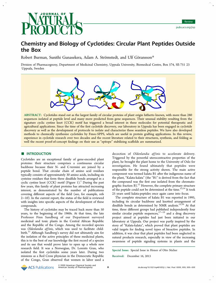
ABSTRACT: Cyclotides stand out as the largest family of circular proteins of plant origin hitherto known, with more than 280sequences isolated at peptide level and many more predicted from gene sequences. Their unusual stability resulting from thesignature cyclic cystine knot (CCK) motif has triggered a broad interest in these molecules for potential therapeutic andagricultural applications. Since the time of the first cyclotide discovery, our laboratory in Uppsala has been engaged in cyclotidediscovery as well as the development of protocols to isolate and characterize these seamless peptides. We have also developedmethods to chemically synthesize cyclotides by Fmoc-SPPS, which are useful in protein grafting applications. In this review,experience in cyclotide research over two decades and the recent literature related to their structures, synthesis, and folding aswell the recent proof-of-concept findings on their use as “epitope” stabilizing scaffolds are summarized.
■ INTRODUCTION
Cyclotides are an exceptional family of gene-encoded plantproteins: their structure comprises a continuous circularbackbone because their N- and C-termini are joined by apeptide bond. That circular chain of amino acid residuestypically consists of approximately 30 amino acids, including sixcysteine residues that form three disulfide bonds arranged in acyclic cystine knot (CCK) motif (Figure 1).1 During the pastfew years, this family of plant proteins has attracted increasinginterest, as demonstrated by the number of publicationscovering different aspects of the field (see, for example, refs2−10). In the current report, the status of the field is reviewedwith insights into specific aspects of the development of thesecompounds.The history of cyclotides may be traced back more than 50
years, to the beginning of the 1960s. At that time, the lateProfessor Finn Sandberg of our Department surveyedmedicinal and toxic plants in the Central African Republicand the Republic of the Congo. Among the recorded specieswas Oldenlandia af f inis, which was used to facilitate child-birth.11 Although Sandberg’s survey did not ultimately aim forthe isolation of the active principles of these medicinal plants,this is to the best of our knowledge the first record of a speciesand its use that would prove later to open up a whole newresearch field. It was a Norwegian, Dr. Lorents Gran, whoisolated the first cyclotides some years later. During twomissions as a Red Cross physician in the Democratic Republicof the Congo, Gran observed that women in labor used a
decoction of Oldenlandia af f inis to accelerate delivery.Triggered by the powerful uterocontractive properties of theplant, he brought the plant home to the University of Oslo forinvestigation. He found ultimately that peptides wereresponsible for the strong activity shown. The main activecomponent was termed kalata B1 after the indigenous name ofthe plant, “Kalata-kalata” (the “B1” is derived from the fact thatthe compound was the first one isolated from the chromato-graphic fraction B).12 However, the complete primary structureof the peptide could not be determined at the time.13,14 It took25 years until kalata-peptides once again came into focus.The complete structure of kalata B1 was reported in 1995,
including its circular backbone and knotted arrangement ofdisulfide bonds as determined by NMR analysis.15,16 At thattime, three different groups had published independently foursimilar circular peptide sequences,17−19 and a drug discoveryproject aimed at peptides had just been initiated in ourlaboratory at Uppsala. Our project was partly inspired by thestory of “Kalata-kalata”, which proved that plant peptides arevalid targets for finding novel types of bioactive peptides. Inaddition, it was clear that plant peptides had been neglected innatural products research, especially in view of the increasingawareness of peptide signaling systems in plants and the
Special Issue: Special Issue in Honor of Otto Sticher
Received: December 16, 2013
Review
pubs.acs.org/jnp
© XXXX American Chemical Society andAmerican Society of Pharmacognosy A dx.doi.org/10.1021/np401055j | J. Nat. Prod. XXXX, XXX, XXX−XXX
development of plants and plant cells as protein expressionsystems.A protocol for the isolation of a highly purified polypeptide
fraction from plant biomass was developed.20 In particular, thisprotocol was used for the isolation of the first suite of cyclotidesfrom one species, and the results confirmed them as a family ofpeptides.5 Hence, we reconnected to Sandberg’s discoveries inAfrica some decades earlier. The peptide family has grownquickly since then, and the collective term cyclotidesaftercyclo-peptideswas suggested by one of the leaders in the field,Professor David Craik, in 1999.1 Since then, our group hasfocused our efforts to understand the chemistry and biology ofcyclotides and to explore their possible use for biotechnological,pharmaceutical, and agricultural applications.
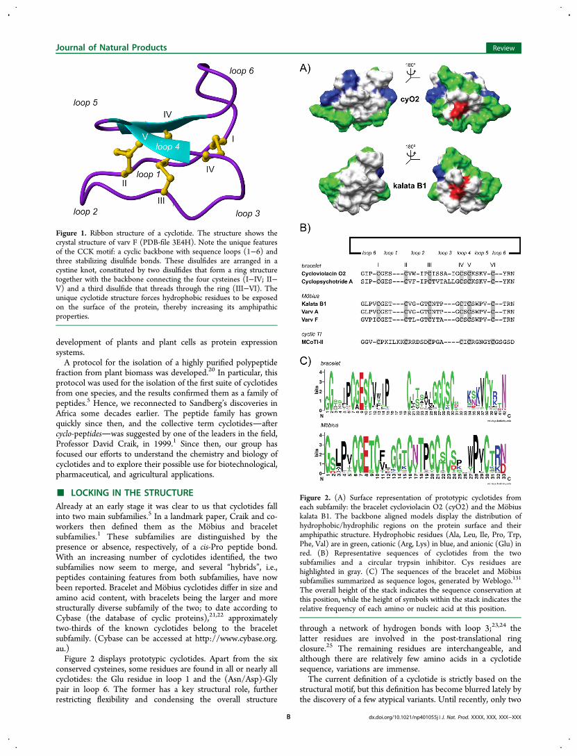
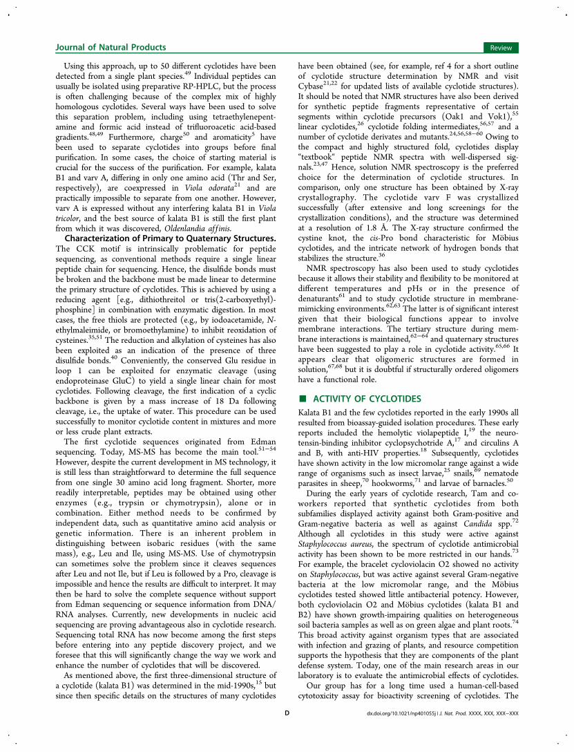
■ LOCKING IN THE STRUCTUREAlready at an early stage it was clear to us that cyclotides fallinto two main subfamilies.5 In a landmark paper, Craik and co-workers then defined them as the Mobius and braceletsubfamilies.1 These subfamilies are distinguished by thepresence or absence, respectively, of a cis-Pro peptide bond.With an increasing number of cyclotides identified, the twosubfamilies now seem to merge, and several “hybrids”, i.e.,peptides containing features from both subfamilies, have nowbeen reported. Bracelet and Mobius cyclotides differ in size andamino acid content, with bracelets being the larger and morestructurally diverse subfamily of the two; to date according toCybase (the database of cyclic proteins),21,22 approximatelytwo-thirds of the known cyclotides belong to the braceletsubfamily. (Cybase can be accessed at http://www.cybase.org.au.)Figure 2 displays prototypic cyclotides. Apart from the six
conserved cysteines, some residues are found in all or nearly allcyclotides: the Glu residue in loop 1 and the (Asn/Asp)-Glypair in loop 6. The former has a key structural role, furtherrestricting flexibility and condensing the overall structure
through a network of hydrogen bonds with loop 3;23,24 thelatter residues are involved in the post-translational ringclosure.25 The remaining residues are interchangeable, andalthough there are relatively few amino acids in a cyclotidesequence, variations are immense.The current definition of a cyclotide is strictly based on the
structural motif, but this definition has become blurred lately bythe discovery of a few atypical variants. Until recently, only two
Figure 1. Ribbon structure of a cyclotide. The structure shows thecrystal structure of varv F (PDB-file 3E4H). Note the unique featuresof the CCK motif: a cyclic backbone with sequence loops (1−6) andthree stabilizing disulfide bonds. These disulfides are arranged in acystine knot, constituted by two disulfides that form a ring structuretogether with the backbone connecting the four cysteines (I−IV; II−V) and a third disulfide that threads through the ring (III−VI). Theunique cyclotide structure forces hydrophobic residues to be exposedon the surface of the protein, thereby increasing its amphipathicproperties.
Figure 2. (A) Surface representation of prototypic cyclotides fromeach subfamily: the bracelet cycloviolacin O2 (cyO2) and the Mobiuskalata B1. The backbone aligned models display the distribution ofhydrophobic/hydrophilic regions on the protein surface and theiramphipathic structure. Hydrophobic residues (Ala, Leu, Ile, Pro, Trp,Phe, Val) are in green, cationic (Arg, Lys) in blue, and anionic (Glu) inred. (B) Representative sequences of cyclotides from the twosubfamilies and a circular trypsin inhibitor. Cys residues arehighlighted in gray. (C) The sequences of the bracelet and Mobiussubfamilies summarized as sequence logos, generated by Weblogo.131
The overall height of the stack indicates the sequence conservation atthis position, while the height of symbols within the stack indicates therelative frequency of each amino or nucleic acid at this position.
Journal of Natural Products Review
dx.doi.org/10.1021/np401055j | J. Nat. Prod. XXXX, XXX, XXX−XXXB

linear variants of cyclotides had been isolated from cyclotide-expressing plants that clearly have the same genetic origin as“true” cyclotides, but they lack the amino acid residues that areneeded for cyclization.26,27 At present, several more of suchlinear cyclotide-like sequences have been found, and these arenow referred to as “acyclotides” or “uncyclotides”.28,29
Furthermore, among the many trypsin-inhibitory cystineknotted proteins that have been isolated from the cucurbit(Cucurbitaceae) plant family, a few have a circular backboneand, thus, fulfill the criteria for inclusion in the cyclotidefamily.30,31 However, they have more sequence similarity withtheir linear counterparts (i.e., linear cucurbit trypsin inhibitors)than with cyclotides in general, and they miss some keyresidues that are found in the common cyclotides, e.g., thestructurally important Glu residue. As such, they are sometimessimply referred to as “cyclic knottins”.32
It is interesting to note that the disulfide connectivity was thesubject of a long debate, as a consequence of the inherentdifficulties of assigning disulfides in molecules with closelypacked cysteines. The cystine knot structure of kalata B1 wasfirst deduced by NMR spectroscopy,15,23,33 but has also beenquestioned by results obtained using this same technique.34
Nevertheless, today the cystine knot is undisputed and has beenverified chemically by partial reduction of disulfides combinedwith stepwise alkylation35 and by X-ray crystallography.36
■ CYCLOTIDES ARE ALL AROUND USSince the discovery of the first cyclotide in Oldenlandia af f inis,about 280 different cyclotides have been isolated from morethan 40 plant species. Patterns in occurrence are beginning tobe unraveled, but the vast majority of cyclotides have beendiscovered in the violet (Violaceae) and coffee (Rubiaceae)families. Recently, cyclotides have also been found in Clitoriaternatea of the pea (Fabaceae) family37,38 and Petunia x hybridaof the potato (Solanaceae) family.28,39 The distribution ofcyclotides within the Fabaceae and Solanaceae at large is yetunknown.Although the Rubiaceae is a large family of plants, with 600
genera and over 13 000 species, cyclotides have been found in
only a minority of species in this family and the distribution isconcentrated within a few tribes.40,41 The Violaceae familyincludes 22 genera and approximately 930 species; these arepredominantly tropical, growing as perennial herbs, shrubs, andtrees or treelets.42,43 The Violaceae has so far proved to be therichest source of cyclotides, with these compounds having beenfound in all species investigated. Remarkably, these two primarycyclotide-bearing plant families are not closely related to oneanother. For this reason, it is likely that cyclotides may be foundmore widely in the plant kingdom. Outside the Violaceae,Rubiaceae, Solanaceae, and Fabaceae, cyclotide-like genes,44,45
sequences, and linear peptides46 have been identified inPoaceae, but no circular cyclotides have been detected amongthese plants. A chemical screen has also shown that members ofthe family Apocynaceae (closely related to Rubiaceae) containsmall proteins with six Cys residues, which possibly couldindicate the presence of cyclotides.40
Developing Methods for Cyclotide Detection andIsolation. The early methods for extracting and fractionatingcyclotides from plant biomass were time- and resource-consuming and developed to isolate polypeptides in general.20
Over time, knowledge has grown regarding the cyclotides, andnew isolation methods have been developed incrementally, withmuch improved yields and more simplified procedures.7,47 Forexample, in our laboratory, the isolation procedure before finalpurification with reversed-phase HPLC has been trimmeddown to include only three steps (extraction with 60%methanol in water, liquid−liquid extraction with dichloro-methane, and reversed-phase solid-phase extraction, RP-SPE)instead of five more laborious steps in the original protocol.20
The current approach for detecting and characterizing novelcyclotides is illustrated in Scheme 1. To evaluate if a plantpossibly contains cyclotides, a fast small-scale extraction usingonly milligrams of plant material is advised before going to alarge extraction or for screening purposes.48 The extract directlyor after RP-SPE can then be analyzed using an LC-MS system.Long retention times coupled with masses ranging from 2800to 3500 strongly suggest further studies.40,48
Scheme 1. Schematic Overview of the Procedures To Detect Novel Cyclotides by Chemical Meansa
aTo first evaluate the cyclotide content of a plant, an aqueous extract is made and analyzed using liquid chromatography−mass spectrometry (LC-MS). After isolation of pure cyclotides by HPLC, the Cys residues should be reduced and alkylated so that the sample can be digested enzymatically,yielding linear products that can be sequenced further by MS-MS. The sequence determination can be elusive, but in combination with amino acidanalysis full sequence coverage is usually obtained. To obtain a complete 3-D structure, NMR spectroscopy or X-ray crystallography can be used.
Journal of Natural Products Review
dx.doi.org/10.1021/np401055j | J. Nat. Prod. XXXX, XXX, XXX−XXXC
Using this approach, up to 50 different cyclotides have beendetected from a single plant species.49 Individual peptides canusually be isolated using preparative RP-HPLC, but the processis often challenging because of the complex mix of highlyhomologous cyclotides. Several ways have been used to solvethis separation problem, including using tetraethylenepent-amine and formic acid instead of trifluoroacetic acid-basedgradients.48,49 Furthermore, charge50 and aromaticity5 havebeen used to separate cyclotides into groups before finalpurification. In some cases, the choice of starting material iscrucial for the success of the purification. For example, kalataB1 and varv A, differing in only one amino acid (Thr and Ser,respectively), are coexpressed in Viola odorata21 and arepractically impossible to separate from one another. However,varv A is expressed without any interfering kalata B1 in Violatricolor, and the best source of kalata B1 is still the first plantfrom which it was discovered, Oldenlandia af f inis.Characterization of Primary to Quaternary Structures.
The CCK motif is intrinsically problematic for peptidesequencing, as conventional methods require a single linearpeptide chain for sequencing. Hence, the disulfide bonds mustbe broken and the backbone must be made linear to determinethe primary structure of cyclotides. This is achieved by using areducing agent [e.g., dithiothreitol or tris(2-carboxyethyl)-phosphine] in combination with enzymatic digestion. In mostcases, the free thiols are protected (e.g., by iodoacetamide, N-ethylmaleimide, or bromoethylamine) to inhibit reoxidation ofcysteines.35,51 The reduction and alkylation of cysteines has alsobeen exploited as an indication of the presence of threedisulfide bonds.40 Conveniently, the conserved Glu residue inloop 1 can be exploited for enzymatic cleavage (usingendoproteinase GluC) to yield a single linear chain for mostcyclotides. Following cleavage, the first indication of a cyclicbackbone is given by a mass increase of 18 Da followingcleavage, i.e., the uptake of water. This procedure can be usedsuccessfully to monitor cyclotide content in mixtures and moreor less crude plant extracts.The first cyclotide sequences originated from Edman
sequencing. Today, MS-MS has become the main tool.51−54
However, despite the current development in MS technology, itis still less than straightforward to determine the full sequencefrom one single 30 amino acid long fragment. Shorter, morereadily interpretable, peptides may be obtained using otherenzymes (e.g., trypsin or chymotrypsin), alone or incombination. Either method needs to be confirmed byindependent data, such as quantitative amino acid analysis orgenetic information. There is an inherent problem indistinguishing between isobaric residues (with the samemass), e.g., Leu and Ile, using MS-MS. Use of chymotrypsincan sometimes solve the problem since it cleaves sequencesafter Leu and not Ile, but if Leu is followed by a Pro, cleavage isimpossible and hence the results are difficult to interpret. It maythen be hard to solve the complete sequence without supportfrom Edman sequencing or sequence information from DNA/RNA analyses. Currently, new developments in nucleic acidsequencing are proving advantageous also in cyclotide research.Sequencing total RNA has now become among the first stepsbefore entering into any peptide discovery project, and weforesee that this will significantly change the way we work andenhance the number of cyclotides that will be discovered.As mentioned above, the first three-dimensional structure of
a cyclotide (kalata B1) was determined in the mid-1990s,15 butsince then specific details on the structures of many cyclotides
have been obtained (see, for example, ref 4 for a short outlineof cyclotide structure determination by NMR and visitCybase21,22 for updated lists of available cyclotide structures).It should be noted that NMR structures have also been derivedfor synthetic peptide fragments representative of certainsegments within cyclotide precursors (Oak1 and Vok1),55
linear cyclotides,26 cyclotide folding intermediates,56,57 and anumber of cyclotide derivates and mutants.24,56,58−60 Owing tothe compact and highly structured fold, cyclotides display“textbook“ peptide NMR spectra with well-dispersed sig-nals.23,47 Hence, solution NMR spectroscopy is the preferredchoice for the determination of cyclotide structures. Incomparison, only one structure has been obtained by X-raycrystallography. The cyclotide varv F was crystallizedsuccessfully (after extensive and long screenings for thecrystallization conditions), and the structure was determinedat a resolution of 1.8 Å. The X-ray structure confirmed thecystine knot, the cis-Pro bond characteristic for Mobiuscyclotides, and the intricate network of hydrogen bonds thatstabilizes the structure.36
NMR spectroscopy has also been used to study cyclotidesbecause it allows their stability and flexibility to be monitored atdifferent temperatures and pHs or in the presence ofdenaturants61 and to study cyclotide structure in membrane-mimicking environments.62,63 The latter is of significant interestgiven that their biological functions appear to involvemembrane interactions. The tertiary structure during mem-brane interactions is maintained,62−64 and quaternary structureshave been suggested to play a role in cyclotide activity.65,66 Itappears clear that oligomeric structures are formed insolution,67,68 but it is doubtful if structurally ordered oligomershave a functional role.
■ ACTIVITY OF CYCLOTIDESKalata B1 and the few cyclotides reported in the early 1990s allresulted from bioassay-guided isolation procedures. These earlyreports included the hemolytic violapeptide I,19 the neuro-tensin-binding inhibitor cyclopsychotride A,17 and circulins Aand B, with anti-HIV properties.18 Subsequently, cyclotideshave shown activity in the low micromolar range against a widerange of organisms such as insect larvae,25 snails,69 nematodeparasites in sheep,70 hookworms,71 and larvae of barnacles.50
During the early years of cyclotide research, Tam and co-workers reported that synthetic cyclotides from bothsubfamilies displayed activity against both Gram-positive andGram-negative bacteria as well as against Candida spp.72
Although all cyclotides in this study were active againstStaphylococcus aureus, the spectrum of cyclotide antimicrobialactivity has been shown to be more restricted in our hands.73
For example, the bracelet cycloviolacin O2 showed no activityon Staphylococcus, but was active against several Gram-negativebacteria at the low micromolar range, and the Mobiuscyclotides tested showed little antibacterial potency. However,both cycloviolacin O2 and Mobius cyclotides (kalata B1 andB2) have shown growth-impairing qualities on heterogeneoussoil bacteria samples as well as on green algae and plant roots.74
This broad activity against organism types that are associatedwith infection and grazing of plants, and resource competitionsupports the hypothesis that they are components of the plantdefense system. Today, one of the main research areas in ourlaboratory is to evaluate the antimicrobial effects of cyclotides.Our group has for a long time used a human-cell-based
cytotoxicity assay for bioactivity screening of cyclotides. The
Journal of Natural Products Review
dx.doi.org/10.1021/np401055j | J. Nat. Prod. XXXX, XXX, XXX−XXXD
compounds varv A, varv F, and cycloviolacin O2 showed potentactivity in a panel of 10 different human tumor cell lines at lowcorrelations with the activity patterns varying from anticancerdrugs in clinical use today,75 suggesting a mode of actiondifferent from that of known anticancer drugs. Additionally, acomparison was made between the cytotoxic activity of varv Aand cycloviolacin O2 against tumor cells from patients andnormal lymphocytes from healthy human subjects. In thisstudy, the cyclotides showed an almost 10-fold selective toxicityagainst leukemia cells.75 Subsequently, it was shown that thecytotoxic effect on tumor cell lines can be distinguishedmorphologically within a few minutes.76 Such a rapid process isindicative of necrosis, while apoptosis requires activation ofseveral intracellular cascades as well as gene expression resultingin longer onset periods. Both the rapid kinetics and the broadspectrum of the toxic effect produced by many cyclotides pointtoward membrane disruption as responsible for activity.76
Cyclotides Interact with Cellular Membranes. Mostbiological activities assigned to cyclotides concern toxicity on abroad range of organisms, including mammalian tumor cells,75
nematodes,77 insect epithelial cells,78 bacteria,72,73 fungi,72 andenveloped viruses.18 One thing these organisms have incommon is their phospholipid membrane: the loss ofmembrane integrity inevitably leads to death of whatever lifeform is encased by it.In order for cyclotides to exert their membrane-disrupting
activity, they must have a high affinity for the target membrane.The pronounced amphiphilic and frequently cationic (in thebracelet subfamily) nature of cyclotides is shared by mostantimicrobial peptides. These qualities confer the hydrophobicand electrostatic forces that drive adsorption of cyclotidestoward the lipid core and anionic surface of the membranes.79
Other characteristics that promote membrane adsorptioninclude tryptophan affinity for the glyceryl/carbonyl region ofthe phospholipids,80 which anchor peptides at the membraneinterfaces, and membrane-induced formation of intramolecularhydrogen bonds that are stabilized by the hydrophobicenvironment of the membrane.79,81 Although cyclotides havethe possibility of benefiting from both these characteristics, forexample, the semiconserved tryptophan next to the cis-Proamong the Mobius cyclotides, their exceptionally rigid structurecannot be expected to undergo much further hydrogen bondingas a consequence of membrane adsorption, with the possibleexception of the loops of the hydrophobic patch in certaincircumstances. However, the same rigid structure not onlyimproves enzymatic stability but also forces hydrophobicresidues to the surface of the molecule, thereby improvingtheir amphiphilic properties. From these aspects, cyclotidesappear as ideal antimicrobial peptide structures.
Several models of membrane-disrupting mechanisms havebeen suggested for lytic peptides, including specific poreformations and more general curvature stress generatedperforations. A peptide multimer formation with a definedpore diameter has been claimed as the membrane-disruptingmechanism of kalata B1 based on stepwise current increase in apatch clamp experiment.66 However, due to lack of supportingdata, combined with a report showing that kalata B1 does notreadily self-associate,67 conclusive evidence for such a specificpore formation has yet to be produced.The membrane composition is of special importance for the
lytic activity of cyclotides. Some selectivity in cytotoxicityagainst leukemic lymphocytes rather than normal lymphocyteshas been shown for cycloviolacin O2.75 Tumor cell membraneshave a higher negative charge density with 3−7 times morephosphatidylserine, which is to a large extent located on theouter leaflet of the membrane.82 In addition, a decrease incholesterol content can be expected to some degree for manytumor cell lines;83 both characteristics are likely consequencesof rapid cell division. Curiously, it has been reported thatcholesterol content significantly increases lytic potency forequivalent bulk concentrations of kalata B1 on modelmembranes.66 This is unprecedented in the field of membranelytic peptides and also contrary to our experience with the samepeptide.84 It should be noted that the area of the targetmembrane affects the amount of peptide needed to reach theadsorption threshold required for lysis. Hence, the peptide-to-lipid ratio dictates the activity in membrane leakage assays ofmolecules with high adsorption isotherms such as cyclotidesand not the total concentration. Therefore, it is difficult tocompare selectivity of lytic potential between different celltypes and microbes with certainty, and even between modelmembrane systems, if care is not taken to ensure equivalenttarget areas.85
Cyclotides belong to a small group of peptides having affinityfor phosphatidylethanolamine (PE) membrane lipids, which is aconserved feature among both subfamilies of cyclotides.86
Although this affinity has been known for some time forMobius cyclotides,87,88 the importance of this affinity for theirlytic activity and its structural key elements was revealed onlyrecently.84,88 The bracelet cyclotide cycloviolacin O2 wasshown to preferentially disrupt PE-containing membranesrather than those with phosphatidylcholine (PC), with thisactivity dependent on the conserved Glu residue.84 Theunderlying mechanism for this selective lytic activity could beascribed to a higher adsorption on PE-containing membranesdue to structural affinity,88 and a subsequent specific extractionof PE lipids. This results in thinning of the membrane, which inturn makes it permeable and sensitive to peptide-induced
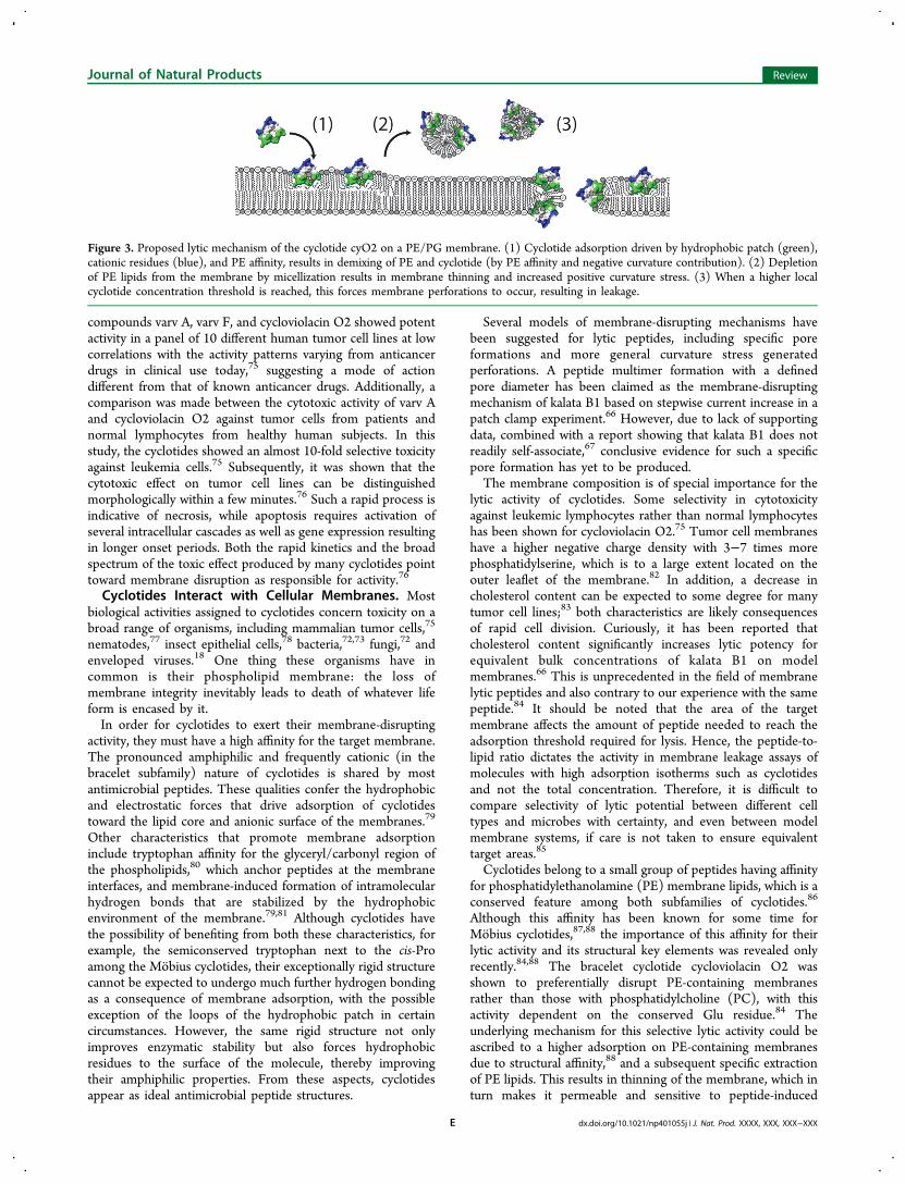
Figure 3. Proposed lytic mechanism of the cyclotide cyO2 on a PE/PG membrane. (1) Cyclotide adsorption driven by hydrophobic patch (green),cationic residues (blue), and PE affinity, results in demixing of PE and cyclotide (by PE affinity and negative curvature contribution). (2) Depletionof PE lipids from the membrane by micellization results in membrane thinning and increased positive curvature stress. (3) When a higher localcyclotide concentration threshold is reached, this forces membrane perforations to occur, resulting in leakage.
Journal of Natural Products Review
dx.doi.org/10.1021/np401055j | J. Nat. Prod. XXXX, XXX, XXX−XXXE
leakage. To our knowledge, cyclotides are the first peptides thathave been demonstrated to display PE-selective lytic activityand to mediate selective extraction of phospholipids from amembrane.84 Cyclotide affinity for PE-containing membranescould be linked subsequently with activity of biologicalmembranes on erythrocytes, bacteria, and enveloped viruses.86
Selectivity between the human membrane model PC/cholesterol (60:40) and E. coli model membranes [PE/phosphatidylglycine (PG)/diphosphatidylglycine (dPG),67:23:10] was 100-fold in lytic activity for cycloviolacin O2,predominately due to PE selectivity, but also by a combinationof electrostatic attraction to the anionic phospholipids and thelack of cholesterol of the bacterial membrane.84 The resultsindicated that when a cyclotide binds to the membrane, itassociates with PE lipids, promoted by both specific affinity andby favorable packing parameters associated with peptides thatexpand the lipid headgroup region. The high local concen-tration of cyclotides and the lipid demixing that follows result inmembrane thinning and curvature stress, which ultimatelyresults in perforations, possibly by toroidal pores or fissureslined by cyclotides (Figure 3).Structure−Activity Relationships. Naturally occurring
cyclotides vary in size between 28 and 37 amino acid residuesand have a net charge between −2 and +3. Cyclotides with apositive net charge have been found generally to be moremembrane active than those that are neutral or negativelycharged.49,75,89,90 This may be seen as a natural observationsince biomembranes normally carry a net negative charge ofvarious densities. The importance of this electrostatic attractionis seen clearly for the cationic residues in the bracelet family, asexemplified by experiments where successive charge neutraliza-tion on cycloviolacin O2 may be correlated with declines intumor cell cytotoxicity.91
Furthermore, Ala mutated variants of Glu in kalata B1 havebeen shown to be inactive in hemolytic and insecticidalassays.65 Structural studies reveal that the carboxylic acid of Gluis oriented toward the interior of the peptide and is responsiblefor maintaining structure rather than peptide surface electro-statics.24 In the bracelet family, the Glu side chain formshydrogen bonds with the backbone structure of loop 3, ahydrophobic sequence forming a helical structure thatotherwise would be distorted. As such, the Glu is importantfor membrane adsorption and possibly also for aggregation andspecific interactions, but via loop 3. The interactions of the Gluare present also in the Mobius subfamily. However, in this caseloop 3 is hydrophilic and not directly involved with membraneadsorption. Substitution of any residue in the highly conservedthree residues of loop 1 (including the conserved Glu) with Ala
abolished the lytic activity of kalata B1, as did mutations ofhydrophobic residues in loop 5.66
Most cyclotides carry an aromatic residue (usuallytryptophan): bracelets in loop 2 and Mobius in loop 5. Interms of membrane adsorption this confers a membraneinterface anchoring residue, which is reasonable since boththese loops are hydrophobic and would be expected toconstitute the patch directly associated with the membraneon adsorption of the respective subfamily. When summarizingthe activities of native cyclotides, the conclusion is that in orderto correlate the structures of cyclotides with their potency andto understand their mechanisms of action, it is necessary toconsider their structure as a whole rather than focusingspecifically on single residues. So far, no systematic study hasbeen performed to monitor these whole-molecule SARs (orQSARs), but a tool for developing this is being investigated inour laboratory.So will the cell membrane and phosphatidylethanolamine
prove to be the final molecular target of cyclotides? Althoughmembrane disruption may explain most biological andpharmacological activities, the current model does not explainall effects of cyclotides. For example, the seemingly specificneurotensin-binding of cyclopsychotride A may be explained bycyclotide binding to the lipids present in the receptor/membrane preparation. However, it is difficult, if notimpossible, to explain the reversible paralysis of barnacle larvaein the antifouling assay.50 Indeed, our group was able recentlyto show the first functional indication of a secondary targetother than the membrane/membrane disruption, namely,DNA-damaging effect at a concentration 100 times lowerthan the lytic concentration.92 This is intriguing because at themiddle concentration (i.e., 10 times lower than the lyticconcentration) peptides showed neither lytic nor DNA-damaging effects. In addition, it has been shown that kalataB1 and MCoTI-I/II do cross the cell membrane, at subtoxicconcentrations.93−95 More recently, cyclotide activity came fullcircle when Gruber and co-workers demonstrated that acyclotide (kalata B7) indeed does bind to the oxytocicreceptor.96 As such, even though there is now a clear hypothesisfor how cyclotides affect the membrane, we are only at thebeginning of understanding the whole spectrum of cyclotidefunction and activity.
■ CYCLOTIDE PRODUCTION: IN PLANTS ANDLABORATORIES
Cyclotides comprise one of the few classes of naturalmacrocyclic gene products discovered to date.97 Analysis ofcyclotide precursor sequences obtained from cDNA have
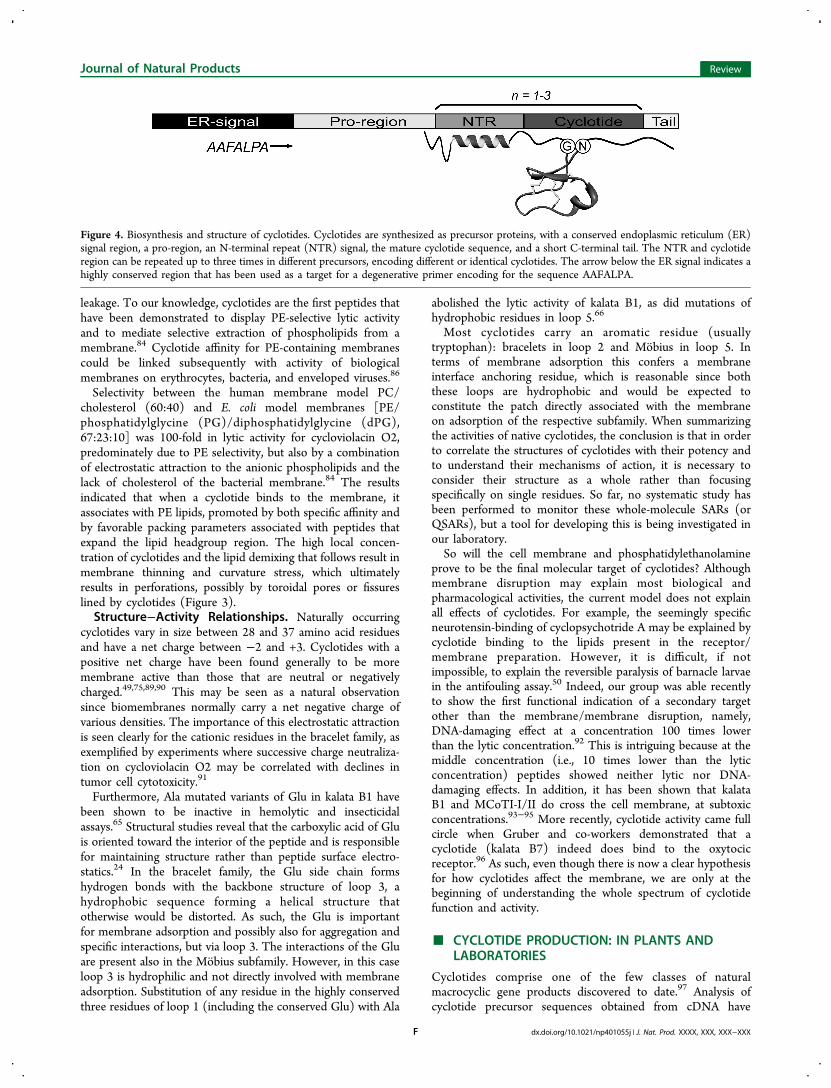
Figure 4. Biosynthesis and structure of cyclotides. Cyclotides are synthesized as precursor proteins, with a conserved endoplasmic reticulum (ER)signal region, a pro-region, an N-terminal repeat (NTR) signal, the mature cyclotide sequence, and a short C-terminal tail. The NTR and cyclotideregion can be repeated up to three times in different precursors, encoding different or identical cyclotides. The arrow below the ER signal indicates ahighly conserved region that has been used as a target for a degenerative primer encoding for the sequence AAFALPA.
Journal of Natural Products Review
dx.doi.org/10.1021/np401055j | J. Nat. Prod. XXXX, XXX, XXX−XXXF
shown that the genes encoding them consist of an endoplasmicreticulum (ER) signal domain, a pro-region, and one to threemature cyclotide domains, each preceded by an N-terminalrepeat (NTR) sequence.25,48,98,99 Figure 4 shows the cleavagepoints of a schematic cyclotide after a Lys/Gly/Asn residue inthe NTR sequence and the Asn or Asp in the cyclotide domain.Details of the processing of the precursors, involving oxidativefolding, excision of the mature cyclotide sequence, and head-to-tail cyclization, are not fully understood nor is the order of theevents concerned. However, an asparaginyl-endoproteinase(AEP) has been suggested to be involved in the cleavage ofthe C-terminal tail and simultaneous cyclization of the cyclotidedomain, at least for the prototypic cyclotide kalata B1.100−102
Additionally, a protein-disulfide isomerase seems to play amajor role in the oxidative folding of cyclotides throughreshuffling (isomerization) of disulfide bonds.103,104
Current insights into cyclotide biosynthesis have beenreviewed recently,6 but some of the points that need to beaddressed are highlighted here. None of the functional studiesin planta have used plants that express cyclotides; only thecommon model plants (Nicotiana benthamiana, N. tabacum,and/or Arabidopsis thaliana) have been used. Indeed,expression of the cyclotide precursor protein in these plantsresults in a cyclic, correctly folded peptide, but in low yields.Accumulating evidence suggests that cyclotide biosynthesisshares common features with both of the smaller circulartrypsin inhibitory peptides from sunflower (e.g., SFTI-1) andthe cyclic cystine knotted trypsin inhibitory peptides from thefamily Cucurbitaceae (e.g., McoTI-I/II). This involvescyclization by hijacking existing AEPs,31,105 but conclusiveevidence is still lacking for plant species expressing cyclotides.As such, until definite proof emerges showing that AEP activityis the common denominator for the biosynthesis of cyclotides,our opinion is that one should not limit the search to AEPsonly. This is of particular importance in light of the recentdiscovery of the first specific peptide cyclase,106 which has beenfound in Saponaria vaccaria (Caryophyllaceae). Although thesubstrate in this case is a shorter precursor and the products arecyclic peptides of only five to eight amino acid residues, thisstudy demonstrates clearly that one should not rule out theoccurrence of specific enzymes also for the biosynthesis ofcyclotides.A more detailed understanding of the biosynthesis is of
fundamental scientific interest, but it may also pave the roadtoward more efficient systems for the screening and productionof cyclotides.107 Camarero and co-workers have producedsuccessfully folded (cucurbit cyclotide) MCoTI-II, usingEscherichia coli,108 and intein fusion protein, which generatescyclic and folded kalata B1 in a one-pot reaction afterpurification of the fusion protein.109 Very recently, peptideligands specific for the VEGF-A binding site on neuropilin-1were identified by screening a ∼6 × 10⁹-membered E. colidisplay library of disulfide-rich peptides derived from thecyclotide kalata B1.110 However, these peptides were neithercyclic nor folded, but the study shows proof of concept of atechnique to find cyclotide-like binders that then can bedeveloped further. Still, there is no example of the productionof a non-natural cyclotide in planta. Cell suspension cultures ofOldenlandia af f inis have been established, expressing yields ofup to 0.7 mg of kalata B1 per liter of medium,111 but plant cellcultures have yet to be proven as a viable alternative for theproduction of bioactive cyclotide mutants.112 To access
cyclotide chemistry in the laboratory, solid-phase peptidesynthesis (SPPS) is still unrivaled.
Solid-Phase Peptide Synthesis. Intriguingly, cyclotidesare amenable to chemical synthesis despite their knotted andcircular nature. Deriving a mature cyclotide from the buildingblock amino acids involves the synthesis of the linear cyclotideprecursor, cyclization of its N- and C-termini, and formation ofthe native disulfides to achieve the correct cystine knottopology. Currently, the linear cyclotide precursors aresynthesized chemically by standard solid-phase peptide syn-thesis based on either tert-butoxycarbonyl (Boc) or 9H-fluoren-9-ylmethoxycarbonyl (Fmoc) chemistry. For a long time, nativeas well as grafted cyclotides have been synthesized predom-inantly by Boc-SPPS.113−115 However, in recent years there hasbeen a greater inclination to develop Fmoc-SPPS-basedprotocols that do not require handling of potentially hazardouschemicals (e.g., TFA for deprotection and the use of HF forfinal cleavage as in Boc-SPPS).Once the linear peptide is obtained, the next step is to ligate
its N- and C-termini to obtain the cyclized peptide. Mostcommonly, this is achieved using a C-terminal thioester and anN-terminal Cys via native chemical ligation,116 which remainsthe most robust synthesis approach to date.113,114,117−119
Peptide thioesters can be assembled readily on resin usingBoc-SPPS, because the thioester group withstands the acidicdeprotection step used. On the contrary, it is challenging tosynthesize thioester peptides by Fmoc-SPPS, because thioestersare intrinsically unstable also to the weak base that is used fordeprotection (e.g., piperidine). The simplest way to overcomethis problem is by generating/activating the C-terminalthioester group once the peptide exits the Fmoc synthesisroute, but peptides attached to the resin via a C-terminalsulfonamide linker can be activated to generate a C-terminalthioester. This approach has been used in cyclotide syn-thesis.120 Similarly, a C-terminally attached thioester group canbe generated in solution on a protected peptide that is cleavedoff from the resin.118 In both cases, cyclization via nativechemical ligation is ultimately achieved as the requirements ofC-terminal thioester and a N-terminal cysteine are met.Very recently, our group has adapted the N-acylurea-
mediated approach for native chemical ligation121 for micro-wave-assisted SPPS for cyclotide syntheses.122 The use ofmicrowave irradiation enhances efficiency and purity of thesynthetic peptides. With this method, a linear peptide isassembled on a C-terminal diaminobenzoic acid linker, whichupon activation and acylation leads to an N-acylurea/N-benzymidazolinone (Nbz) moiety. In thiol-rich environments,the Nbz-peptide undergoes in situ thioesterification and nativechemical ligation to give a cyclized peptide by native chemicalligation.The manual cyclotide synthesis protocols have also been
improved with the assistance of microwaves and automation toaccelerate the speed of peptide synthesis during the amino acidcoupling and thioesterification steps.115,122 Presumably, thio-esterification of the linear peptide leads to a vigorous “thia-zip”reaction in which the cyclic peptide backbone is zipped fromthe C- to the N-terminal in a series of thiol and thiolactoneexchanges between the carbonyl group of the C-terminalthioester and the intertwining Cys side chains. Ultimately, thehead-to-tail cyclized peptide is derived by a spontaneous S,Nacyl-migration from the terminal Cys side chain to the peptidebackbone. The characteristic feature of this thioester-mediatedstrategy is that cyclization of the peptide chain occurs prior to
Journal of Natural Products Review
dx.doi.org/10.1021/np401055j | J. Nat. Prod. XXXX, XXX, XXX−XXXG
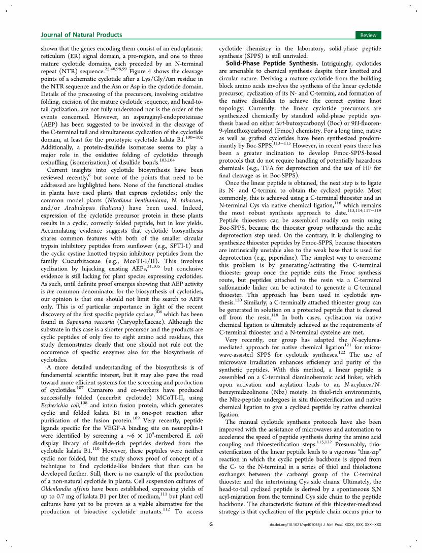
oxidation of the Cys residues. In fact, cyclization of the twotermini of the cyclotide precursor appears to have a dramaticeffect on folding, by enhancing the propensity for the correctfolding.117 The cyclized peptide then attains its native disulfideconnectivity under basic (∼pH 8) folding conditions. However,obtaining the correct native cystine knot topology of cyclotidesis not always straightforward. This problem can be tackledalready at the peptide synthesis stage by selectively protectingside chains of the Cys residues using chemically distinctprotecting groups.32,72,114 This allows the disulfides in thepeptide to be formed in two consecutive steps, with disulfidesCysI−CysIV and CysIII−CysVI formed first and CysII−CysVformed later, thus limiting the number of misfolded disulfideisomers.A synthetic approach that has only rarely been used is Cys
oxidization prior to cyclization of the peptide backbone, asexemplified in the early kalata B1 synthesis attempts.117 In thisinstance, folding of the peptide chain into the native cystineknot topology preorganizes the N- and C-termini in closeproximity. However, the use of this strategy has been limitedbecause specific achiral residues are required at the termini ofthe linear chain to prevent racemization and also due to thepossibility of side reactions at the Lys and Asp residues duringcyclization.117 The different syntheses strategies are outlined inFigure 5.No general sequence-dependent synthesis problems have
been reported for cyclotides, with the exception being the cycliccucurbit trypsin inhibitor MCoTI-II that contains an Asp-Glymotif.118,120 This motif is prone to aspartimide formation,which results from a ring-closure between the β-carboxy sidechain of Asp and the nitrogen of the α-carboxamide and isdependent on the total exposure time of the growing peptidechain to the piperidine used in Fmoc-SPPS and by extensivefolding times. However, MCoTI-II has been successfullysynthesized by suppressing aspartimide formation, by theintroduction of the Asp-Gly dipeptide with the Gly backbonenitrogen protected by 2-hydroxy-4-methoxybenzyl or 2,4-dimethoxybenzyl protecting groups.118
A chemoenzymatic biomimetic strategy has also been utilizedin the synthesis of MCoTI-I and MCoTI-II analogues.120
Taking advantage of the linear cyclotide backbone acid bearingP1 residue at the C-terminus, which is a viable substrate forprotease-mediated ligation, the synthesis of the cyclic backbonewithout the need for a C-terminal thioester has been achieved.In this approach, the open chain MCoTI analogues were firstrefolded to the cystine knot topology and subsequently cyclizedusing polymer-supported proteases. The method has, however,not been used widely, but it is not unlikely that AEPs or evenspecific cyclization enzymes might find use in future cyclotidesyntheses.Tying the Knot. Folding refers to the oxidative formation of
the native cystine knot and establishment of the native three-dimensional molecular topology. This appears to be highlydependent on the inherent propensity of each cyclotide toadopt the cystine knot. Understanding cyclotide folding is offundamental interest as well as important from an applicationsperspective, as cyclotides are potentially valuable scaffolds indrug design.Insights have been obtained into the in vitro cyclotide folding
pathways by folding experiments, although the mechanisms ofin vivo cyclotide synthesis and folding are only beginning to beunraveled.100,101 A single cyclotide precursor with six Cysresidues can adopt potentially 75 different disulfide con-
formations (15 one-disulfide, 45 two-disulfide, and 15 three-disulfide species), and it is fascinating that a cyclotide precursornavigates its way through the correct folding pathway into thefinal native fold under optimized folding conditions.In general, it appears that folding of Mobius and MCoTI
cyclic knottins are easier when compared to bracelet cyclotides.Both Mobius and MCoTI fold conveniently into the nativeconformation in buffers maintained at pH ∼8. The yield may insome cases be improved by adding cosolvents (e.g., 2-propanol) to increase hydrophobicity in the buffers.123 Forkalata B1, it appears that 2-propanol is needed to stabilize thedistinct hydrophobic patch of residues that becomes exposed tothe surface upon folding. Importantly, in the absence of 2-propanol, kalata B1 folds at a slower rate, leaving the flexibledisulfide intermediates susceptible to deamidation at thesensitive Asn-Gly.123 2-Propanol is not required for MCoTI-II folding, presumably because the native form has morehydrophilic amino acids exposed at its surface. The rate andyield are heavily influenced by the redox potential of the buffer,which may be controlled by the addition of glutathione(reduced and oxidized). Redox potential is also influenced by“external” factors, e.g., the amount of air (oxygen) dissolved inthe solution or in contact with the solution. As such, it is goodpractice to degas folding buffers, seal reaction tubes with argonor nitrogen, and use freshly prepared solutions of glutathione.
Figure 5. Schematic illustration of different cyclotide synthesisstrategies. (A) Thioester-mediated Boc-SPPS strategy. (B) Boc-SPPSwithout thioesterification. (C) Boc-SPPS using selective disulfideprotection. (D) Thioester-mediated Fmoc-SPPS folding.
Journal of Natural Products Review
dx.doi.org/10.1021/np401055j | J. Nat. Prod. XXXX, XXX, XXX−XXXH
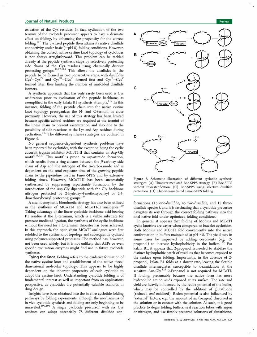
In addition to these obvious differences in the folding buffers,there are also fundamental differences in the major foldingintermediates between different cyclotide subfamilies, asdepicted in Figure 6. A major native-like two-disulfide
intermediate, des(CysI-CysIV) or 2SS(IIa), is a kinetic trappresent during the Mobius oxidative folding.56 This inter-mediate is not the direct precursor to the native form and doesnot readily refold into the native conformation, unless with theassistance of glutathione, which facilitates disulfide reshuffling.In MCoTI-II oxidative folding, a similar major two-disulfideintermediate with a native-like structure is present, which,unlike kalata B1, easily converts to the native form and hencethe direct precursor to the native form. Another two-disulfideintermediate, des(CysII-CysV) or 2SS(IIb), is present duringkalata B1 unfolding. This intermediate has been suggested asthe possible direct precursor during oxidative refolding.Structural analysis indicates the disulfide CysII−CysV is themost surface-exposed disulfide in kalata B1 and the easiest bondto be broken, indicating that 2SS(IIb) could be the most likelycandidate to be the direct precursor to the native form. It isinteresting to note that this intermediate does not have anative-like structure, as it elutes on RP-HPLC very close to the
reduced form rather than to the native form, although it hastwo native disulfide bonds (CysI−CysIV and CysIII−CysVI).Bracelet cyclotides appear more problematic with respect to
their in vitro folding. They do not simply fold under theoptimized conditions for Mobius or MCoTI and generally givea range of misfolded products during their folding. However,they have been successfully folded, by selective Cys protectionduring synthesis, as previously mentioned. Furthermore, ourgroup has established optimized in vitro folding conditions forthe prototypical bracelet cycloviolacin O2, which included thepresence of detergents and folding at lower temperatures.123
That cyclotide, which is the only bracelet cyclotide that hasbeen studied in any detail, shows a major misfoldedintermediate with three non-native disulfides. The native formrequires a major conformational change, but also breaking ofthe disulfide bonds to form the native form. It is alleged thatboth detergents and cosolvents act as stabilizers for the nativecyclotide conformation by interacting with the hydrophobicresidues that are exposed to the surface upon folding.Despite the conserved cystine knot, why are the folding
pathways of these proteins so different? For Mobius cyclotidesand the cucurbit cyclotides (McoTI-I/II), there are differencesin size and sequence of individual loops. Mainly the size of thecystine knot, which is larger in MCoTI-II, appears to beresponsible for the differences in the folding pathways. Despitesimilar structures of the backbones and overall sizes of themolecules, the degree and the location of the hydrophobic andcharged resides are different in Mobius and bracelet cyclotides.Mainly residues in loops 5 and 6 form the hydrophobic patch inkalata B1, but the corresponding loops are more hydrophilicand charged in the bracelet cyclotide cycloviolacin O2.24
Furthermore, a large hydrophobic patch involving loop 2 andan α-helix in loop 3 is present in cycloviolacin O2. Interestingly,the corresponding part is hydrophilic in kalata B1.23 As a resultof these structural differences, it is suggested that cycloviolacinO2 requires not only a hydrophobic cosolvent but alsodetergents to stabilize the more amphipathic character of itsnative form. Thus, specific structural characteristics ofindividual cyclotides appear to dictate their folding behavior.Although some details can start to be revealed and someattempts to systematic studies have been done,123,124 it stillcannot be predicted if and how cyclotides will fold or not.
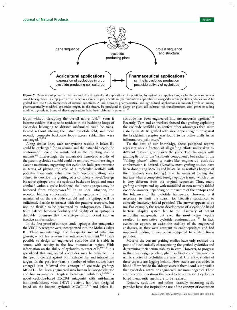
■ A CIRCLE OUTSIDE THE BOXSo can the fundamental knowledge of these cyclic peptides betranslated into applied sciences? For drug design, as well as inthe field of natural product chemistry, peptides and proteins aresometimes considered as rather fragile biomolecules that areeasily degraded by chemical and biotic factors. Currently, thisview is changing. Clearly, the circular backbone and the cystineknot confer an extreme structural stability that lacks parallels inother protein families. The main natural role of this structureappears to be in plant defense, most notably against insectpests, but cyclotides do have the potential to be exploited inmany ways in both agricultural and pharmaceutical applications(Figure 7).With the rising number of hypervariable cyclotide sequences
and supporting evidence for their ultrastable scaffold, cyclotideshave been implicated as ideal molecular templates for thedesign of pharmaceutical and agricultural agents. As a first lineof evidence for the flexibility of the cyclotide template, Daly etal. designed acylic permutants of kalata B1, to prove that thecyclotide backbone could be opened in four of the backbone
Figure 6. Overview of oxidative folding pathways of cyclotides. Fullyreduced cyclotides are folded via the main folding intermediates(containing two or three disulfide bonds, 2SS or 3SS) to the nativestructure. Kalata B1, a representative member of the Mobius subfamily,has a native-like main folding intermediate [2SS(IIa)], which is not adirect precursor to the native form, during its oxidative folding. A two-disulfide intermediate (2SS-IIb), present during kalata B1 unfolding,has been suggested as the possible direct precursor during kalata B1oxidative refolding. Cycloviolacin O2, representing the braceletsubfamily, appears to fold via 3SS intermediates with non-nativedisulfide bonds. The cyclic knottin MCoTI-II also has a native-like 2SSfolding intermediate, but this is in fact the direct precursor to thenative form.
Journal of Natural Products Review
dx.doi.org/10.1021/np401055j | J. Nat. Prod. XXXX, XXX, XXX−XXXI
loops, without disrupting the overall native fold.59 Soon itbecame evident that specific residues in the backbone loops ofcyclotides belonging to distinct subfamilies could be trans-located without altering the native cyclotide fold, and morerecently complete backbone loops across subfamilies wereexchanged.60,124
Along similar lines, each noncysteine residue in kalata B1could be exchanged for an alanine and the native-like cyclotideconformation could be maintained in the resulting alaninemutants.65 Interestingly, the undesirable hemolytic activity ofthe parent cyclotide scaffold could be removed with these singlealanine mutations, suggesting that cyclotides hold great promisein terms of playing the role of a molecular scaffold withpotential therapeutic value. The term “epitope grafting” wascoined to describe the grafting of a completely novel/foreignbioactive epitope onto the cyclotide backbone loops, and onceconfined within a cyclic backbone, the linear epitopes may beharbored from exoproteases.125 In an ideal situation, thereceptor binding conformation of the epitope will still bemaintained on the cyclotide scaffold and the epitope will besufficiently flexible to interact with the putative receptors, butnot too flexible to be penetrated by endoproteases. Thus, afinite balance between flexibility and rigidity of an epitope isdesirable to ensure that the epitope is not locked into aninactive conformation.In the first proof-of-concept study, epitopes that antagonize
the VEGF-A receptor were incorporated into the Mobius kalataB1. These mutants target the therapeutic area of antiangio-genesis, which has relevance in anticancer treatment.125 It waspossible to design an engineered cyclotide that is stable inserum, with activity in the low micromolar region. Withinformation on the ability of cyclotides to enter cells,93−95 it isspeculated that engineered cyclotides may be valuable in atherapeutic context against both extracellular and intracellulartargets. In the past few years, a number of other studies haveemerged that followed this concept of cyclotide grafting:MCoTI-II has been engineered into human leukocyte elastaseand human mast cell tryptase beta-based inhibitors;126,127 anovel cyclotide-based CXCR4 antagonist with anti-humanimmunodeficiency virus (HIV)-1 activity has been designedbased on the knottin cyclotide MCoTI-I;128 and kalata B1
cyclotide has been engineered into melanocortin agonists.129
Recently, Tam and co-workers showed that grafting exploitingthe cyclotide scaffold also confers other advantages than merestability: kalata B1 grafted with an epitope antagonistic againstthe bradykinin receptor was found to be active orally in aninflammatory pain assay.10
To the best of our knowledge, these published reportsrepresent only a fraction of all grafting efforts undertaken bydifferent research groups over the years. The challenges withgrafting lie not in the “synthesis component”, but rather in the“folding phase” when a native-like engineered cyclotideconformation is desired. (Notably, most grafting studies havebeen done using McoTIs and kalata B1 as scaffolds because oftheir relatively easy folding.) The challenges of folding alsoincrease when a completely foreign epitope is used, which oftenis very different from the original sequence. Thus, manygrafting attempts end up with misfolded or non-natively foldedcyclotide isomers, depending on the nature of the epitopes andthe tolerance of the cyclotide framework. However, is itnecessary to limit the search for bioactive substances tocorrectly (natively) folded peptides? The answer appears to beno. For example, the recent development of a cyclotide-basedbacterial display system led to the discovery of potentneuropilin antagonists, but even the most active peptideresulted in non-native cyclotide conformations.110 In fact,cyclization appears to assist the stability of the engineeredanalogues, as they were resistant to endopeptidases and hadimproved binding to neuropilin compared to control linearpeptides.Most of the current grafting studies have only reached the
point of biochemically characterizing the grafted cyclotides anddetermining their serum stability in vitro. However, to progressin the drug design pipeline, pharmacokinetic and pharmacody-namic studies of cyclotides are essential. Currently, studies ofthese aspects are lagging behind. How stable are cyclotides inblood? How fast do the kidneys excrete them? And is it possiblethat cyclotides, native or engineered, are immunogenic? Theseare the critical questions that need to be addressed if cyclotide-based therapeutic agents are to be realized.Notably, cyclotides and other naturally occurring cyclic
peptides have also inspired the use of the concept of cyclization
Figure 7. Overview of potential pharmaceutical and agricultural applications of cyclotides. In agricultural applications, cyclotide gene sequencescould be expressed in crop plants to enhance resistance to pests, while in pharmaceutical applications biologically active peptide epitopes could begrafted into the CCK framework of natural cyclotides. A link between pharmaceutical and agricultural applications is indicated with an arrow;pharmaceutically modified cyclotides might, in the future, be produced in plants or plant cell cultures, via transformation with genes encodingmodified cyclotides. Some of these applications have been claimed in patents.132
Journal of Natural Products Review
dx.doi.org/10.1021/np401055j | J. Nat. Prod. XXXX, XXX, XXX−XXXJ
to linear peptides. One example is the recent study of a naturalα-conotoxin, Vc1.1, with exciting potential for the treatment ofneuropathic pain (by inhibiting the HVA Ca2+ channel currentsvia activation of GABAB receptors).130 By engineering a cyclicVc1.1 analogue, it was possible to increase the stability ingastric fluid, simulated intestinal fluid, and human serum.Whether cyclotides will be further developed and commer-
cially used remains to be seen. The patents on cyclotides mainlyconcern aspects of cyclotide activity or inventions regardingcyclotide genes. Using this knowledge, developments toward anagricultural application have been made; for example, acyclotide gene has been transferred to crop plants in anattempt to improve natural defenses against pests.101 Thesuccessful start, i.e., production of transgenic plants, ispromising, but the studies so far have relied on the insertionof only a single gene. Coexpression with auxiliary proteins suchas folding and circularization enzymes from cyclotide-bearingplants has the potential to increase the yields. Another potentialapplication is to directly use the pesticide effects of cyclotidesdirectly to inhibit the growth of bacteria, algae, and fungi.Cyclotides have already been shown to have a potent, nontoxic,and reversible effect against fouling barnacles.50
However, the importance of the fundamental aspects ofscience should not be underestimated, nor should it beforgotten that the best source of new knowledge likely iscuriosity alone. Without curiosity as the driving force,cyclotides may still be awaiting discovery. In fact, it is alsothe fundamental features of the cyclotides that attract the mostinterest: namely, the cyclic cystine knot and its inherentstability and rigidity. It is these features that define this peptidecircle as a molecule outside the box.
■ AUTHOR INFORMATIONCorresponding Author*Tel: +46 184715031. Fax: +46 18509101. E-mail: [email protected] authors declare no competing financial interest.
■ ACKNOWLEDGMENTSWork by the authors has been supported by the SwedishResearch Council (621-2007-5167) and the Swedish Founda-tion for Strategic Research (F06-0058). We thank current andformer members of the group and collaborators. U.G. thanksDr. P. Claeson and Prof. L. Bohlin for their guidance during thefirst year of cyclotide research in Uppsala, Prof. G. Samuelssonfor his inspiring early work in the field of plant peptides, andthe late Prof. F. Sandberg for his enthusiastic support.
■ DEDICATIONDedicated to Prof. Dr. Otto Sticher, of ETH-Zurich, Zurich,Switzerland, for his pioneering work in pharmacognosy andphytochemistry.
■ REFERENCES(1) Craik, D. J.; Daly, N. L.; Bond, T.; Waine, C. J. Mol. Biol. 1999,294, 1327−1336.(2) Schroeder, C. I.; Swedberg, J. E.; Craik, D. J. Curr. Protein Pept.Sci. 2013, 14, 532−552.(3) Poth, A. G.; Chan, L. Y.; Craik, D. J. Biopolymers 2013, 100, 480−491.(4) Ireland, D. C.; Clark, R. J.; Daly, N. L.; Craik, D. J. J. Nat. Prod.2010, 73, 1610−1622.
(5) Goransson, U.; Luijendijk, T.; Johansson, S.; Bohlin, L.; Claeson,P. J. Nat. Prod. 1999, 62, 283−286.(6) Craik, D. J.; Malik, U. Curr. Opin. Chem. Biol. 2013, 17, 546−554.(7) Goransson, U.; Svangard, E.; Claeson, P.; Bohlin, L. Curr. ProteinPept. Sci. 2004, 5, 317−329.(8) Craik, D. J.; Swedberg, J. E.; Mylne, J. S.; Cemazar, M. ExpertOpin. Drug Discovery 2012, 7, 179−194.(9) Ji, Y.; Majumder, S.; Millard, M.; Borra, R.; Bi, T.; Elnagar, A. Y.;Neamati, N.; Shekhtman, A.; Camarero, J. A. J. Am. Chem. Soc. 2013,135, 11623−11633.(10) Wong, C. T.; Rowlands, D. K.; Wong, C. H.; Lo, T. W.;Nguyen, G. K.; Li, H. Y.; Tam, J. P. Angew. Chem., Int. Ed. 2012, 51,5620−5624.(11) Sandberg, F. In Cahiers de la Maboke; Buffon: Paris, 1965; Vol.III, Fascicule 1, p 27.(12) Gran, L. Lloydia 1973, 36, 174−178.(13) Gran, L. Acta Pharmacol. Toxicol. 1973, 33, 400−408.(14) Sletten, K.; Gran, L. Medd. Nor. Farm. Selsk. 1973, 35, 69−82.(15) Saether, O.; Craik, D. J.; Campbell, I. D.; Sletten, K.; Juul, J.;Norman, D. G. Biochemistry 1995, 34, 4147−4158.(16) Pallaghy, P. K.; Nielsen, K. J.; Craik, D. J.; Norton, R. S. ProteinSci. 1994, 3, 1833−1839.(17) Witherup, K. M.; Bogusky, M. J.; Anderson, P. S.; Ramjit, H.;Ransom, R. W.; Wood, T.; Sardana, M. J. Nat. Prod. 1994, 57, 1619−1625.(18) Gustafson, K. R.; Sowder, R. C.; Henderson, L. E.; Parsons, I.C.; Kashman, Y.; Cardellina, J. H.; McMahon, J. B.; Buckheit, L. K.;Pannell, L. K.; Boyd, M. R. J. Am. Chem. Soc. 1994, 116, 9337−9338.(19) Schopke, T.; Hasan, A. M. I.; Kraft, R.; Otto, A.; Hiller, K. Sci.Pharm. 1993, 61, 145−153.(20) Claeson, P.; Goransson, U.; Johansson, S.; Luijendijk, T.;Bohlin, L. J. Nat. Prod. 1998, 61, 77−81.(21) Wang, C. K.; Kaas, Q.; Chiche, L.; Craik, D. J. Nucleic Acids Res.2008, 36, D206−D210.(22) Mulvenna, J. P.; Wang, C.; Craik, D. J. Nucleic Acids Res. 2006,34, D192−D194.(23) Rosengren, K. J.; Daly, N. L.; Plan, M. R.; Waine, C.; Craik, D. J.J. Biol. Chem. 2003, 278, 8606−8616.(24) Goransson, U.; Herrmann, A.; Burman, R.; Haugaard-Jonsson,L. M.; Rosengren, K. J. ChemBioChem 2009, 10, 2354−2360.(25) Jennings, C.; West, J.; Waine, C.; Craik, D.; Anderson, M. Proc.Natl. Acad. Sci. U.S.A. 2001, 98, 10614−10619.(26) Ireland, D. C.; Colgrave, M. L.; Nguyencong, P.; Daly, N. L.;Craik, D. J. J. Mol. Biol. 2006, 357, 1522−1535.(27) Gerlach, S. L.; Burman, R.; Bohlin, L.; Mondal, D.; Goransson,U. J. Nat. Prod. 2010, 73, 1207−1213.(28) Poth, A. G.; Mylne, J. S.; Grassl, J.; Lyons, R. E.; Millar, A. H.;Colgrave, M. L.; Craik, D. J. J. Biol. Chem. 2012, 287, 27033−27046.(29) Nguyen, G. K.; Lim, W. H.; Nguyen, P. Q.; Tam, J. P. J. Biol.Chem. 2012, 287, 17598−17607.(30) Hernandez, J. F.; Gagnon, J.; Chiche, L.; Nguyen, T. M.;Andrieu, J. P.; Heitz, A.; Trinh Hong, T.; Pham, T. T.; Le Nguyen, D.Biochemistry 2000, 39, 5722−5730.(31) Mylne, J. S.; Chan, L. Y.; Chanson, A. H.; Daly, N. L.; Schaefer,H.; Bailey, T. L.; Nguyencong, P.; Cascales, L.; Craik, D. J. Plant Cell2012, 24, 2765−2778.(32) Chiche, L.; Heitz, A.; Gelly, J. C.; Gracy, J.; Chau, P. T.; Ha, P.T.; Hernandez, J. F.; Le-Nguyen, D. Curr. Protein Pept. Sci. 2004, 5,341−349.(33) Nair, S. S.; Romanuka, J.; Billeter, M.; Skjeldal, L.; Emmett, M.R.; Nilsson, C. L.; Marshall, A. G. Biochim. Biophys. Acta 2006, 1764,1568−1576.(34) Skjeldal, L.; Gran, L.; Sletten, K.; Volkman, B. F. Arch. Biochem.Biophys. 2002, 399, 142−148.(35) Goransson, U.; Craik, D. J. J. Biol. Chem. 2003, 278, 48188−48196.(36) Wang, C. K.; Hu, S. H.; Martin, J. L.; Sjogren, T.; Hajdu, J.;Bohlin, L.; Claeson, P.; Goransson, U.; Rosengren, K. J.; Tang, J.; Tan,N. H.; Craik, D. J. J. Biol. Chem. 2009, 284, 10672−10683.
Journal of Natural Products Review
dx.doi.org/10.1021/np401055j | J. Nat. Prod. XXXX, XXX, XXX−XXXK
(37) Nguyen, G. K.; Zhang, S.; Nguyen, N. T.; Nguyen, P. Q.; Chiu,M. S.; Hardjojo, A.; Tam, J. P. J. Biol. Chem. 2011, 286, 24275−24287.(38) Poth, A. G.; Colgrave, M. L.; Lyons, R. E.; Daly, N. L.; Craik, D.J. Proc. Natl. Acad. Sci. U.S.A. 2011, 108, 10127−10132.(39) Poth, A. G.; Colgrave, M. L.; Philip, R.; Kerenga, B.; Daly, N. L.;Anderson, M.; Craik, D. J. ACS Chem. Biol. 2010, 6, 345−355.(40) Gruber, C. W.; Elliott, A. G.; Ireland, D. C.; Delprete, P. G.;Dessein, S.; Goransson, U.; Trabi, M.; Wang, C. K.; Kinghorn, A. B.;Robbrecht, E.; Craik, D. J. Plant Cell 2008, 20, 2471−2483.(41) Koehbach, J.; Attah, A. F.; Berger, A.; Hellinger, R.; Kutchan, T.M.; Carpenter, E. J.; Rolf, M.; Sonibare, M. A.; Moody, J. O.; Wong, G.K.; Dessein, S.; Greger, H.; Gruber, C. W. Biopolymers 2013, 100,438−452.(42) Hekking, W. H. A. Proc. K. Ned. Akad. Wet., Ser. C: Biol. Med. Sci.1984, 87, 121−130.(43) Hekking, W. H. A. Flora 1988, 180, 345−376.(44) Mulvenna, J. P.; Mylne, J. S.; Bharathi, R.; Burton, R. A.; Shirley,N. J.; Fincher, G. B.; Anderson, M. A.; Craik, D. J. Plant Cell 2006, 18,2134−2144.(45) Basse, C. W. Plant Physiol. 2005, 138, 1774−1784.(46) Nguyen, G. K.; Lian, Y.; Pang, E. W.; Nguyen, P. Q.; Tran, T.D.; Tam, J. P. J. Biol. Chem. 2013, 288, 3370−3380.(47) Craik, D. J.; Henriques, S. T.; Mylne, J. S.; Wang, C. K. MethodsEnzymol. 2012, 516, 37−62.(48) Herrmann, A.; Burman, R.; Mylne, J. S.; Karlsson, G.; Gullbo, J.;Craik, D. J.; Clark, R. J.; Goransson, U. Phytochemistry 2008, 69, 939−952.(49) Ireland, D. C.; Colgrave, M. L.; Craik, D. J. Biochem. J. 2006,400, 1−12.(50) Goransson, U.; Sjogren, M.; Svangard, E.; Claeson, P.; Bohlin, L.J. Nat. Prod. 2004, 67, 1287−1290.(51) Goransson, U.; Broussalis, A. M.; Claeson, P. Anal. Biochem.2003, 318, 107−117.(52) Broussalis, A. M.; Goransson, U.; Coussio, J. D.; Ferraro, G.;Martino, V.; Claeson, P. Phytochemistry 2001, 58, 47−51.(53) Colgrave, M. L.; Poth, A. G.; Kaas, Q.; Craik, D. J. Biopolymers2010, 94, 592−601.(54) Hashempour, H.; Koehbach, J.; Daly, N. L.; Ghassempour, A.;Gruber, C. W. Amino Acids 2013, 44, 581−595.(55) Dutton, J. L.; Renda, R. F.; Waine, C.; Clark, R. J.; Daly, N. L.;Jennings, C. V.; Anderson, M. A.; Craik, D. J. J. Biol. Chem. 2004, 279,46858−46867.(56) Daly, N. L.; Clark, R. J.; Craik, D. J. J. Biol. Chem. 2003, 278,6314−6322.(57) Cemazar, M.; Joshi, A.; Daly, N. L.; Mark, A. E.; Craik, D. J.Structure 2008, 16, 842−851.(58) Heitz, A.; Avrutina, O.; Le-Nguyen, D.; Diederichsen, U.;Hernandez, J. F.; Gracy, J.; Kolmar, H.; Chiche, L. BMC Struct. Biol.2008, 8, 54.(59) Daly, N. L.; Craik, D. J. J. Biol. Chem. 2000, 275, 19068−19075.(60) Clark, R. J.; Daly, N. L.; Craik, D. J. Biochem. J. 2006, 394, 85−93.(61) Colgrave, M. L.; Craik, D. J. Biochemistry 2004, 43, 5965−5975.(62) Shenkarev, Z. O.; Nadezhdin, K. D.; Lyukmanova, E. N.; Sobol,V. A.; Skjeldal, L.; Arseniev, A. S. J. Inorg. Biochem. 2008, 102, 1246−1256.(63) Shenkarev, Z. O.; Nadezhdin, K. D.; Sobol, V. A.; Sobol, A. G.;Skjeldal, L.; Arseniev, A. S. FEBS J. 2006, 273, 2658−2672.(64) Wang, C. K.; Colgrave, M. L.; Ireland, D. C.; Kaas, Q.; Craik, D.J. Biophys. J. 2009, 97, 1471−1481.(65) Simonsen, S. M.; Sando, L.; Rosengren, K. J.; Wang, C. K.;Colgrave, M. L.; Daly, N. L.; Craik, D. J. J. Biol. Chem. 2008, 283,9805−9813.(66) Huang, Y. H.; Colgrave, M. L.; Daly, N. L.; Keleshian, A.;Martinac, B.; Craik, D. J. J. Biol. Chem. 2009, 284, 20699−20707.(67) Nourse, A.; Trabi, M.; Daly, N. L.; Craik, D. J. J. Biol. Chem.2004, 279, 562−570.(68) Rosengren, K. J.; Daly, N. L.; Harvey, P. J.; Craik, D. J.Biopolymers 2013, 100, 453−460.
(69) Plan, M. R.; Saska, I.; Cagauan, A. G.; Craik, D. J. J. Agric. FoodChem. 2008, 56, 5237−5241.(70) Colgrave, M. L.; Kotze, A. C.; Huang, Y. H.; O’Grady, J.;Simonsen, S. M.; Craik, D. J. Biochemistry 2008, 47, 5581−5589.(71) Colgrave, M. L.; Kotze, A. C.; Kopp, S.; McCarthy, J. S.;Coleman, G. T.; Craik, D. J. Acta Trop. 2009, 109, 163−166.(72) Tam, J. P.; Lu, Y. A.; Yang, J. L.; Chiu, K. W. Proc. Natl. Acad.Sci. U.S.A. 1999, 96, 8913−8918.(73) Pranting, M.; Loov, C.; Burman, R.; Goransson, U.; Andersson,D. I. J. Antimicrob. Chemother. 2010, 65, 1964−1971.(74) Ovesen, R. G.; Brandt, K. K.; Goransson, U.; Nielsen, J.;Hansen, H. C.; Cedergreen, N. Environ. Toxicol. Chem. 2011, 30,1190−1196.(75) Lindholm, P.; Goransson, U.; Johansson, S.; Claeson, P.;Gullbo, J.; Larsson, R.; Bohlin, L.; Backlund, A. Mol. Cancer Ther.2002, 1, 365−369.(76) Svangard, E.; Burman, R.; Gunasekera, S.; Lovborg, H.; Gullbo,J.; Goransson, U. J. Nat. Prod. 2007, 70, 643−647.(77) Colgrave, M. L.; Kotze, A. C.; Ireland, D. C.; Wang, C. K.;Craik, D. J. ChemBioChem 2008, 9, 1939−1945.(78) Barbeta, B. L.; Marshall, A. T.; Gillon, A. D.; Craik, D. J.;Anderson, M. A. Proc. Natl. Acad. Sci. U.S.A. 2008, 105, 1221−1225.(79) Stromstedt, A.; Ringstad, L.; Schmiidtchen, A.; Malmsten, M.Curr. Opin. Colloid Interface Sci. 2010, 15, 467−478.(80) de Planque, M. R.; Bonev, B. B.; Demmers, J. A.; Greathouse, D.V.; Koeppe, R. E., 2nd; Separovic, F.; Watts, A.; Killian, J. A.Biochemistry 2003, 42, 5341−5348.(81) Wieprecht, T.; Apostolov, O.; Beyermann, M.; Seelig, J. J. Mol.Biol. 1999, 294, 785−794.(82) Utsugi, T.; Schroit, A. J.; Connor, J.; Bucana, C. D.; Fidler, I. J.Cancer Res. 1991, 51, 3062−3066.(83) Hendrich, A. B.; Michalak, K. Curr. Drug Targets 2003, 4, 23−30.(84) Burman, R.; Stromstedt, A. A.; Malmsten, M.; Goransson, U.Biochim. Biophys. Acta 2011, 1808, 2665−2673.(85) Wimley, W. C. ACS Chem. Biol. 2010, 5, 905−917.(86) Henriques, S. T.; Huang, Y. H.; Castanho, M. A.; Bagatolli, L.A.; Sonza, S.; Tachedjian, G.; Daly, N. L.; Craik, D. J. J. Biol. Chem.2012, 287, 33629−33643.(87) Kamimori, H.; Hall, K.; Craik, D. J.; Aguilar, M. I. Anal. Biochem.2005, 337, 149−153.(88) Henriques, S. T.; Huang, Y. H.; Rosengren, K. J.; Franquelim,H. G.; Carvalho, F. A.; Johnson, A.; Sonza, S.; Tachedjian, G.;Castanho, M. A.; Daly, N. L.; Craik, D. J. J. Biol. Chem. 2011, 286,24231−24241.(89) Ireland, D. C.; Wang, C. K.; Wilson, J. A.; Gustafson, K. R.;Craik, D. J. Biopolymers 2008, 90, 51−60.(90) Svangard, E.; Goransson, U.; Hocaoglu, Z.; Gullbo, J.; Larsson,R.; Claeson, P.; Bohlin, L. J. Nat. Prod. 2004, 67, 144−147.(91) Herrmann, A.; Svangard, E.; Claeson, P.; Gullbo, J.; Bohlin, L.;Goransson, U. Cell. Mol. Life Sci. 2006, 63, 235−245.(92) Yeshak, M. Y.; Goransson, U.; Burman, R.; Hellman, B. Mutat.Res. 2012, 747, 176−181.(93) Greenwood, K. P.; Daly, N. L.; Brown, D. L.; Stow, J. L.; Craik,D. J. Int. J. Biochem. Cell Biol. 2007, 39, 2252−2264.(94) Contreras, J.; Elnagar, A. Y.; Hamm-Alvarez, S. F.; Camarero, J.A. J. Controlled Release 2011, 155, 134−143.(95) Cascales, L.; Henriques, S. T.; Kerr, M. C.; Huang, Y. H.; Sweet,M. J.; Daly, N. L.; Craik, D. J. J. Biol. Chem. 2011, 286, 36932−36943.(96) Koehbach, J.; O’Brien, M.; Muttenthaler, M.; Miazzo, M.;Akcan, M.; Elliott, A. G.; Daly, N. L.; Harvey, P. J.; Arrowsmith, S.;Gunasekera, S.; Smith, T. J.; Wray, S.; Goransson, U.; Dawson, P. E.;Craik, D. J.; Freissmuth, M.; Gruber, C. W. Proc. Natl. Acad. Sci. U.S.A.2013, 110, 21183−21188.(97) Arnison, P. G.; Bibb, M. J.; Bierbaum, G.; Bowers, A. A.; Bugni,T. S.; Bulaj, G.; Camarero, J. A.; Campopiano, D. J.; Challis, G. L.;Clardy, J.; Cotter, P. D.; Craik, D. J.; Dawson, M.; Dittmann, E.;Donadio, S.; Dorrestein, P. C.; Entian, K. D.; Fischbach, M. A.;Garavelli, J. S.; Goransson, U.; Gruber, C. W.; Haft, D. H.;
Journal of Natural Products Review
dx.doi.org/10.1021/np401055j | J. Nat. Prod. XXXX, XXX, XXX−XXXL
Hemscheidt, T. K.; Hertweck, C.; Hill, C.; Horswill, A. R.; Jaspars, M.;Kelly, W. L.; Klinman, J. P.; Kuipers, O. P.; Link, A. J.; Liu, W.;Marahiel, M. A.; Mitchell, D. A.; Moll, G. N.; Moore, B. S.; Muller, R.;Nair, S. K.; Nes, I. F.; Norris, G. E.; Olivera, B. M.; Onaka, H.;Patchett, M. L.; Piel, J.; Reaney, M. J.; Rebuffat, S.; Ross, R. P.; Sahl, H.G.; Schmidt, E. W.; Selsted, M. E.; Severinov, K.; Shen, B.; Sivonen,K.; Smith, L.; Stein, T.; Sussmuth, R. D.; Tagg, J. R.; Tang, G. L.;Truman, A. W.; Vederas, J. C.; Walsh, C. T.; Walton, J. D.; Wenzel, S.C.; Willey, J. M.; van der Donk, W. A. Nat. Prod. Rep. 2013, 30, 108−160.(98) Zhang, J.; Liao, B.; Craik, D. J.; Li, J. T.; Hu, M.; Shu, W. S. Gene2009, 431, 23−32.(99) Simonsen, S. M.; Sando, L.; Ireland, D. C.; Colgrave, M. L.;Bharathi, R.; Goransson, U.; Craik, D. J. Plant Cell 2005, 17, 3176−3189.(100) Saska, I.; Gillon, A. D.; Hatsugai, N.; Dietzgen, R. G.; Hara-Nishimura, I.; Anderson, M. A.; Craik, D. J. J. Biol. Chem. 2007, 282,29721−29728.(101) Gillon, A. D.; Saska, I.; Jennings, C. V.; Guarino, R. F.; Craik,D. J.; Anderson, M. A. Plant J. 2008, 53, 505−515.(102) Conlan, B. F.; Gillon, A. D.; Barbeta, B. L.; Anderson, M. A.Am. J. Bot. 2011, 98, 2018−2026.(103) Gruber, C. W.; Cemazar, M.; Heras, B.; Martin, J. L.; Craik, D.J. Trends Biochem. Sci. 2006, 31, 455−464.(104) Gruber, C. W.; Cemazar, M.; Clark, R. J.; Horibe, T.; Renda, R.F.; Anderson, M. A.; Craik, D. J. J. Biol. Chem. 2007, 282, 20435−20446.(105) Mylne, J. S.; Colgrave, M. L.; Daly, N. L.; Chanson, A. H.;Elliott, A. G.; McCallum, E. J.; Jones, A.; Craik, D. J. Nat. Chem. Biol.2011, 7, 257−259.(106) Barber, C. J.; Pujara, P. T.; Reed, D. W.; Chiwocha, S.; Zhang,H.; Covello, P. S. J. Biol. Chem. 2013, 288, 12500−12510.(107) Borra, R.; Camarero, J. A. Biopolymers 2013, 100, 502−509.(108) Camarero, J. A.; Kimura, R. H.; Woo, Y. H.; Shekhtman, A.;Cantor, J. ChemBioChem 2007, 8, 1363−1366.(109) Kimura, R. H.; Tran, A. T.; Camarero, J. A. Angew. Chem., Int.Ed. 2006, 45, 973−976.(110) Getz, J. A.; Cheneval, O.; Craik, D. J.; Daugherty, P. S. ACSChem. Biol. 2013, 8, 1147−1154.(111) Dornenburg, H. Biopolymers 2010, 94, 602−610.(112) Dornenburg, H. Biotechnol. Lett. 2008, 30, 1311−1321.(113) Tam, J. P.; Lu, Y. A. Tetrahedron Lett. 1997, 38, 5599−5602.(114) Tam, J. P.; Lu, Y. A. Protein Sci. 1998, 7, 1583−1592.(115) Cemazar, M.; Craik, D. J. J. Pept. Sci. 2008, 14, 683−689.(116) Dawson, P. E.; Muir, T. W.; Clark-Lewis, I.; Kent, S. B. Science1994, 266, 776−779.(117) Daly, N. L.; Love, S.; Alewood, P. F.; Craik, D. J. Biochemistry1999, 38, 10606−10614.(118) Park, S.; Gunasekera, S.; Leta Aboye, T.; Goransson, U. Int. J.Pept. Res. Ther. 2010, 16, 167−176.(119) Clark, R. J.; Craik, D. J. Biopolymers 2010, 94, 414−422.(120) Thongyoo, P.; Roque-Rosell, N.; Leatherbarrow, R. J.; Tate, E.W. Org. Biomol. Chem. 2008, 6, 1462−1470.(121) Blanco-Canosa, J. B.; Dawson, P. E. Angew. Chem., Int. Ed.2008, 47, 6851−6855.(122) Gunasekera, S.; Aboye, T. L.; Madian, W. A.; El-Seedi, H. R.;Goransson, U. Int. J. Pept. Res. Ther. 2013, 19, 43−54.(123) Aboye, T. L.; Clark, R.; Burman, R.; Bajona, M.; Craik, D. J.;Goransson, U. Antioxid. Redox Signaling 2011, 14, 77−86.(124) Gunasekera, S.; Daly, N. L.; Clark, R. J.; Craik, D. J. Antioxid.Redox Signaling 2009, 11, 971−980.(125) Gunasekera, S.; Foley, F. M.; Clark, R. J.; Sando, L.; Fabri, L. J.;Craik, D. J.; Daly, N. L. J. Med. Chem. 2008, 51, 7697−7704.(126) Thongyoo, P.; Bonomelli, C.; Leatherbarrow, R. J.; Tate, E. W.J. Med. Chem. 2009, 52, 6197−6200.(127) Sommerhoff, C. P.; Avrutina, O.; Schmoldt, H. U.; Gabrijelcic-Geiger, D.; Diederichsen, U.; Kolmar, H. J. Mol. Biol. 2010, 395, 167−175.
(128) Aboye, T. L.; Ha, H.; Majumder, S.; Christ, F.; Debyser, Z.;Shekhtman, A.; Neamati, N.; Camarero, J. A. J. Med. Chem. 2012, 55,10729−10734.(129) Eliasen, R.; Daly, N. L.; Wulff, B. S.; Andresen, T. L.; Conde-Frieboes, K. W.; Craik, D. J. J. Biol. Chem. 2012, 287, 40493−40501.(130) Clark, R. J.; Jensen, J.; Nevin, S. T.; Callaghan, B. P.; Adams, D.J.; Craik, D. J. Angew. Chem., Int. Ed. Engl. 2010, 49, 6545−6548.(131) Crooks, G. E.; Hon, G.; Chandonia, J. M.; Brenner, S. E.Genome Res. 2004, 14, 1188−1190.(132) Smith, A. B.; Daly, N. L.; Craik, D. J. Expert Opin. Ther. Pat.2011, 21, 1657−1672.
Journal of Natural Products Review
dx.doi.org/10.1021/np401055j | J. Nat. Prod. XXXX, XXX, XXX−XXXM