Embed Size (px)
Citation preview
Proc. Natl. Acad. Sci. USAVol. 75, No. 12, pp. 5964-5968, December 1978Biochemistry
Origin ofDNA replication in papovavirus chromatin is recognizedby endogenous endonuclease
(simian virus 40 and polyoma virus nucleoprotein complexes/gel electrophoresis/restriction endonuclease digestion/physical maps)
WALDEMAR WALDECK, BERND FOHRING*, KAMAL CHOWDHURY, PETER GRUSSt, AND GERHARD SAUER
Institut fur Virusforschung, Deutsches Krebsforschungszentrum, Im Neuenheimer Feld 280, 6900 Heidelberg, West Germany
Communicated by Wolfgang Beermann, September 25, 1978
ABSTRACT Isolated simian virus 40 (SV40) and polyomanucleoprotein complexes contain endonuclease that, under invitro conditions, converts part (up to 30%) of the covalentlyclosed superhelical DNA to full-length linear rods. The positionsof the cleavage sites within the genomes of SV40 and polyomawere determined by digestion with various single-cut restrictionendonucleases and subsequent agarose gel electrophoresis ofthe cleavage products. Both SV40 and polyoma covalently closedsuperhelical DNA were cleaved open at their respective originsof DNA replication (±75 base pairs). The full-length linear DNArods whose ends map adjacent to the origin of DNA replicationcould also be isolated by sodium dodecyl sulfate/phenol ex-traction both from SV40-infected permissive cells and frompurified SV40 virions. These data reveal the resence of aunique structure of the papovavirus chromatin cose to the ini-tiation site of DNA replication.
Isolated papovavirus chromatin has been used as a simple modelfor the study of the fine structure of chromosomes (1-5). A closesimilarity between the structural organizations of viral andeukaryotic chromatins was revealed with the main featurebeing that beaded globular structures termed "nucleosomes"(6), which consist of cellular histones (5, 7) are aligned along theDNA filaments on a small number of distinct alternative posi-tions (4).
Viral chromatin also has been successfully used to determinethe in vitro factors and conditions that are required for DNAreplication. The availability of soluble nucleoprotein complexesderived from the nuclei of simian virus 40 (SV40)-infected cells(8) has fostered such studies (8, 9). A number of enzymes suchas a and y DNA polymerases (10, 11) and RNA polymerase (12)are associated with papovavirus nucleoprotein complexes. Aswill be shown in this report, there is, in addition, an endonu-clease activity present that introduces one double-strand breakinto the viral DNA. In the case of both SV40 and polyoma DNA,the position that is susceptible to the endonuclease activitywithin the chromatin was shown to be located at the origin ofreplication. This finding has revealed a specific structure of thepapovavirus chromatin in a biologically important region ofthe genome.
MATERIALS AND METHODSVirus and Cells. SV40 strain Rh 911 was grown on CV-1 cells
as described (13). Propagation of polyoma virus and infectionof 3T6 cells were carried out as described (14). For isolation ofnucleoprotein complexes the cells were infected at a multi-plicity of 10 plaque-forming units per cell.
Preparation of Nucleoprotein Complexes. Nuclei wereisolated from SV40-infected CV-1 cells (107 cells in a 15-cmpetri dish, 38 hr after infection) and from polyoma-infected 3T6
cells (2 X 10⁷ cells in a 15-cm petri dish, 26 hr after infection)as described (14). For preparation of nuclear extracts as initiallydescribed by Su and De Pamphilis (8) the nuclei were sus-pended in 2 ml of modified hypotonic extraction buffer (10mMHepes, pH 8.0/1 mM MgCl2/0.5 mM CaCl2/1 mM di-thiothreitol/1 mM phenylmethylsulfonyl fluoride). Nucleo-protein complexes were extracted by incubating the mixturefor 1 hr at 0°C (in wet ice) with occasional agitation. The nucleiwere removed from the supernate, which was stored at 00C,by centrifugation for 5 min at 8000 X g, resuspended in 1 mlof extraction buffer, and eluted again for 1 hr, as describedabove. After removal of the nuclei by centrifugation for 5 minat 8000 X g, the supernates containing the nucleoproteincomplexes were pooled and stored at -70°C.
Isolation of DNA from Nucleoprotein Complexes. Thenucleoprotein complex preparations were mixed with an equalvolume of 1.2% sodium dodecyl sulfate containing 10 mMEDTA, and the DNA was extracted by addition of buffer-sat-urated phenol/chloroform, 1:1 (vol/vol). The aqueous phasewas adjusted to 1 M NaCl and the DNA was precipitated byaddition of 2 vol of undenatured ethanol. After pelleting, theDNA was suspended in 20mM Tris, pH 7.5, and incubated at37°C for 30 min with RNase (10 ,ig/ml) that previously hadbeen heated for 15 min at 850 C.
Preparation of Radiolabeled Viral DNA. 3H-Labeled DNAfrom infected cells was prepared as described (13).
Purification of SV40 Virion DNA. CV-1 cells in roller vesselswere infected with SV40 strain Rh 911 at a multiplicity of 0.1plaque-forming unit per cell. One week after infection theculture medium was adjusted to pH 9.0 with Na2HCO3 andthen the cells were frozen and thawed three times. The celldebris was pelleted by centrifugation in a Christ-HeraeusMinifuge at 2000 rpm for 15 min. The supernate was adjustedto 0.5 M NaCl and polyethylene glycol was added to 10%(wt/vol). The suspension was incubated at 40C overnight, afterwhich the polyethylene glycol was pelleted by centrifugationat 3000 rpm for 15 min in a Christ-Heraeus IV KS centrifuge.One-half milliliter of the pellet was layered on top of a CsClgradient (1.34 g/cm3 in 10 mM Tris, pH 7.5) for band equi-librium centrifugation in a Beckman SW 65 rotor at 4°C and40,000 rpm for 48 hr. The virions banding at a density of 1.34g/cm3 were harvested and dialyzed against 2000 vol of 20mMTris, pH 7.5. After a 15-min treatment at 370C with 1% sodiumdodecyl sulfate/20 mM Tris/10mM EDTA, the viral DNA wasextracted with buffered phenol.
Abbreviations: SV40, simian virus 40; nick, single-strand break; FOIDNA, covalently closed superhelical DNA; FOII DNA, double-stranded circular DNA containing a nick in one of the strands; FOIIIDNA, double-stranded linear rods of unit length.* Present address: Rutgers University, Department of Pathology,Piscataway, NJ 08854.
t Present address: National Institutes of Health, Bethesda, MD20014.
5964
The publication costs of this article were defrayed in part by pagecharge payment. This article must therefore be hereby marked "ad-vertisement" in accordance with 18 U. S. C. §1734 solely to indicatethis fact.

Proc. Natl. Acad. Sci. USA 75 (1978) 5965
Purification of Intracellular SV40 Double-Stranded LinearDNA Rods of Unit Length (FOIII DNA). Isolation of intra-cellular SV40 FOIII DNA was performed as detailed (13).Briefly, SV40 DNA was selectively extracted and purified ina CsCI/ethidium bromide gradient. The DNA contained in thelight band of the gradient (30 ,gg) was electrophoresed in a 1.4%agarose gel (1.0 X 9 X 0.3 cm) for 90 min at 100 V. Afterstaining of the DNA within the gel with ethidium bromide aphotograph was taken and the section containing the FOIIIDNA was excised. The DNA was recovered from the agaroseas described (13).Agarose Gel Electrophoresis. The DNA was analyzed by
electrophoresis in vertical 1% or 1.4% agarose (SeaKem, MCIBiomedical, Rockland, ME) gels as described (15). After elec-trophoresis the gels were stained in electrophoresis buffercontaining ethidium bromide at 0.5 ,ug/ml for 10 min andphotographed under UV light using Polaroid film type 667.
Blotting. Denaturation and transfer of DNA from the slabgels to nitrocellulose sheets (Schleicher and Schiill, BA 85) wereperformed essentially as described by Southern (16).
Hybridization with 32P-labeled nick-translated DNA (17)was carried out for 24 hr at 650C. After hybridization, the filterswere washed extensively in 75 mM NaCI/7.5 mM sodium ci-trate, pH 7.0, at 650C and dried overnight at 370C. Filters wereoverlaid with Kodak Codirex film for 2 days.
RESULTSFOIII DNA Is Generated In Vitro in Nucleoprotein
Complexes. Isolated polyoma and SV40 nucleoprotein com-plexes contained covalently closed superhelical DNA (FOIDNA) and some double-stranded circular DNA containing anick in one of the strands (FOII DNA) but no detectable FOIIIDNA (Fig; 1, lanes a and d). However, as early as 2 min afterincubation in extraction buffer, linear FOIII DNA could bedetected (Fig. 1, lanes b and c). As indicated in Fig. 2, the rel-ative amount of SV40 FOIII DNA increased with time andappeared to reach a plateau by 30 min of incubation. Thesekinetics indicate that FOIII is generated rather rapidly and thatonly a fraction of the viral DNA molecules, not exceeding 30%,can be rendered linear by introduction of a double-strand break.The simultaneous presence of FOI, FOII, and FOIII DNA innucleoprotein complexes that had been incubated at 37°C invitro is shown in Fig. 1, which also indicates that the majorproduct of the reaction is FOII DNA.The appearance of FOIII DNA in the presence of FOI DNA
1 FOII
_- _F01~~~~F111
__OOI. 0
FIG. 1. Agarose slab gel electrophoresis of viral DNA isolatedfrom nucleoprotein complexes. The nucleoprotein complexes wereisolated from polyoma-infected 3T6 cells and incubated in the ex-traction buffer for 10 min either at 0°C (lane a) or at 37°C (lane b).SV40 nucleoprotein complexes isolated from CV-1 cells were incu-bated in the extraction buffer for 10 min either at 37°C (lane c) or at0°C (lane d). Electrophoresis of the purified DNA was carried out foreither 2 hr (polyoma DNA) or 5 hr (SV40 DNA). Direction of elec-trophoresis was from top to bottom.
Time, min
FIG. 2. Kinetics of appearance of SV40 FOIII DNA in nucleo-protein complexes at 37°C in vitro. Nucleoprotein complexes wereincubated at 37°C in extraction buffer. At the indicated periods oftime, 20-IAI aliquots were withdrawn and, after phenol-extraction, theDNA was analyzed in ethidium bromide-stained agarose slab gels.From photographs (under UV light), densitometer tracings wereobtained (Joyce-Loebl densitometer) from which the relative amountof FOII DNA was calculated.
is incompatible with the assumption that the conversion of FOIDNA to FOIII DNA might occur by introduction of randomnicks into both strands of the DNA, leading eventually to adouble-strand break. If so, one would expect first an increasingamount of FOII DNA concomitant with a decrease of FOIDNA. Only after complete conversion of FOI DNA to FOIIDNA should linear FOIII DNA appear if random nicking bynucleases were driving the reaction.
Essentially the same kinetics of conversion of FOI DNA toFOIII DNA were noticed when polyoma nucleoprotein com-plexes were incubated at 37°C (data not shown).Determination of Cleavage Site. The above data suggest
a mechanism that cleaves open FOI DNA by a double-strandbreak. To locate the position of opening on the viral genome,FOIII DNA was generated by incubation of the nucleoproteincomplexes for 30 min at 37°C followed by DNA extraction withphenol. Then it was digested with single-cut restriction en-donucleases to assess whether two discrete cleavage productswere formed. The isolated SV40 DNA (FOI, FOII, and FOIII)which served as a substrate for single-cut restriction endonu-cleases is shown in Fig. 3, lane a. Digestion of an aliquot of thisDNA with BamHI generated FOIII DNA by introduction ofone double-strand break into circular FOI and FOII DNA atmap position 0.17 and, most interestingly, two fragments (seearrows) which must have arisen from specific cleavage of theFOIIIDNA. As estimated from a comparison with the sizemarkers (HindIII SV40DNA fragments shown in lane d), thesize of these fragments is 55.5 and 44.5% of the length of SV40DNA. Another single-cut enzyme that cleaves SV40DNA atmap position 0.67 coincident with the origin of DNA replication(18), Bgl I, generated only FOIIIDNA. Based upon these data,the site of opening of SV40DNA in chromatin can be assignedto the origin of DNA replication at map position 0.67.
Investigation of FOIIIDNA isolated from polyoma chro-matin with single-cut restriction endonucleases revealed es-sentially the same results as those seen with SV40. When di-gested with BamHI, which cleaves the polyoma DNA at mapposition 0.58 (19), two fragments were formed whose molecularweights were 88 and 12% of the length of polyoma DNA (Fig.3, lane f). The larger fragment is indicated by the arrow (bothin the stained gel and in the autoradiograph in the lower partof the figure); the smaller fragment cannot be visualized,probably because of its small size. Furthermore, EcoRI, whichcleaves polyoma DNA at map position 0 (20) generates, besidesFOIII DNA, two fragments (arrows in Fig. 3, lane g). Theywere 71 and 29% of the length of polyoma DNA.
Biochemistry: Waldeck et al.
5966 Biochemistry: Waldeck et al.
Mr x lo06
-00~ 3.6
-oo- 2.5- 2.0
* -_ 1.6
- 1.1
-- ---|-3
Ty
FIG. 3. Agarose slab gel electrophoresis of single-cut restrictionendonuclease digestion products of viral DNA isolated from nucleo-protein complexes. SV40 (lanes a-d) and polyoma (lanes e-h)nucleoprotein complex preparations were incubated for 30 min at37°C in extraction buffer. Then the DNA was purified, digested tocompletion with restriction endonucleases that introduce one dou-ble-strand break and analyzed by agarose slab gel electrophoresis.Lanes: a, undigested aliquot of the SV40 FOI, FOII, and FOIII DNA;b, after digestion with BamHI; c, after digestion with BgI I; d, Hin-dIII-generated SY40 DNA fragments obtained after cleavage of FOIDNA, coelectrophoresed as size markers; e, polyoma FOI and FOIIDNA from nucleoprotein complexes incubated at 0°C, f, BamHI-digested polyoma DNA; g, EcoRI-cleaved polyoma DNA; h, an un-
digested aliquot of g preparation. (Lower Right) Autoradiograph ofthe polyoma DNA in the gel which had been transferred after dena-turation to a nitrocellulose filter and hybridized with nick-translated32P-labeled polyoma DNA. The fragments resulting from cleavageof FOIII DNA with single-cut endonucleases are pointed out by thearrows. Direction of electrophoresis was from top to bottom.
These data indicate that the position at which FOIII DNAwas cleaved by endonuclease is the origin of polyoma DNAreplication at map position 0.71 (20, 21).
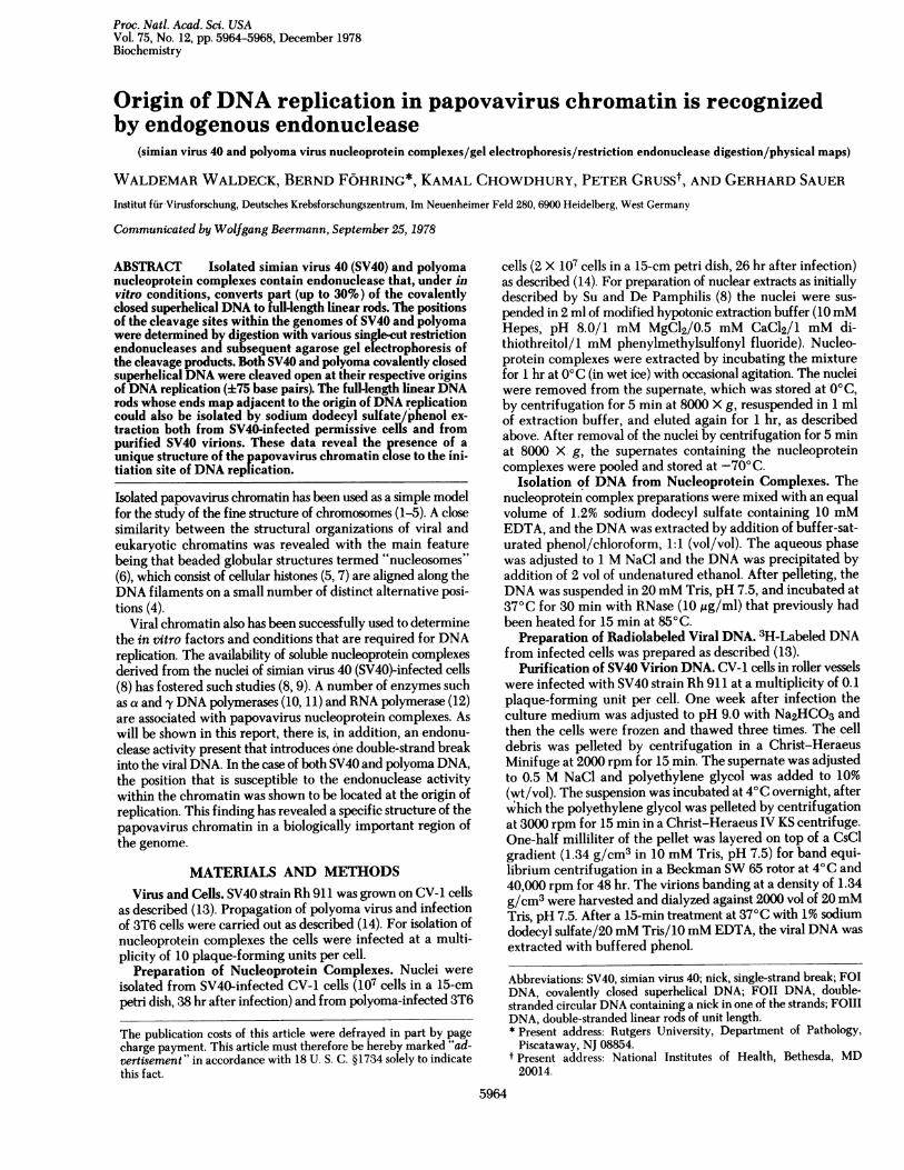
Specifically Cleaved SV40 FOIII DNA Is Present in Pro-ductively Infected Cells. We have previously shown thepresence of FOIII DNA in SV40-infected cells (13). To testwhether this population contains molecules whose ends mapat or adjacent to the origin of SV40 DNA replication, we se-
lectively isolated [as detailed recently (13)] the FOIII DNA fromproductively infected cells by dye/buoyant density gradientcentrifugation and by preparative agarose gel electrophoresis.The purified FOIII DNA was subjected to digestion with var-
ious single-cut restriction enzymes and the resulting cleavageproducts were electrophoresed into agarose and then detectedby blot hybridization with nick-translated SV40 [32P]DNA.When treated with Bgl I, the FOIII DNA substrate remainedunaltered and no cleavage products could be discerned (Fig.4). Two cleavage products were obtained after EcoRI treat-ment, with molecular weights of 2.5 X 10⁶ and 1.1 X 10⁶. Thelarger product formed a diffuse band, suggesting that theopening had occurred in a short region rather than within a
specific site. This notion is corroborated by the data obtained
a b c d
FIG. 4. Autoradiograph of single-cut restriction endonucleasedigestion products of intracellular SV40 FOIII DNA. From produc-tively infected CV-1 cells, the SV40 FOIII DNA was selectively iso-lated as described (13) and subjected to agarose slab gel electropho-resis after complete digestion with Bgl I (lane a), EcoRI (lane b), HpaII (lane c), and BamHI (lane d). After electrophoresis the DNA wasdenatured, transferred to a nitrocellulose filter, and hybridized withnick-translated SV40 [32P]DNA. The molecular weights of the frag-ments were estimated by comparison of their relative mobilities withthe distances migrated by authentic SV40 DNA restriction fragmentsof known molecular weights in the same gel (not shown here). Direc-tion of electrophoresis was from top to bottom.
after Hpa II cleavage which also generated a diffuse bandslightly smaller than unit length. The resulting small productis not revealed, probably because it migrated out of the gel.Finally, BamHI digestion produced two fragments, 2 X 10⁶ and1.6 X 106 daltons.These results show the presence of SV40 FOIII DNA mole-
cules in infected cells whose ends terminate either at or in closevicinity to the origin of DNA replication.
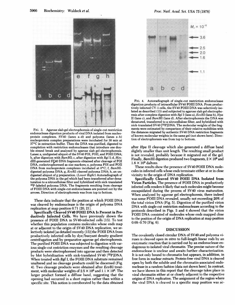
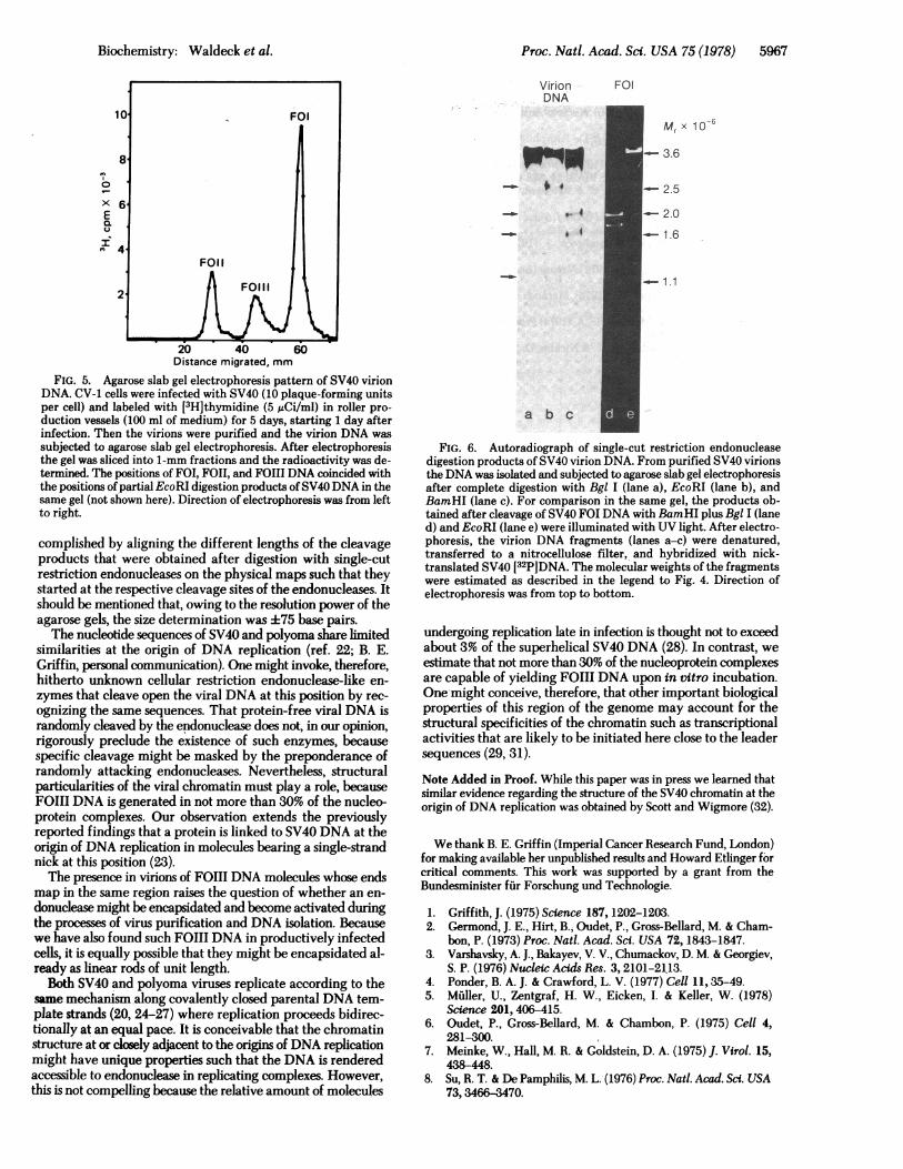
Specifically Cleaved SV40 FOIII DNA Isolated fromVirion Particles. The presence of FOIII DNA in productivelyinfected cells renders it likely that such molecules might becomeencapsidated during the process of SV40 virus maturation.When analyzed by agarose gel electrophoresis, there indeedwas some FOIII DNA revealed, usually not exceeding 20% ofthe total virion DNA (Fig. 5). Digestion of the purified virionDNA with single-cut restriction endonucleases according to theprotocols described in Figs. 3 and 4 showed that the virionFOIII DNA consisted of molecules whose ends mapped closeto the position of the origin of DNA replication at map position0.69-0.70 (Fig. 6).
DISCUSSIONThe covalently closed circular DNA of SV40 and polyoma vi-ruses is cleaved open in vitro to full-length linear rods by anenzymatic reaction that is carried out by an endonuclease en-dogenous to isolated viral chromatin. The precise nature of theendonuclease is unclear and awaits further characterization.It is not only bound to chromatin but appears, in addition, infree form in nuclear extracts. Protein-free viral DNA is cleavedopen by both the soluble and the chromatin-associated endo-nuclease in a random fashion (data not shown here). In contrast,we have'shown in this report that the cleavage takes place inviral chromatin either at or closely adjacent to the respectiveorigins of DNA replication. The assignment of the region wherethe viral DNA is cleaved to a specific map position was ac-
I
Proc. Natl. Acad. Sci. USA 75 (1978)
4
Biochemistry: Waldeck et al.
X 6E
I-~~~~F0
2- FO11l
20 40 60Distance migrated, mm
FIG. 5. Agarose slab gel electrophoresis pattern of SV40 virionDNA. CV-1 cells were infected with SV40 (10 plaque-forming unitsper cell) and labeled with [3H]thymidine (5 uCi/ml) in roller pro-duction vessels (100 ml of medium) for 5 days, starting 1 day afterinfection. Then the virions were purified and the virion DNA wassubjected to agarose slab gel electrophoresis. After electrophoresisthe gel was sliced into 1-mm fractions and the radioactivity was de-termined. The positions of FOI, FOII, and FO1l DNA coincided withthe positions of partial EcoRI digestion products ofSV40DNA in thesame gel (not shown here). Direction of electrophoresis was from leftto right.
complished by aligning the different lengths of the cleavageproducts that were obtained after digestion with single-cutrestriction endonucleases on the physical maps such that theystarted at the respective cleavage sites of the endonucleases. Itshould be mentioned that, owing to the resolution power of theagarose gels, the size determination was +75 base pairs.The nucleotide sequences of SV40 and polyoma share limited
similarities at the origin of DNA replication (ref. 22; B. E.Griffin, personal communication). One might invoke, therefore,hitherto unknown cellular restriction endonuclease-like en-zymes that cleave open the viral DNA at this position by rec-ognizing the same sequences. That protein-free viral DNA israndomly cleaved by the endonuclease does not, in our opinion,rigorously preclude the existence of such enzymes, becausespecific cleavage might be masked by the preponderance ofrandomly attacking endonucleases. Nevertheless, structuralparticularities of the viral chromatin must play a role, becauseFOIII DNA is generated in not more than 30% of the nucleo-protein complexes. Our observation extends the previouslyreported findings that a protein is linked to SV40 DNA at theorigin of DNA replication in molecules bearing a single-strandnick at this position (23).The presence in virions of FOIII DNA molecules whose ends
map in the same region raises the question of whether an en-donuclease might be encapsidated and become activated duringthe processes of virus purification and DNA isolation. Becausewe have also found such FOIII DNA in productively infectedcells, it is equally possible that they might be encapsidated al-ready as linear rods of unit length.
Both SV40 and polyoma viruses replicate according to thesame mechanism along covalently closed parental DNA tem-plate strands (20, 24-27) where replication proceeds bidirec-tionally at an equal pace. It is conceivable that the chromatinstructure at or closely adjacent to the origins of DNA replicationmight have unique properties such that the DNA is renderedaccessible to endonuclease in replicating complexes. However,this is not compelling because the relative amount of molecules
Proc. Natl. Acad. Sci. USA 75 (1978) 5967
FOI
Mr x 10 6
- 3.6
- 2.5
- 2.0- 1.6
Virio nDNA
WM_-I 6 X
_.0 , J
_.0 , I
1.1
a b c
FIG. 6. Autoradiograph of single-cut restriction endonucleasedigestion products of SV40 virion DNA. From purified SV40 virionsthe DNA was isolated and subjected to agarose slab gel electrophoresisafter complete digestion with Bgl I (lane a), EcoRI (lane b), andBamHI (lane c). For comparison in the same gel, the products ob-tained after cleavage of SV40 FOI DNA with BamHI plus Bgl I (laned) and EcoRI (lane e) were illuminated with UV light. After electro-phoresis, the virion DNA fragments (lanes a-c) were denatured,transferred to a nitrocellulose filter, and hybridized with nick-translated SV40 [32P]DNA. The molecular weights of the fragmentswere estimated as described in the legend to Fig. 4. Direction ofelectrophoresis was from top to bottom.
undergoing replication late in infection is thought not to exceedabout 3% of the superhelical SV40 DNA (28). In contrast, weestimate that not more than 30% of the nucleoprotein complexesare capable of yielding FOII DNA upon in vitro incubation.One might conceive, therefore, that other important biologicalproperties of this region of the genome may- account for thestructural specificities of the chromatin such as transcriptionalactivities that are likely to be initiated here close to the leadersequences (29, 31).
Note Added in Proof. While this paper was in press we learned thatsimilar evidence regarding the structure of the SV40 chromatin at theorigin of DNA replication was obtained by Scott and Wigmore (32).
We thank B. E. Griffin (Imperial Cancer Research Fund, London)for making available her unpublished results and Howard Etlinger forcritical comments. This work was supported by a grant from theBundesminister ffir Forschung und Technologie.
1. Griffith, J. (1975) Science 187, 1202-1203.2. Germond, J. E., Hirt, B., Oudet, P., Gross-Bellard, M. & Cham-
bon, P. (1973) Proc. Natl. Acad. Sci. USA 72, 1843-1847.3. Varshavsky, A. J., Bakayev, V. V., Chumackov, D. M. & Georgiev,
S. P. (1976) Nucleic Acids Res. 3, 2101-2113.4. Ponder, B. A. J. & Crawford, L. V. (1977) Cell 11, 35-49.5. Muller, U., Zentgraf, H. W., Eicken, I. & Keller, W. (1978)
Science 201, 406-415.6. Oudet, P., Gross-Bellard, M. & Chambon, P. (1975) Cell 4,
281-300.7. Meinke, W., Hall, M. R. & Goldstein, D. A. (1975) J. Virol. 15,
438-448.8. Su, R. T. & De Pamphilis, M. L. (1976) Proc. Natl. Acad. Sci. USA
73,3466-3470.
5968 Biochemistry: Waldeck et al.
9. Edenberg, H. J., Waqar, M. A. & Hubermann, J. A. (1977) Nu-cleic Acids Res. 4, 3083-3096.
10. Otto, B. & Fanning, E. (1978) Nucleic Acids Res. 5, 1713-1728.
11. Edenberg, H. J., Anderson, S. & De Pamphilis, M. L. (1978) J.Biol. Chem. 253,3273-3279.
12. Green, M. H. & Brooks, T. L. (1976) Virology 72, 110-120.13. Gruss, P. & Sauer, G. (1977) J. Virol. 21, 365-378.14. Winnacker, E. L., Magnusson, G. & Reichard, P. (1972) J. Mol.
Biol. 72, 523-537.15. Tegtmeyer, P. & Macasaet, F. (1972) J. Virol. 10, 599-604.16. Southern, E. (1975) J. Mol. Biol. 98,503-517.17. Botchan, M., Topp, W. & Sambrook, J. (1976) Cell 9, 269-
287.18. Nathans, D. & Danna, K. J. (1972) Nature (London) New Biol.
236,200-202.19, Griffin, B. E. & Fried, M. (1976) Methods Cancer Res. 12,
49-86.20. Griffin, B. E., Fried, M. & Cowie, A. (1974) Proc. Natl. Acad. Sci.
USA 71,2077-2081.21. Crawford, L. V., Robbins, A. K. & Nicklin, D. M. (1976) J. Gen.
Virol. 25, 133-142.
Proc. Nati. Acad. Sci. USA 75 (1978)
22. Dhar, R., Subramanian, K. N., Pan, J. & Weissman, S. M. (1977)J. Biol. Chem. 252,368-376.
23. Kasamatsu, H. & Wu, M. (1976) Proc. Nati. Acad. Scf. USA 73,1945-1949.
24. Fareed, G. C., Garon, C. F. & Salzman, N. P. (1972) J. Virol. 10,484-491.
25. Sebring, E. D., Kelly, T. J., Thoren, M. M. & Salzman, N. P. (1971)J. Virol. 8,478-490.
26. Jaenisch, R. & Levine, A. (1971) Virology 44,488-493.27. Danna, K. J. & Nathans, D. (1972) Proc. Nati. Acad. Sd. USA 69,
3097-3100.28. Levine, A. J., Kang, H. S. & Billheimer, F. E. (1970) J. Mol. Biol.
56,549-568.29. Bratosin, S., Horowitz, M., Laub, 0. & Aloni, Y. (1978) Cell 13,
783-790.30. Aloni, Y., Dhar, R., Laub, O., Horowitz, M. & Khoury, G. (1977)
Proc. Nati. Acad. Sd. USA 74,3686-3690.31. Berk, A. J. & Sharp, P. A. (1978) Proc. NatI. Acad. Sci. USA 75,
1274-1278.32. Scott, W. A. & Wigmore, D. J. (1979) Cell, in press.