Embed Size (px)
Citation preview
Biobenefication of Sishen Hematite Iron Ore, using bacterial cultures to
remove potassium (Muscovite) and phosphorous (Apatite)
By
HEINRICH GEYER
A thesis submitted in partial fulfillment of the requirements for the degree of
MAGISTER SCIENTIAE
In the Faculty of Natural and Agricultural Sciences, Department of Microbiology and
Plant Pathology, University of Pretoria, Pretoria, South Africa
December 2008
Supervisor: Prof. T.E. Cloete
©© UUnniivveerrssiittyy ooff PPrreettoorriiaa
i
―I hereby certify that the thesis submitted to the University of Pretoria for the degree of MSc.
(Microbiology) is my own work and has not previously been submitted by me in respect of a
degree at any other tertiary institution.‖
―Hiermee verklaar ek dat die verhandeling wat ek hiermee aan die Universiteit van Pretoria vir
die MSc. (Mikrobiobiologie)-graad voorlê, my eie werk is en nie vantevore deur my aan enige
ander tersiêre inrigting vir enige graad voorgelê is nie.‖
Signed:
Date:
ii
―Bacteria represent the world's greatest success story. They are today and have always been the
modal organisms on earth; they cannot be nuked to oblivion and will outlive us all. This time is
their time, not the "age of mammals" as our textbooks chauvinistically proclaim. But their price
for such success is permanent relegation to a micro-world, and they cannot know the joy and
pain of consciousness. We live in a universe of trade-offs; complexity and persistence do not
work well as partners.‖
Stephen Jay Gould (Eight Little Piggies, 1993)

iii
Dedication
This thesis is dedicated to my grandparents and mother for their life of dedication, support and
overwhelming love that inspired me to overcome the perils in life and reach my dreams.
iv
Acknowledgements
Prof Eugene Cloete, from the Department of Microbiology and Plant Pathology, University of
Pretoria, for his unstinted mentorship and guidance throughout the years of this study.
Danie Krige, from Kumba Iron Ore, Ltd. for his technical assistance and advice.
Groy Grunewaldt, from Kumba Iron Ore, Ltd. for his technical assistance and advice.
Dr Sabine Verryn, from the Department of Geology, University of Pretoria, for her valuable
assistance with all qualitative mineralogy analysis.
Maggie Loubser, from the Department of Geology, University of Pretoria, for her valuable
assistance with all quantitative mineralogy analysis.
Dr Karen Surridge, from the Department of Microbiology and Plant Pathology, University of
Pretoria, for her valuable assistance with all molecular assays.
Dr Peter Williams, from the Department of Microbiology and Plant Pathology, University of
Pretoria, for his valuable assistance and contribution towards the completion of this project.
My colleagues and friends for their friendship and support.
Alicia van der Merwe for being the best friend I ever had.
Kumba Iron Ore, Ltd. for financial assistance.
The National Research Foundation of South Africa has provided financial support.
v
List of conference contributions
Year
2006: Williams, P.J., Geyer, H., Surridge, A.K.J., Katabua, J., Cloete, T.E. Bacterial
population study of the industrial wastewater and iron ore of the Sishen Iron Ore
mine. 14th
Biennial SASM Conference, CSIR Convention Centre, Pretoria,
South Africa, 9-12 April 2006.
2008: Geyer, H. and Cloete, T.E. Biobenefication of Sishen Hematite Iron Ore using
indigenous bacterial cultures. Dti Technology Awards, Bloemfontein, 30 - 31
October 2008.
vi
Table of contents
CHAPTER 1 .............................................................................................................................. - 1 -
1.1 BACKGROUND ............................................................................................................... - 1 -
1.2 ALKALI BEARING MINERALS .................................................................................. - 3 -
1.2.1 Hematite...................................................................................................................... - 4 -
1.2.2 Muscovite ................................................................................................................... - 4 -
1.2.3 Apatite ........................................................................................................................ - 5 -
1.2.4 Biotite ......................................................................................................................... - 5 -
1.2.5 K-feldspar ................................................................................................................... - 6 -
1.2.6 Illite ............................................................................................................................. - 7 -
1.2.7 Mineral dissolution ..................................................................................................... - 7 -
1.3 IRON ORE PRODUCTION ............................................................................................... 11
1.3.1 Effect of alkali .............................................................................................................. 12
1.3.2 Importance of phosphorous and potassium to living cells ........................................... 13
CHAPTER 2 ................................................................................................................................ 19
2.1 INTRODUCTION ............................................................................................................... 19
2.2 IRON OXIDIZING BACTERIA ........................................................................................ 22
2.2.1 General characteristics .................................................................................................. 22
2.2.2 Leaching mechanisms................................................................................................... 24
2.2.2.1 Ferrous oxidation ................................................................................................... 24
2.2.2.2 Sulfur as an energy source ..................................................................................... 26
2.2.3 Iron oxidizing bacteria currently used in the biomining industry ................................ 27
2.2.3.1 Acidithiobacillus .................................................................................................... 27
2.2.3.2 Acidithiobacillus ferrooxidans ............................................................................... 27
2.2.3.3 Acidithiobacillus thiooxidans................................................................................. 30
2.2.3.4 Acidithiobacillus caldus ......................................................................................... 30
vii
2.2.3.5 Thiobacillus............................................................................................................ 30
2.2.3.6 Leptospirillum ........................................................................................................ 31
2.2.3.7 Acidiphilium ........................................................................................................... 32
2.2.4 Iron oxidizing bacteria with potential use in the biomining industry ........................... 32
2.2.4.1 Sulfobacillus ........................................................................................................... 32
2.2.4.2 Ferroplasma............................................................................................................ 33
2.2.4.3 Sulfolobus ............................................................................................................... 34
2.2.4.4 Gallionella ferruginea ........................................................................................... 34
2.2.4.5 Metallosphaera ...................................................................................................... 34
2.2.4.6 Acidianus................................................................................................................ 35
2.3 ACID MINE DRAINAGE (AMD) ..................................................................................... 36
2.3.1 Forms of sulfur in rocks ................................................................................................ 39
2.3.2 Alkalinity in rocks ........................................................................................................ 41
2.3.3 Treatment strategies ...................................................................................................... 41
2.4 DIRECT VS INDIRECT BIOLEACHING ........................................................................ 46
2.4.1 Reaction pathway ......................................................................................................... 48
2.4.1.1 Thiosulfate pathway ............................................................................................... 48
2.4.1.2 Polysulfide pathway ............................................................................................... 50
2.4.1.3 Hydrogen sulfide pathway ..................................................................................... 50
2.4.1.4 Attachment to mineral surfaces ............................................................................. 50
2.4.1.5 Importance of exopolysaccharides in bioleaching ................................................. 51
2.4.1.6 Role of ferric/ferrous iron ...................................................................................... 52
2.5 FACTORS AFFECTING BIOLEACHING ....................................................................... 53
2.5.1 Microbial, physiochemical and process parameters ..................................................... 54
2.5.2 Properties of minerals ................................................................................................... 55
viii
2.6 INDUSTRIAL BIOLEACHING......................................................................................... 56
2.6.1 Heap and dump leaching .............................................................................................. 57
2.6.2 Tank leaching ............................................................................................................... 58
2.7 INDUSTRIAL BIOLEACING PROCESSES .................................................................... 59
2.7.1 BIOX ® ........................................................................................................................ 60
2.7.2 GEOCOATTM
............................................................................................................... 63
2.7.3 GEOLEACHTM
............................................................................................................. 64
2.7.4 BioCOPTM
..................................................................................................................... 65
2.7.5 BacTech/Mintek process .............................................................................................. 65
2.8 CONCLUSION ................................................................................................................... 66
CHAPTER 3 ................................................................................................................................ 67
3.1 INTRODUCTION ............................................................................................................... 68
3.2 MATERIALS AND METHODS ........................................................................................ 71
3.2.1 Iron Ore......................................................................................................................... 72
3.2.2 Leaching ....................................................................................................................... 72
3.2.3 Quantitative and Qualitative mineralogy ...................................................................... 73
3.2.4 Scanning electron microscopy (SEM) ................................................................................. 73
3.3 RESULTS AND DISCUSSION ......................................................................................... 74
3.3.1 Quantitative and Qualitative mineralogy ...................................................................... 74
3.3.2 Scanning electron microscopy .................................................................................... 101
3.4 CONCLUSION ................................................................................................................. 102
CHAPTER 4 .............................................................................................................................. 104
4.1 INTRODUCTION ............................................................................................................. 104
4.2 MATERIALS AND METHODS ...................................................................................... 107
4.2.1 Iron Ore....................................................................................................................... 107
4.2.2 Culture ........................................................................................................................ 107
4.2.3 Leaching ..................................................................................................................... 108
ix
4.2.4 DNA extraction........................................................................................................... 108
4.2.5 Polymerase chain reaction (PCR) ............................................................................... 109
4.2.6 Denaturing gradient gel electrophoresis (DGGE) ...................................................... 109
4.2.7 Sequencing.................................................................................................................. 110
4.2.8 Quantitative mineralogy ............................................................................................. 111
4.2.9 Scanning electron microscopy (SEM) ........................................................................ 111
4.3 RESULTS AND DISCUSSION ....................................................................................... 111
4.3.1 pH measurement ......................................................................................................... 111
4.3.2 Quantitative mineralogy ............................................................................................. 114
4.3.3 Sequencing.................................................................................................................. 123
4.3.4 Scanning electron microscopy .................................................................................... 124
4.4 CONCLUSION ................................................................................................................. 126
CHAPTER 5 .............................................................................................................................. 127
5.1 INTRODUCTION ............................................................................................................. 128
5.2 MATERIALS AND METHODS ...................................................................................... 130
5.2.1 Iron Ore....................................................................................................................... 130
5.2.2 Cultures ....................................................................................................................... 130
5.2.3 Inoculum media .......................................................................................................... 131
5.2.4 Leaching ..................................................................................................................... 131
5.2.5 pH calibration ............................................................................................................. 131
5.2.6 DNA extraction........................................................................................................... 131
5.2.7 Polymerase chain reaction .......................................................................................... 132
5.2.8 Denaturing gradient gel electrophoresis (DGGE) ...................................................... 133
5.2.9 Sequencing.................................................................................................................. 133
5.2.10 Qualitative mineralogy ............................................................................................. 133
5.2.11 Scanning electron microscopy .................................................................................. 134
x
5.3 RESULTS AND DISCUSSION ....................................................................................... 134
5.3.1 pH measurement ......................................................................................................... 134
5.3.2 Quantitative mineralogy ............................................................................................. 136
5.3.3 Denaturing gradient electrophoresis (DGGE) ............................................................ 146
5.3.4 Scanning electron microscopy .................................................................................... 148
5.4 CONCLUSION ................................................................................................................. 150
CHAPTER 6 .............................................................................................................................. 151
6.1 INTRODUCTION ............................................................................................................. 152
6.2 MATERIALS AND METHODS ...................................................................................... 154
6.2.1 Iron ore ....................................................................................................................... 154
6.2.2 Enrichment media ....................................................................................................... 154
6.2.3 pH measurements ....................................................................................................... 156
6.2.4 DNA extraction........................................................................................................... 156
6.2.5 Polymerase chain reaction .......................................................................................... 156
6.2.6 Denaturing gradient gel electrophoresis (DGGE) ...................................................... 157
6.2.7 Sequencing.................................................................................................................. 157
6.2.8 Qualitative mineralogy ............................................................................................... 158
6.2.9 Scanning electron microscopy .................................................................................... 158
6.3 RESULTS AND DISCUSSION ....................................................................................... 159
6.3.1 pH measurement ......................................................................................................... 159
6.3.2 Quantitative mineralogy ............................................................................................. 161
6.3.3 Indigenous bacterial cultures ...................................................................................... 173
6.3.4 Scanning electron microscopy .................................................................................... 185
6.4 CONCLUSION ................................................................................................................. 186
CHAPTER 7 .............................................................................................................................. 187
CHAPTER 8 .............................................................................................................................. 189
xi
List of Tables
Table Title Page
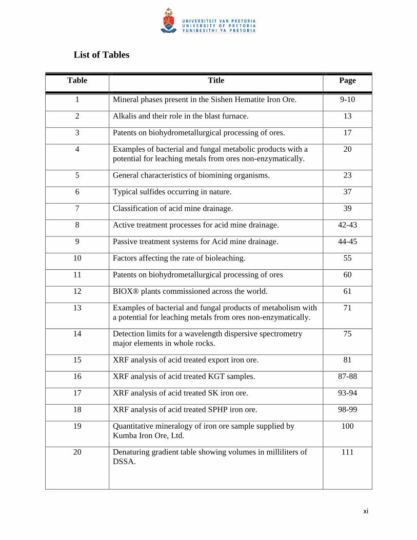
1 Mineral phases present in the Sishen Hematite Iron Ore. 9-10
2 Alkalis and their role in the blast furnace. 13
3 Patents on biohydrometallurgical processing of ores. 17
4 Examples of bacterial and fungal metabolic products with a
potential for leaching metals from ores non-enzymatically.
20
5 General characteristics of biomining organisms. 23
6 Typical sulfides occurring in nature. 37
7 Classification of acid mine drainage. 39
8 Active treatment processes for acid mine drainage. 42-43
9 Passive treatment systems for Acid mine drainage. 44-45
10 Factors affecting the rate of bioleaching. 55
11 Patents on biohydrometallurgical processing of ores 60
12 BIOX® plants commissioned across the world. 61
13 Examples of bacterial and fungal products of metabolism with
a potential for leaching metals from ores non-enzymatically.
71
14 Detection limits for a wavelength dispersive spectrometry
major elements in whole rocks.
75
15 XRF analysis of acid treated export iron ore. 81
16 XRF analysis of acid treated KGT samples. 87-88
17 XRF analysis of acid treated SK iron ore. 93-94
18 XRF analysis of acid treated SPHP iron ore. 98-99
19 Quantitative mineralogy of iron ore sample supplied by
Kumba Iron Ore, Ltd.
100
20 Denaturing gradient table showing volumes in milliliters of
DSSA.
111
xii
21 Average pH measurement of iron oxidizing leaching
experiment.
113
22 XRF analysis of A. ferrooxidans (ATCC 13598) treated
export iron sample.
116
23 XRF analysis of L. ferrooxidans (ATCC 49879) treated export
sample.
119
24 XRF analysis of L. ferriphilum (ATCC 49881) treated export
sample.
120
25 XRF analysis of Sulfobacillus thermosulfidooxidans treated
export sample.
121
26 XRF analysis of mix iron oxidizing bacteria leaching of export
sample.
122
27 pH measurement of heterotrophic bacterial leaching
experiments.
136
28 XRF analysis of Bacillus megaterium leached export iron ore. 141
29 XRF analysis of Bacillus cereus leached export iron ore. 142
30 XRF analysis of Bacillus subtilis leached export samples. 144
31 XRF analysis of Pseudomonas putida leached export sample. 145
32 XRF analysis of heterotrophic leached export sample. 146
33 Enrichment media for amplifying indigenous bacterial
cultures.
156
34 Average pH measurement for indigenous bacterial leaching
experiments.
160
35 Indigenous bacterial leaching of export iron ore. 166
36 Indigenous bacterial leaching of KGT sample. 170
37 Indigenous bacterial leaching of SK sample. 173
38 Indigenous bacteria identified from study. 176
39 Survey of each identified bacterial culture. 177-182
xiii
List of Figures
Figure Title Page
1 The Sishen Iron Ore Mine located in the Northern Cape, South
Africa.
1
2 Illustration of the different mineral phases present in the
Sishen iron ore.
4
3 Blast furnace diagram. 12
4 Illustration of bacteria selected from literature. 16
5 Model of the iron oxidation electron transport pathway of A.
ferrooxidans.
25
6 Model of the sulfur oxidation electron transport pathway of A.
ferrooxidans.
26
7 Schematic representation of the chemistry during in situ
electrolytic cultivation of iron-oxidizing bacteria.
35
8 Illustration of the contact leaching mechanism of the indirect
leaching attack as catalyzed by a pyrite attached A.
ferrooxidans cell.
47
9 Cycle of oxidative pyrite degradation by bacterial and/or
chemical leaching.
49
10 Heap-leaching operation at Cerro Colorado in Chile. 58
11 Illustration of copper leaching. 59
12 A typical BIOX® process flow sheet. 63
13 X-ray powder diffraction analyses of untreated export ore. 77
14 X-ray powder diffraction analyses of untreated KGT
(conglomerate) iron ore.
86
15 X-ray powder diffraction analyses of untreated SK (shale) iron
ore.
92
16 X-ray powder diffraction analyses of untreated SPHP iron ore. 97
17 Scanning electron microscopy analysis of acid treated KGT
iron ore.
101
18 Denaturing gradient gel electrophoresis (DGGE) of iron
oxidizing bacterial cultures.
123
19 Scanning electron microscopy images portraying biofilm
formation on the surface of the iron ore sample.
124
20 Contamination of heterotrophic leaching experiment with
unknown cultures.
147
21 Denaturing gradient gel of amplified genomic DNA from
heterotrophic leaching experiments showing dominant strains
present in leaching solution.
148
22 Scanning electron microscopy images of heterotrophic leached
export iron ore.
149
23 Denaturing gradient gel electrophoresis of amplified genomic
DNA from indigenous cultures.
174
24 Scanning electron microscopy images of indigenous bacterial
leached samples.
186
List of abbreviations
Al – aluminum
AMD – Acid mine drainage
ATP – Adenosine triphosphate
Ca – Calcium
Cl – Chlorine
DGGE – denaturing gradient gel electrophoresis
DNA – Deoxyribonucleic acid
Fe – Iron
g – gram
h – hour
HiPIP – High redox potential iron oxidase
K – potassium
KGT – Conglomerate ore
Li – Lithium
Mg – Magnesium
mg – Milligram
min – minute
Mtpa – million ton per annum
n/d – not determined
Ni – Nickel
P – Phosphate
PCR – polymerase chain reaction
RNA – Ribonucleic acid
rpm – revolution per minute
Rus –rusticyanin
Si – silicon
Summary
Kumba Iron Ore, Ltd. is the world‘s fourth largest supplier of sea-borne iron ore and currently
operates two mines in South Africa namely: the Sishen mine in the Northern Cape and
Thabazimbi mine in Limpopo.
The Sishen mine, located at the northern end of the Maremane anticline where the bulk of the
hematite ore is buried beneath younger cover lithologies, was our focus area. Here the iron
resources are made up by laminated and massive ore bodies that belong to the Asbestos Hills
Subgroup. These ore bodies are overlain by conglomerates, shales, flagstone and quartzite. The
alkalis, potassium and phosphorous, are common constituents of iron ore, which is known to
have a deleterious effect on the manufacturing of iron and steel. Therefore steel making
companies charge penalties when purchasing iron ore concentrates with alkali concentrations
above predetermined levels.
To ensure that the export batches at the Sishen mine stay within set limits, the ores from different
batches (with alkali concentration greater and below set limits) are mixed to produce a batch
which meet requirements. However this solution will soon become ineffective as the low alkali
ore is progressively depleted. Conventional methods used to treat high alkali ores include pyro-
and hydrometallurgical methods. These approaches have several limitations such as poor
product recovery, involvement of high process and energy cost and an increase in pollution load
of water resources. Therefore necessitating research and development of alternative cheap and
environment friendly procedures, which could supplement or replace conventional methods to
ensure that mining stays economically feasible at the Sishen Iron Ore mine.
The application of microorganisms to minng practices is collectively referred to as
biohydrometallurgy and includes bioleaching and biooxidation processes. The phrase
bioleaching refers to the conversion of an insoluble metal (typically a metal sulfide) into a
soluble form (typically a metal sulfate), via microbial activity. When metals are extracted into
solution, the process is referred to as bioleaching, whereas if the metal remains in the mineral, it
is referred to as biooxidation. The latter term biobeneficiation refers to the selective dissolution
of undesired minerals from the ores by direct or indirect action of microbes, thereby enriching
the desirable mineral content. Therefore the objective of this study was to determine whether
bacteria (naturally occurring on the ore or introduced species) could be used to selectively
remove the alkalis from the iron ore mined at Sishen. The species evaluated were able to change
the solution pH and/or form biofilms, which is assumed to have affected mineral mobilization.
Data obtained during this study suggests that the composition of the ore plays a significant role
in its susceptibility to bioleaching. Furthermore we also found that the indigenous cultures were
more effective than the introduced species to mobilize the alkalis, which could possibly be
ascribed to an adaptation of the microbes present.
These preliminary results suggest that bioleaching is an effective alternative cost effective
approach to treat iron ore and could possibly be implemented in future into the mining schedule
at Sishen.
Page | - 1 -
CHAPTER 1
GENERAL INTRODUCTION
1.1 BACKGROUND
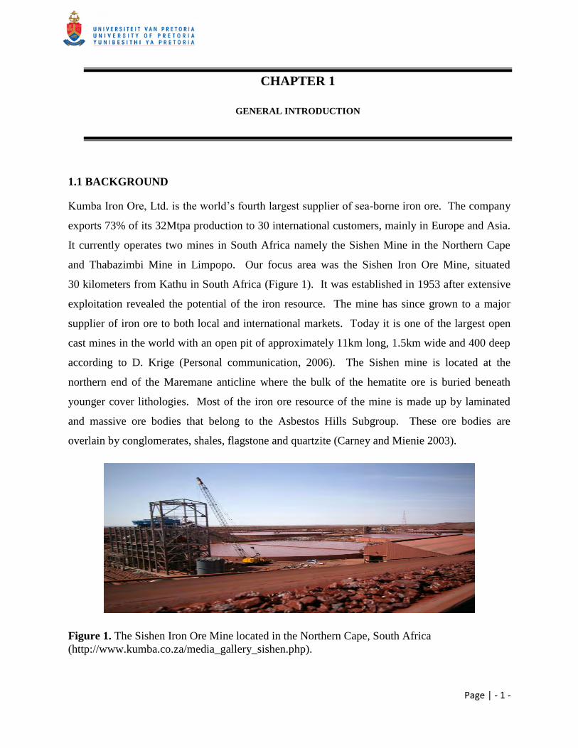
Kumba Iron Ore, Ltd. is the world‘s fourth largest supplier of sea-borne iron ore. The company
exports 73% of its 32Mtpa production to 30 international customers, mainly in Europe and Asia.
It currently operates two mines in South Africa namely the Sishen Mine in the Northern Cape
and Thabazimbi Mine in Limpopo. Our focus area was the Sishen Iron Ore Mine, situated
30 kilometers from Kathu in South Africa (Figure 1). It was established in 1953 after extensive
exploitation revealed the potential of the iron resource. The mine has since grown to a major
supplier of iron ore to both local and international markets. Today it is one of the largest open
cast mines in the world with an open pit of approximately 11km long, 1.5km wide and 400 deep
according to D. Krige (Personal communication, 2006). The Sishen mine is located at the
northern end of the Maremane anticline where the bulk of the hematite ore is buried beneath
younger cover lithologies. Most of the iron ore resource of the mine is made up by laminated
and massive ore bodies that belong to the Asbestos Hills Subgroup. These ore bodies are
overlain by conglomerates, shales, flagstone and quartzite (Carney and Mienie 2003).
Figure 1. The Sishen Iron Ore Mine located in the Northern Cape, South Africa
(http://www.kumba.co.za/media_gallery_sishen.php).
CHAPTER 1 INTRODUCTION
Page | - 2 -
The mining industry is constantly confronted with several difficulties such as depletion of high
grade minerals, worsening metal prices and mounting operation costs (Jain and Sharma 2004).
In the recent past, iron ore was a low priced commodity which discouraged industrial adoption of
hydrometallurgical beneficiation of these ores. At present an increase in global steel production
has increased the requirement for iron ore, with a consequent increase in the price for this
commodity, making hydrometallurgical approaches viable (Delvasto et al., 2008).
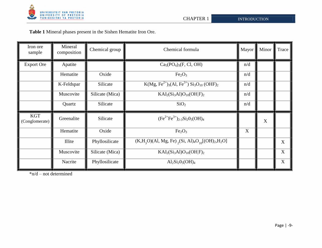
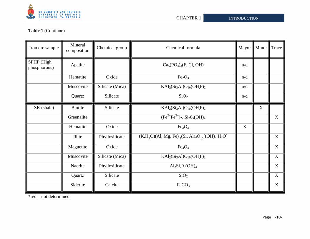
The iron ore mined at the Sishen Iron Ore Mine contains several different mineral phases
(Table 1) which includes potassium and phosphorous. These alkalis are known to have
deleterious effects on the manufacturing of iron and steel (Delvasto et al., 2008), therefore,
steel-making companies charge penalties when purchasing iron ore concentrates with alkali
concentrations above predetermined levels. The limits are determined by the steel making
companies and ranges from 0.25% mass in Japan to 0.55% mass in Switzerland for potassium
allowed in the iron ore concentrates. Kumba Iron Ore, Ltd., has an industrial set limit of 0.24%
potassium allowed in their export ore according to C. Taljaard (Personal communication, 2006).
The ore mined at different sits at the mine have varying composition and therefore different
alkali concentrations. To ensure that their export batches stay within this set limit, the ores from
different batches (with potassium >0.24% and <0.24%) are mixed to produce an average
potassium value of below 0.24% according to D. Krige (Personal communication, 2006).
However this is only a temporary solution as the low potassium ore (< 0.24%) is progressively
depleted according to D. Krige (Personal communication, 2006). Certain pyro – and
hydrometallurgical methods can be applied to decrease the alkali concentration
(Cheng et al., 1999; Kokal et al., 2003), however there are several drawbacks when using these
methods such as: poor product recovery, involvement of high process and energy cost and an
increase in pollution load of water resources (Jain and Sharma 2004). Thus an alternative,
natural and economical feasible process is required to aid conventional methods (such as pyro-
and hydrometallurgy processes) to remove unwanted alkalis from the ores concentrates mined at
Sishen.
CHAPTER 1 INTRODUCTION
Page | - 3 -
Biohydrometallurgy1 is an option for the removal of the deleterious phosphate and potassium, as
it is well established that many microorganisms are capable of mobilizing these minerals,
especially in nutrient limited environments (Banfield et al., 1999; Nautiyal 1999).
1.2 ALKALI BEARING MINERALS
The Sishen Iron Ore Mine is located at the Northern end of the Maremane anticline, where the
bulk of the hematite iron ore is buried beneath younger cover lithologies. The ore bodies are
overlain with conglomerates, shales, flagstones and quartzite (Carney and Mienie 2003). Kumba
Iron Ore, Ltd. supplied some of these samples for the bioleaching experiments. The samples
were labeled and characterized (Table 1) as follow: SK (shale with high potassium
concentrations), KGT (conglomerates with high phosphorous concentration), SPHP (sample with
high phosphorous concentration) and Export Ore (alkali concentrations at accepted limits)2.
Mineralogy of the samples was determined by Kumba Iron Ore Ltd. according to
B. Ntsoelengoe (Personal communication, 2006) and as part of the thesis objectives.
Following is a brief description of the mineral phases detected in the different iron ore samples,
as to familiarize the reader with the terms used in subsequent chapters. Emphasis was placed on
minerals containing the alkali‘s, potassium and phosphorous as well as hematite which was
found to be a mayor constituent of all the iron ore samples.
1.2.1 Hematite
Hematite is the mineral form of iron (III) oxide, which serves as a major supply of iron to
industry (Jorgenson and Kirk 2003). It crystallizes in the rhombohedral3 system and has the
same crystal arrangement as ilmenite (FeTiO3) and corundum (Al2O3) (Harrison et al., 2006).
Various forms of the mineral exist namely kidney ore, martite, iron rose and specularite
1 Biohydrometallurgical processes include bioleaching and biobenefication (Ehrlich 1991).
3 The term rhombohedral is used in crystallography. This crystal system is one of the seven lattice point groups,
named after the two dimensional rhombus (http://en.wikipedia.org/wiki/Rhombohedral).
CHAPTER 1 INTRODUCTION
Page | - 4 -
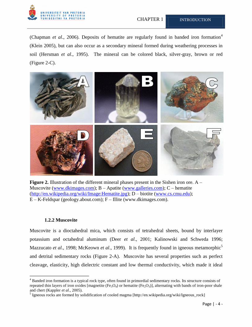
(Chapman et al., 2006). Deposits of hematite are regularly found in banded iron formation4
(Klein 2005), but can also occur as a secondary mineral formed during weathering processes in
soil (Hersman et al., 1995). The mineral can be colored black, silver-gray, brown or red
(Figure 2-C).
Figure 2. Illustration of the different mineral phases present in the Sishen iron ore. A –
Muscovite (www.dkimages.com); B – Apatite (www.galleries.com); C – hematite
(http://en.wikipedia.org/wiki/Image:Hematite.jpg); D – biotite (www.cs.cmu.edu);
E – K-Feldspar (geology.about.com); F – Illite (www.dkimages.com).
1.2.2 Muscovite
Muscovite is a dioctahedral mica, which consists of tetrahedral sheets, bound by interlayer
potassium and octahedral aluminum (Deer et al., 2001; Kalinowski and Schweda 1996;
Mazzucato et al., 1998; McKeown et al., 1999). It is frequently found in igneous metamorphic5
and detrital sedimentary rocks (Figure 2-A). Muscovite has several properties such as perfect
cleavage, elasticity, high dielectric constant and low thermal conductivity, which made it ideal
4 Banded iron formation is a typical rock type, often found in primordial sedimentary rocks. Its structure consists of
repeated thin layers of iron oxides [magnetite (Fe3O4) or hematite (Fe2O3)], alternating with bands of iron-poor shale
and chert (Kappler et al., 2005). 5 Igneous rocks are formed by solidification of cooled magma [http://en.wikipedia.org/wiki/Igneous_rock]
CHAPTER 1 INTRODUCTION
Page | - 5 -
for many electrical, electronic and thermal insulation processes (Deer et al., 2001). Rausell and
coworkers (1965) determined the sensitivity of muscovite to potassium concentration in solution.
They discovered that muscovite would not release interlayer potassium when it is placed in a
dilute electrolyte solution. Moreover, Pal and coworkers (2001) demonstrated that if biotite and
muscovite were both present in soil, no potassium dissolution from muscovite would occur.
A 0.1mg/l potassium concentration in solution was found to inhibit the exchange of potassium in
muscovite with other ions in solution (vermiculation) (Wilson 2004).
1.2.3 Apatite
Apatite is the most common phosphate mineral in nature (Figure 2-B). It occurs as an accessory
phase in igneous, metamorphic6 and sedimentary rocks (Welch et al., 2002). It is also produced
by biological systems, where it is a major constituent of tooth enamel and bone material. Apatite
is a group of phosphate minerals than can be categorized as three different minerals, namely
hydroxylapatite, fluoroapatite and chlorapatite. The fluorine, chlorine and hydroxyl ions are able
to substitute each other freely, as they are commonly found in the same specimen (Amethyst
Galleries‘ Mineral Gallery, 1996). Apatite is somewhat insoluble at near neutral pH (Welch et
al., 2002), however its reactivity and solubility is determined by its composition (Cazalbou et al.,
2004). It is known that fluoroapatite is less soluble than hydroxyapatite, however the solubility
and reactivity of apatite can be increase if there is a carbonate substitution into the phosphate site
(Welch et al., 2002).
1.2.4 Biotite
Biotite is a common phyllosilicate (or layered alumino-silicate) mineral within the mica group
(Douche 1993). It occurs as an essential, accessory or secondary mineral in most plutonic and
volcanic igneous rocks of crystal origin (Deer et al., 2001). Like other mica minerals, it has a
6 Metamorphic rock is the result of the transformation of an existing rock type
(http://en.wikipedia.org/wiki/Metamorphic_rocks).
CHAPTER 1 INTRODUCTION
Page | - 6 -
highly perfect basal cleavage and consists of flexible sheets or lamellae7, which can easily flake
off. The weathering of micas such as feldspar and biotite has been intensively studied due to
their importance as potassium source for plants (Wilson 2004). Weathering of biotite can
progress in one of the following ways: congruent (destruction of the mineral surface) and
incongruent (transformation into vermiculite by release of interlayer potassium) (Calvaruso et
al., 2006). The dissolution of biotite is thought to be diffusion-controlled and therefore depends
on the potassium concentration in the bulk solution (Wilson 2004). The sheets in the crystal
structure of biotite are made up of iron, magnesium, aluminum, silicon, oxygen and hydrogen
ions that are weakly bound together by potassium. The interlayer potassium can be substituted in
part by sodium, calcium, barium. Magnesium can be completely replaced by ferrous iron and
ferric iron and in part by titanium and manganese (Deer et al., 2001). Biotite is often referred to
as the iron mica as it contains more iron than phlogopite (Hashemi-Nezhad 2005). It has a
monoclinic crystal system8, with tabular to prismatic crystals with a pinacoid termination. The
structure contains four prism faces and two pinacoid faces to form a pseudohexagonal crystal.
The mineral can appear greenish to brown or black and even yellow when it is weathered
(Figure 2-D).
1.2.5 K-feldspar
Feldspars crystallize from magma in both intrusive and extrusive igneous rocks. It also occurs as
compact minerals, veins, and may also be present in many types of metamorphic rock. Feldspars
ubiquity and varying composition has led to its use as the primary tool in classifying igneous
rocks. It is however absent in certain rocks such as ultrabasic and rare alkaline rocks.
Compositions between NaAlSi3O8 and KAlSi3O8 are referred to as alkali feldspar and those
between NaAlSi3O8 and CaAl2Si2O8 as plagioclase. Alkali feldspar is white or colorless when
pure but appears pink when contaminated with iron (Figure 2-E) (Deer at al., 2001). The
structure consists of cross-linked, ‗double-crankshaft‘ chains of Si4+
and Al3+
tetrahedral with
charge compensating such as sodium, potassium and calcium occupying small cavities in the
7 A lamella is a gill-shaped structure: fine sheets of material held adjacent one another, with fluid in-between-(or
simply 'welded'-plates)[ http://en.wikipedia.org/wiki/Lamellae_(materials)] 8 In crystallography, the monoclinic crystal system is one of the 7 lattice point groups.
(http://en.wikipedia.org/wiki/Monoclinic_crystal_system)
CHAPTER 1 INTRODUCTION
Page | - 7 -
framework (Han and Lee 2005). Alkali feldspar can be weathered into a secondary mineral
kaolinite [Al2Si2O5(OH)4] under the influence of H+ ions. The low solubility of aluminum at pH
above 4.5 is a major reason for the slow dissolution rate of feldspar (Landeweert et al., 2001).
Experimental data has shown that dissolution of feldspar can either be surface-controlled or
leached-layer control. In the latter, incongruent9 dissolution occurs, thus certain ions will go
preferentially into solution, whereas others will remain in the solid crystalline phase. According
to this mechanism, a cation-depleted coating of silicon and aluminum (leached layer) builds up
around the feldspar and controls dissolution (Wilson 2004). The possible existence of a surface
controlled mechanism was illustrated by Holdren and Stillings during feldspar dissolution
experiments, where no leached layer occured (Wilson 2004).
1.2.6 Illite
Illite (hydromuscovite or hydromica) is a non-expanding, clay-sized phyllosilicate or layer
alumino-silicate that commonly occurs in sediments, soils and argillaceous sedimentary rocks
(Figure 2-F). Its structure consists of repeated tetrahedron – octahedron – tetrahedron layers.
The interlayer space is largely occupied by hydrated potassium cations which is possibly
responsible for the absence of swelling during reactions. Illite is structurally similar to
muscovite (Section 1.2.2) or sericite [KAl2(OH)2(AlSi3O10)], with more silicon, magnesium, iron
and water, and less tetrahedral aluminum and interlayer potassium in its structure. Illite occurs
as an alternation product of muscovite and feldspar during weathering
(Mengel and Uhlenbecker 1993).
1.2.7 Mineral dissolution
Mineral dissolution is thought to progress via two hypothetical mechanisms namely transport-
and surface-controlled, which is influenced by different rate-limiting steps. With transport-
controlled dissolution, the diffusion of the reactants or weathering products progresses through a
diffusion layer, towards the mineral surface or the bulk solution (Scheckel et al., 2005). Here the
9 Incongruent dissolution – Ions in the mineral lattice do not dissolve according to their stochiometric ration.
CHAPTER 1 INTRODUCTION
Page | - 8 -
reaction is controlled by the rate of diffusion or advection (Brantley 2004). Surface controlled
(interface-controlled) dissolution is limited by chemical reactions at the surface of the mineral
(Scheckel and Impellitteri 2005). As the reaction is controlled by the dissolution of the mineral,
an increase in diffusion would have no effect on the reaction rate due to an absence of a
concentration gradient at the mineral-water interface (Brantley 2004). Laboratory and field
experiments suggest that silicate dissolution under environmental conditions is surface-
controlled; however the discovery of a leached layer in many dissolving silicates has again raised
the question whether dissolution is either transport or surface-controlled
(Brantley 2004; Wilson 2004).
CHAPTER 1 INTRODUCTION
Page | -9-
Table 1 Mineral phases present in the Sishen Hematite Iron Ore.
Iron ore
sample
Mineral
composition Chemical group Chemical formula Mayor Minor Trace
Export Ore Apatite Ca5(PO4)3(F, Cl, OH) n/d
Hematite Oxide Fe2O3 n/d
K-Feldspar Silicate K(Mg, Fe2+
)3(Al, Fe3+
) Si3O10 (OHF)2 n/d
Muscovite Silicate (Mica) KAI2(Si3Al)O10(OH,F)2 n/d
Quartz Silicate SiO2 n/d
KGT (Conglomerate)
Greenalite Silicate (Fe2+
Fe3+
)2-3Si205(OH)4 X
Hematite Oxide Fe2O3 X
Illite Phyllosilicate (K,H3O)(Al, Mg, Fe)
2(Si, Al)4O10
[(OH)2,H2O] X
Muscovite Silicate (Mica) KAI2(Si3Al)O10(OH,F)2 X
Nacrite Phyllosilicate Al2Si205(OH)4 X
*n/d – not determined
CHAPTER 1 INTRODUCTION
Page | -10-
Table 1 (Continue)
Iron ore sample Mineral
composition Chemical group Chemical formula Mayor Minor Trace
SPHP (High
phosphorous) Apatite Ca5(PO4)3(F, Cl, OH) n/d
Hematite Oxide Fe2O3 n/d
Muscovite Silicate (Mica) KAI2(Si3Al)O10(OH,F)2 n/d
Quartz Silicate SiO2 n/d
SK (shale) Biotite Silicate KAl2(Si3Al)O10(OH,F)2 X
Greenalite (Fe2+
Fe3+
)2-3Si205(OH)4 X
Hematite Oxide Fe2O3 X
Illite Phyllosilicate (K,H3O)(Al, Mg, Fe)
2(Si, Al)4O10
[(OH)2,H2O] X
Magnetite Oxide Fe3O4 X
Muscovite Silicate (Mica) KAl2(Si3Al)O10(OH,F)2 X
Nacrite Phyllosilicate Al2Si205(OH)4 X
Quartz Silicate SiO2 X
Siderite Calcite FeCO3 X
*n/d – not determined
CHAPTER 1 INTRODUCTION
Page | -11-
1.3 IRON ORE PRODUCTION
Pyrometallurgical processes have been used for decades for the recovery of base metal values
such as copper, lead, zinc, etc. from scrap and secondary materials. The traditional process
includes: reverberatory -, rotary kiln (short rotary furnace), electric-, slag fuming – and blast
furnaces. In addition to these methods new, more intense pyrometallurgical processes have been
designed and include the following: Flash smelting, Bath smelting and Top blown rotary
converter (Hancock and Peacey 1994). Following is a brief overview of iron production using a
blast furnce, as well as the importance of alkalis in iron manufacturing and living cells. The
importance of the alkalis to living organisms is covered in this section, as the main objective of
this study is to remove the unwanted alkalis from the ores via microbial processes.
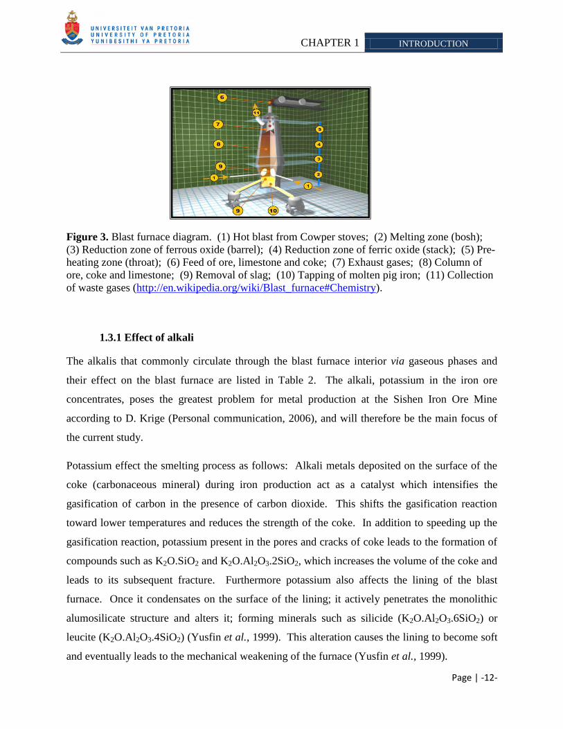
The iron ore is smelted in a blast furnace at 1500°C (Figure 3). Ore, coke (carbonaceous
material) and limestone flux are continuously supplied through the top of the furnace
(Figure 3-6), while air (or pure oxygen) is blown into the bottom of the chamber (Figure 3-1).
The main chemical reaction producing molten iron is: Fe2O3 + 3CO → 2Fe + 3CO2. The air (or
pure oxygen) blown into the furnace reacts with the coke (carbon source) to produce carbon
monoxide and heat. The carbon monoxide then reacts with the iron oxide (found in the iron ore)
to produce molten iron and carbon dioxide. The carbon dioxide, unreacted carbon monoxide and
nitrogen from the air passes up through the furnace as fresh feed material travels down into the
reaction zone (Figure 3-2). As the material travels downward, the counter-current gases preheat
the feed, decompose the limestone to calcium oxide and begin to reduce the iron oxides
(Figure 3-5). ―Four uptakes‖ allow the gases to exit the furnace (Figure 3-7 and Figure 3-11).
The gases are passed through a ―dust catcher‖, to allow small particles to be removed, and a gas
cooling unit, before the gas is released into the atmosphere. The end products, molten metal and
slag phases are tapped from the bottom of the furnace (Figure 3-10).
CHAPTER 1 INTRODUCTION
Page | -12-
Figure 3. Blast furnace diagram. (1) Hot blast from Cowper stoves; (2) Melting zone (bosh);
(3) Reduction zone of ferrous oxide (barrel); (4) Reduction zone of ferric oxide (stack); (5) Pre-
heating zone (throat); (6) Feed of ore, limestone and coke; (7) Exhaust gases; (8) Column of
ore, coke and limestone; (9) Removal of slag; (10) Tapping of molten pig iron; (11) Collection
of waste gases (http://en.wikipedia.org/wiki/Blast_furnace#Chemistry).
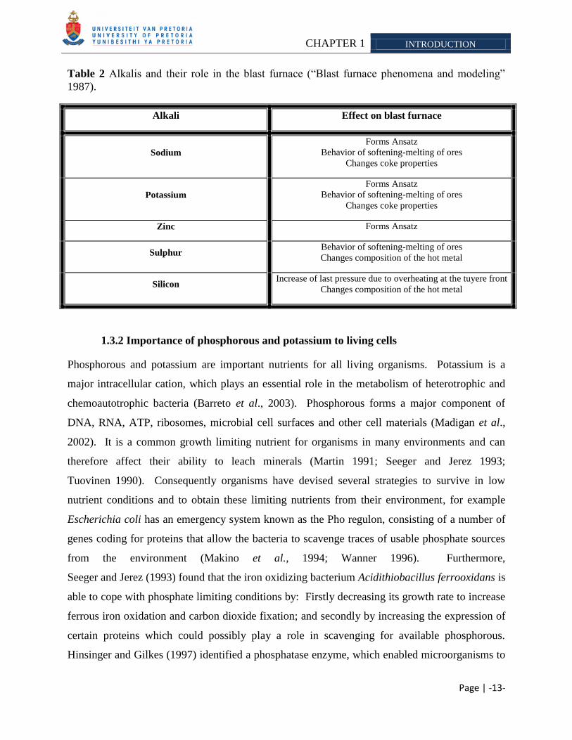
1.3.1 Effect of alkali
The alkalis that commonly circulate through the blast furnace interior via gaseous phases and
their effect on the blast furnace are listed in Table 2. The alkali, potassium in the iron ore
concentrates, poses the greatest problem for metal production at the Sishen Iron Ore Mine
according to D. Krige (Personal communication, 2006), and will therefore be the main focus of
the current study.
Potassium effect the smelting process as follows: Alkali metals deposited on the surface of the
coke (carbonaceous mineral) during iron production act as a catalyst which intensifies the
gasification of carbon in the presence of carbon dioxide. This shifts the gasification reaction
toward lower temperatures and reduces the strength of the coke. In addition to speeding up the
gasification reaction, potassium present in the pores and cracks of coke leads to the formation of
compounds such as K2O.SiO2 and K2O.Al2O3.2SiO2, which increases the volume of the coke and
leads to its subsequent fracture. Furthermore potassium also affects the lining of the blast
furnace. Once it condensates on the surface of the lining; it actively penetrates the monolithic
alumosilicate structure and alters it; forming minerals such as silicide (K2O.Al2O3.6SiO2) or
leucite (K2O.Al2O3.4SiO2) (Yusfin et al., 1999). This alteration causes the lining to become soft
and eventually leads to the mechanical weakening of the furnace (Yusfin et al., 1999).
CHAPTER 1 INTRODUCTION
Page | -13-
Table 2 Alkalis and their role in the blast furnace (―Blast furnace phenomena and modeling‖
1987).
1.3.2 Importance of phosphorous and potassium to living cells
Phosphorous and potassium are important nutrients for all living organisms. Potassium is a
major intracellular cation, which plays an essential role in the metabolism of heterotrophic and
chemoautotrophic bacteria (Barreto et al., 2003). Phosphorous forms a major component of
DNA, RNA, ATP, ribosomes, microbial cell surfaces and other cell materials (Madigan et al.,
2002). It is a common growth limiting nutrient for organisms in many environments and can
therefore affect their ability to leach minerals (Martin 1991; Seeger and Jerez 1993;
Tuovinen 1990). Consequently organisms have devised several strategies to survive in low
nutrient conditions and to obtain these limiting nutrients from their environment, for example
Escherichia coli has an emergency system known as the Pho regulon, consisting of a number of
genes coding for proteins that allow the bacteria to scavenge traces of usable phosphate sources
from the environment (Makino et al., 1994; Wanner 1996). Furthermore,
Seeger and Jerez (1993) found that the iron oxidizing bacterium Acidithiobacillus ferrooxidans is
able to cope with phosphate limiting conditions by: Firstly decreasing its growth rate to increase
ferrous iron oxidation and carbon dioxide fixation; and secondly by increasing the expression of
certain proteins which could possibly play a role in scavenging for available phosphorous.
Hinsinger and Gilkes (1997) identified a phosphatase enzyme, which enabled microorganisms to
Alkali Effect on blast furnace
Sodium
Forms Ansatz
Behavior of softening-melting of ores
Changes coke properties
Potassium
Forms Ansatz
Behavior of softening-melting of ores
Changes coke properties
Zinc Forms Ansatz
Sulphur Behavior of softening-melting of ores
Changes composition of the hot metal
Silicon Increase of last pressure due to overheating at the tuyere front
Changes composition of the hot metal
CHAPTER 1 INTRODUCTION
Page | -14-
cleave phosphates from organophosphates, thereby enabling them to utilize it. However, when
orthophosphate and organophosphates are limited in the environment, microorganisms are forced
to scavenge for other nutrient sources (Wilson 2004). Rogers et al. (1998) found that if feldspar
contained trace amounts of phosphorous as apatite inclusions, it would be highly colonized by
microorganisms and caused the mineral surface to become etched. They further discovered that
the feldspar samples which, did not contain apatite inclusions but had a similar bulk composition,
were not colonized by microorganisms. Therefore they concluded that it is possible that the
microorganism could identify the phosphatase in the mineral and actively remove and
incorporate the phosphates into their metabolism. Several reports have demonstrated that
microorganisms are able to accelerate the release of dissolved phosphate to solution from rock
phosphate by producing inorganic and organic acids (Drever and Vance 1994; Hinsinger and
Gilkes 1997; Jennings 1995; Margolis and Moreno 1992). Therefore we hypothesize that when
microorganisms are grown under potassium and phosphorous limiting conditions, they could
possibly remove these limiting nutrients from the Sishen hematite iron ore.
1.4 EXPERIMENTAL APPROACH
The mining industry constantly faces various challenges such as depletion of high grade
minerals, worsening metal prices and mounting operation costs (Jain and Sharma 2004). In the
past, iron ore was a low priced product which discouraged industrial adoption of
hydrometallurgical beneficiation of these ores. However due to an increase in global steel
production, the requirement of iron ore increased, with a consequent increase in the commodities
value, making research and application of alternative processing and treatment processes viable
(Delvasto et al., 2008).
The iron ore mined at the Sishen Iron Ore Mine contains several phases of minerals (Apatite,
biotite, illite and muscovite) which contains the alkali‘s potassium and phosphorous. Alkali‘s
have a deleterious effect on the manufacturing of iron and steel (Delvasto et al., 2008), forcing
companies to charge penalties when purchasing iron ore concentrates with alkali concentrations
above certain levels. This urged preliminary research into an alternative, cheap and
environmentally friendly approach to treat high alkali ore bodies. Kumba Iron Ore, Ltd. faces a
severe crisis within the next 3-4 years, as the low alkali ore becomes more depleted according to
CHAPTER 1 INTRODUCTION
Page | -15-
R. Grunewaltd (Personal communication, 2006). This spurred additional research into
optimizing the current approaches reported in this thesis. Our preliminary experiments were to
test the ability of iron oxidizing, heterotrophic (Figure 4) and indigenous bacteria to remove the
unwanted substances (biobenefication10
). The applicability of biohydrometallurgy to remove
alkali from iron ore was tested during this study. Several reports supported the hypothesis that
bacteria might be able to mobilize the alkalis.
Microbes and minerals are closely linked, such that one often cannot exist without the other in
nature (Lower et al., 2001). Several researchers have commented on the role that
microorganisms play in the cycling of elements and sorption of metals (Langley and Beveridge
1999), the dissolution of minerals (Banfield and Hamers 1997; Barker et al., 1998; Bennett et al.,
2001; Edwards et al., 1998; Stone 1997) and mineral crystallization (Fortin and Beveridge 1997;
Fortin et al., 1997; Warren and Ferris 1998). Experiments done by Bennett (2001) provided
evidence that silicate weathering by bacteria is sometimes driven by the nutrient requirements of
the microbial consortium. This suggested that the weathering of a mineral may be influenced by
its nutritional potential, with the microorganisms destroying only the beneficial minerals. The
researchers further commented that the microorganisms could thus directly benefit from the
weathering of specific minerals. Other research supporting the findings showed that
microorganisms would colonize various different minerals such as feldspar (Bennett et al., 2001;
Rogers et al., 1998), when nutrient became limiting in the environment. More recently
Rogers and Bennett (2004), further supported the idea that microbes would preferentially leach
minerals with beneficial nutrients. They found that in petroleum contaminated aquifers; silicate
glasses that contained phosphorous and iron were preferentially colonized compared to glasses
that did not contain these nutrients.
Krebs et al. (1997) listed three principles that enable microorganism to leach and mobilize
metals from solid material, namely: (i) redox potential; (ii) the formation of organic and
inorganic acids; and (iii) the excretion of complexing agents.
In 1997 Bosecker commented that the dissolution of non-sulfidic ores can be achieved by
exploiting the metabolic capabilities of heterotrophic microorganisms (Table 3). These
10
Biobeneficiation refers to the selective dissolution of undesired minerals from the ores by direct or indirect action
of microbes, thereby enriching the desirable mineral content (Deo et al., 2001; Vasan et al., 2001)
CHAPTER 1 INTRODUCTION
Page | -16-
organisms produce metabolites that are possibly able to solubilize and/or aid in solubilizing
oxide, silicate, carbonate and hydroxide minerals (Ivarson 1981; Jain and Sharma 2004).
Therefore we chose bacteria with different nutritional requirement (chemolithotrophic and
heterotrophic) to conduct our experiments. The specific strains for this study will be discussed in
subsequent chapters. Furthermore the ability of indigenous bacteria to remove the alkali from
the ore was tested, as it is hypothesized that these organisms might be adapted to scavenge for
limiting nutrients. The most important parameter included in the heterotrophic (Chapter 5) and
indigenous (Chapter 6) leaching experiments, is to starve the cells for potassium, as this could
possibly increase the expression of genes that might enable the organisms to remove the nutrient
from the ore.
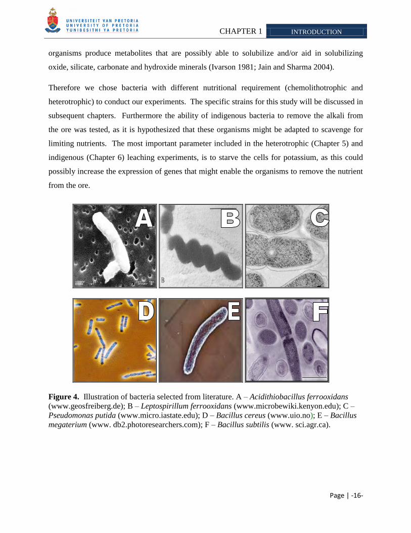
Figure 4. Illustration of bacteria selected from literature. A – Acidithiobacillus ferrooxidans
(www.geosfreiberg.de); B – Leptospirillum ferrooxidans (www.microbewiki.kenyon.edu); C –
Pseudomonas putida (www.micro.iastate.edu); D – Bacillus cereus (www.uio.no); E – Bacillus
megaterium (www. db2.photoresearchers.com); F – Bacillus subtilis (www. sci.agr.ca).
CHAPTER 1 INTRODUCTION
Page | -17-
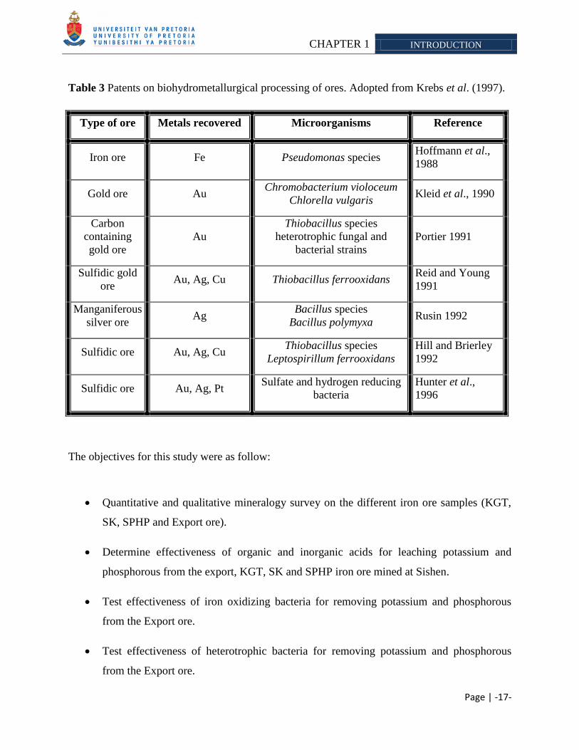
Table 3 Patents on biohydrometallurgical processing of ores. Adopted from Krebs et al. (1997).
Type of ore Metals recovered Microorganisms Reference
Iron ore Fe Pseudomonas species Hoffmann et al.,
1988
Gold ore Au Chromobacterium violoceum
Chlorella vulgaris Kleid et al., 1990
Carbon
containing
gold ore
Au
Thiobacillus species
heterotrophic fungal and
bacterial strains
Portier 1991
Sulfidic gold
ore Au, Ag, Cu Thiobacillus ferrooxidans
Reid and Young
1991
Manganiferous
silver ore Ag
Bacillus species
Bacillus polymyxa Rusin 1992
Sulfidic ore Au, Ag, Cu Thiobacillus species
Leptospirillum ferrooxidans
Hill and Brierley
1992
Sulfidic ore Au, Ag, Pt Sulfate and hydrogen reducing
bacteria
Hunter et al.,
1996
The objectives for this study were as follow:
Quantitative and qualitative mineralogy survey on the different iron ore samples (KGT,
SK, SPHP and Export ore).
Determine effectiveness of organic and inorganic acids for leaching potassium and
phosphorous from the export, KGT, SK and SPHP iron ore mined at Sishen.
Test effectiveness of iron oxidizing bacteria for removing potassium and phosphorous
from the Export ore.
Test effectiveness of heterotrophic bacteria for removing potassium and phosphorous
from the Export ore.
CHAPTER 1 INTRODUCTION
Page | -18-
Survey the effectiveness of indigenous bacteria to remove potassium and phosphorous
from KGT, Export and SK samples.
Determine bacterial community of indigenous bacteria enriched for during the
biobenefication experiment.
Page | -19-
CHAPTER 2
LITERATURE REVIEW
IRON OXIDIZING BACTERIA AND THEIR IMPORTANCE IN THE BIOMINING INDUSTRY
2.1 INTRODUCTION
Biohydrometallurgical processes include bioleaching and biobenefication (Ehrlich 1991). The
phrase bioleaching refers to the conversion of an insoluble metal (typically a metal sulfide) into a
soluble form (typically a metal sulfate), via microbial activity (Rawlings 2002). When metals
are extracted into solution, the process is typically referred to as bioleaching, whereas if the
metal remains in the mineral, it is referred to as biooxidation (Rawlings 2005). The latter term
biobenefication refers to a process in which microorganisms are used to selectively remove
undesirable mineral components from ores (Ehrlich 1991). The solubilization process is
considered to be largely a chemical process, with the microorganisms providing the chemicals
(Table 4) and the space where the mineral dissolution reaction occurs (Gehrke et al., 1998;
Rawlings 2005). These processes utilize biogeochemical activities of which various bacteria and
fungi are capable. The function of bacteria is to act as a catalyst of the dissolution reaction or
generator of metabolic products, which causes chemical dissolution. Whereas fungi exclusively
act as a generator of metabolic products that causes chemical dissolution of metal values
(Ehrlich 1991).
Bioleaching is a technology that is applicable to metal extractions from samples such as:
low-grade ores, ore benefication, coal benefication, metal detoxification and recovery
of metals from waste materials (Bosecker 2001; Jain and Sharma 2004). Except for
its industrial application to raw materials, microbial leaching has several
other potential uses such as remediation of mining sites, treatment of metal containing
waste products and detoxification of sewage sludge (Bosecker 2001).
CHAPTER 2 LITERATURE REVIEW
Page | -20-
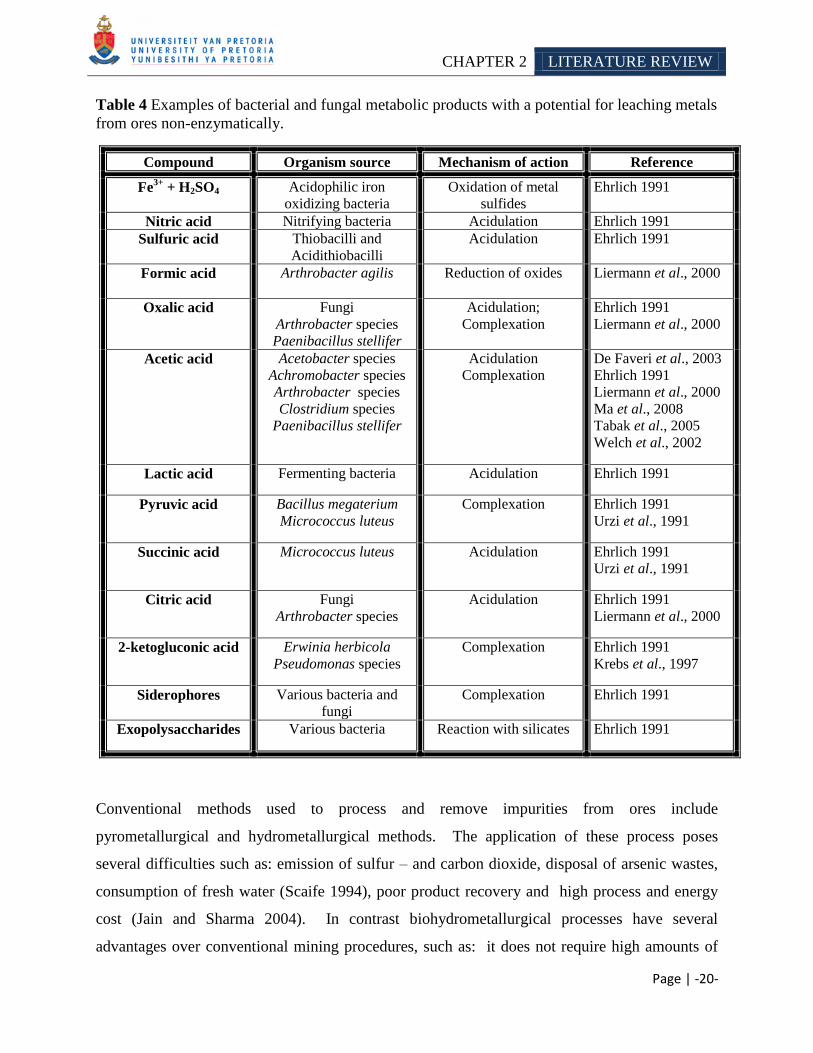
Table 4 Examples of bacterial and fungal metabolic products with a potential for leaching metals
from ores non-enzymatically.
Compound Organism source Mechanism of action Reference
Fe3+
+ H2SO4 Acidophilic iron
oxidizing bacteria
Oxidation of metal
sulfides
Ehrlich 1991
Nitric acid Nitrifying bacteria Acidulation Ehrlich 1991
Sulfuric acid Thiobacilli and
Acidithiobacilli
Acidulation Ehrlich 1991
Formic acid Arthrobacter agilis
Reduction of oxides Liermann et al., 2000
Oxalic acid Fungi
Arthrobacter species
Paenibacillus stellifer
Acidulation;
Complexation
Ehrlich 1991
Liermann et al., 2000
Acetic acid Acetobacter species
Achromobacter species
Arthrobacter species
Clostridium species
Paenibacillus stellifer
Acidulation
Complexation
De Faveri et al., 2003
Ehrlich 1991
Liermann et al., 2000
Ma et al., 2008
Tabak et al., 2005
Welch et al., 2002
Lactic acid Fermenting bacteria Acidulation Ehrlich 1991
Pyruvic acid Bacillus megaterium
Micrococcus luteus
Complexation Ehrlich 1991
Urzi et al., 1991
Succinic acid Micrococcus luteus Acidulation Ehrlich 1991
Urzi et al., 1991
Citric acid Fungi
Arthrobacter species
Acidulation Ehrlich 1991
Liermann et al., 2000
2-ketogluconic acid Erwinia herbicola
Pseudomonas species
Complexation Ehrlich 1991
Krebs et al., 1997
Siderophores Various bacteria and
fungi
Complexation Ehrlich 1991
Exopolysaccharides Various bacteria Reaction with silicates Ehrlich 1991
Conventional methods used to process and remove impurities from ores include
pyrometallurgical and hydrometallurgical methods. The application of these process poses
several difficulties such as: emission of sulfur – and carbon dioxide, disposal of arsenic wastes,
consumption of fresh water (Scaife 1994), poor product recovery and high process and energy
cost (Jain and Sharma 2004). In contrast biohydrometallurgical processes have several
advantages over conventional mining procedures, such as: it does not require high amounts of
CHAPTER 2 LITERATURE REVIEW
Page | -21-
energy as with roasting and smelting; the process does not produce harmful gaseous emissions
such as sulfur dioxide (Rawlings 2002), the technology is relatively inexpensive and safe (Jain
and Sharma 2004). However acid mine drainage can be generated by certain microorganisms,
which in turn can harm the environment if it is not properly controlled (Olson et al., 2003;
Rawlings 2005). Bioleaching will however not completely replace conventional methods,
because: firstly the bioleaching process does not recover precious metals from the ores which are
often an essential component in the profitability of the operation; and secondly when ore bodies
do not contain sufficient acid consuming minerals, the residual acids generated have to be
neutralized during the leaching process, thus increasing the operational cost (Dreshner 2004).
Leaching of sulfidic minerals using chemolithotrophic bacteria are the best studied and
commercially exploited biotechnology today (Jain and Sharma 2004). Iron oxidizing bacteria
have been applied in laboratory and/or large scale heap processes to solubilize insoluble metal
sulfates of copper (Brierley and Brierley 2001; Pinches et al., 1997; Qiu et a. 2005), cobalt, gold
(Aswegen 1993; Olson 1994), nickel (Dew and Miller 1997), zinc (Kai et al., 2000) and uranium
(Khalid et al., 1993) have been solubilized in the laboratory or in large-scale heap or tank
aeration processes (Amankwah 2005; Rawlings 2002; Rawlings et al., 2003). The metabolic
capability of iron oxidizing bacteria has been exploited in several designed industrial scale
processes such as the BIOX® process (Van Aswegen et al., 2006), GEOCOATTM
(Harvey et al., 2002), BacTechTM
process (Olson et al., 2003) and the BioCOPTM
process
(Batty and Rorke 2006). These microorganisms produce chemicals such as ferric iron and
sulfuric acid from the ferrous iron and sulfur contained in the mineral or solution
(Rawlings et al., 2003). The ferric ion serves as an oxidation attack on the mineral
(Takai et al., 2001), whereas sulfuric acid is responsible for a proton attack (Rawlings 2005).
Furthermore bacteria are able to leach minerals possibly via two proposed systems namely
‗contact leaching‘ and ‗non-contact leaching‘. With contact leaching biofilms are produced.
Here the extracellular polymeric substance can function in the following way: it can mediate
attachment to a (metal) sulfide surface and it may concentrate the ferric ions by complexation
through uronic acids or other residues at the mineral surface, thereby allowing an oxidative
attack on the sulfide (Sand and Gehrke 2006).
CHAPTER 2 LITERATURE REVIEW
Page | -22-
The focus of this review is the iron oxidizing bacteria currently used in industry. Reference will
be made to: general characteristic of biomining organisms; factors affecting leaching (both
microbial and physiochemical); microbial oxidation and leaching mechanisms; iron oxidizing
bacteria currently used in industry and candidates with future prospects; and acid mine drainage
formation and treatment strategies.
2.2 IRON OXIDIZING BACTERIA
2.2.1 General characteristics
The microorganisms currently employed in biomining operations have several characteristics in
common (Table 5). These abilities enable them to leach the minerals or impurities from the ores
and enable them to survive in the biomining process environment. For example with a passive
process such as heap leaching (Section 2.6.1), all the requirements of the leaching organisms are
met. The air in the heap provides the carbon source in the form of carbon dioxide and the
preferred electron acceptor (O2). The mineral leached supplies the electron donor (ferrous iron
and/or inorganic sulfur) (Section 2.2.2) and the water used to irrigate the heap, functions as the
growth medium (Rawlings 2002). Some species of biomining organisms are able to fix nitrogen
from the air (Section 2.2.3); however, they may not be able to do so in a highly aerated
environment (Rawlings 2002). In commercial processes, small quantities of inexpensive,
fertilizer-grade, ammonium sulfate and potassium phosphate may be added to ensure that
nutrient limitation does not occur (Rawlings 2002). Due to the few requirements of these
organisms, it has become economical feasible to leach low-grade ore.
CHAPTER 2 LITERATURE REVIEW
Page | -23-
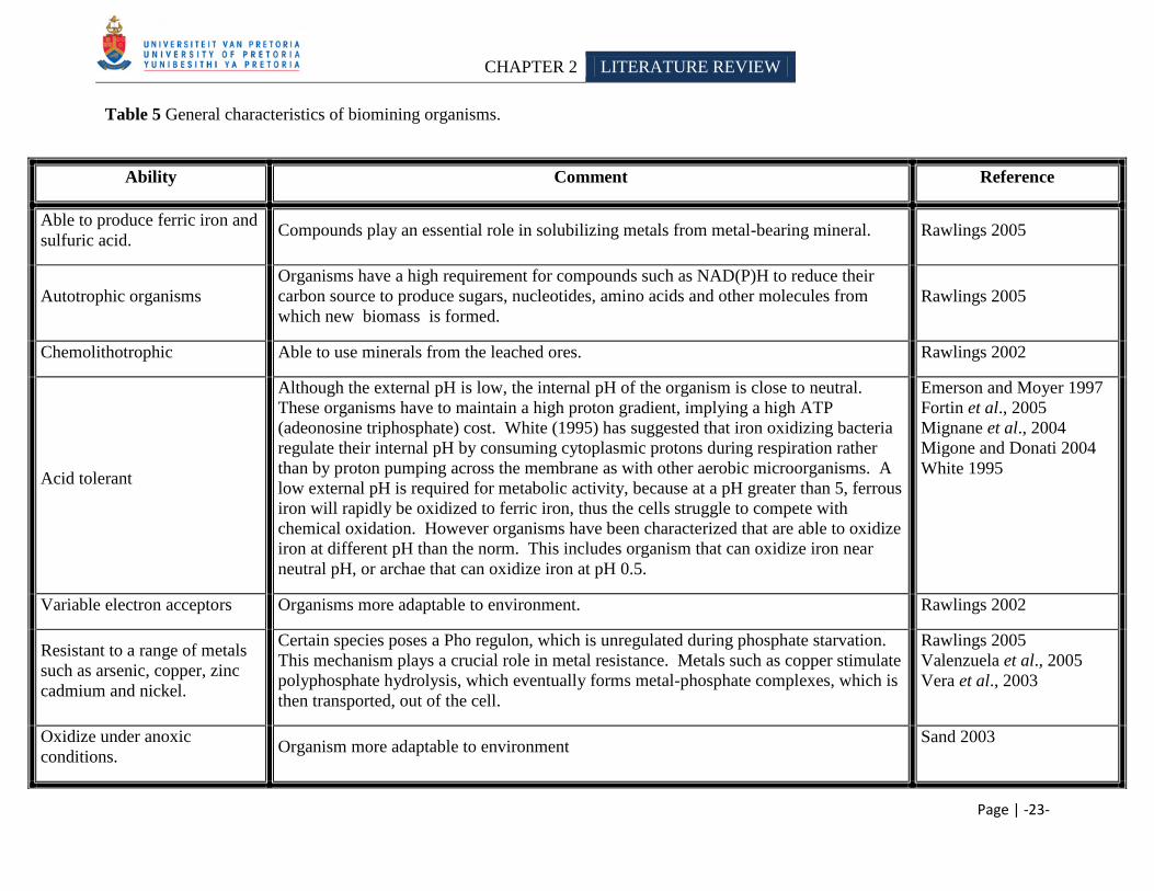
Table 5 General characteristics of biomining organisms.
Ability Comment Reference
Able to produce ferric iron and
sulfuric acid. Compounds play an essential role in solubilizing metals from metal-bearing mineral. Rawlings 2005
Autotrophic organisms
Organisms have a high requirement for compounds such as NAD(P)H to reduce their
carbon source to produce sugars, nucleotides, amino acids and other molecules from
which new biomass is formed.
Rawlings 2005
Chemolithotrophic Able to use minerals from the leached ores. Rawlings 2002
Acid tolerant
Although the external pH is low, the internal pH of the organism is close to neutral.
These organisms have to maintain a high proton gradient, implying a high ATP
(adeonosine triphosphate) cost. White (1995) has suggested that iron oxidizing bacteria
regulate their internal pH by consuming cytoplasmic protons during respiration rather
than by proton pumping across the membrane as with other aerobic microorganisms. A
low external pH is required for metabolic activity, because at a pH greater than 5, ferrous
iron will rapidly be oxidized to ferric iron, thus the cells struggle to compete with
chemical oxidation. However organisms have been characterized that are able to oxidize
iron at different pH than the norm. This includes organism that can oxidize iron near
neutral pH, or archae that can oxidize iron at pH 0.5.
Emerson and Moyer 1997
Fortin et al., 2005
Mignane et al., 2004
Migone and Donati 2004
White 1995
Variable electron acceptors Organisms more adaptable to environment. Rawlings 2002
Resistant to a range of metals
such as arsenic, copper, zinc
cadmium and nickel.
Certain species poses a Pho regulon, which is unregulated during phosphate starvation.
This mechanism plays a crucial role in metal resistance. Metals such as copper stimulate
polyphosphate hydrolysis, which eventually forms metal-phosphate complexes, which is
then transported, out of the cell.
Rawlings 2005
Valenzuela et al., 2005
Vera et al., 2003
Oxidize under anoxic
conditions. Organism more adaptable to environment
Sand 2003
CHAPTER 2 LITERATURE REVIEW
Page | -24-
2.2.2 Leaching mechanisms
Bioleaching is the use of microorganisms to solubilize minerals from ores (Rawlings 2002). The
solubilization is thought to be mainly a chemical process, with the organisms supplying the
chemicals and the space in which the reaction occurs (Gehrke et al., 1998; Rawlings 2005). The
iron oxidizing bacteria supplies ferric iron and/or acids, typically sulfuric acid (proton hydrolysis
attack), which acts upon the minerals (Rawlings 2005; Takai et al., 2001). Following is a
general description of the ferrous (production of ferric iron) and sulfur oxidation (production of
sulfuric acid) mechanisms of iron oxidizing bacteria.
2.2.2.1 Ferrous oxidation
Under aerobic conditions, ferrous iron (Fe2+
) is spontaneously oxidized to ferric iron (Fe3+
),
unless the pH is low (White 1995). Lacey and Lawson (1970) found that if the solution pH was
below 2, the oxidation kinetics of ferrous ion to ferric iron is low; however when A. ferrooxidans
was inoculated into the solution the reaction increased 5 to 6 times. The ferrous/ferric iron redox
couple has a positive standard electrode potential and therefore only oxygen can act as a natural
electron acceptor (Reaction 1) and iron as an electron donor under aerobic conditions
(Rawlings 2005).
Reaction 1: Fe2+
+ H+ + ½O2 Fe
3+ + ½H2O
Acidophilic bacteria are able to use ferrous iron as an electron acceptor. The difference between
the ferrous/ferric iron and oxygen/water redox couples are small, and only one mole of electrons
are released per mole of iron (Reaction 1), therefore a lot of ferrous iron is needed to sustain
bacterial life. The large quantities of ferrous iron required are not transported into the cell, but
only deliver its electron to a carrier situated in the cell envelope. The iron oxidation mechanism
of Acidithiobacillus ferrooxidans has been extensively studied (Rawlings 2005). The proposed
model is illustrated in figure 5. A. ferrooxidans contains a rus operon, which is suspected to
encode for the electron transport chain that is used during oxidation (Appia-Ayme et al., 1999).
This operon consists of genes for an aa3-type cytochrome oxidase (Kai et al., 1992;
Yarzábal et al., 2002), an outermembrane located cytochrome-c (Cyc2) (Sugio et al., 1998), a c4
type cytochrome (Cavazza et al., 1996) , a copper-containing protein rysticyanin and an open
CHAPTER 2 LITERATURE REVIEW
Page | -25-
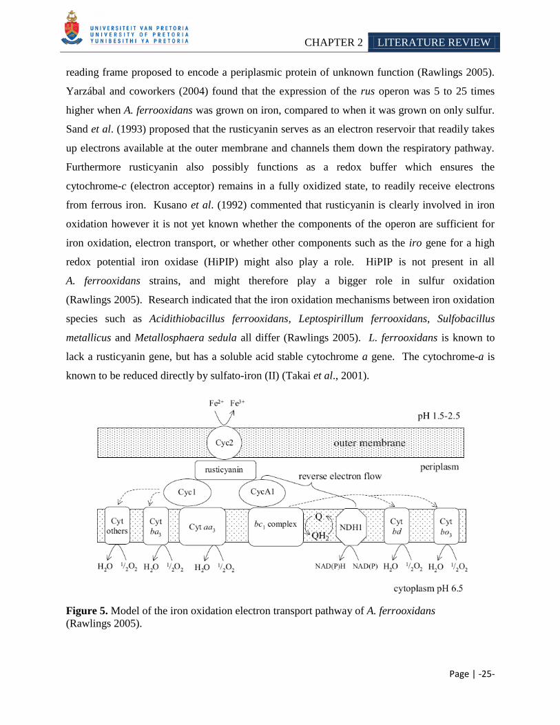
reading frame proposed to encode a periplasmic protein of unknown function (Rawlings 2005).
Yarzábal and coworkers (2004) found that the expression of the rus operon was 5 to 25 times
higher when A. ferrooxidans was grown on iron, compared to when it was grown on only sulfur.
Sand et al. (1993) proposed that the rusticyanin serves as an electron reservoir that readily takes
up electrons available at the outer membrane and channels them down the respiratory pathway.
Furthermore rusticyanin also possibly functions as a redox buffer which ensures the
cytochrome-c (electron acceptor) remains in a fully oxidized state, to readily receive electrons
from ferrous iron. Kusano et al. (1992) commented that rusticyanin is clearly involved in iron
oxidation however it is not yet known whether the components of the operon are sufficient for
iron oxidation, electron transport, or whether other components such as the iro gene for a high
redox potential iron oxidase (HiPIP) might also play a role. HiPIP is not present in all
A. ferrooxidans strains, and might therefore play a bigger role in sulfur oxidation
(Rawlings 2005). Research indicated that the iron oxidation mechanisms between iron oxidation
species such as Acidithiobacillus ferrooxidans, Leptospirillum ferrooxidans, Sulfobacillus
metallicus and Metallosphaera sedula all differ (Rawlings 2005). L. ferrooxidans is known to
lack a rusticyanin gene, but has a soluble acid stable cytochrome a gene. The cytochrome-a is
known to be reduced directly by sulfato-iron (II) (Takai et al., 2001).
Figure 5. Model of the iron oxidation electron transport pathway of A. ferrooxidans
(Rawlings 2005).
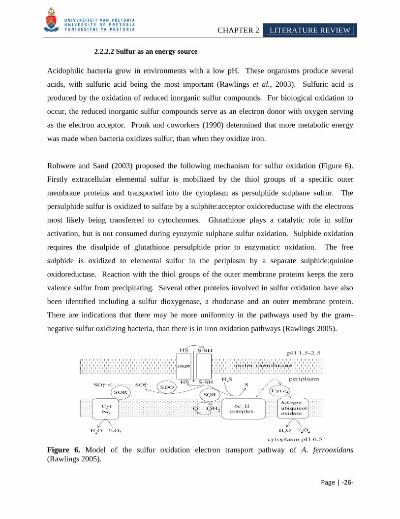
CHAPTER 2 LITERATURE REVIEW
Page | -26-
2.2.2.2 Sulfur as an energy source
Acidophilic bacteria grow in environments with a low pH. These organisms produce several
acids, with sulfuric acid being the most important (Rawlings et al., 2003). Sulfuric acid is
produced by the oxidation of reduced inorganic sulfur compounds. For biological oxidation to
occur, the reduced inorganic sulfur compounds serve as an electron donor with oxygen serving
as the electron acceptor. Pronk and coworkers (1990) determined that more metabolic energy
was made when bacteria oxidizes sulfur, than when they oxidize iron.
Rohwere and Sand (2003) proposed the following mechanism for sulfur oxidation (Figure 6).
Firstly extracellular elemental sulfur is mobilized by the thiol groups of a specific outer
membrane proteins and transported into the cytoplasm as persulphide sulphane sulfur. The
persulphide sulfur is oxidized to sulfate by a sulphite:acceptor oxidoreductase with the electrons
most likely being transferred to cytochromes. Glutathione plays a catalytic role in sulfur
activation, but is not consumed during eynzymic sulphane sulfur oxidation. Sulphide oxidation
requires the disulpide of glutathione persulphide prior to enzymaticc oxidation. The free
sulphide is oxidized to elemental sulfur in the periplasm by a separate sulphide:quinine
oxidoreductase. Reaction with the thiol groups of the outer membrane proteins keeps the zero
valence sulfur from precipitating. Several other proteins involved in sulfur oxidation have also
been identified including a sulfur dioxygenase, a rhodanase and an outer membrane protein.
There are indications that there may be more uniformity in the pathways used by the gram-
negative sulfur oxidizing bacteria, than there is in iron oxidation pathways (Rawlings 2005).
Figure 6. Model of the sulfur oxidation electron transport pathway of A. ferrooxidans
(Rawlings 2005).
CHAPTER 2 LITERATURE REVIEW
Page | -27-
2.2.3 Iron oxidizing bacteria currently used in the biomining industry
Iron oxidizing bacteria have been used in different commercial processes such as the BIOX®,
BACTECHTM
, GEOCOATTM
and BACFOXTM
process. These organisms have been isolated
from various locations such as: sulfur springs (Maeda et al., 1999), hot-spring biomats
(Belkova et al., 2004), acid mine drainage sites (Fowler et al., 1999; Maeda et al., 1999;
Rawlings 2005), corroded concrete (Maeda et al., 1999; Terunobu et al., 1999), roots from
Wetland plants (Emerson et al., 1999) and water systems (Coetser and Cloete 2005), etc. The
following section will assess iron oxidizing bacteria found in non-sterile commercial or pilot
scale processes.
2.2.3.1 Acidithiobacillus
These organisms were previously placed in the genus Thiobacillus, but due to 16S rRNA
sequence results, it became clear that this genus contained sulfur oxidizing bacteria that belonged
to α-, β and γ-division of the Proteobacteria (Lane et al., 1992). To solve this problem, the
genus was subdivided (Kelly and Wood 2002) and a new genus Acidithiobacillus was created,
which includes all the highly acidophilic organisms. This new genus includes the organisms
Acidithiobacillus ferrooxidans, Acidithiobacillus thiooxidans and Acidithiobacillus caldus.
These bacteria appear to be ubiquitous and have been isolated from various sites such as: sulfur
springs (Maeda et al., 1999), hot-spring biomats (Belkova et al., 2004), acid mine drainage sites
(Fowler et al., 1999; Maeda et al., 1999; Rawlings 2005), corroded concrete (Maeda et al., 1999;
Terunobu et al., 1999), roots from Wetland plants (Emerson et al., 1999) and water systems
(Coetser and Cloete 2005).
2.2.3.2 Acidithiobacillus ferrooxidans
Acidithiobacillus ferrooxidans was first isolated from an acid mine in 1947
(Colmer and Hinkle 1947). It was the first bacterium discovered that was able to oxidize
minerals (Rawlings 2002) and is to date the most studied (Battaglia et al., 1997). It is a
mesophilic, rod-shaped, gram-negative, motile (Ohmura et al., 1996) and non-sporeforming
bacterium. It has an optimal pH range of 1.5-2.5 and temperature range of 28-37°C
(Niemelä et al., 1994; Razzell et al., 1962; Solisio et al., 2002), with a maximum of 46°C
(Nemati et al., 1989). The low pH makes the environment inhospitable to other bacteria
CHAPTER 2 LITERATURE REVIEW
Page | -28-
(Mason and Rice 2002). Although the external pH is low, the internal pH of the organism is
close to neutral. Therefore these organisms have to maintain a high proton gradient implying a
high ATP (adenosine triphosphate) cost. White (1995) suggested that iron oxidizing bacteria
regulate their internal pH by consuming cytoplasmic protons during respiration rather than by
proton pumping across the membrane as with other aerobic microorganism. A low external pH
is required for metabolic activity, because at a pH greater than 5, ferrous iron will rapidly be
oxidized to ferric iron, thus the cells struggle to compete with chemical oxidation.
Kupka and Kupsăvo (1999) demonstrated that the oxidation of ferrous ions by A. ferrooxidans
decreased when the pH exceeded 2.5.
A. ferrooxidans obtains energy by oxidizing ferrous iron and sulfur, and does not favor
environments in which the redox potential is high (high ferric ion to ferrous ion ratio)
(Rawlings et al., 1999). A. ferrooxidans is able to obtain energy from alternative sources such
as: i) hydrogen, with oxygen as terminal electron acceptor under aerobic conditions
(Barreto et al., 2003; Drobner et al., 1990) or under anaerobic conditions by dissimilatory
reduction of ferric iron (Ohmura et al., 1999; Pronk et al., 1991) and ii) formate with oxygen or
ferric iron as terminal electron acceptor (Pronk et al., 1991). The enzyme, hydrogenase, enables
A. ferrooxidans to use hydrogen as an energy source. When hydrogen is utilized, the optimal pH
range shifts to 3.0-5.8. A. ferrooxidans assimilates carbon dioxide (its main carbon source) via
the Calvin Benson cycle (Levicán et al., 2002), which is catalyzed by the ribulose-biphosphate
carboxylase enzyme. The nitrogen requirement of the organism is met through ammonium
compounds present in the biomining mediums (Ramírez et al., 2004). The G+C mole ratio of A.
ferrooxidans isolates are between 57%-59% and belong to possibly 4 different DNA-DNA
hybridization groups (Rawlings 2002).
The composition of its extracellular polymeric substance (EPS) changes depending on whether
ferric iron or sulfur is present. A. ferrooxidans is sensitive to sodium (Lavalle et al., 2005) and
organic matter (Fournier et al., 1998; Gu et al., 2007; Manning 1975; Rawlings 2002;
Sand et al., 1992). Strains have been isolated that showed low tolerance to sodium, however its
growth is completely inhibited by the presence of 10 000 mg/l glucose, regardless of the initial
cell density and in spite of favorable pH and aeration conditions (Marchand 2003). Its sensitivity
to organic matter, makes it hard to grow in the laboratory, however several different medias has
CHAPTER 2 LITERATURE REVIEW
Page | -29-
been developed: 9k media (Ishigaki et al., 2005; Kai et al., 2000; Park et al., 2005;
Tyagi et al., 1993), ISP media (Manning 1975), Sodium thiosulfate media (Colmer et al., 1950)
and Leathen media (Chan et al., 2003). A two layer technique has also been developed, where
the iron oxidizing bacterium is grown along with heterotrophic bacteria, which removes the
compounds that are toxic to the iron oxidizer (Johnson 1995; Rawlings 2002).
A. ferrooxidans is resistant to a wide range of metals such as copper (Alvarez and Jerez 2004),
zinc, silver, mercury (Barreto et al., 2003), arsenic (Nies and Silver 1995) and uranium
(Pronk et al., 1991). Solisio et al. (2002) applied this ability to leach zinc and aluminum from
industrial waste sludge. Several researchers have commented that the polyphosphate degradation
of A. ferrooxidans might play a role in metal detoxification. Furthermore research has
determined that Acidithiobacillus resistance to arsenate greatly depends on its arsenic resistance
ars operon (Nies and Silver 1995) and also on the phosphate concentration present in the growth
media, as arsenate is a structural analog of phosphate and will therefore be readily taken up by
the cell during starvation. Also copper ions stimulate polyphosphate degradation and phosphate
efflux, again supporting a model for metal detoxification in which heavy metals stimulate
polyphosphate hydrolysis and the metal-phosphate complexes formed would be transported out
of the cell as part of a possibly functional heavy metal tolerance mechanism (Alvarez and Jerez
2004).
Researchers have demonstrated that the existence and importance of a hydrogen sulphide:ferric
ion oxidoreductase and sulfate:ferric iron oxidoreductase in sulfur oxidation. These enzymes use
ferric iron as an electron acceptor in the oxidation of elemental sulfur and sulfite (Sugio 1992).
Sugio et al. (1996) found that these enzymes were responsible for the solubilization of copper
from copper concentrates (Sugio et al., 1996). Research by Kai et al. (2000) further supported
the existence of these enzymes. They demonstrated that A. ferrooxidans was able to leach zinc
sulfide and manganese dioxide simultaneously. The ferric iron oxidized the zinc sulfide and the
ferrous iron reduced the manganese oxide. This allowed the consumption of the oxidizing and
reducing agents to decrease and the possibility that these two minerals could be simultaneously
electrowon. The researchers commented that during simultaneous leaching of metal oxides and
metal sulfides, the consumption of oxidizing and reducing agents decreases.
CHAPTER 2 LITERATURE REVIEW
Page | -30-
2.2.3.3 Acidithiobacillus thiooxidans
A. thiooxidans is nutritionally very similar to A. ferrooxidans; however it is not able to oxidize
ferrous iron and is therefore limited to using reduced sulfur compounds as an electron donor.
A. thiooxidans is a mesophilic (Rawlings 2002), motile by flagella (Ohmura et al., 1996) and
grows in a pH range of 0.5-5.5. The organism has a G + C ratio of 53% with a DNA-DNA
similarity with A. ferrooxidans of about 20% or less (Rawlings 2002).
2.2.3.4 Acidithiobacillus caldus
Acidithiobacillus caldus is able to oxidize reduces sulfur compounds by not ferric iron (Hallberg
and Lindstrőm 1994). Dopson and Lindstrőm (2004) isolated this organism from a bioleaching
community that grew at 45°C on pyrite, arsenical pyrite and chalcopyrite. This is a moderate
thermophilic organism which has an optimum temperature of about 45°C. Some strains are able
to grow mixotrophically using yeast extract or glucose (Rawlings 2002).
2.2.3.5 Thiobacillus
Thiobacillus species were found to be satellite organisms, which grew with A. ferrooxidans.
Furthermore, researchers demonstrated that Thiobacilli was able to grow mixotrophically on
tetrathionate with glucose, which could therefore aid A. ferrooxidans growth in the presence of
glucose (Ehrlich 1996). Tuffin and coworkers (2005) isolated a tronsposon TnAtcArs that carries
a set of arsenic resistance genes from Acidithiobacillus caldus which was islated from a
commercial biooxidation treatment plant of arsenopyrite. Research found that the conditions in
the tank made this organism highly resistant. Other species of acidophilic Thiobacilli have been
described by Huber and Stetter (1990) which includes Thiobacillus prosperus and T. cuprinus.
Thiobacillus prosperus is a new group of halotolerant metal mobilizing bacteria. T. cuprinus is a
facultative chemolithotrophic bacterium which can oxidize metal sulfides but cannot oxidize
ferrous iron. Bosecker (1997) demonstrated that these organisms can preferentially mobilize
copper from chalcopyrite, which makes them ideal candidates for future bioleaching technology.
CHAPTER 2 LITERATURE REVIEW
Page | -31-
2.2.3.6 Leptospirillum
Leptospirillum was first isolated in 1972 from a copper leach dump and coal spoil heap in
Armendia (Inoue et al., 2000). The bacteria of this genus are quite similar to that of
Acidithiobacillus. They are also acid tolerant, with the ability to grow in a pH range of between
1.5-1.8 (Rawlings 2002); gram negative, autotrophic (Johnson et al., 2006), spiral shaped,
mesophilic, non-sporeforming (Johnson et al., 1992), motile by means of flagella
(Ohmura et al., 1996) and chemolithoautrophic (Rawlings 2002). Based on 16S rRNA sequence
analysis, this genus does not form part of the division Proteobacteria, but rather Nitrospora
(Lane et al., 1992). All Leptospirilli are only able to use ferrous iron as an electron donor,
resulting in a higher affinity for ferrous iron compared to Acidithiobacilli (Rawlings 2002). The
growth of Acidithiobacillus ferrooxidans is inhibited above 36 mM ferric iron concentration
whereas L. ferrooxidans is resistant even to 500 mM ferric iron concentration (Dave 2008;
Mignane et al., 2004). Leptospirilli have not been reported to grow on sulfur-salt medium,
however they are able to efficiently degrade pyrite (FeS2) (Sugio et al., 1994). This organism
also poses the hydrogen sulphide:ferric ion oxidoreductase, as A. ferrooxidans, which catalyzes
the oxidation of elemental sulfur with ferric ion as an electron acceptor to give ferrous ion and
the bisulfite ion. However research showed that during sulfur oxidation, the organism produces
bisulfate as an intermediate in low quantities, which inhibits the hydrogen sulphide:ferric ion
oxidoreductase, thereby inhibiting sulfur oxidation in Leptospirilli (Sugio et al., 1994).
Leptospirilli have a G+C content of 49-51%, with four known species (Rawlings 2002).
DNA-DNA hybridization between the groups is 11% or less (Coram and Rawlings 2002).
L. ferrooxidans belongs to the 49-51% G +C group, with L. ferriphilum belonging to the higher
G + C group (Rawlings 2002). The fourth specie was isolated from an abandoned pyrite mine by
Bond and coworkers (2000). Leptospirilli have been found widely in biooxidation processes,
usually in combination with a sulfur oxidizing bacterium such as Acidithiobacillus thiooxidans or
A. caldus (Rawlings 2002). Moreover, Tuffin et al. (2006) also isolated a set of arsenic
resistance genes from a highly arsenic resistant strain of L. ferriphilum, as with Acidithiobacillus
caldus.
CHAPTER 2 LITERATURE REVIEW
Page | -32-
2.2.3.7 Acidiphilium
These are gram-negative, acid tolerant heterotrophs. These organisms are not primarily involved
in mineral decomposition (Hallberg and Johnson 2001), but they are included in this section
because they have been detected in batch reactors (Goebel and Stackebrandt 1994) and have
frequently been found growing near A. ferrooxidans, where they are believed to feed on the
organic waste products produced by the iron and sulfur oxidizing bacteria (Rawlings 2002).
These organisms have been used in the laboratory to cultivate A. ferrooxidans, in the overlay
plating technique (Section 2.2.3.2), to remove presumably organic toxins form the upper layer,
allowing the organic matter intolerant iron and sulfur oxidizing bacteria to grow (Johnson 1995).
These organisms are also able to use ferric iron as an electron acceptor, at low oxygen tension,
thus regenerating ferrous iron (Rawlings 2002; Romero et al., 2003).
2.2.4 Iron oxidizing bacteria with potential use in the biomining industry
Mineral oxidation is a mixture of chemistry and biology. Current biomining operations run at
temperatures ranging from ambient to 40°C. Certain minerals such as chalcopyrite can only be
economically leached at elevated temperature. Thus organisms able grow and leach at these
elevated temperatures will be valuable in future bioleaching processes (Rawlings 2002). At
temperatures exceeding 65°C, the biomining consortia are dominated by Archaea rather than
bacteria, with species of Sulfolobus and Metallosphaera being most prominent
(Norris et al., 2000). The following organisms discussed are likely to contribute to the
functioning of these new bioleaching process (Rawlings 2002).
2.2.4.1 Sulfobacillus
Sulfobacilli are gram positive, endospore-forming, non-motile, moderate thermophilic
acidophiles, which can grow in a temperature range of 28-60°C, with an optimum around 50°C.
It grows in a pH range from 1.9 – 3.0 with an optimum range of 1.9 - 2.4 (Rawlings 2002;
Watling et al., 2008). These organisms have been isolated from heaps of mineral waste and
biomining operations (Rawlings 2002). They are able to grow autotrophically or
CHAPTER 2 LITERATURE REVIEW
Page | -33-
heterotrophically. When they grow autotrophically they use ferrous iron, reduced inorganic
sulfur compounds or sulfide minerals as electron donors (Norris et al., 1996).
S. thermosulfidooxidans cannot use sulfate as a source of sulfur and therefore must be provided
with reduced sulfur. Sulfobacilli are not able to efficiently fix carbon dioxide, and therefore need
elevated CO2 in the atmosphere (yeast extract can be added to the media) (Rawlings 2002).
Glucose can serve as a carbon and energy source for Sulfobacilli when growing
heterotrophically. These microorganisms are also able to grow in the absence of oxygen, using
ferric iron as an electron acceptor and either organic or inorganic sulfur compounds as electron
donor. Four species have been distinguished using phylogenetic and physiological
characteristics: Sulfobacillus thermosulfidooxidans (DSM 9293), S. acidophilus (DSM 10332),
S. sibiricus (DSM 17363) and S. thermotolerance (DSM 17362). S. acidophilus (Norris et al.,
1996) can be differentiated from the other species by being capable of autotrophic growth on
sulfur with addition of carbon dioxide enriched air. S. sibiricus (Melamud et al., 2003), can be
differentiated from the other species by its: higher optimal growth temperature, ability to grow
on thiosulfate or tetrathionate as sole energy source and inability to grow heterotrophically.
S. thermotolerance (Bogdanova et al., 2006) has larger cells and a lower optimal temperature.
A possible fifth specie of the genus Sulfobacillus has been isolated from a continuous
bioleaching tank, charged with cobaltiferous pyrite (Johnson et al., 2007).
2.2.4.2 Ferroplasma
Ferroplasma are archaea. These organisms are mesophilic, pleomorphic in shape, lack cell walls
and grows optimally at a temperature of 33°C and pH of 1.7 (Rawlings 2002).
Ferroplasma acidiphilum was isolated from a pilot plant bioreactor in Kazakstan, which treated
arsenopyrite/pyrite (Golyshina et al., 2000). This organism is able to oxidize ferrous iron but not
sulfur and appears to be obligatory aerobic (Rawlings 2002).
CHAPTER 2 LITERATURE REVIEW
Page | -34-
2.2.4.3 Sulfolobus
The first thermophilic acidophile isolated and characterized were Sulfolobus acidocaldarius.
These organisms are obligated thermophilic autotrophic archaea that obtains energy by oxidizing
ferrous iron, reduced inorganic sulfur compounds or sulfide ore (Das et al., 1999 as cited in
Brandl 2001; Rawlings 2002). They have an optimal growth temperature in the range of 65-
70°C. From a biomining perspective, there is particular interest in thermoacidophiles, such as
Sulfolobus metallicus, Acidianus spp. and Metallosphaera spp, that mobilize metals from sulfidic
minerals (Johnson 2006). The bioleaching kinetics of thermophiles is known to be higher than
that of the mesophiles (Das et al., 1999 as cited in Brandl 2001; Rawlings 2002).
2.2.4.4 Gallionella ferruginea
This is an aerobic, neutralophilic iron oxidizer. It that has been obtained in purified enrichment
and pure culture in the laboratory and it grows at very low oxygen tensions in opposing gradients
of oxygen and ferrous iron (Hanert 1992). Hallbeck et al. (1993) have found that this organism
was able to grow lithotrophically on iron, however there is also proof that this organism can
grow mixotrophically.
2.2.4.5 Metallosphaera
Metallosphaera are aerobic iron and sulfur oxidizing chemolithotrophs archaea that are able to
grow on complex organics such as casamino acids or yeast extract, but not on sugars.
Researchers demonstrated that Metallosphaera sedula is able to grow in a pH range of 1.0-4.5
and oxidize a variety of minerals at temperature 80-85°C (Rawlings 2002). Norris (1997) found
Metallosphaera like organisms to be the most efficient organisms to leach chalcopyrite at high
temperatures. However, 16s rRNA sequence analysis placed some disagreement on these
finding as to which are more Sulfolobus or Metallosphaera like (Norris et al., 2000).
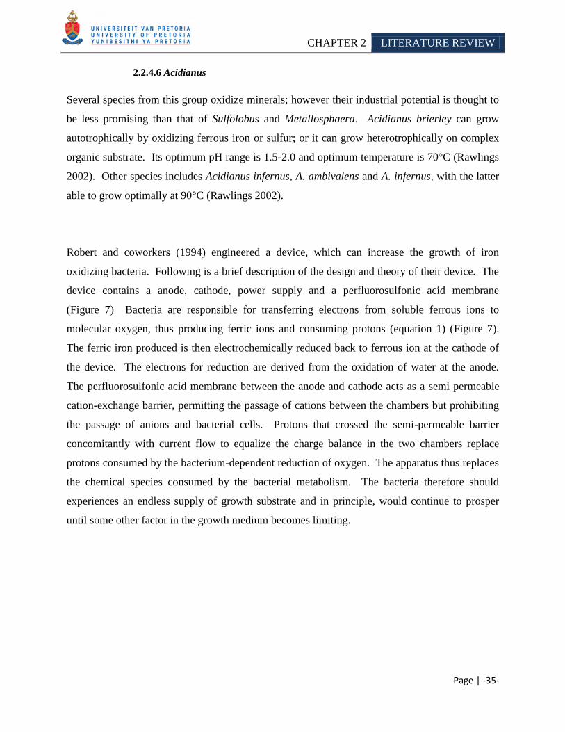
CHAPTER 2 LITERATURE REVIEW
Page | -35-
2.2.4.6 Acidianus
Several species from this group oxidize minerals; however their industrial potential is thought to
be less promising than that of Sulfolobus and Metallosphaera. Acidianus brierley can grow
autotrophically by oxidizing ferrous iron or sulfur; or it can grow heterotrophically on complex
organic substrate. Its optimum pH range is 1.5-2.0 and optimum temperature is 70°C (Rawlings
2002). Other species includes Acidianus infernus, A. ambivalens and A. infernus, with the latter
able to grow optimally at 90°C (Rawlings 2002).
Robert and coworkers (1994) engineered a device, which can increase the growth of iron
oxidizing bacteria. Following is a brief description of the design and theory of their device. The
device contains a anode, cathode, power supply and a perfluorosulfonic acid membrane
(Figure 7) Bacteria are responsible for transferring electrons from soluble ferrous ions to
molecular oxygen, thus producing ferric ions and consuming protons (equation 1) (Figure 7).
The ferric iron produced is then electrochemically reduced back to ferrous ion at the cathode of
the device. The electrons for reduction are derived from the oxidation of water at the anode.
The perfluorosulfonic acid membrane between the anode and cathode acts as a semi permeable
cation-exchange barrier, permitting the passage of cations between the chambers but prohibiting
the passage of anions and bacterial cells. Protons that crossed the semi-permeable barrier
concomitantly with current flow to equalize the charge balance in the two chambers replace
protons consumed by the bacterium-dependent reduction of oxygen. The apparatus thus replaces
the chemical species consumed by the bacterial metabolism. The bacteria therefore should
experiences an endless supply of growth substrate and in principle, would continue to prosper
until some other factor in the growth medium becomes limiting.
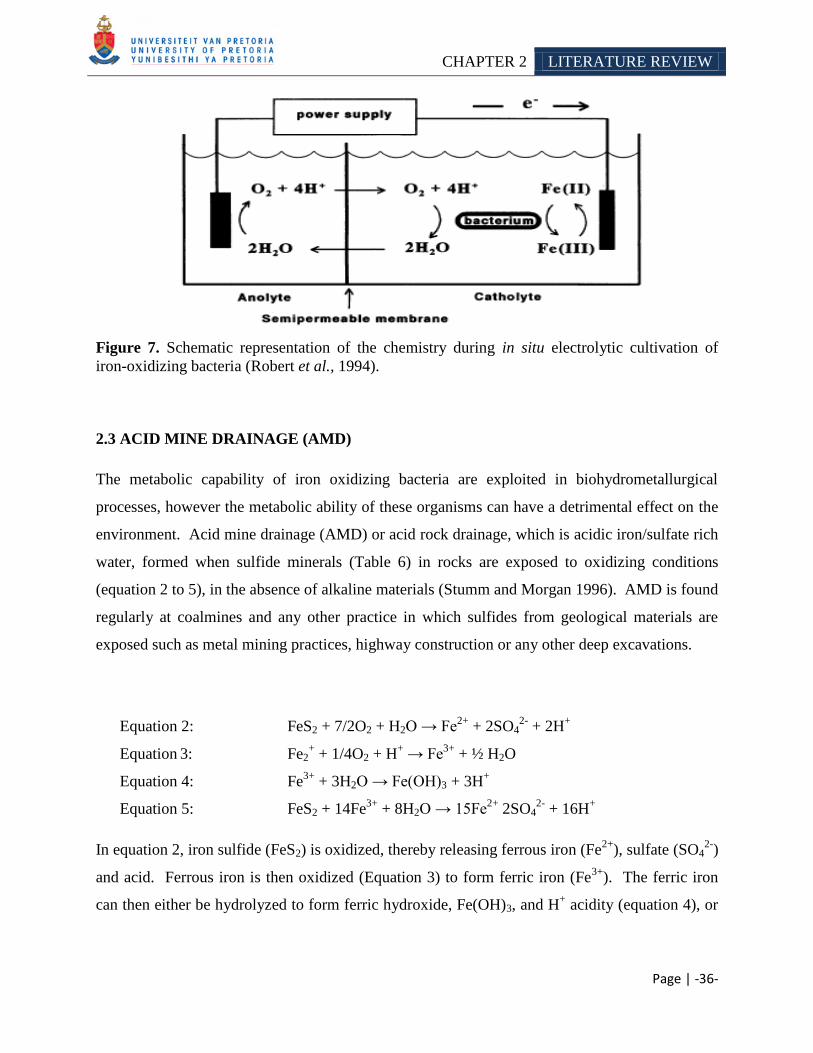
CHAPTER 2 LITERATURE REVIEW
Page | -36-
Figure 7. Schematic representation of the chemistry during in situ electrolytic cultivation of
iron-oxidizing bacteria (Robert et al., 1994).
2.3 ACID MINE DRAINAGE (AMD)
The metabolic capability of iron oxidizing bacteria are exploited in biohydrometallurgical
processes, however the metabolic ability of these organisms can have a detrimental effect on the
environment. Acid mine drainage (AMD) or acid rock drainage, which is acidic iron/sulfate rich
water, formed when sulfide minerals (Table 6) in rocks are exposed to oxidizing conditions
(equation 2 to 5), in the absence of alkaline materials (Stumm and Morgan 1996). AMD is found
regularly at coalmines and any other practice in which sulfides from geological materials are
exposed such as metal mining practices, highway construction or any other deep excavations.
Equation 2: FeS2 + 7/2O2 + H2O → Fe2+
+ 2SO42-
+ 2H+
Equation 3: Fe2
+ + 1/4O2 + H
+ → Fe
3+ + ½ H2O
Equation 4: Fe3+
+ 3H2O → Fe(OH)3 + 3H+
Equation 5: FeS2 + 14Fe3+
+ 8H2O → 15Fe2+
2SO42-
+ 16H+
In equation 2, iron sulfide (FeS2) is oxidized, thereby releasing ferrous iron (Fe2+
), sulfate (SO42-
)
and acid. Ferrous iron is then oxidized (Equation 3) to form ferric iron (Fe3+
). The ferric iron
can then either be hydrolyzed to form ferric hydroxide, Fe(OH)3, and H+ acidity (equation 4), or
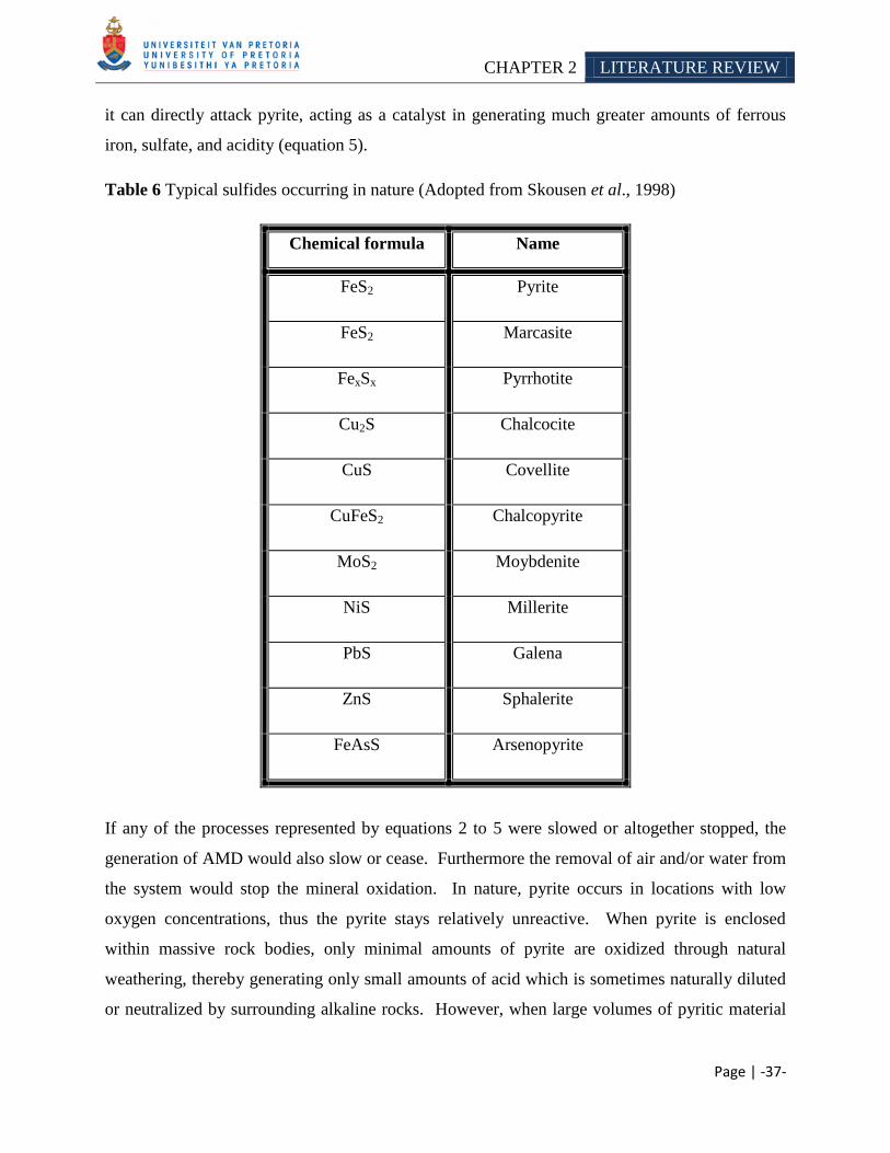
CHAPTER 2 LITERATURE REVIEW
Page | -37-
it can directly attack pyrite, acting as a catalyst in generating much greater amounts of ferrous
iron, sulfate, and acidity (equation 5).
Table 6 Typical sulfides occurring in nature (Adopted from Skousen et al., 1998)
Chemical formula Name
FeS2 Pyrite
FeS2 Marcasite
FexSx Pyrrhotite
Cu2S Chalcocite
CuS Covellite
CuFeS2 Chalcopyrite
MoS2 Moybdenite
NiS Millerite
PbS Galena
ZnS Sphalerite
FeAsS Arsenopyrite
If any of the processes represented by equations 2 to 5 were slowed or altogether stopped, the
generation of AMD would also slow or cease. Furthermore the removal of air and/or water from
the system would stop the mineral oxidation. In nature, pyrite occurs in locations with low
oxygen concentrations, thus the pyrite stays relatively unreactive. When pyrite is enclosed
within massive rock bodies, only minimal amounts of pyrite are oxidized through natural
weathering, thereby generating only small amounts of acid which is sometimes naturally diluted
or neutralized by surrounding alkaline rocks. However, when large volumes of pyritic material
CHAPTER 2 LITERATURE REVIEW
Page | -38-
are fractured and exposed to oxidizing conditions, which can occur in mining or other major land
disturbances, the pyrite reacts, and water dissolves and moves the reaction products
(Equation 2 to 5) into ground and surface water sources.
Under many conditions' equation 2 is the rate-limiting step in pyrite oxidation because the
conversion of ferrous iron to ferric iron is slow at pH values below 5 under abiotic conditions
(Skousen et al., 1998). However, iron oxidizing and sulfur oxidizing bacteria can greatly
accelerate this reaction, which aids the formation of AMD. Acidophiles commonly found in
AMD include Leptospirillum ferrooxidans and other Leptospirilli, Acidithiobacillus
ferrooxidans, A. thiooxidans and A. caldus. Heterotrophs such as Acidiphilium species are also
found in acid mine drainage, in close association with chemolithotrophs
(de Wolf-Durand et al., 1997; Hallberg and Johnson 2001). The importance of bacteria in
accelerating AMD formation, spurred research into bactericides to control the microbial
population and hopefully prevent or treat AMD (Table 8).
The availability of oxygen also limits the formation of acid mine drainage. Spoils with low
porosity and permeability, as with those composed of soft shales, often only form AMD in the
upper part where enough oxygen is present. In porous and permeable spoil composed of coarse
sandstone, air convection driven by the heat generated by pyrite oxidation may provide high
amounts of oxygen deep into the spoil (Guo et al., 1994, Guo and Cravotta 1996).
Alkaline mine drainage can also be formed. Here water has a pH above 6, with dissolved metals
that can create acids via reaction 2 and 3 (mentioned before). Skousen and Ziemkiewicz (1996)
grouped mine drainage into several basic types according to their various different properties or
stages of formation (Table 7).
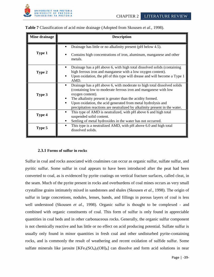
CHAPTER 2 LITERATURE REVIEW
Page | -39-
Table 7 Classification of acid mine drainage (Adopted from Skousen et al., 1998).
Mine drainage Description
Type 1
Drainage has little or no alkalinity present (pH below 4.5).
Contains high concentrations of iron, aluminum, manganese and other
metals.
Type 2
Drainage has a pH above 6, with high total dissolved solids (containing
high ferrous iron and manganese with a low oxygen content).
Upon oxidation, the pH of this type will drease and will become a Type 1
AMD.
Type 3
Drainage has a pH above 6, with moderate to high total dissolved solids
(containing low to moderate ferrous iron and manganese with low
oxygen content).
The alkalinity present is greater than the acidity formed.
Upon oxidation, the acid generated from metal hydrolysis and
precipitation reactions are neutralized by alkalinity present in the water.
Type 4 This type of AMD is neutralized, with pH above 6 and high total
suspended solid content.
Settling of metal hydroxides in the water has not occurred.
Type 5 This type is a neutralized AMD, with pH above 6.0 and high total
dissolved solids.
2.3.1 Forms of sulfur in rocks
Sulfur in coal and rocks associated with coalmines can occur as organic sulfur, sulfate sulfur, and
pyritic sulfur. Some sulfur in coal appears to have been introduced after the peat had been
converted to coal, as is evidenced by pyrite coatings on vertical fracture surfaces, called cleat, in
the seam. Much of the pyrite present in rocks and overburdens of coal mines occurs as very small
crystalline grains intimately mixed in sandstones and shales (Skousen et al., 1998). The origin of
sulfur in large concretions, nodules, lenses, bands, and fillings in porous layers of coal is less
well understood (Skousen et al., 1998). Organic sulfur is thought to be complexed - and
combined with organic constituents of coal. This form of sulfur is only found in appreciable
quantities in coal beds and in other carbonaceous rocks. Generally, the organic sulfur component
is not chemically reactive and has little or no effect on acid producing potential. Sulfate sulfur is
usually only found in minor quantities in fresh coal and other undisturbed pyrite-containing
rocks, and is commonly the result of weathering and recent oxidation of sulfide sulfur. Some
sulfate minerals like jarosite [KFe3(SO4)2(OH)6] can dissolve and form acid solutions in near
CHAPTER 2 LITERATURE REVIEW
Page | -40-
surface environments. Pyritic or sulfide sulfur is the dominant form of sulfur in the majority of
coal and associated rocks. It is the sulfur form of greatest concern. Of all the sulfide minerals that
may be present, iron disulfides predominate and are the major acid producers. Accordingly, the
maximum potential acidity (MPA) of a fresh overburden sample correlates closely with the
pyritic sulfur content. Studies indicated that variations in total sulfur contents of overburden
samples reflect similar variations in pyritic sulfur content. Several types of pyritic sulfur are
known based on physical appearance, and are classed into six groups: 1) primary massive,
2) plant replacement pyrite, 3) primary euhedral pyrite, 4) secondary cleat (joint) coats, 5) mossy
pitted pyrite, and 6) framboidal pyrite. Caruccio et al. (1988) provide an extensive review of the
different forms, morphologies, and reactivity of pyritic materials. The equations for pyrite
oxidation show that a material containing 1% sulfur, all as pyrite, would yield upon complete
reaction an amount of sulfuric acid that requires 31.25 mg of CaCO3 to neutralize the material
(Sobec et al., 1978). When sulfur in the rock is exclusively pyrite, the total sulfur content of the
rock accurately quantifies the acid-producing potential, if it were all to react. When organic or
sulfate sulfur are present in significant amounts in partially weathered rocks, total sulfur
measurements overestimate the amount of acid that will be formed upon oxidation. Therefore,
correction for sulfates and organic sulfur naturally present in some overburdens or resulting from
partial weathering of pyritic materials may be necessary to increase accuracy in predicting the
acid-producing potential of materials containing mixed sulfur species (Skousen et al., 1998).
The rate of pyrite oxidation depends on numerous variables (Skousen et al., 1998) such as:
reactive surface area of pyrite
form of pyritic sulfur
oxygen concentrations
solution pH
catalytic agents
the presence of Thiobacillus bacteria. The possibility of identifying and quantifying the
effects of these and other controlling factors with all the various rock types in a field
setting is unlikely.
CHAPTER 2 LITERATURE REVIEW
Page | -41-
2.3.2 Alkalinity in rocks
The natural base content of overburden materials (alkali and alkaline earth cations, commonly
present as carbonates or exchangeable cations on clays) is important in evaluating the future
neutralization potential (NP) of the materials. The amount of alkaline material in unweathered
overburden may be sufficient to equal or overwhelm the acid-producing potential of the material.
Of the many types of alkaline compounds present in rocks, carbonates (specifically calcite and
dolomite) are the primary alkaline compounds, which occur, in sufficient quantities to be
considered as effective deterrents to AMD generation. In overburden containing both alkaline
and pyritic material, the alkaline material may be sufficient to reduce oxidation from occurring
or to neutralize the acid formed from pyrite. Higher alkalinities also help control bacteria and
restrict solubility of ferric iron, which are both known to accelerate acid generation. Although a
number of factors must be considered, a balance of the acid-producing potential and neutralizing
capacity of an overburden sample will indicate whether acidity or alkalinity is expected in the
material upon complete weathering (Skousen et al., 1998).
2.3.3 Treatment strategies
There are various options available to remediate AMD, which may be divided into those that use
either chemicals or biological mechanisms to neutralize acidity and remove metals from solution.
Both abiotic and biotic systems include those that are classified as active (Table 8) and passive
(Table 9). Active treatment systems require continuous inputs of resources to sustain the
process, whereas passive systems require relative little resource input once in operation
(Johnson and Hallberg 2005). Active treatment systems are very effective; however the
treatments are expensive when cost of equipment, chemicals and manpower are considered
(Skousen et al., 1998). Due to the high cost of active systems, passive treatment systems have
been developed that do not require continuous chemical inputs and take advantage of naturally
occurring chemical and biological processes to clean contaminated mine waters, however these
systems also suffer from several drawback as discussed below. These systems include
constructed wetlands, anoxic limestone drains and vertical flow systems such as successive
alkalinity producing systems, limestone ponds and open limestone channels. Compared to active
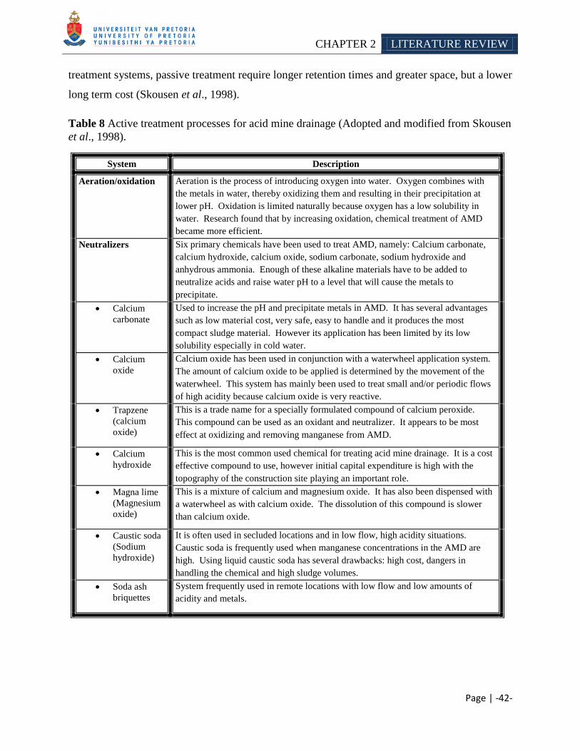
CHAPTER 2 LITERATURE REVIEW
Page | -42-
treatment systems, passive treatment require longer retention times and greater space, but a lower
long term cost (Skousen et al., 1998).
Table 8 Active treatment processes for acid mine drainage (Adopted and modified from Skousen
et al., 1998).
System Description
Aeration/oxidation Aeration is the process of introducing oxygen into water. Oxygen combines with
the metals in water, thereby oxidizing them and resulting in their precipitation at
lower pH. Oxidation is limited naturally because oxygen has a low solubility in
water. Research found that by increasing oxidation, chemical treatment of AMD
became more efficient.
Neutralizers Six primary chemicals have been used to treat AMD, namely: Calcium carbonate,
calcium hydroxide, calcium oxide, sodium carbonate, sodium hydroxide and
anhydrous ammonia. Enough of these alkaline materials have to be added to
neutralize acids and raise water pH to a level that will cause the metals to
precipitate.
Calcium
carbonate
Used to increase the pH and precipitate metals in AMD. It has several advantages
such as low material cost, very safe, easy to handle and it produces the most
compact sludge material. However its application has been limited by its low
solubility especially in cold water.
Calcium
oxide
Calcium oxide has been used in conjunction with a waterwheel application system.
The amount of calcium oxide to be applied is determined by the movement of the
waterwheel. This system has mainly been used to treat small and/or periodic flows
of high acidity because calcium oxide is very reactive.
Trapzene
(calcium
oxide)
This is a trade name for a specially formulated compound of calcium peroxide.
This compound can be used as an oxidant and neutralizer. It appears to be most
effect at oxidizing and removing manganese from AMD.
Calcium
hydroxide
This is the most common used chemical for treating acid mine drainage. It is a cost
effective compound to use, however initial capital expenditure is high with the
topography of the construction site playing an important role.
Magna lime
(Magnesium
oxide)
This is a mixture of calcium and magnesium oxide. It has also been dispensed with
a waterwheel as with calcium oxide. The dissolution of this compound is slower
than calcium oxide.
Caustic soda
(Sodium
hydroxide)
It is often used in secluded locations and in low flow, high acidity situations.
Caustic soda is frequently used when manganese concentrations in the AMD are
high. Using liquid caustic soda has several drawbacks: high cost, dangers in
handling the chemical and high sludge volumes.
Soda ash
briquettes
System frequently used in remote locations with low flow and low amounts of
acidity and metals.
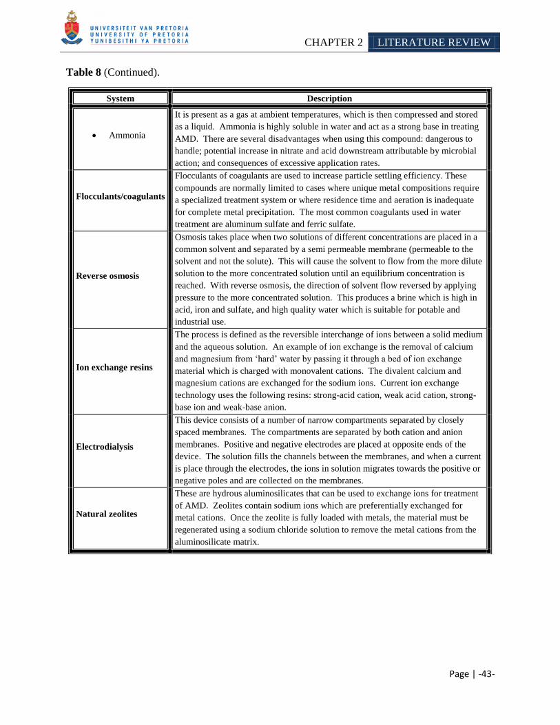
CHAPTER 2 LITERATURE REVIEW
Page | -43-
Table 8 (Continued).
System Description
Ammonia
It is present as a gas at ambient temperatures, which is then compressed and stored
as a liquid. Ammonia is highly soluble in water and act as a strong base in treating
AMD. There are several disadvantages when using this compound: dangerous to
handle; potential increase in nitrate and acid downstream attributable by microbial
action; and consequences of excessive application rates.
Flocculants/coagulants
Flocculants of coagulants are used to increase particle settling efficiency. These
compounds are normally limited to cases where unique metal compositions require
a specialized treatment system or where residence time and aeration is inadequate
for complete metal precipitation. The most common coagulants used in water
treatment are aluminum sulfate and ferric sulfate.
Reverse osmosis
Osmosis takes place when two solutions of different concentrations are placed in a
common solvent and separated by a semi permeable membrane (permeable to the
solvent and not the solute). This will cause the solvent to flow from the more dilute
solution to the more concentrated solution until an equilibrium concentration is
reached. With reverse osmosis, the direction of solvent flow reversed by applying
pressure to the more concentrated solution. This produces a brine which is high in
acid, iron and sulfate, and high quality water which is suitable for potable and
industrial use.
Ion exchange resins
The process is defined as the reversible interchange of ions between a solid medium
and the aqueous solution. An example of ion exchange is the removal of calcium
and magnesium from ‗hard‘ water by passing it through a bed of ion exchange
material which is charged with monovalent cations. The divalent calcium and
magnesium cations are exchanged for the sodium ions. Current ion exchange
technology uses the following resins: strong-acid cation, weak acid cation, strong-
base ion and weak-base anion.
Electrodialysis
This device consists of a number of narrow compartments separated by closely
spaced membranes. The compartments are separated by both cation and anion
membranes. Positive and negative electrodes are placed at opposite ends of the
device. The solution fills the channels between the membranes, and when a current
is place through the electrodes, the ions in solution migrates towards the positive or
negative poles and are collected on the membranes.
Natural zeolites
These are hydrous aluminosilicates that can be used to exchange ions for treatment
of AMD. Zeolites contain sodium ions which are preferentially exchanged for
metal cations. Once the zeolite is fully loaded with metals, the material must be
regenerated using a sodium chloride solution to remove the metal cations from the
aluminosilicate matrix.
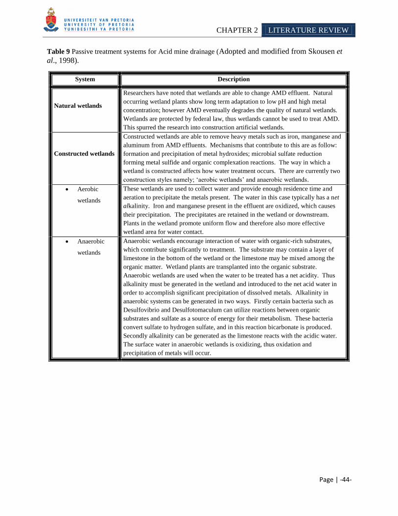
CHAPTER 2 LITERATURE REVIEW
Page | -44-
Table 9 Passive treatment systems for Acid mine drainage (Adopted and modified from Skousen et
al., 1998).
System Description
Natural wetlands
Researchers have noted that wetlands are able to change AMD effluent. Natural
occurring wetland plants show long term adaptation to low pH and high metal
concentration; however AMD eventually degrades the quality of natural wetlands.
Wetlands are protected by federal law, thus wetlands cannot be used to treat AMD.
This spurred the research into construction artificial wetlands.
Constructed wetlands
Constructed wetlands are able to remove heavy metals such as iron, manganese and
aluminum from AMD effluents. Mechanisms that contribute to this are as follow:
formation and precipitation of metal hydroxides; microbial sulfate reduction
forming metal sulfide and organic complexation reactions. The way in which a
wetland is constructed affects how water treatment occurs. There are currently two
construction styles namely; ‗aerobic wetlands‘ and anaerobic wetlands.
Aerobic
wetlands
These wetlands are used to collect water and provide enough residence time and
aeration to precipitate the metals present. The water in this case typically has a net
alkalinity. Iron and manganese present in the effluent are oxidized, which causes
their precipitation. The precipitates are retained in the wetland or downstream.
Plants in the wetland promote uniform flow and therefore also more effective
wetland area for water contact.
Anaerobic
wetlands
Anaerobic wetlands encourage interaction of water with organic-rich substrates,
which contribute significantly to treatment. The substrate may contain a layer of
limestone in the bottom of the wetland or the limestone may be mixed among the
organic matter. Wetland plants are transplanted into the organic substrate.
Anaerobic wetlands are used when the water to be treated has a net acidity. Thus
alkalinity must be generated in the wetland and introduced to the net acid water in
order to accomplish significant precipitation of dissolved metals. Alkalinity in
anaerobic systems can be generated in two ways. Firstly certain bacteria such as
Desulfovibrio and Desulfotomaculum can utilize reactions between organic
substrates and sulfate as a source of energy for their metabolism. These bacteria
convert sulfate to hydrogen sulfate, and in this reaction bicarbonate is produced.
Secondly alkalinity can be generated as the limestone reacts with the acidic water.
The surface water in anaerobic wetlands is oxidizing, thus oxidation and
precipitation of metals will occur.
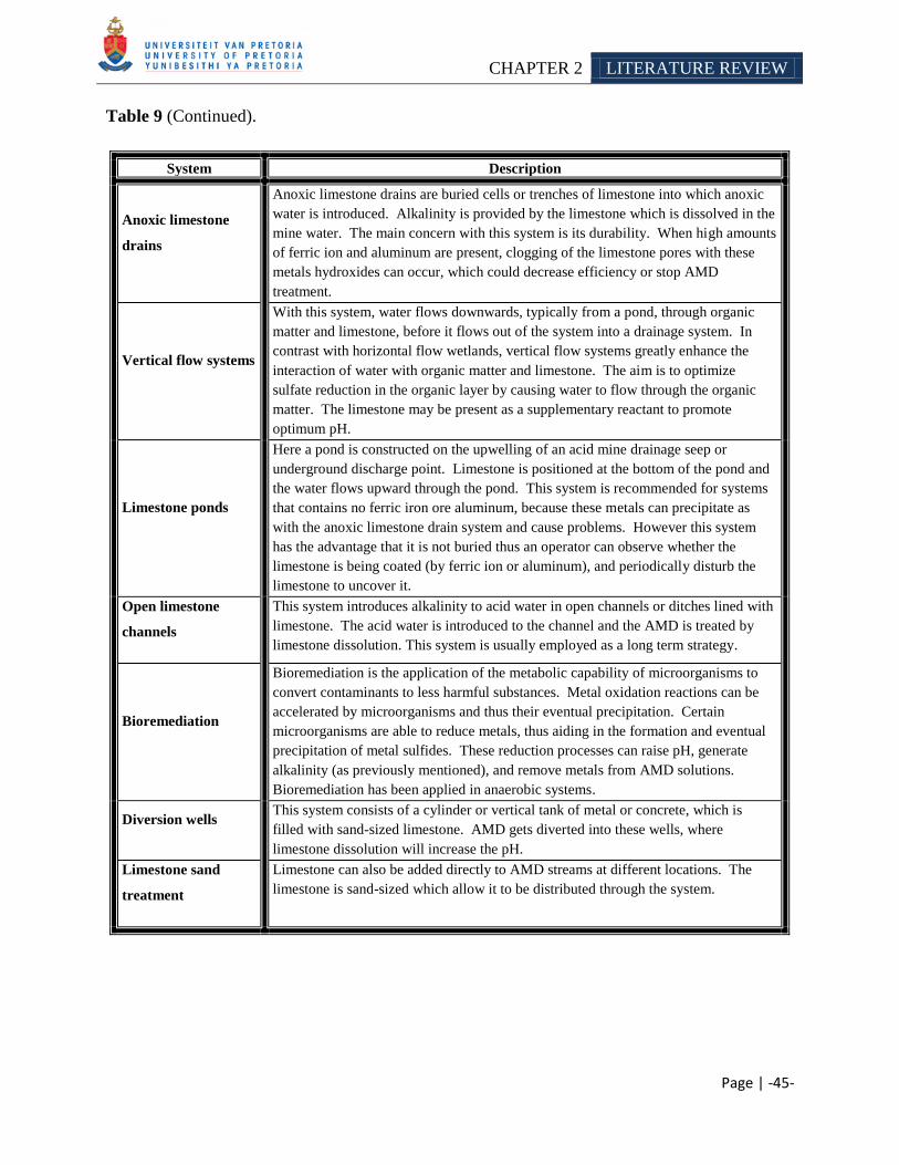
CHAPTER 2 LITERATURE REVIEW
Page | -45-
Table 9 (Continued).
System Description
Anoxic limestone
drains
Anoxic limestone drains are buried cells or trenches of limestone into which anoxic
water is introduced. Alkalinity is provided by the limestone which is dissolved in the
mine water. The main concern with this system is its durability. When high amounts
of ferric ion and aluminum are present, clogging of the limestone pores with these
metals hydroxides can occur, which could decrease efficiency or stop AMD
treatment.
Vertical flow systems
With this system, water flows downwards, typically from a pond, through organic
matter and limestone, before it flows out of the system into a drainage system. In
contrast with horizontal flow wetlands, vertical flow systems greatly enhance the
interaction of water with organic matter and limestone. The aim is to optimize
sulfate reduction in the organic layer by causing water to flow through the organic
matter. The limestone may be present as a supplementary reactant to promote
optimum pH.
Limestone ponds
Here a pond is constructed on the upwelling of an acid mine drainage seep or
underground discharge point. Limestone is positioned at the bottom of the pond and
the water flows upward through the pond. This system is recommended for systems
that contains no ferric iron ore aluminum, because these metals can precipitate as
with the anoxic limestone drain system and cause problems. However this system
has the advantage that it is not buried thus an operator can observe whether the
limestone is being coated (by ferric ion or aluminum), and periodically disturb the
limestone to uncover it.
Open limestone
channels
This system introduces alkalinity to acid water in open channels or ditches lined with
limestone. The acid water is introduced to the channel and the AMD is treated by
limestone dissolution. This system is usually employed as a long term strategy.
Bioremediation
Bioremediation is the application of the metabolic capability of microorganisms to
convert contaminants to less harmful substances. Metal oxidation reactions can be
accelerated by microorganisms and thus their eventual precipitation. Certain
microorganisms are able to reduce metals, thus aiding in the formation and eventual
precipitation of metal sulfides. These reduction processes can raise pH, generate
alkalinity (as previously mentioned), and remove metals from AMD solutions.
Bioremediation has been applied in anaerobic systems.
Diversion wells This system consists of a cylinder or vertical tank of metal or concrete, which is
filled with sand-sized limestone. AMD gets diverted into these wells, where
limestone dissolution will increase the pH.
Limestone sand
treatment
Limestone can also be added directly to AMD streams at different locations. The
limestone is sand-sized which allow it to be distributed through the system.
CHAPTER 2 LITERATURE REVIEW
Page | -46-
2.4 DIRECT VS INDIRECT BIOLEACHING
In 1964 Silverman and Ehrlich introduced the phrase ―direct leaching‖ which described a
hypothesized enzymatic reaction taking place between an attached cell and an underlying
mineral surface (Edwards et al., 2001; Sand et al., 1995). However, it is now generally accepted
that this mechanism does not exist (Sand and Gehrke 2006). The ―indirect leaching‖ mechanism
now consist of two sub-mechanisms namely the ―contact‖ and ―non-contact‖ mechanism
(Rawlings 2002; Sand and Gehrke 2006). Non-contact leaching describes the oxidation of
ferrous iron to ferric iron by planktonic bacteria. The resulting ferric iron acts on the mineral
surface where it is reduced and then re-enters the cycle to be oxidized to ferrous iron by the
planktonic bacteria (Sand and Gehrke 2006).
Contact leaching takes into account that most of the microbial cells are attached to the surface of
sulfide minerals and by means of ferric iron ions, which are complexed in their slime/glycocalyx
or extracellur polymeric substances (EPS), they begin to degrade the sulfide minerals
(Schippers and Sand 1999). Figure 8 illustrates the proposed contact mechanism of the indirect
leaching approach. The model depicts that the dissolution of the mineral takes place at an
interface between the bacterial cell wall and the mineral surface. This interfacial process is
controlled by electrochemical parameters. The microorganisms contributes to the
electrochemical parameters by generating oxidizing agents such as ferric iron ions, and also by
subsequently oxidizing the sulfur compounds resulting from the dissolution of the mineral
(Rawlings 2002).
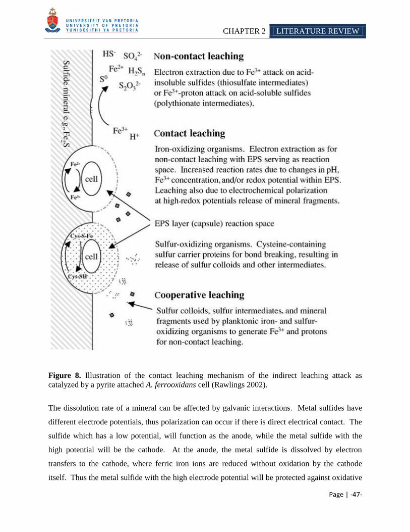
CHAPTER 2 LITERATURE REVIEW
Page | -47-
Figure 8. Illustration of the contact leaching mechanism of the indirect leaching attack as
catalyzed by a pyrite attached A. ferrooxidans cell (Rawlings 2002).
The dissolution rate of a mineral can be affected by galvanic interactions. Metal sulfides have
different electrode potentials, thus polarization can occur if there is direct electrical contact. The
sulfide which has a low potential, will function as the anode, while the metal sulfide with the
high potential will be the cathode. At the anode, the metal sulfide is dissolved by electron
transfers to the cathode, where ferric iron ions are reduced without oxidation by the cathode
itself. Thus the metal sulfide with the high electrode potential will be protected against oxidative
CHAPTER 2 LITERATURE REVIEW
Page | -48-
attack as long as there is an electrical contact and will thus be dissolved at a slower rate than the
galvanically unprotected sulfide (Rossi 1990).
2.4.1 Reaction pathway
Several experimental results have indicated that the reaction pathway for metal sulfide
dissolution is not determined by the crystal structure of the mineral (Sand et al., 2001; Schippers
and Sand 1999). The only decisive factor is the reactivity of the mineral with protons. For
example, research has found that the acid non-soluble monosulfides such as molybdenite and
tungstenite are degraded by the same mechanism as the acid non-soluble disulfide pyrite, but
structurally similar disulfides pyrite and the acid soluble hauerite are dissolved by different
mechanisms (Sand et al., 2001; Schippers and Sand 1999).
2.4.1.1 Thiosulfate pathway
The thiosulfate pathway describes the degradation of acid non-soluble metal sulfides such as
pyrite, molybdenite and tungstenite (Schippers and Sand 1999, Schippers et al., 1996). Pyrite is
dissolved as a result of electron extraction by hydrated ferric ions from the crystal lattice
(Equation 6).
Equation 6: FeS2 + 6[Fe(H2O)6]3+
+ 9H2O = S2O23-
+ 7[Fe(H2O6]2+
+ 6H+
Thiosulfate, by means of a series of reactions, will subsequently form either sulfate and protons
or higher polythionates and elemental sulfur (Figure 9). A number of reactions of the thiosulfate
degradation pathway have been found to be mediated by enzymes. Researchers have isolated an
enzyme from A. ferrooxidans which is responsible for the oxidation of thiosulfate to tetrathionate
(thiosulfate dehydrogenase) (Sand and Gehrke 2006). The enzyme responsible for the hydrolysis
of tetrathionate (tetrathionate hydrolyses) has also been isolated from A. ferrooxidans
(de Jong et al., 1997; Sugio et al., 1996; Tano et al., 1996). However the degree to which
enzymatic catalysis contributes to thiosulfate degradation remains to be determined. Schippers
and coworkers (1999) determined that the amount of biomass produced during pyrite dissolution,
indicates that some of the reactions must release electrons at a level sufficient for cytochrome β-
flavine reduction. This would permit the two oxidation phosphorylation reactions, whereas
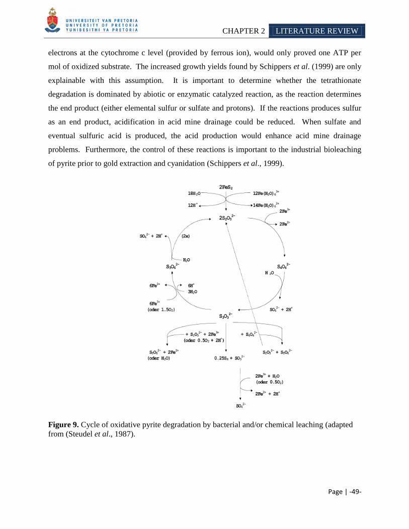
CHAPTER 2 LITERATURE REVIEW
Page | -49-
electrons at the cytochrome c level (provided by ferrous ion), would only proved one ATP per
mol of oxidized substrate. The increased growth yields found by Schippers et al. (1999) are only
explainable with this assumption. It is important to determine whether the tetrathionate
degradation is dominated by abiotic or enzymatic catalyzed reaction, as the reaction determines
the end product (either elemental sulfur or sulfate and protons). If the reactions produces sulfur
as an end product, acidification in acid mine drainage could be reduced. When sulfate and
eventual sulfuric acid is produced, the acid production would enhance acid mine drainage
problems. Furthermore, the control of these reactions is important to the industrial bioleaching
of pyrite prior to gold extraction and cyanidation (Schippers et al., 1999).
Figure 9. Cycle of oxidative pyrite degradation by bacterial and/or chemical leaching (adapted
from (Steudel et al., 1987).
CHAPTER 2 LITERATURE REVIEW
Page | -50-
2.4.1.2 Polysulfide pathway
The polysulfide pathway, describes the metal sulfide degradation of acid soluble sulfide minerals
such as hauerite, sphalerite, galena, chalcopyrite and arsenopyrite. The degradation of these
minerals yields a polysulfide intermediate (Schippers and Sand 1999). The crystal lattice is
attacked by a combined action of protons and ferric ions. Polarization of a surface sulfides are
induced by the protons, which enables the release of the sulfide from the crystal lattice
concomitantly with electron transfer to a ferric ion (Schippers and Sand 1999). The first free
intermediate of the proton attack is an H2S+ cation radical, which after subsequent reactions
forms a polysulfide molecule. The polysulfide compound decomposes to H2S+ and elemental
sulfur. Bacteria regenerate the protons by further oxidizing the elemental sulfur to sulfuric acid
(Steudel 1996).
2.4.1.3 Hydrogen sulfide pathway
When ferric ions are absent, readily acid-soluble metal sulfides such as sphalerite can be
dissolved by simple acid attack liberating hydrogen sulfide. Hydrogen sulfide is further oxidized
by sulfur-oxidizing bacteria to sulfate. This reaction releases a proton, which can enter the cycle
again to dissolve more metal sulfides. For example, Chan and Suzuki (1993) found that
A. ferrooxidans is able to solubilize hydrogen sulfide to elemental sulfur. Moreover,
Porro et al. (1997) reported that A. ferrooxidans was able to leach covellite in the absence of
iron, supporting the hydrogen sulfide pathway.
2.4.1.4 Attachment to mineral surfaces
Bacteria are able to form biofilms on the surface of minerals. Biofilm formation is a multistep
process which is influenced by many factors, including the specific mineralogy of the rock,
solution chemistry (pH, ionic strength) and the characteristics of the microorganism
(hydrophobicity, surface charge) (Characklis 1989, Banfield and Hamers 1997;
Little et al., 1997). The attachment of the cells to the surface can occur via random processes
such as diffusion and convection or more specifically via chemotaxis. Chemotaxis is the active
CHAPTER 2 LITERATURE REVIEW
Page | -51-
orientation of a bacterium to a chemical gradient (Jerez 2001). Several researchers studied the
chemotatic response of bacteria towards metal ions and other compounds, for example: research
by Chakraborty and Roy (1992) and Acuňa et al. (1992). The organisms can detect the site of
metal sulfide dissolution by sensing a dissolution gradient of dissolved ferrous ions and/or
sulfate. This allows the bacterium to respond to the attractant/repellent by swimming away or
towards the increased concentration. When the bacterium reaches the surface, the cell can attach
by mostly an unknown process (Sand and Gehrke 2006). Blake and coworkers (2001) concluded
from their research that aporusticyanin could possibly play an important role in the early
attachment of A. ferrooxidans to mineral surfaces. Moreover, exopolysaccharides produced by
the microbes also, play an important role in attachment of the leaching bacterium.
The exopolysaccharides formed by iron oxidizing bacteria has a net positive charge and at acidic
pH, pyrite has a negative charge (Devasia et al., 1993; Ohmura et al., 1993; Hallman 1992).
Thus, attachment of a positively charged bacterium to a negatively charged surface may take
place due to electrostatic interaction between the two surfaces. Lazar (2004) found that both
electrostatic and hydrophobic interactions could be responsible for the tight adhesion of cells to
mineral surfaces (as cited in Sand and Gehrke 2006). The specificity of exopolysaccharides for
various metal sulfides has not been termed. Gehrke et al. (1998) has however indicated that the
composition of the EPS of A. ferrooxidans changed when the cells were grown in different
substrates. Sulfur-grown cells had more lipids, less neutral sugars and only trace glucuronic acid
as compared to those of pyrite grown cells.
2.4.1.5 Importance of exopolysaccharides in bioleaching
The attachment of bacterial cells to the surface of metals is thought to be predominately
mediated by the exopolysaccharides surrounding the cells (Gehrke et al., 1998;
Rojas et al., 1995; Tributsch 2001). Research by Gehrke et al. (2001) indicated that the
attachment does not occur randomly (Gehrke et al., 2001). Moreover, Sand et al. (2001)
demonstrated using atomic force microscopy that the cells of A. ferrooxidans preferentially
attached to mineral sites with visible surface defects and that mineral dissolution possibly occurs
in the exopolysaccharides. Malinovskaya et al. (1990) found that acid polysaccharide bacterial
CHAPTER 2 LITERATURE REVIEW
Page | -52-
slimes promoted silicate dissolution; however Welch and Van Devivere (1994) found that EPS
could also inhibit the dissolution of feldspar under various conditions. The EPS layer formed is
thought to breach the gap between the outer membrane of the cell and the surface of the metal
surface. Currently there is a proposed electrochemical mechanism for bioleaching of metal
sulfides, which is supported by two plausible hypotheses and illustrates the importance of the
EPS layer (Medvedev and Stuchebrukhov 2001). The first assumption is that there is an electron
tunneling effect present in the EPS. It is known that electrons can bridge distances up to 2nm by
tunneling from one electron hole to another. Therefore the distance of ferric ions from the pyrite
(sulfide mineral) surface cannot exceed this distance, if the assumption is to be correct. When
we consider the distance between the cell membrane and the substrate surface, this assumption
seems to be reasonably sound and could explain the reduction of the ferric ion. The second
hypothesis is that ferrous ion-glucuronic acid complexes are not stable and this allows the ferrous
ions to migrate in the EPS space. If the ferrous ion diffuses towards the outer membrane it will
be reoxidized by the enzymatic system of the cells. Thus they serve as a substrate and can enter
the oxidation/reduction cycle again (Medvedev and Stuchebrukhov 2001).
2.4.1.6 Role of ferric/ferrous iron
Researchers have found that the amount of EPS-bound ferric ions varies with the metabolic
activity of the leaching bacteria. Cells with ample amounts of iron ions and glucuronic acid in
their exopolymers exhibit higher oxidation activity than those with low amounts of these
constituents. This observation has also been made for microbial strains such as A. ferrooxidans,
L. ferrooxidans and L. ferriphilum. The ferrous ions oxidation mechanism between
L. ferrooxidans and A. ferrooxidans seems to be completely different (Blake et al., 1993),
however the attachment of the cells to the surface of metal sulfides and EPS formation prior to
the onset of leaching is achieved by similar mechanisms (Gehrke et al., 1995;
Gehrke et al., 1998). Moreover, glucuronic acids and ferric ions are key components of the
exopolymers of both species. Research by Edwards and coworkers (2001) has indicated that the
ferric ions within the EPS-layers are connected to the metabolism of the bacteria, due to its
complex formation on the surface of the cell.
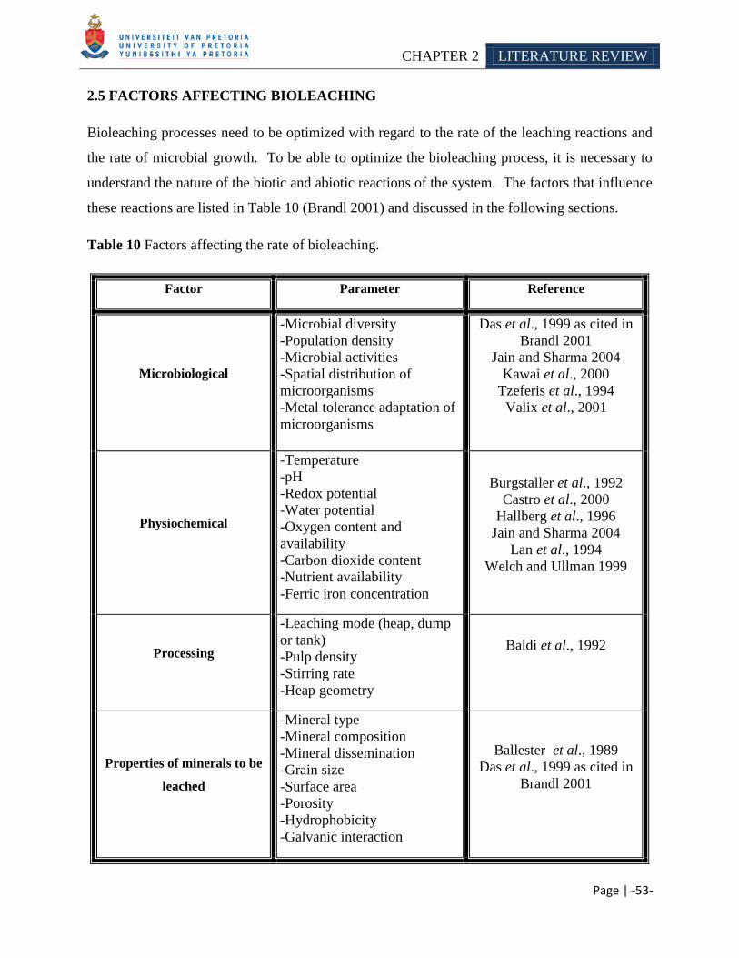
CHAPTER 2 LITERATURE REVIEW
Page | -53-
2.5 FACTORS AFFECTING BIOLEACHING
Bioleaching processes need to be optimized with regard to the rate of the leaching reactions and
the rate of microbial growth. To be able to optimize the bioleaching process, it is necessary to
understand the nature of the biotic and abiotic reactions of the system. The factors that influence
these reactions are listed in Table 10 (Brandl 2001) and discussed in the following sections.
Table 10 Factors affecting the rate of bioleaching.
Factor Parameter Reference
Microbiological
-Microbial diversity
-Population density
-Microbial activities
-Spatial distribution of
microorganisms
-Metal tolerance adaptation of
microorganisms
Das et al., 1999 as cited in
Brandl 2001
Jain and Sharma 2004
Kawai et al., 2000
Tzeferis et al., 1994
Valix et al., 2001
Physiochemical
-Temperature
-pH
-Redox potential
-Water potential
-Oxygen content and
availability
-Carbon dioxide content
-Nutrient availability
-Ferric iron concentration
Burgstaller et al., 1992
Castro et al., 2000
Hallberg et al., 1996
Jain and Sharma 2004
Lan et al., 1994
Welch and Ullman 1999
Processing
-Leaching mode (heap, dump
or tank)
-Pulp density
-Stirring rate
-Heap geometry
Baldi et al., 1992
Properties of minerals to be
leached
-Mineral type
-Mineral composition
-Mineral dissemination
-Grain size
-Surface area
-Porosity
-Hydrophobicity
-Galvanic interaction
Ballester et al., 1989
Das et al., 1999 as cited in
Brandl 2001
CHAPTER 2 LITERATURE REVIEW
Page | -54-
2.5.1 Microbial, physiochemical and process parameters
From a microbial aspect, parameters such as their diversity, density, activity, etc. can influence
the rate of bioleaching (Jain and Sharma 2004). Das and coworkers (1999) found that the iron
and sulfur oxidation rate from different microbial species and strains differ. The researchers
contemplated that the observed difference in oxidation rates could be due to several factors such
as change in morphology, difference in the chemical composition of the lipopolysaccharides,
difference in nutrient metabolism and the microorganisms DNA base ratio. Moreover they stated
that the concentration of the microbial cells also influences the oxidation rate. An increase in
bacterial numbers should hypothetically increase the oxidation rate. Therefore, various different
approaches have been developed to increase the bacterial numbers during a bioleaching process,
such as: membrane filtration or centrifugation of bacterial solution, followed by inoculation in a
smaller volume; use of a biofilm type reactor called BACFOX (bacterial film oxidation) or using
the organisms‘ affinity for a mineral. As an example of the latter, Acidithiobacillus and other
acidophilic microorganisms have a special affinity towards jarosite. Therefore, in a reactor, if a
thin film of jarosite can be developed under suitable conditions, the microorganism would get the
necessary site for surplus growth (Das et al., 1999 as cited in Brandl 2001).
The type of ore leached can also affect the bacterial number and therefore the oxidation rate.
When metals are leached from ores and wastes, the toxicity of certain metals may influence the
rate of leaching. Thus the use of metal tolerant species could enhance the leaching process
(Valix et al., 2001b). Research has suggested that high concentration of heavy metals act as a
broad protoplasmic poison which also denatures proteins and nucleic acids (Avakyan 1994).
Avakyan (1994) showed the ability of microorganisms to survive in a heavy metal environment
by selecting mutants at high concentrations of heavy metals. Several other researchers have also
shown the possibility of isolating microbial strains resistant to heavy metals, for example: Nickel
tolerant strains (Tzeferis et al., 1994), nickel and cobalt tolerant strains (Valix et al., 2001) and
aluminum tolerant strains (Kawai et al., 2000).
Oxygen concentration is another important parameter for bioleaching. In oxidation process it
serves as the electron acceptor (Rawlings 2002). In a heap leaching operation (Section 2.6.1),
steps can be taken to ensure adequate oxygen supply, by constructing the heap to ensure
optimum aeration and/or by installing an aeration system. With stirred tank bioleaching
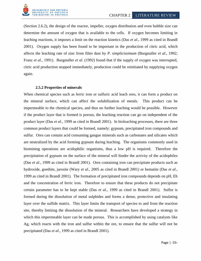
CHAPTER 2 LITERATURE REVIEW
Page | -55-
(Section 2.6.2), the design of the reactor, impeller, oxygen distribution and even bubble size can
determine the amount of oxygen that is available to the cells. If oxygen becomes limiting in
leaching reactions, it imposes a limit on the reaction kinetics (Das et al., 1999 as cited in Brandl
2001). Oxygen supply has been found to be important in the production of citric acid, which
affects the leaching rate of zinc from filter dust by P. simplicissimum (Burgstaller et al., 1992;
Franz et al., 1991). Burgstaller et al. (1992) found that if the supply of oxygen was interrupted,
citric acid production stopped immediately, production could be reinitiated by supplying oxygen
again.
2.5.2 Properties of minerals
When chemical species such as ferric iron or sulfuric acid leach ores, it can form a product on
the mineral surface, which can affect the solubilization of metals. This product can be
impermeable to the chemical species, and thus no further leaching would be possible. However
if the product layer that is formed is porous, the leaching reaction can go on independent of the
product layer (Das et al., 1999 as cited in Brandl 2001). In bioleaching processes, there are three
common product layers that could be formed, namely: gypsum, precipitated iron compounds and
sulfur. Ores can contain acid consuming gangue minerals such as carbonates and silicates which
are neutralized by the acid forming gypsum during leaching. The organisms commonly used in
biomining operations are acidophilic organisms, thus a low pH is required. Therefore the
precipitation of gypsum on the surface of the mineral will hinder the activity of the acidophiles
(Das et al., 1999 as cited in Brandl 2001). Ores containing iron can precipitate products such as
hydroxide, goethite, jarosite (Wary et al., 2005 as cited in Brandl 2001) or hematite (Das et al.,
1999 as cited in Brandl 2001). The formation of precipitated iron compounds depends on pH, Eh
and the concentration of ferric iron. Therefore to ensure that these products do not precipitate
certain parameter has to be kept stable (Das et al., 1999 as cited in Brandl 2001). Sulfur is
formed during the dissolution of metal sulphides and forms a dense, protective and insulating
layer over the sulfide matrix. This layer limits the transport of species to and from the reaction
site, thereby limiting the dissolution of the mineral. Researchers have developed a strategy in
which this impermeable layer can be made porous. This is accomplished by using catalysts like
Ag, which reacts with the iron and sulfur within the ore, to ensure that the sulfur will not be
precipitated (Das et al., 1999 as cited in Brandl 2001).
CHAPTER 2 LITERATURE REVIEW
Page | -56-
Galvanic interactions can also affect the rate of biooxidation. Das and coworkers (1999) found
that the dissolution of pyrite (FeS2) is faster than similar minerals such as sphalerite (ZnS),
galena (PbS) or chalcopyrite (CuFeS2). Moreover when pyrite is leached along with one of the
before mentioned minerals, the rate of dissolution decreases. Das and coworkers (1999) ascribed
the results to galvanic interaction which is explained as follow: the sulfide minerals are
considered semiconductors and can act as electron carriers. Due to the electronic nature of the
minerals, the sulfide dissolution reaction can be considered as two half cells (anode and cathode).
Each of the sulphide minerals has its own rest potential depending on various factors such as
crystallographic structure, solution composition and ionic concentration. If pyrite is in intimate
contact with sphalerite, then pyrite, having the higher rest potential will act as cathode and
sphalerite as the anode. Therefore sphalerite would be anodically dissolved whereas pyrite
would be cathodically protected. So galvanic interactions not only increase the dissolution of
anodic sulphide minerals but preferentially the leach of a particular mineral (Das et al., 1999 as
cited in Brandl 2001; Mason and Rice 2002).
2.6 INDUSTRIAL BIOLEACHING
There are two different leaching approaches used in the biomining industry, namely heap and
tank leaching. The approaches selected, depends on the type of mineral to be leached
(Rawlings 2002). Stirred tanks (continuous flow) are distinguished from heap leaching, by
homogeneous and constant growth conditions, which selects for rapidly growing microbes and
are therefore usually dominated by two or three species. Heap leaching is characterized by a
heterogeneous growth environment which can change with the age of the heap and therefore the
microbial community tends to be more diverse. Here the microorganisms grow as a biofilm
which is not subject to washout as with tank leaching (Rawlings and Johnson 2007). Following
is a general description of these processes, as well as examples where they have been used.
CHAPTER 2 LITERATURE REVIEW
Page | -57-
2.6.1 Heap and dump leaching
Bioleaching has been practiced for centuries. Initially, leaching was initiated by simply
irrigating a waste ore dump. This was seen as a low technology process, with low metal
recovery efficiency. However, since the early 1960s, metal recovery processes were made more
efficient by constructing and irrigating specially designed heaps (Rawlings et al., 2003;
Rawlings 2005; Schnell 1997). To construct a heap, ore is piled onto an impermeable base and
supplied with an efficient leach liquor, distribution and collection system. Acidic leaching
solution is percolated through the heaped ore and microbes growing on the surface of the mineral
produce ferric iron and acids that result in mineral dissolution and metal solubilization. Aeration
can be active or passive, with the former pipes have to be installed that blows air from the
bottom of the heap (Figure 10). In the latter case air is drawn into the heap due to the flow of
liquid (Rawlings et al., 2003; Rawlings 2005). Oxygen is a very important component for
effective leaching (Section 2.5), due to its use as an electron acceptor. Thus the leaching kinetics
can become limiting if this component is lacking. Moreover, the supply of air also supplies the
organism with the necessary carbon source. The metal-containing leach solutions that drain from
the heap are collected and sent for metal recovery (Figure 11). The heaps are cheaper to
construct than tank reactors, and are thus suited for the leaching of low-grade ores
(Bennett et al., 2004; Rawlings et al., 2003; Rawlings 2005). However the heap leaching
process is more difficult to control. It is difficult to aerate the process and gradients of pH and
nutrients from inside the heaps which affect bacterial growth and eventually the leaching kinetics
(Rawlings 2005). If the pH rises above 2.5, ferric iron precipitation might occur; that will hinder
bioleaching (Rawlings et al., 2003) by forming an impermeable product layer (Section 2.5.2).
Furthermore, due to the low-grade of the ore, it might also contain compounds such as silicates,
which can limit the leaching rate.
Heap leaching has been applied mainly to the treatment of copper ores (Figure 11)
(Rawlings et al., 2003). However it can also be applied to the pretreatment of gold bearing ores
(Rawlings et al., 2003). Several advances have been made to the heap leaching process, but here
the target has mainly been from an engineering perspective and not microbiological
(Rawlings 2002).
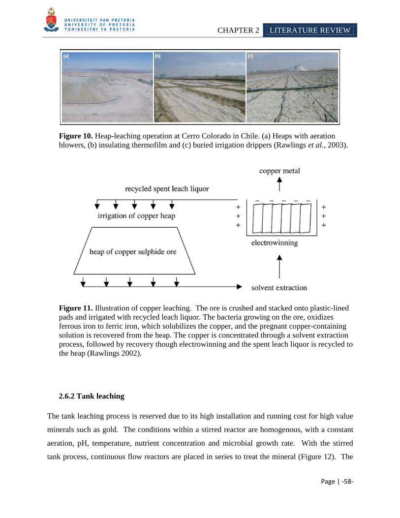
CHAPTER 2 LITERATURE REVIEW
Page | -58-
Figure 10. Heap-leaching operation at Cerro Colorado in Chile. (a) Heaps with aeration
blowers, (b) insulating thermofilm and (c) buried irrigation drippers (Rawlings et al., 2003).
Figure 11. Illustration of copper leaching. The ore is crushed and stacked onto plastic-lined
pads and irrigated with recycled leach liquor. The bacteria growing on the ore, oxidizes
ferrous iron to ferric iron, which solubilizes the copper, and the pregnant copper-containing
solution is recovered from the heap. The copper is concentrated through a solvent extraction
process, followed by recovery though electrowinning and the spent leach liquor is recycled to
the heap (Rawlings 2002).
2.6.2 Tank leaching
The tank leaching process is reserved due to its high installation and running cost for high value
minerals such as gold. The conditions within a stirred reactor are homogenous, with a constant
aeration, pH, temperature, nutrient concentration and microbial growth rate. With the stirred
tank process, continuous flow reactors are placed in series to treat the mineral (Figure 12). The
CHAPTER 2 LITERATURE REVIEW
Page | -59-
ore or finely milled mineral concentrate is added to the first tank together with inorganic nutrient
in the form of ammonia and phosphate containing fertilizer (Rawlings et al., 2003). The mineral
suspension flows through a series of highly aerated tanks that are temperature and pH controlled
(Rawlings 1995; Rawlings 2002; Van Aswegen et al., 1991). Mineral dissolution takes days in a
stirred tank reactor, compared to months with a heap reactor (Rawlings 1997). The major
constraint with tank leaching is the quantity of solids that can be maintained in suspension. The
liquid can become too thick in the reactor, thereby decreasing gas transfer, which will decrease
the oxygen concentration and finally affect the leaching kinetics (Rawlings et al., 2003).
Examples of industrial use of the tank leaching process includes the leaching of gold containing
arsenopyrite at the Fairview mine (Barberton, South Africa) and Ashanti goldfields in Ghana;
and treatment of cobalt-containing pyrite at Kasese (Uganda) (Rawlings 2005). Rawlings et al.
(1999b) found that in a continuous-flow, stirred tank reactor the ferric iron concentration remains
high, thus it appears that A. ferrooxidans appears to be less important than a combination of
Leptospirillum and A. ferrooxidans or A. caldus.
2.7 INDUSTRIAL BIOLEACING PROCESSES
The metabolic capabilities of microorganisms have been exploited commercially to treat various
types of ores (Table 11). Following is a brief description of processes that utilizes a consortium
of iron oxidizing bacteria. These processes include the BIOX®, GEOCOATTM
, GEOLEACHTM
,
BIOCOPTM
and the BacTech/Mintek process.
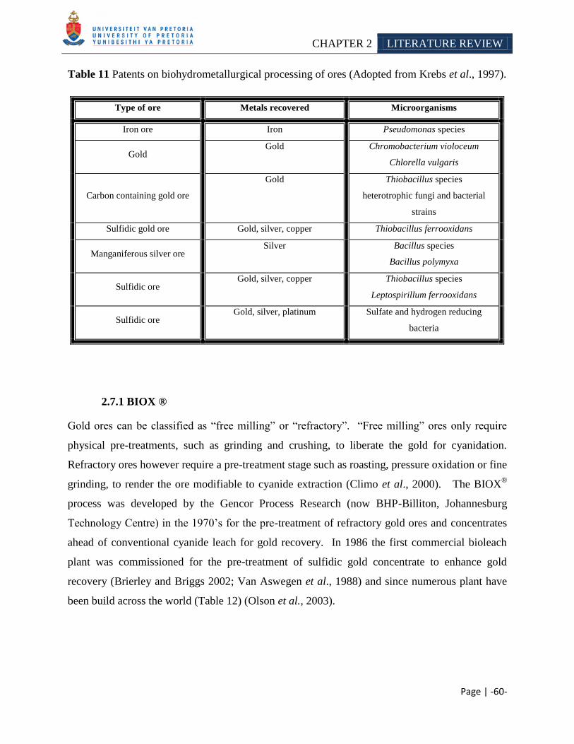
CHAPTER 2 LITERATURE REVIEW
Page | -60-
Table 11 Patents on biohydrometallurgical processing of ores (Adopted from Krebs et al., 1997).
Type of ore Metals recovered Microorganisms
Iron ore Iron Pseudomonas species
Gold Gold Chromobacterium violoceum
Chlorella vulgaris
Carbon containing gold ore
Gold Thiobacillus species
heterotrophic fungi and bacterial
strains
Sulfidic gold ore Gold, silver, copper Thiobacillus ferrooxidans
Manganiferous silver ore Silver Bacillus species
Bacillus polymyxa
Sulfidic ore Gold, silver, copper Thiobacillus species
Leptospirillum ferrooxidans
Sulfidic ore Gold, silver, platinum Sulfate and hydrogen reducing
bacteria
2.7.1 BIOX ®
Gold ores can be classified as ―free milling‖ or ―refractory‖. ―Free milling‖ ores only require
physical pre-treatments, such as grinding and crushing, to liberate the gold for cyanidation.
Refractory ores however require a pre-treatment stage such as roasting, pressure oxidation or fine
grinding, to render the ore modifiable to cyanide extraction (Climo et al., 2000). The BIOX®
process was developed by the Gencor Process Research (now BHP-Billiton, Johannesburg
Technology Centre) in the 1970‘s for the pre-treatment of refractory gold ores and concentrates
ahead of conventional cyanide leach for gold recovery. In 1986 the first commercial bioleach
plant was commissioned for the pre-treatment of sulfidic gold concentrate to enhance gold
recovery (Brierley and Briggs 2002; Van Aswegen et al., 1988) and since numerous plant have
been build across the world (Table 12) (Olson et al., 2003).
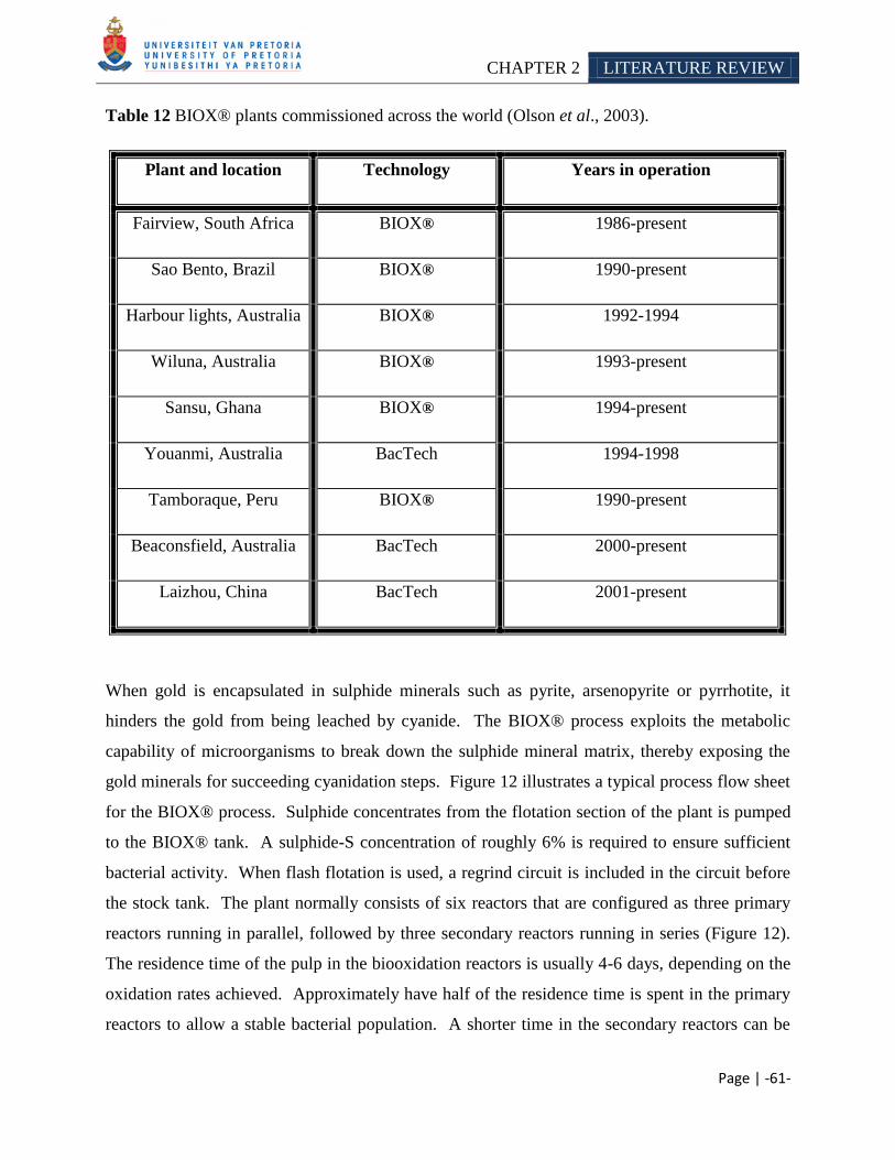
CHAPTER 2 LITERATURE REVIEW
Page | -61-
Table 12 BIOX® plants commissioned across the world (Olson et al., 2003).
Plant and location Technology Years in operation
Fairview, South Africa BIOX® 1986-present
Sao Bento, Brazil BIOX® 1990-present
Harbour lights, Australia BIOX® 1992-1994
Wiluna, Australia BIOX® 1993-present
Sansu, Ghana BIOX® 1994-present
Youanmi, Australia BacTech 1994-1998
Tamboraque, Peru BIOX® 1990-present
Beaconsfield, Australia BacTech 2000-present
Laizhou, China BacTech 2001-present
When gold is encapsulated in sulphide minerals such as pyrite, arsenopyrite or pyrrhotite, it
hinders the gold from being leached by cyanide. The BIOX® process exploits the metabolic
capability of microorganisms to break down the sulphide mineral matrix, thereby exposing the
gold minerals for succeeding cyanidation steps. Figure 12 illustrates a typical process flow sheet
for the BIOX® process. Sulphide concentrates from the flotation section of the plant is pumped
to the BIOX® tank. A sulphide-S concentration of roughly 6% is required to ensure sufficient
bacterial activity. When flash flotation is used, a regrind circuit is included in the circuit before
the stock tank. The plant normally consists of six reactors that are configured as three primary
reactors running in parallel, followed by three secondary reactors running in series (Figure 12).
The residence time of the pulp in the biooxidation reactors is usually 4-6 days, depending on the
oxidation rates achieved. Approximately have half of the residence time is spent in the primary
reactors to allow a stable bacterial population. A shorter time in the secondary reactors can be
CHAPTER 2 LITERATURE REVIEW
Page | -62-
tolerated, where the sulphide oxidation is completed. Nutrients such as potassium, phosphorous
and nitrogen are added to the primary reactors to increase bacterial growth. The microbial
community usually includes the following species, namely: Acidithiobacillus ferrooxidans,
Acidithiobacillus thiooxidans and Leptospirillum ferrooxidans; however the cultures are not
controlled in the process, but rather left to adapt to the concentrate and operating conditions
(Olson et al., 2003). Rawlings et al. (1999) found that Acidithiobacillus caldus and L.
ferriphilum dominated the microbial community when arsenopyrite was biooxidized at 40°C.
Tuffin et al. (2005) isolated A. caldus from a biooxidation plant, where arsenopyrite was
processed, that was highly resistant to arsenic. Air is injected into the BIOX® process to supply
oxygen to the biooxidation process. When sulphide minerals such as pyrite are oxidized, acids
are produced. However biooxidation of arsenopyrite and pyrrhotite consumes acids, thus the pH
(optimum range 1.2-1.8) in the BIOX® process has to be regulated using lime and sulfuric acid
(van Aswegen et al., 2006). The product of the BIOX® process contains high concentrations of
dissolved ions and must be washed in a three-stage countercurrent decantation circuit before
cyanide leaching (Figure 12).
Research conducted by Amankwah and coworkers (2005) enabled more gold to be extracted.
They tested the effect of a two stage bacterial pretreatment process for double refractory gold
ores (ores that contain both sulfides and carbonaceous material). The gold ores were first treated
with chemolithotrophic bacteria to oxidize the sulfides and in a second stage carbonaceous
matter was removed using Streptomyces setonii. It is well known that carbonaceous material is
not oxidized by the pretreatment steps and continue to serve as a preg-robber during cyanidation.
Using this additional step, 13.6% more gold could be extracted from the ores.
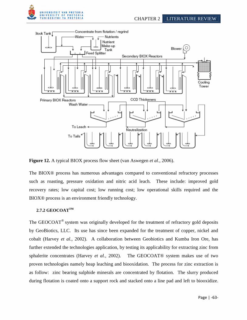
CHAPTER 2 LITERATURE REVIEW
Page | -63-
Figure 12. A typical BIOX process flow sheet (van Aswegen et al., 2006).
The BIOX® process has numerous advantages compared to conventional refractory processes
such as roasting, pressure oxidation and nitric acid leach. These include: improved gold
recovery rates; low capital cost; low running cost; low operational skills required and the
BIOX® process is an environment friendly technology.
2.7.2 GEOCOATTM
The GEOCOAT®
system was originally developed for the treatment of refractory gold deposits
by GeoBiotics, LLC. Its use has since been expanded for the treatment of copper, nickel and
cobalt (Harvey et al., 2002). A collaboration between Geobiotics and Kumba Iron Ore, has
further extended the technologies application, by testing its applicability for extracting zinc from
sphalerite concentrates (Harvey et al., 2002). The GEOCOAT® system makes use of two
proven technologies namely heap leaching and biooxidation. The process for zinc extraction is
as follow: zinc bearing sulphide minerals are concentrated by flotation. The slurry produced
during flotation is coated onto a support rock and stacked onto a line pad and left to biooxidize.
CHAPTER 2 LITERATURE REVIEW
Page | -64-
Low-pressure blowers are used to supply sufficient air to the system and to remove excess heat.
The concentrated slurry adheres strongly to the support rock and does not wash out during
irrigation of the heap. The cultures inoculated into the heap, depends upon the desired operating
temperature. Sulphide oxidizing bacteria such as Acidithiobacillus caldus (DSMZ strain 8584),
Sulfobacillus thermosulfidooxidans (DSMZ strain 9293 and 1192), Acidianus brierleyi
(DSMZ strains 1651 and 6334), Acidianus infernus (DSMZ strain 3191), Metallosphaera sedula
(ATCC strain 3390) Sulfolobus acidocaldarius (ATCC strain 49426) Sulfolobus shibatae
(DSMZ strain 5389) and Sulfolobus metallicus (DSMZ strain 6482) have been successfully
applied in the GEOCOAT® process (Harvey et al., 2002). Nutrients for the microorganisms are
added to the heap by recirculating solutions. During the biooxidation process, the sulphides in
the concentrated slurry are oxidized and the solubilized zinc, iron, arsenic and sulfates are
removed from the heap via the recirculating solution. A portion of this recirculated solution is
used for metal recovery. Once the biooxidation process is complete, the coated rocks may be
unloaded from the pad and recycled or replaced with fresh support rock. The concentrates can
then be removed and screened for precious metals (Harvey et al., 2002).
The GEOCOAT® process has a number of advantages over conventional refractory processes
and hydrometallurgical base metal recovery systems, such as: low operating cost due to its
simplicity; lower construction cost, because it does not require exotic materials; ability to
economically process low grade ores and the GEOCOAT® process is safe, because there is no
necessity for high pressures (Harvey et al., 2002).
2.7.3 GEOLEACHTM
The GEOLEACHTM
process was developed by GeoBiotics, LLC, for treatment of base metal
sulphide ores (www.geobiotics.com/GeoL_Process.cfm). The process utilizes iron and sulfur
oxidizing microorganisms to assist in oxidizing and leaching sulfide minerals in a constructed
whole ore heap environment. The microbial community includes mesophilic microorganisms
such as Thiobacillus ferrooxidans, Thiobacillus thiooxidans, and Leptospirillum ferrooxidans,
and thermophilic microorganisms such as the Archaea Sulfolobus and Acidianus
(www.geobiotics.com/GeoL_Process.cfm). The GEOLEACHTM
process utilizes the HotHeapTM
CHAPTER 2 LITERATURE REVIEW
Page | -65-
technology in which the process retains their chemical reaction heat so that the heap can operate
at high temperatures. As with copper leaching, high temperatures are necessary to leach this ore
economically. Aeration of the heap is controlled according to the oxidation rate within the heap
and irrigation of the heap is controlled according to the temperature inside the heap. GeoBiotics,
LLC commented that the GEOLEACHTM
process can be used to economically recover zinc,
cobalt and nickel from sulfide ore (www.geobiotics.com/GeoL_Process.cfm).
2.7.4 BioCOPTM
BioCOP™ Process is owned by BHP Billiton. The BioCOP™ Process has been specifically
developed for treatment of concentrates not suitable for commercial smelting due to the content
of deleterious elements, such as arsenic, which is harmful to the environment. The process
utilizes thermophilic microorganisms operating at temperatures up to 80 °C to leach copper
sulphide mineral concentrates. The copper is subsequently recovered by conventional solvent
extraction and electrowinning, producing a high value copper metal product
(Batty and Rorke 2006).
2.7.5 BacTech/Mintek process
An alternative to the BIOX® process is the Bacox process developed by BacTech (Australia)
(Miller 1997). The BacTech process is similar to the BIOX® process, with the major difference
being that the BacTech process is operated at 50°C. Moderately thermophilic bacteria therefore
carry out biooxidation of concentrates; however the exact composition of the microbial
consortium is unclear (Franzmann and Williams 1997). The BacTech process has the added
advantage over the BIOX® process in that less cooling is required however the solubility of
oxygen and carbon dioxide is less soluble at these higher temperatures (Rawlings 2004).
CHAPTER 2 LITERATURE REVIEW
Page | -66-
2.8 CONCLUSION
Biomining exploits the metabolic capabilities of microorganisms for industrial scale mineral
processing operations. Conventional treatment methods which include pyro - and
hydrometallurgical methods have several limitations that encouraged research into alternative
methods. Here biohydrometallurgical processes have proved to be useful; with several industrial
scale plants such as the BIOX®, BacTech, BioCOPTM
, GEOLEACHTM
and GEOCOATTM
currently in operation. These systems employ iron oxidizing bacteria such as A. ferrooxidans, L.
ferrooxidans, L. ferriphilum, etc. which can leach minerals via a ‗contact‘ or ‗non-contact
mechanism‘. Several factors however affect the efficiency of leaching such as: physiochemical
parameters, mineral characteristics and microbial strain variations. Except for its industrial
bioleaching application, microbial leaching capabilities have also been exploited in
bioremediation operations. Apart from their beneficial application in bioleaching, the metabolic
capability of iron oxidizing bacteria, can also negatively affect nature, in the form of acid mine
drainage. Here the microorganism accelerates the certain reactions which cause acid mine
drainage to form more easily. Numerous active and passive strategies have been developed to
control or prevent the formation of acid water.
Biomining is a safe, low energy and inexpensive technology. It may not completely replace
conventional processing methods, but its application has made mining more economical feasible
and environmentally friendly.
CHAPTER 3 ACID LEACHING
Page | -67-
CHAPTER 3
REMOVAL OF POTASSIUM AND PHOSPHOROUS FROM SISHEN HEMATITE IRON ORE USING
VARIOUS (IN)ORGANIC ACIDS
Abstract
The Sishen Iron Ore Mine is situated in the Northern Cape, South Africa, and forms part
of the northern end of the Maremane anticline where the bulk of the hematite is buried
beneath younger cover lithologies. The iron ore bodies mined are overlain by
conglomerates, shales, flagstone and quartzite. Potassium and phosphorous are common
constituents of the iron ore bodies and are known to have deleterious effect on iron and
steel manufacturing. Therefore steel making companies charge penalties when purchasing
iron ore with alkali concentrations above predetermined limits. Conventional methods
used to treat high alkali ore have several drawbacks such as poor product recovery, high
running cost and increase pollution of water thus an alternative approach is needed to
supplement/replace these methods. Biohydrometallurgy is a natural alternative approach
which has been applied commercially to extract or treat various ore bodies. Here we aim
to assess the ability of (in)organic acids, known to be produced by microorganisms, to
solubilize potassium and phosphorous from different iron ore samples mined at Sishen.
We found that the composition of the ore particle plays a crucial role in the ability of each
acid to solubilize potassium and phosphorous. Moreover, iron ore particles that contained
different combinations of the potassium bearing mineral phase’s muscovite, illite,
K-feldspar and biotite were solubilized differently by the acids tested. The organic acids
tested were able to remove more alkalis than the inorganic acids, which is ascribed to their
ability to bind minerals, thereby decreasing its saturation state in solution.
Keywords: Iron ore, Acid leaching, Muscovite, Illite, K-feldspar, Biotite, Apatite
______________________________________________________________________________
CHAPTER 3 ACID LEACHING
Page | -68-
3.1 INTRODUCTION
The Sishen Iron Ore Mine is one of the major suppliers of iron ore to both local and international
markets. It is situated in the Northern Cape (South Africa) and forms part of the northern end of
the Maremane anticline where the bulk of the hematite ore is buried beneath younger cover
lithologies. The majority of the iron ore resource of the mine is made up of laminated and
massive ore bodies that belong to the Asbestos Hills Subgroup. These ore bodies are overlain by
conglomerates, shales, flagstone and quartzite (Carney and Mienie 2003). The alkalis, potassium
and phosphorous, are common constituents of iron ore and have deleterious effects on the
manufacturing of iron and steel (Delvasto et al., 2008). Therefore, steel making companies
charge penalties when purchasing iron ore concentrates with alkali concentrations above
predetermined levels according to D. Krige (Personal communication, 2006).
Kumba Iron Ore, Ltd. has an industrial set limit of 0.24% for potassium and phosphorous
allowed in their export ore according to D. Krige (Personal communication, 2006). To ensure
that their batches stay within acceptable levels; the ores from different excavation sites
(with alkalis‘ >0.24% and <0.24%) are mixed to produce a batch with an average
potassium/phosphorous level below 0.24% (Dukino et al., 2000). This approach is however a
temporary solution as the low potassium/phosphorous ore (< 0.24%) is becoming progressively
depleted according to R. Grunewaldt (Personal communication, 2006). Alternative methods that
can be employed to treat high impurity ore include pyro- and hydrometallurgical processes
(Cheng et al., 1999; Kokal et al., 2003). However, the application of these methods poses
several problems such as poor product recovery, involvement of high process and energy cost
and an increase in pollution load of water (Jain and Sharma 2004). Therefore a low
maintenance, environment friendly and efficient approach is needed, that can supplement
conventional approaches and be incorporated into the mining schedule at minimal cost.
Primary rock-forming minerals can be chemically weathered by water, acids, complexing agents
and oxygen into dissolved substances and secondary residual minerals. These reactions can be
mediated by living organisms (Landeweert et al., 2001). It is well established that many
microorganisms are capable of mobilizing minerals from various sources in nature especially in
CHAPTER 3 ACID LEACHING
Page | -69-
nutrient limited environments (Banfield et al., 1999; Nautiyal 1999; Paris et al., 1996). Plant
roots and their associated rhizosphere microorganisms produce low molecular weight organic
compounds such as aliphatic acids and aldehydes, phenols, sugar acids and amino acids, all of
which may accelerate mineral weathering (Drever and Vance 1994). Several reports have
demonstrated that microorganisms or their metabolites could solubilize minerals from ores.
Leyval and Berthelin demonstrated in 1991 that rhizosphere microorganism, associated with pine
roots, could leach potassium from phlogopite by possibly producing organic acids.
Jennings (1995) commented on the importance of organic acids in fungal nutrition and
physiology. He stated that apart from their possible utilization as carbon and energy source, they
also contribute to intracellur osmotic potential, charge balance and pH homeostasis.
Margolis and Moreno (1992) found that apatite dissolution was enhanced in the presence of
organic acids.
Schnitzer and Kodama (1976) showed that fulvic acid was able to solubilize iron, magnesium,
potassium, aluminum and silicon from biotite, phlogopite and muscovite, by forming complexes
between the carboxyl/phenolic group of the acid and the mineral. Moreover, they also
commented that the mineral with the highest iron concentration was the most susceptible to
solubilization by fulvic acid. According to Schnitzer and Kodama (1976), iron ions are transition
metal ions and contain low crystal field stabilization energy; therefore fulvic acid can readily
remove it. The complexing of iron with fulvic acid affected the structural stability of the mineral
and thus other mineral constituents could also be readily released. Growing evidence has
suggested that microbial metabolites, especially acids, can be applied to leach various metals
from different ore bodies (Ehrlich 1991; Jain and Sharma 2004). However, there is still
controversy in literature regarding this (Drever and Stillings 1997; Jones et al., 2003). Drever
and Stillings (1997) listed three different ways in which organic acids and their anions might
affect mineral weathering rates, namely: the organic acids can change the dissolution rate of the
mineral far from equilibrium by decreasing the solution pH or by forming complexes at the
mineral surface; secondly they can affect the saturation state of the solution with respect to the
mineral and lastly the organic acid can affect the speciation in solution of ions such as aluminum
that affect minerals dissolution themselves (Table 13). For example, citric acid contains several
carboxyl groups which tends to donate protons (H+), resulting in a negatively charged carboxyl
CHAPTER 3 ACID LEACHING
Page | -70-
group. The protons attack the mineral surface (acidulation/hydrolysis reaction), while the
negatively charged carboxyl group can form stable complexes with cations thus removing it from
solution (Sayer and Gadd 2001). If the organic acid, for example citric or oxalic acid, contains
two or more electron donor groups so that one or more rings are formed, during the reaction with
a metal, the organic acid can be termed a chelating agent and the resulting complexes termed
metal chelates (Gadd 1999; Martell and Calvin 1952).
Here we aim to asses the aptitude of known microbially produced (in)organic acids to solubilize
potassium and phosphorous from the various ore bodies mined at Sishen. Abiotic tests are
known to be more reproducible than biotic tests, therefore these preliminary results will aid us in
determining the effectiveness of microbial leaching in subsequent steps.
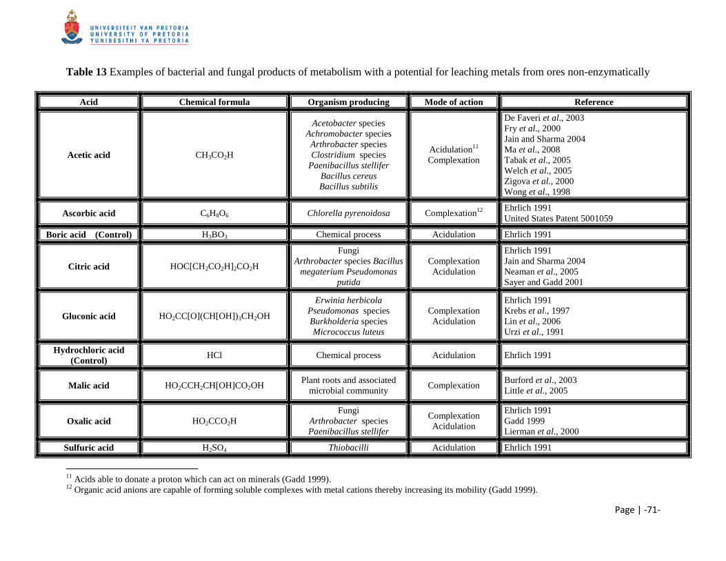
Page | -71-
Table 13 Examples of bacterial and fungal products of metabolism with a potential for leaching metals from ores non-enzymatically
Acid Chemical formula Organism producing Mode of action Reference
Acetic acid CH3CO2H
Acetobacter species
Achromobacter species
Arthrobacter species
Clostridium species
Paenibacillus stellifer
Bacillus cereus
Bacillus subtilis
Acidulation11
Complexation
De Faveri et al., 2003
Fry et al., 2000
Jain and Sharma 2004
Ma et al., 2008
Tabak et al., 2005
Welch et al., 2005
Zigova et al., 2000
Wong et al., 1998
Ascorbic acid C6H8O6 Chlorella pyrenoidosa Complexation12
Ehrlich 1991
United States Patent 5001059
Boric acid (Control) H3BO3 Chemical process Acidulation Ehrlich 1991
Citric acid HOC[CH2CO2H]2CO2H
Fungi
Arthrobacter species Bacillus
megaterium Pseudomonas
putida
Complexation
Acidulation
Ehrlich 1991
Jain and Sharma 2004
Neaman et al., 2005
Sayer and Gadd 2001
Gluconic acid HO2CC[O](CH[OH])3CH2OH
Erwinia herbicola
Pseudomonas species
Burkholderia species
Micrococcus luteus
Complexation
Acidulation
Ehrlich 1991
Krebs et al., 1997
Lin et al., 2006
Urzi et al., 1991
Hydrochloric acid
(Control) HCl Chemical process Acidulation Ehrlich 1991
Malic acid HO2CCH2CH[OH]CO2OH Plant roots and associated
microbial community Complexation
Burford et al., 2003
Little et al., 2005
Oxalic acid HO2CCO2H
Fungi
Arthrobacter species
Paenibacillus stellifer
Complexation
Acidulation
Ehrlich 1991
Gadd 1999
Lierman et al., 2000
Sulfuric acid H2SO4 Thiobacilli Acidulation Ehrlich 1991
11
Acids able to donate a proton which can act on minerals (Gadd 1999). 12
Organic acid anions are capable of forming soluble complexes with metal cations thereby increasing its mobility (Gadd 1999).
Page | -72-
3.2 MATERIALS AND METHODS
3.2.1 Iron Ore
The majority of ore resources at the Sishen Iron ore Mine, Northern Cape, South Africa is made
up of laminated and massive ore bodies that belong to the Asbestos Hills Subgroup. These ore
bodies are overlain by conglomerates, shales, flagstone and quartzite (Carney and Mienie 2003).
Kumba Iron Ore, Ltd. supplied various different types of iron ore samples from the Sishen Mine
for leaching, and labeled them as follow according to B. Ntsoelengoe (Personal communication,
2006): export (alkali levels within set limits); SK (shale - high potassium),
SPHP (high phosphorous) and KGT (conglomerate – high potassium). Prior to leaching, the
samples were rinsed with distilled water in a sieve (< 1mm) to remove small debris which could
hamper subsequent analysis, followed by sterilization at 121°C for 15 min.
3.2.2 Leaching
The ability of different organic acids to solubilize potassium/phosphorous from the export iron
ore sample was evaluated by adding 50g of sterilized ore to 100ml of each acid (1M) in 500 ml
Erlenmeyer flasks, followed by incubation at 30°C and a duplicate at 60°C for 5 days (Table 15).
The ability of different (in)organic acid to solubilize potassium/phosphorous from the SK, KGT
or SPHP samples were assessed by adding 50g of sterilized ore to 100ml of each acid (1M) in
500ml Erlenmeyer flasks, followed by incubation at 30°C and a duplicate at 60°C for 5 days
(Table 16, 17 and 18).
After 5 days the iron ores were removed from the various acids and washed three times with
distilled water. The samples were then placed in glass petridishes and dried in an oven at 100°C
overnight.
CHAPTER 3 ACID LEACHING
Page | -73-
3.2.3 Quantitative and Qualitative mineralogy
Treated samples were rinsed with distilled water to remove residual acids. Quantitative and
qualitative mineralogy of iron ore samples were preformed as described by Loubser and Verryn
(2008). 50 g of each iron ore samples was ground to <75μm in a tungsten carbide milling vessel,
followed by roasting at 1000°C to determine the loss of ignition. 1g of the milled sample was
then added to 6g Li2B4O7 and fused into glass beads. The remaining sample was pressed (2 tons)
into a powder briquette for minor element analysis. Major element analyses were done on the
fused bead using an ARL9400XP+ spectrometer (wavelength dispersive). Mineral phases were
determined using the PANalytical X'Pert PRO X-Ray Powder diffractometer.
Quantitative and qualitative mineralogy of untreated iron ore samples were determined with
X-ray powder diffraction (XRD) and X-ray fluorescence (XRF) at the X-ray analytical facility of
the University of Pretoria, Pretoria, South Africa. Alkali removal from the ore was determined
by measuring the residual alkali contained in the ore after treatment using XRF.
3.2.4 Scanning electron microscopy (SEM)
Acid treated KGT samples were rinsed with distilled water to remove residual acids, followed by
incubation at 50°C overnight to dry the samples.
KGT samples were scatter-coated with gold using, a Polaron Equipment LTD SEM Autocoating
unit E5200, to make the samples conductive. Samples were observed with a JEOL 5800LV
scanning electron microscope at an accelerating voltage of 5kV.
CHAPTER 3 ACID LEACHING
Page | -74-
3.3 RESULTS AND DISCUSSION
3.3.1 Quantitative and Qualitative mineralogy
Export iron ore samples were treated with various acids, as several mineral weathering studies
have demonstrated their ability to increase the rate of the mineral dissolution reaction and affect
its stoichiometry, over a wide pH range (Drever and Stillings 1997; Jones et al., 2003;
Stillings et al., 1996; Wogelius and Walther 1991). Drever and Stillings (1997) listed three
different ways in which organic acids and their anions can affect mineral weathering rates,
namely: the organic acids can change the dissolution rate of the mineral far from equilibrium by
decreasing the solution pH or by forming complexes at the mineral surface; secondly they can
affect the saturation state of the solution with respect to the mineral and lastly the organic acid
can affect the speciation in solution of ions such as aluminum that affect minerals dissolution
themselves (Table 13). Two models have been proposed to describe mineral dissolution via
protonation and complexation. Firstly a surface complexation model, where protonation of oxide
minerals takes place on the mineral surface. Protonation of oxygen and hydroxyl groups on the
mineral surface changes the electrostatic interactions between the metal and oxygen
subsequently leading to polarization (Brantley 2004). Thus, electrons are shifted towards the
more electronegative oxygen, consequently weakening the bonding that holds the metal in the
mineral lattice. The formation of surface proton complexes is therefore a requirement for surface
controlled dissolution of minerals (Scheckel et al., 2005). This model has been applied to
silicate dissolution and researchers have proposed that protonation of the terminal aluminum
hydroxyl sites controls the dissolution of feldspar (Brantley 2004). The second model is the
ligand promoted dissolution, but due to disagreement among scientist in interpreting data for
silicate dissolution, no model has been well established (Brantley 2004). One model that
possibly explains the mechanism of organic ligand-promoted dissolution is related to surface
complexation reactions. Here the attachment of a ligand on the hydroxyl-binding site of a
mineral oxide is thought to polarize the metal, which consequently shifts electrons towards the
metal, weakening the bonding between the metal and the mineral lattice (Brantley, 2004). The
detachment of the metal-ligand complex is thought to be the rate-limiting step in
ligand-promoted dissolution of oxide minerals (Furrer and Stumm1986).
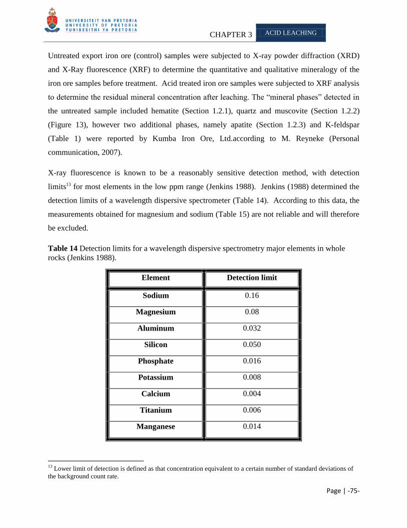
CHAPTER 3 ACID LEACHING
Page | -75-
Untreated export iron ore (control) samples were subjected to X-ray powder diffraction (XRD)
and X-Ray fluorescence (XRF) to determine the quantitative and qualitative mineralogy of the
iron ore samples before treatment. Acid treated iron ore samples were subjected to XRF analysis
to determine the residual mineral concentration after leaching. The ―mineral phases‖ detected in
the untreated sample included hematite (Section 1.2.1), quartz and muscovite (Section 1.2.2)
(Figure 13), however two additional phases, namely apatite (Section 1.2.3) and K-feldspar
(Table 1) were reported by Kumba Iron Ore, Ltd.according to M. Reyneke (Personal
communication, 2007).
X-ray fluorescence is known to be a reasonably sensitive detection method, with detection
limits13 for most elements in the low ppm range (Jenkins 1988). Jenkins (1988) determined the
detection limits of a wavelength dispersive spectrometer (Table 14). According to this data, the
measurements obtained for magnesium and sodium (Table 15) are not reliable and will therefore
be excluded.
Table 14 Detection limits for a wavelength dispersive spectrometry major elements in whole
rocks (Jenkins 1988).
Element Detection limit
Sodium 0.16
Magnesium 0.08
Aluminum 0.032
Silicon 0.050
Phosphate 0.016
Potassium 0.008
Calcium 0.004
Titanium 0.006
Manganese 0.014
13
Lower limit of detection is defined as that concentration equivalent to a certain number of standard deviations of
the background count rate.
CHAPTER 3 ACID LEACHING
Page | -76-
Four untreated export iron ore samples (control) were analyzed with XRF, to give a more
accurate estimate of the batch sample (Table 15). Focus will be placed mainly on the residual
alkali, as these minerals have a deleterious effect in steel manufacturing (Delvasto et al., 2008).
Results of treated samples were expected to exceed the control sample due to sample variation;
therefore a range was determined which included the minimum and maximum amount of alkali
measured in the batch untreated sample. From this data set we can possibly determine the
amount of alkali removed due to acid leaching and excluded data sets possibly influenced by
sample variation. Moreover, the effect of acid leaching on silicon and aluminum will be
investigated due to a report by Mojallali and Weed (1978) which demonstrated that a loss of
aluminum and silicon caused structural rearrangement of a mineral. Wilson and Jones (1983)
found that a decrease in magnesium also shows a rearrangement of a mineral. From XRD
analysis (Table 1), the minerals in which potassium occurs also includes aluminum, therefore it
is likely that potassium or aluminum/silicon might be solubilized indirectly due to the action of
the acid on the other mineral as discovered by Scott and Amonette (1988).
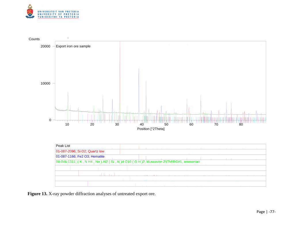
Page | -77-
Figure 13. X-ray powder diffraction analyses of untreated export ore.
Position [°2Theta] 10 20 30 40 50 60 70 80
Counts
0
10000
20000 Export iron ore sample
Peak List
01-087-2096; Si O2; Quartz low 01-087-1166; Fe2 O3; Hematite 00-046-1311; ( K , N H4 , Na ) Al2 ( Si , Al )4 O10 ( O H )2; Muscovite-2\ITM\RG#1, ammonian
Page | -78-
They demonstrated that the micas biotite and muscovite undergo structural changes and possible
dissolution under low pH conditions or in the presence of an organic chelating agent. Moreover,
they also found that other cations are ejected along with potassium from the interlayer of the
mica. Landeweert et al. (2001) commented that oxalate and citrate are able to form stable
complexes with cations such as aluminum, thus decreasing the solution concentration and
thereby increasing the weathering rate of minerals containing aluminum.
From the above, we examined the following minerals and used the calculated parameters during
this discussion: Al2O3 (1.15% - 1.56%), SiO2 (1.92% - 2.80%), K2O (0.16% - 0.18%) and P2O5
(0.13% - 0.17%). Concentrations of each mineral removed below these parameters will be
reported as ―large quantities14
‖ removed. Silicon was present in quartz, muscovite, biotite, etc.
in all of the samples analyzed (Table 1). Quartz is known to dissolve slowly, due to the high
activation energy needed to break the silicon-oxygen bond (Brehm et al., 2005 as cited in Wilson
2004). We did not determine the source of silicon dissolution, as it was outside the scope of this
study; however it is assumed that the silicon released was from the less resistant mineral. Hereon
we accept the average value of the minerals present in the untreated export sample analysed with
XRF as an acceptable representative of the batch sample.
Muscovite and K-feldspar were identified as the major potassium bearing mineral present in the
export sample. Muscovite is an aluminosilicate mineral with compositional affinity to feldspar,
feldspathoids and quartz (Deer et al., 2001; Kalinowski and Schweda 1996;
McKeown et al., 1999). The structure of feldspar consists of cross-linked, ‗double-crankshaft‘
chains of Si4+
and Al3+
tetrahedral with charge compensating such as sodium, potassium and
calcium occupying small cavities in the framework (Han and Lee 2005). Alkali feldspar can be
weathered into a secondary mineral kaolinite [Al2Si2O5(OH)4] under the influence of H+ ions.
The acids assayed during this section of the study are able to aid the solubilization process via
protonation and/or complexation mechanisms. All of the acids tested, except gluconic acid
(60°C), were able to remove potassium so that the residual was below 0.16%. Its inability to
14
A maximum and minum concentration for each mineral present in the untreated sample was determined. Once
the ore was treated with the specific assay, we analyzed it again with XRF. If the residual amount of a specific
mineral was below the minimum concentration, we ascribe it to the assay tested.
CHAPTER 3 ACID LEACHING
Page | -79-
efficiently remove potassium (below 0.16%) is ascribed to sample variation15 and/or mineral
dissemination16, as it was able to remove potassium from the other ore bodies analyzed.
XRF analysis indicated that the selected minerals were solubilized from the export iron ore
(Table 15). The amount of silicon and aluminum removed, illustrates a rearrangement of the
minerals as discussed by Mojallali and Weed (1978). Acetic (30°C), ascorbic (60°C) and
boric acid (30°C and 60°C) were all able to remove aluminum, silicon, potassium and
phosphorous below the set parameters, as indicated previously. Huang and Longo (1992) tested
the dissolution of K-feldspar in oxalic and acetic acid. They found that dissolution of Si and K
from the mineral was increased with decreasing pH. They further discovered that the presence of
Ca and Mg decreased the dissolution of Si and K. The dissolution of feldspar is known to be
affected by various different factors such as twinning lamellae, dislocation, surface roughness
and surface area (Deer et al., 2001). The mechanism suggested by some researchers for mineral
dissolution by organic acids refers to the complexation of aluminum, which can be seen as an
indirect mechanism or ligand-promoted dissolution (Wilson 2004). Preliminary results obtained
during this study and work done as part of a Phd thesis, at the University of Pretoria, justified
repeating the citric and acetic acid experiments. Here XRF data confirmed that acetic and citric
acid were both able to efficiently remove potassium and phosphorous from the export iron ore
sample.
It was thought that the acids that were able to form complexes and attack the minerals with
protons would be able to remove the most alkali as Drever and Stillings (1997) stated that
bidentate acids are more effect than monodentate acids at solubilization minerals, due to stronger
complex formation (Drever and Stillings 1997), however from our data we found that acetic and
citric acid were able to remove comparable amounts. The data suggested that acetic and
gluconic acid, which is known to have protonation and complexation mechanisms
(Ehrlich 1991), were able to remove large concentrations of the alkalis (Table 15). However
boric acid, which is not able to form complexes with the solubilized minerals were able to
remove comparable amounts of alkali. This observation is ascribed to saple variation, as acids
15
Sample contains an alkali concentration higher than the set parameter and therefore no apparent removal will be
reported. 16
Alkali is located in the core of the particle and therefore inaccessible to the acid
CHAPTER 3 ACID LEACHING
Page | -80-
able to form complexes with solubilized minerals showed greater potential with the other
samples.
Most literature reports focused on leaching experiments conducted on almost pure minerals and
some on heterogonous samples such as iron ore. We ascribe the difference in results obtained,
compared to literature, to the varying composition of the ore and/or the accessibility of the
minerals leached as the alkali could be located in the core of the particle. This observation
spurred additional research into determining the effect of particle size on mineral leaching, as
Bosecker (1997) found that a particle size of 42 μm was optimum for leaching. However,
Mortland and Lawton found that potassium released from biotite in a NaCl solution was slower
from fine particles than from large ones (as cited in Wilson 2004). Reichenbach and Rich (1969)
found similar results to Mortland and Lawton when they experimented with different particle
sizes of muscovite and its exchange with BaCl2 at elevated temperatures.
Reichenbach and Rich (1969) proposed that the decreased leaching efficiency with smaller
particles was due to a difference in the exchange mechanism brought about by the splitting of the
particles. The splitting caused a release in stress brought about by the expansion and bending of
the flakes following potassium depletion form mineral, thus resulting in inhibition of further
potassium exchange.
During this study, the effect of extended leaching times were also assayed. Optimum leaching
was obtained after 5 days for most acids tested and were therefore selected as a standard. There
are several possible reasons that only a limited number of minerals were solubilized. Firstly it is
possible that after 5 days all the accessible minerals were removed and only the inaccessible
(inside the core of the particle) remains. Moreover the available protons could also be exhausted
and therefore no further solubilization of the minerals could occur as described by
Welch et al. (2002). Lastly, a saturation state could have been reached in solution and therefore
no further minerals would be solubilized. Scott and Smith (1966) conducted experiments in
which they determined the susceptibility of interlayer potassium in micas to exchange with
sodium. They found that essentially, all the potassium in muscovite, biotite, phlogopite and
vermiculite were exchangeable when the concentration of the potassium in solution was kept
low.
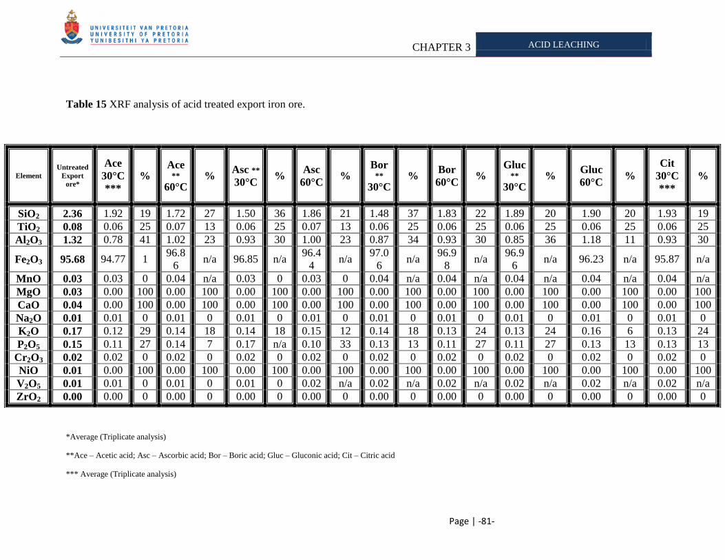
CHAPTER 3 ACID LEACHING
Page | -81-
Table 15 XRF analysis of acid treated export iron ore.
*Average (Triplicate analysis)
**Ace – Acetic acid; Asc – Ascorbic acid; Bor – Boric acid; Gluc – Gluconic acid; Cit – Citric acid
*** Average (Triplicate analysis)
Element
Untreated
Export
ore*
Ace
30°C
***
% Ace
** 60°C
% Asc **
30°C %
Asc
60°C %
Bor **
30°C %
Bor
60°C %
Gluc **
30°C %
Gluc
60°C %
Cit
30°C
***
%
SiO2 2.36 1.92 19 1.72 27 1.50 36 1.86 21 1.48 37 1.83 22 1.89 20 1.90 20 1.93 19
TiO2 0.08 0.06 25 0.07 13 0.06 25 0.07 13 0.06 25 0.06 25 0.06 25 0.06 25 0.06 25
Al2O3 1.32 0.78 41 1.02 23 0.93 30 1.00 23 0.87 34 0.93 30 0.85 36 1.18 11 0.93 30
Fe2O3 95.68 94.77 1 96.8
6 n/a 96.85 n/a
96.4
4 n/a
97.0
6 n/a
96.9
8 n/a
96.9
6 n/a 96.23 n/a 95.87 n/a
MnO 0.03 0.03 0 0.04 n/a 0.03 0 0.03 0 0.04 n/a 0.04 n/a 0.04 n/a 0.04 n/a 0.04 n/a
MgO 0.03 0.00 100 0.00 100 0.00 100 0.00 100 0.00 100 0.00 100 0.00 100 0.00 100 0.00 100
CaO 0.04 0.00 100 0.00 100 0.00 100 0.00 100 0.00 100 0.00 100 0.00 100 0.00 100 0.00 100
Na2O 0.01 0.01 0 0.01 0 0.01 0 0.01 0 0.01 0 0.01 0 0.01 0 0.01 0 0.01 0
K2O 0.17 0.12 29 0.14 18 0.14 18 0.15 12 0.14 18 0.13 24 0.13 24 0.16 6 0.13 24
P2O5 0.15 0.11 27 0.14 7 0.17 n/a 0.10 33 0.13 13 0.11 27 0.11 27 0.13 13 0.13 13
Cr2O3 0.02 0.02 0 0.02 0 0.02 0 0.02 0 0.02 0 0.02 0 0.02 0 0.02 0 0.02 0
NiO 0.01 0.00 100 0.00 100 0.00 100 0.00 100 0.00 100 0.00 100 0.00 100 0.00 100 0.00 100
V2O5 0.01 0.01 0 0.01 0 0.01 0 0.02 n/a 0.02 n/a 0.02 n/a 0.02 n/a 0.02 n/a 0.02 n/a
ZrO2 0.00 0.00 0 0.00 0 0.00 0 0.00 0 0.00 0 0.00 0 0.00 0 0.00 0 0.00 0
CHAPTER 3 ACID LEACHING
Page | -82-
Phosphorous was detected as apatite in the export ore (Table 1). All the acids tested, except
acetic acid at 60°C and ascorbic acid at 30°C were able to remove ―large quantities‖ of
potassium and phosphorous. This can again be attributed to sample variation or mineral
distribution inside the particle, as Welch and coworkers (2002) demonstrated that acetic and
oxalic acid were able to increase apatite dissolution. They attributed the ability of these acids to
solubilize the phosphorous from the mineral to their ability to form complexes with either the
calcium on the mineral surface or in the solution. Moreover, they further commented that the
acids were also effective at near neutral pH, thereby supporting the complexing mechanism as
fewer protons would be present for protonation. Welch and coworkers (2002) further
demonstrated that calcium was preferentially released from apatite than phosphate. They
proposed that the increased release could be due to an ore reactive calcium rich phase such as
carbonate, calcium carbonate or calcium fluoride. Denêvre et al. (1996) found that complexation
is dependent on the relative concentration of the anions and metals in solution, the pH and
stability constants of the various complexes; therefore it is probable that these factors played a
role in our leaching experiments. We found that boric (60°C), gluconic (30°C) and
ascorbic acid (60°C) were able to remove more than 25% of the phosphorous from the export
ore. Gluconic acid is one of the strongest naturally occurring organic acids and has been
associated with bacteria that are able to solubilize high levels of phosphates
(Goldstein and Krishnaraj 2007). Furthermore, boric acid is able to release a proton, which can
attack the mineral surface and aid in solubilization (Sayer and Gadd 2001). Research has
demonstrated that gluconic and ascorbic acid were able to form complexes with the minerals
released, thus affecting the saturation state and the amount of alkali that could be removed
(Drever and Stillings 1997; Goldstein 1995). Moreover, when these acids form complexes,
protons are released which can aid further solubilization of the minerals (Drever and Stillings
1997; Ehrlich 1991).
We found during this section of the study that the organic acids were more effective at removing
the alkali from the ore than the inorganic acids. This is in accordance with other research that
found mineral weathering by organic acids to be 3-5 times higher compared to inorganic acids at
the same pH. This observation was ascribed partly due to the complex forming ability of organic
acids, which lowers the saturation state of cations in solution (Banfield et al., 1999). Moreover,
CHAPTER 3 ACID LEACHING
Page | -83-
researchers indicated that increased temperatures favor the release of phosphorous from apatite
(Kim et al., 2003), which is in accordance with some of the XRF data obtained during this study.
From the XRF data (Table 15), it was observed that none of the manganese, sodium, chromium,
vanadium or zirconium was removed. These minerals do not form a component of the mineral
phases detected in the ore by XRD (Table 1). The inability of the acids to remove these minerals
could be due to the minerals intrinsic characteristics or possible position inside the ore particle.
For example, vanadium is a type of transition metal, which is known to have corrosion resistance
to alkali, sulfuric-, hydrochloric acid and saline solution (Moskalyk and Alfantazi 2003).
Furthermore, all of the magnesium and calcium were removed from the ore (Table 15), which is
ascribed to the leaching mechanisms of the acids tested.
KGT (conglomerate) samples were supplied by Kumba Iron Ore, Ltd. and categorized as a high
potassium/low phosphorous iron ore samples according to B. Ntsoelengoe (Personal
communication, 2006). The susceptibility of this sample to acid leaching was tested at two
different temperatures (Table 16). Four untreated KGT samples (control) were analyzed with
XRF, to give a more accurate estimate of the mineral composition of the batch sample
(Table 15). Focus will be placed mainly on the residual alkali, as these minerals have a
deleterious effect on steel manufacturing (Delvasto et al., 2008). Results of treated samples are
expected to exceed the control sample due to sample variation; therefore a range was determined
which included the minimum and maximum amount of alkali measured in the replicate untreated
sample. Therefore the following minerals and parameters for the export iron ore samples were
used during this study: Al2O3 (1.90 - 2.14); SiO2 (3.90 – 4.90), K2O (0.48 – 0.56) and P2O5
(0.06 – 0.07). Concentrations of each mineral removed below these parameters will be reported
as ―large quantities‖ removed. From this data set we can possibly determine the amount of alkali
removed due to acid leaching and exclude the data sets possibly influenced by sample variation.
Moreover, the effect on silicon and aluminum will also be investigated as these might be
solubilized with the alkalis as stated previously. Scott and Amonette (1988) found that the micas
biotite and muscovite undergo structural changes and possible dissolution under low pH
conditions or in the presence of an organic chelating agent. Moreover, they also found that other
cations are ejected along with potassium from the interlayer of the mica.
CHAPTER 3 ACID LEACHING
Page | -84-
Landeweert et al. (2001) commented that oxalate and citrate are able to form stable complexes
with cations such as aluminum, thus decreasing the solution concentration and thereby increasing
the weathering rate of minerals containing aluminum. Mojallali and Weed (1978) stated that a
loss of aluminum and/or silicon shows a more structural rearrangement of the mineral. Later
Wilson and Jones (1983) found that a decrease in magnesium and iron shows an even more
rearrangement of a mineral.
Untreated KGT samples (control) were subjected to X-ray powder diffraction (XRD) and X-Ray
fluorescence (XRF) to determine the quantitative and qualitative mineralogy of the iron ore
samples initially. Acid treated KGT samples were subjected to XRF analysis to determine the
residual mineral concentration after leaching (Table 16). The ―mineral phases‖ detected in the
untreated sample included hematite, quartz and muscovite (Figure 14), however additional
phases, namely greenalite, illite, etc (Table 1) were reported by Kumba Iron Ore, Ltd.. Silicon
was present in quartz, muscovite, biotite, etc. in all of the samples analyzed (Table 1). Quartz is
known dissolved slowly, due to the high activation energy needed to break the silicon-oxygen
bond (Brehm et al., 2005 as cited in Wilson 2004). We did not determine the source of silicon
dissolution, as it was outside the scope of this study; however it is assumed that the silicon
released was from the less resistant mineral. Jenkins (1988) reported the lower detection limits
of a wavelength dispersive spectrometer (Table 14), therefore sodium and magnesium will be
excluded from further analysis as it is below the limits for the machine.
Potassium was detected as illite and muscovite in the KGT sample (Table 1). Illite is a non-
expanding, clay-sized phyllosilicate or layer alumino-silicate that commonly occurs in
sediments, soils and argillaceous sedimentary rocks. It is structurally similar to muscovite or
sericite [KAl2(OH)2(AlSi3O10)], with more silicon, magnesium, iron and water, with less
tetrahedral aluminum and interlayer potassium. Illite occurs as an alternation product of
muscovite and feldspar during weathering (Mengel and Uhlenbecker 1993).
Acetic (30°C), citric (60°C), sulfuric (30°C) and malic acid (30°C) were able to remove ―large
quantities‖ of potassium and aluminum. The amount of aluminum removed, illustrates a
rearrangement of the minerals (Mojallali and Weed 1978). Han and Lee (2005) found that
Bacillus mucilaginosus, a known potassium solubilizing bacterium, was able solubilize
CHAPTER 3 ACID LEACHING
Page | -85-
potassium from mica, illite and orthoclase by producing organic acids. Acetic, malic and citric
acid are able to form complexes with the minerals released from the ore (Table 16).
During complex formation, protons are released which can aid solubilization of other minerals.
Sulfuric acid and malic acid are able to donate a proton, thus solubilization via protonation
(Drever and Stillings 1997). The inability of the other acids tested to solubilize alkali from the
ore is ascribed to mineral dissemination and sample variation, as certain of the data reported
quantities are outside of the set parameters. Illite and muscovite have similar structures.
Muscovite is known to have considerable resistance toward leaching of interlayer potassium,
compared to trioctahedral micas (such as biotite). It was proposed by Kalinowski and Schweda
(1996) that an inclined orientation of hydroxyl ions in the dioctahedral micas results in stronger
binding of potassium.
XRD analysis of the KGT sample did not detect a phosphorous phase. This is in accordance
with results obtained from Kumba Iron Ore, Ltd. (Table 1). Manganese, sodium, chromium and
vanadium were not leached from the ore by the acids tested. It is assumed that this is due to
mineral dissemination or mineral characteristics for example. Vanadium is a type of transition
metal, which is known to have corrosion resistance to alkali, sulfuric-, hydrochloric acid and
saline solution (Moskalyk and Alfantazi 2003) and therefore we expect it not to be affected, as
with the export iron ore analysis. Magnesium, calcium, nickel and zirconium were not detected
in the KGT sample which is probable that the concentrations of these minerals were below the
detection limit of XRF (Jenkins 1988).
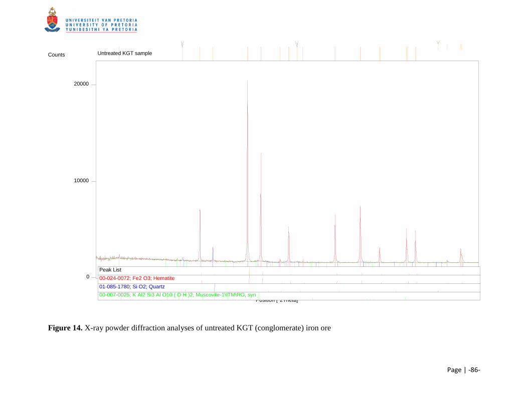
Page | -86-
Figure 14. X-ray powder diffraction analyses of untreated KGT (conglomerate) iron ore
Position [°2Theta]
10 20 30 40 50 60 70 80
Counts
0
10000
20000
Untreated KGT sample
Peak List 00-024-0072; Fe2 O3; Hematite 01-085-1780; Si O2; Quartz 00-007-0025; K Al2 Si3 Al O10 ( O H )2; Muscovite-1\ITM\RG, syn
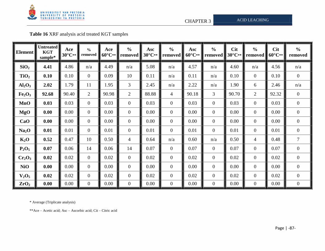
CHAPTER 3 ACID LEACHING
Page | -87-
Table 16 XRF analysis acid treated KGT samples
Element Untreated
KGT
sample*
Ace
30°C** %
removed Ace
60°C**
%
removed
Asc
30°C**
%
removed
Asc
60°C**
%
removed
Cit
30°C**
%
removed
Cit
60°C**
%
removed
SiO2 4.41 4.86 n/a 4.49 n/a 5.08 n/a 4.57 n/a 4.60 n/a 4.56 n/a
TiO2 0.10 0.10 0 0.09 10 0.11 n/a 0.11 n/a 0.10 0 0.10 0
Al2O3 2.02 1.79 11 1.95 3 2.45 n/a 2.22 n/a 1.90 6 2.46 n/a
Fe2O3 92.68 90.40 2 90.98 2 88.88 4 90.18 3 90.70 2 92.32 0
MnO 0.03 0.03 0 0.03 0 0.03 0 0.03 0 0.03 0 0.03 0
MgO 0.00 0.00 0 0.00 0 0.00 0 0.00 0 0.00 0 0.00 0
CaO 0.00 0.00 0 0.00 0 0.00 0 0.00 0 0.00 0 0.00 0
Na2O 0.01 0.01 0 0.01 0 0.01 0 0.01 0 0.01 0 0.01 0
K2O 0.52 0.47 10 0.50 4 0.64 n/a 0.60 n/a 0.50 4 0.48 7
P2O5 0.07 0.06 14 0.06 14 0.07 0 0.07 0 0.07 0 0.07 0
Cr2O3 0.02 0.02 0 0.02 0 0.02 0 0.02 0 0.02 0 0.02 0
NiO 0.00 0.00 0 0.00 0 0.00 0 0.00 0 0.00 0 0.00 0
V2O5 0.02 0.02 0 0.02 0 0.02 0 0.02 0 0.02 0 0.02 0
ZrO2 0.00 0.00 0 0.00 0 0.00 0 0.00 0 0.00 0 0.00 0
* Average (Triplicate analysis)
**Ace – Acetic acid; Asc – Ascorbic acid; Cit – Citric acid
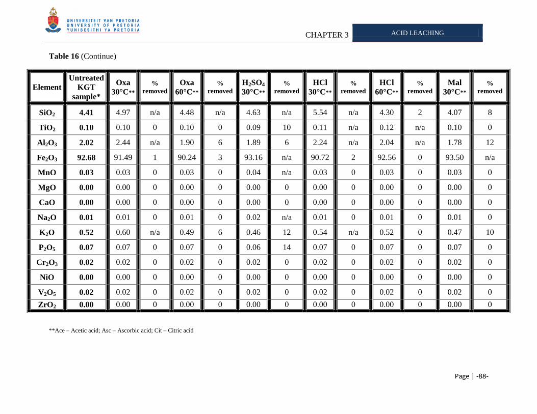
CHAPTER 3 ACID LEACHING
Page | -88-
Table 16 (Continue)
Element
Untreated
KGT
sample*
Oxa
30°C** %
removed Oxa
60°C** %
removed H2SO4
30°C** %
removed HCl
30°C** %
removed HCl
60°C** %
removed Mal
30°C** %
removed
SiO2 4.41 4.97 n/a 4.48 n/a 4.63 n/a 5.54 n/a 4.30 2 4.07 8
TiO2 0.10 0.10 0 0.10 0 0.09 10 0.11 n/a 0.12 n/a 0.10 0
Al2O3 2.02 2.44 n/a 1.90 6 1.89 6 2.24 n/a 2.04 n/a 1.78 12
Fe2O3 92.68 91.49 1 90.24 3 93.16 n/a 90.72 2 92.56 0 93.50 n/a
MnO 0.03 0.03 0 0.03 0 0.04 n/a 0.03 0 0.03 0 0.03 0
MgO 0.00 0.00 0 0.00 0 0.00 0 0.00 0 0.00 0 0.00 0
CaO 0.00 0.00 0 0.00 0 0.00 0 0.00 0 0.00 0 0.00 0
Na2O 0.01 0.01 0 0.01 0 0.02 n/a 0.01 0 0.01 0 0.01 0
K2O 0.52 0.60 n/a 0.49 6 0.46 12 0.54 n/a 0.52 0 0.47 10
P2O5 0.07 0.07 0 0.07 0 0.06 14 0.07 0 0.07 0 0.07 0
Cr2O3 0.02 0.02 0 0.02 0 0.02 0 0.02 0 0.02 0 0.02 0
NiO 0.00 0.00 0 0.00 0 0.00 0 0.00 0 0.00 0 0.00 0
V2O5 0.02 0.02 0 0.02 0 0.02 0 0.02 0 0.02 0 0.02 0
ZrO2 0.00 0.00 0 0.00 0 0.00 0 0.00 0 0.00 0 0.00 0
**Ace – Acetic acid; Asc – Ascorbic acid; Cit – Citric acid
CHAPTER 3 ACID LEACHING
Page | -89-
SK iron ore samples (shale) are known to be high in potassium/low phosphorous. The aptitude
of various acids to remove the alkalis from the SK sample was tested (Table 17). Mineral phases
detected with XRD included muscovite, hematite and quartz (Figure 15). Additional phases
were reported by Kumba staff (Table 1). The alkali potassium was present as biotite, muscovite
and illite. Biotite is a ferromagnesium mineral with compositional affinity to olivine
[(Mg,Fe)2SiO4], pyroxene [XY(Si,Al)2O6]17 and amphiboles18 (Deer et al., 2001). As with other
mica minerals, it has a highly perfect basal cleavage and consists of flexible sheets or lamellae19,
which can easily flake off. Calvaruso et al. (2006) stated that biotite can undergo two types of
weathering namely: congruent (destruction of the mineral surface) and incongruent
(transformation into vermiculite by release of interlayer potassium). The sheets in the crystal
structure of biotite are made up of iron, magnesium, aluminum, silicon, oxygen and hydrogen
ions that are weakly bound together by potassium. The potassium can be substituted in part by
sodium, calcium, barium. Magnesium can be completely replaced by ferrous iron and ferric iron
and in part by titanium and manganese. Biotite is often referred to as the iron mica because it
contains more iron than phlogopite (Hashemi-Nezhad 2005). No phosphorous phases were
detected in the SK sample.
The following parameters for the alkalis and mineral described by Mojallali and Weed (1978)
and Wilson and Jones (1983) were calculated from XRF data of untreated SK sample:
Al2O3 (9.69 – 10.17); SiO2 (11.56 – 12.26); K2O (2.31 – 2.43) and P2O5 (0.03 – 0.04). These
results are in accordance with classification by Kumba Iron Ore, Ltd. staff. Treated samples
analyzed did contain quantities outside the parameters set with the untreated samples (Table 17),
which is attributed to sample variation. Concentrations of each mineral removed below these
parameters will be reported as ―large quantities‖ removed as this will be used to determine which
acids were able to solubilize the most alkali. Alkali removal from the ore was determined by
measuring the residual contained in the ore after the acid treatment and comparing it to the
17
X represents calcium, sodium, ferrous iron, magnesium, zinc, manganese and lithium. Y represents ions such as
chromium, aluminium, ferric iron, magnesium, manganese, scandium, titanium, vanadium.
(http://en.wikipedia.org/wiki/Pyroxene) 18
The main differences between amphiboles and pyroxenes are that amphiboles contain essential hydroxyl (OH) or
halogene (F, Cl) and secondly the basic structure is a double chain of tetrahedra (as opposed to the single chain
structure of pyroxene). (http://en.wikipedia.org/wiki/Amphiboles) 19
A lamella is a gill-shaped structure: fine sheets of material held adjacent one another, with fluid in-between-(or
simply 'welded'-plates)[ http://en.wikipedia.org/wiki/Lamellae_(materials)]
CHAPTER 3 ACID LEACHING
Page | -90-
untreated sample (control). Jenkins (1988) reported the lower detection limits of a wavelength
dispersive spectrometer (Table 14), therefore sodium will be excluded from further analysis. We
accept the average value of the untreated SK sample as an acceptable representative (Table 17).
Acetic (30°C), ascorbic (60°C), sulfuric (30°C and 60°C) and malic acid (30°C) were able to
remove large quantities of potassium from the ore. Only acetic and sulfuric acid were able to
remove = aluminum, potassium and silicon from the sample. The amount of silicon and
aluminum removed, illustrates a rearrangement of the minerals as stated by
Mojallali and Weed (1978). These acids aid solubilization via protonation in the (Ehrlich 1991).
It was expected that citric acid would be able to remove some of the potassium, as it was
effective at removing the alkali from the KGT sample. It is assumed that the difference in
mineral composition of the KGT, SK and export iron ore samples played a role in the amount of
potassium release (Das et al., 1999 as cited in Brandl 2001).
The low concentration of potassium solubilized was expected as the sample contained illite,
biotite and muscovite which are all known to be effected by the potassium concentration in
solution. Rausell and coworkers (1965) determined the sensitivity of muscovite to potassium
concentration in solution. Later Pal and coworkers (2001) demonstrated that if biotite and
muscovite were both present in soil, no potassium dissolution from muscovite would occur,
further supporting findings by Rausell et al. (1965). Also biotite weathering is known to be
diffusion-controlled and depends on the potassium concentration in the solution (Wilson 2004)
and therefore we expected low potassium solubilization to occur. Scott and Smith (1966)
determined the susceptibility of various different micas to exchange their interlayer potassium
with sodium from the surrounding environment. They found that almost all of the interlayer
potassium of muscovite and biotite were exchangeable, as long as the solution potassium
concentration was kept below certain limits. It is therefore possible that the inability of the acids
to remove greater amounts of potassium from the SK (high potassium) sample is due to a
saturation state of potassium reached in solution.
Manganese was removed by oxalic (30°C and 60°C), hydrochloric (30°C), ascorbic (30°C) and
citric acid (30°C and 60°C). It susceptibility to leaching could be due to its possible position on
the surface of the particle. Phosphorous, vanadium and chromium were not leached, possibly
CHAPTER 3 ACID LEACHING
Page | -91-
due to their position closer to the core of the particle (Table 17). Ballester et al. (1989) and Das
et al. (1999) listed several properties of minerals which could influence leaching. These include:
mineral type, gain size, mineral dissemination, etc. Therefore the position of the mineral in the
particle could determine its susceptibility to leaching.
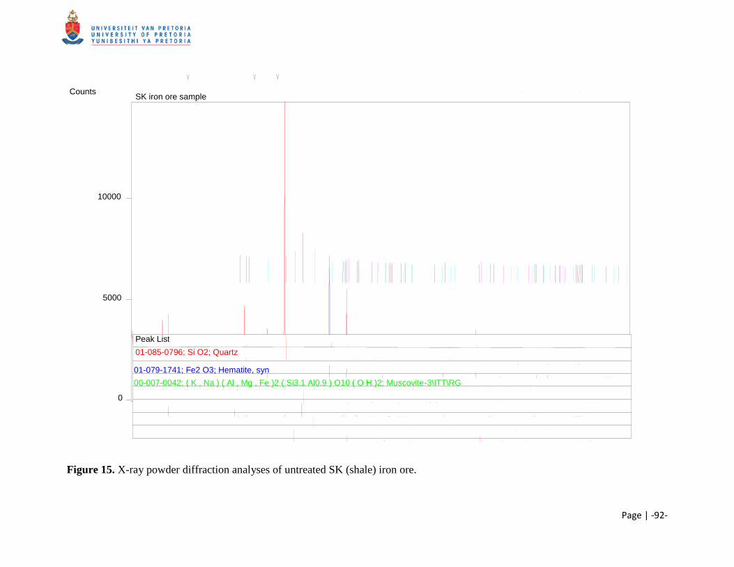
Page | -92-
Figure 15. X-ray powder diffraction analyses of untreated SK (shale) iron ore.
Position [°2Theta]
10 20 30 40 50 60 70 80
Counts
0
5000
10000
SK iron ore sample
Peak List
01-085-0796; Si O2; Quartz
01-079-1741; Fe2 O3; Hematite, syn
00-007-0042; ( K , Na ) ( Al , Mg , Fe )2 ( Si3.1 Al0.9 ) O10 ( O H )2; Muscovite-3\ITT\RG
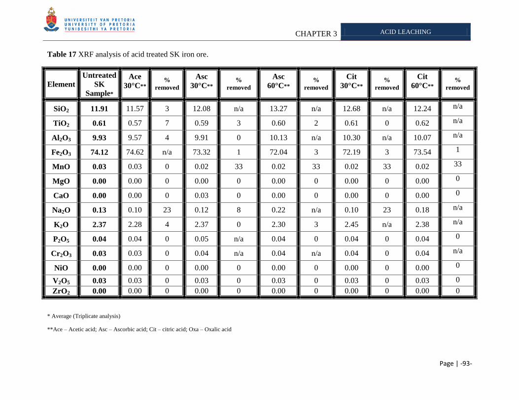
CHAPTER 3 ACID LEACHING
Page | -93-
Table 17 XRF analysis of acid treated SK iron ore.
Element
Untreated
SK
Sample*
Ace
30°C** %
removed
Asc
30°C** %
removed
Asc
60°C** %
removed
Cit
30°C** %
removed
Cit
60°C** %
removed
SiO2 11.91 11.57 3 12.08 n/a 13.27 n/a 12.68 n/a 12.24 n/a
TiO2 0.61 0.57 7 0.59 3 0.60 2 0.61 0 0.62 n/a
Al2O3 9.93 9.57 4 9.91 0 10.13 n/a 10.30 n/a 10.07 n/a
Fe2O3 74.12 74.62 n/a 73.32 1 72.04 3 72.19 3 73.54 1
MnO 0.03 0.03 0 0.02 33 0.02 33 0.02 33 0.02 33
MgO 0.00 0.00 0 0.00 0 0.00 0 0.00 0 0.00 0
CaO 0.00 0.00 0 0.03 0 0.00 0 0.00 0 0.00 0
Na2O 0.13 0.10 23 0.12 8 0.22 n/a 0.10 23 0.18 n/a
K2O 2.37 2.28 4 2.37 0 2.30 3 2.45 n/a 2.38 n/a
P2O5 0.04 0.04 0 0.05 n/a 0.04 0 0.04 0 0.04 0
Cr2O3 0.03 0.03 0 0.04 n/a 0.04 n/a 0.04 0 0.04 n/a
NiO 0.00 0.00 0 0.00 0 0.00 0 0.00 0 0.00 0
V2O5 0.03 0.03 0 0.03 0 0.03 0 0.03 0 0.03 0
ZrO2 0.00 0.00 0 0.00 0 0.00 0 0.00 0 0.00 0
* Average (Triplicate analysis)
**Ace – Acetic acid; Asc – Ascorbic acid; Cit – citric acid; Oxa – Oxalic acid
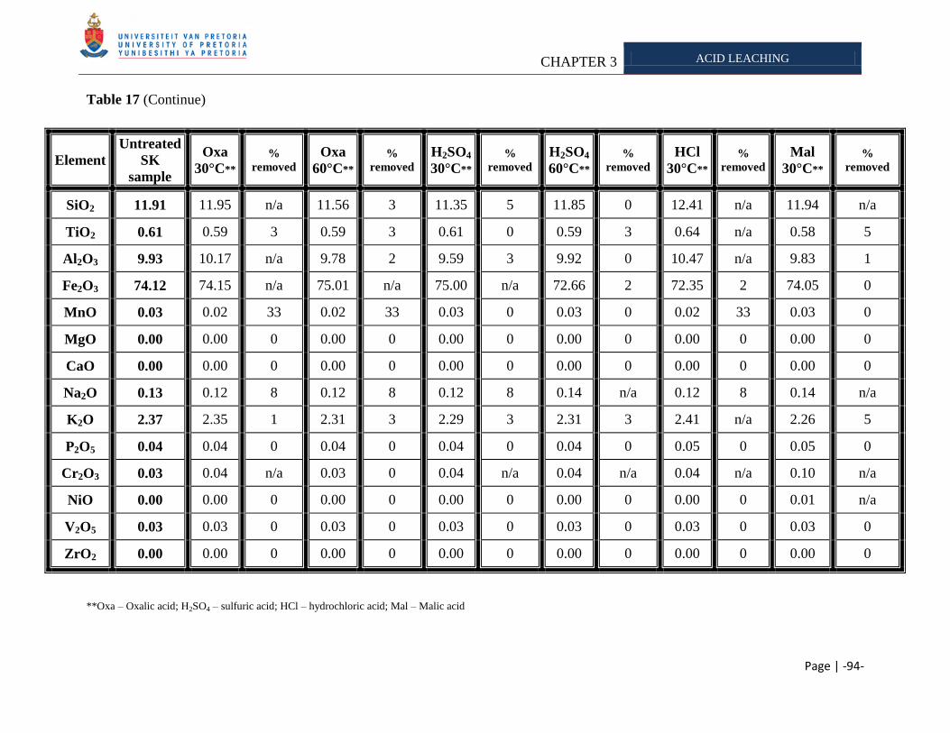
CHAPTER 3 ACID LEACHING
Page | -94-
Table 17 (Continue)
Element
Untreated
SK
sample
Oxa
30°C** %
removed Oxa
60°C** %
removed H2SO4
30°C** %
removed H2SO4
60°C** %
removed HCl
30°C** %
removed Mal
30°C** %
removed
SiO2 11.91 11.95 n/a 11.56 3 11.35 5 11.85 0 12.41 n/a 11.94 n/a
TiO2 0.61 0.59 3 0.59 3 0.61 0 0.59 3 0.64 n/a 0.58 5
Al2O3 9.93 10.17 n/a 9.78 2 9.59 3 9.92 0 10.47 n/a 9.83 1
Fe2O3 74.12 74.15 n/a 75.01 n/a 75.00 n/a 72.66 2 72.35 2 74.05 0
MnO 0.03 0.02 33 0.02 33 0.03 0 0.03 0 0.02 33 0.03 0
MgO 0.00 0.00 0 0.00 0 0.00 0 0.00 0 0.00 0 0.00 0
CaO 0.00 0.00 0 0.00 0 0.00 0 0.00 0 0.00 0 0.00 0
Na2O 0.13 0.12 8 0.12 8 0.12 8 0.14 n/a 0.12 8 0.14 n/a
K2O 2.37 2.35 1 2.31 3 2.29 3 2.31 3 2.41 n/a 2.26 5
P2O5 0.04 0.04 0 0.04 0 0.04 0 0.04 0 0.05 0 0.05 0
Cr2O3 0.03 0.04 n/a 0.03 0 0.04 n/a 0.04 n/a 0.04 n/a 0.10 n/a
NiO 0.00 0.00 0 0.00 0 0.00 0 0.00 0 0.00 0 0.01 n/a
V2O5 0.03 0.03 0 0.03 0 0.03 0 0.03 0 0.03 0 0.03 0
ZrO2 0.00 0.00 0 0.00 0 0.00 0 0.00 0 0.00 0 0.00 0
**Oxa – Oxalic acid; H2SO4 – sulfuric acid; HCl – hydrochloric acid; Mal – Malic acid
CHAPTER 3 ACID LEACHING
Page | -95-
SPHP iron ore was categorized by Kumba Iron Ore, Ltd., as a high phosphorous/low potassium
sample. This is in accordance with our results (Table 18). The mineral phases detected were
(Figure 16): apatite, hematite, muscovite and quartz (Table 1). XRF analysis of the untreated
SPHP iron ore gave the following parameters, as selected for the other samples analyzed:
Al2O3 (0.47 - 0.51); SiO2 (0.57 - 0.83); K2O (0.03 - 0.04) and P2O5 (0.34 - 0.40). Samples
analyzed did contain minerals with quantities outside the parameters (Table 18), which is
attributed to sample variation. Concentrations of each mineral removed below these parameters
will be reported as ―large quantities‖ removed. Alkali removal from the ore was determined by
measuring the residual contained in the ore after the acid treatment and comparing it to the
untreated sample (control). Jenkins (1988) reported the lower detection limits of a wavelength
dispersive spectrometer (Table 14), therefore sodium was excluded from further analysis.
According to XRF analysis, magnesium, calcium, nickel and zirconium were absent from the
sample. The amount of these minerals could possibly be below the detection limit, explaining
their absence from the mineral composition. We accept the average value of the untreated SK
sample as an acceptable representative (Table 18).
There was no amount of potassium removed from the SPHP sample that could exclude the role
of sample variation as none of the samples contained residual amounts of potassium that was
below the set parameters stated previously. There are three possible reasons why none of the
potassium could be removed. Firstly the alkali could have been present in the core of the
particle, therefore inaccessible to leaching; secondly a minute quantity of potassium was
released, which could not be detected by XRF; and lastly sample composition might be highly
variable between batches. Several acid were however able to remove a ―large quantity‖ of
phosphorous. These included the following (Table 18): Citric acid (60°C), oxalic (30°C and
60°C), sulfuric acid (30°C and 60°C), hydrochloric (30°C and 60°C) and malic acid (30°C). The
acetic and ascorbic acids were able to solubilize minerals from the export, KGT and SK sample,
but not from the SPHP sample. This is ascribed to mineral composition and dissemination
within the ore particle. More phosphorous was released from the SPHP sample when the acid
leaching experiments were conducted at elevated temperatures. This is in accordance with
research by Goldstein and Krishnaraj (2007) and Kim et al. (2003). Kim and coworkers (2003)
determined the type of phosphorous found in the sediments of lakes and rivers and the effect of
CHAPTER 3 ACID LEACHING
Page | -96-
temperature on phosphorous solubilization. They found that more phosphorous was released as
the temperature increased. Goldstein and Krishnaraj (2007) discovered that the most common
method by which calcium phosphate are solulized, is by acidulation. All of the acids tested are
able to donate a proton and thus consequently acidify the solution (Ehrlich 1991), which would
therefore influence the mobility of the various minerals in the ore.
Oxalic acid and hydrochloric acid were able to remove a ―large quantity‖ of aluminum. Oxalic
acid is able to aid solubilization by forming complexes with minerals released and by
protonation. Hydrochloric acid is able to donate a proton and thus assist solubilization by a
protonation attack (Table 13). We expected that aluminum and potassium would be leached
simultaneously as with the other iron ore samples tested. This was not the case with the SPHP
sample. We ascribed the results obtained for the amount of aluminum and potassium released to
sample variation or to low concentration of potassium present in the sample, therefore any
removal seem insignificant from XRF analysis. Furthermore, all the effective acids for
solubilizing phosphorous are able to aid solubilization via protonation, which is in accordance
with results from Goldstein and Krishnaraj (2007), which found that calcium phosphate is mostly
solubilized via acidulation.
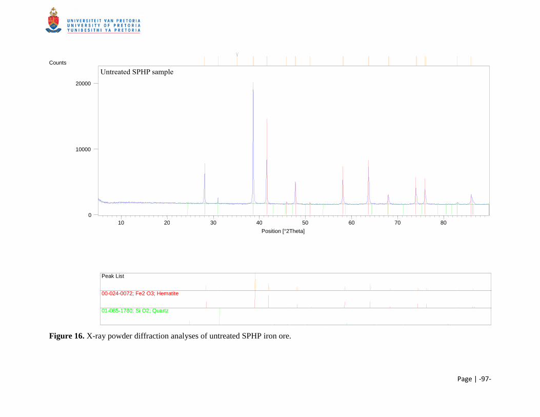
Page | -97-
Figure 16. X-ray powder diffraction analyses of untreated SPHP iron ore.
Position [°2Theta] 10 20 30 40 50 60 70 80
Counts
0
10000
20000
Peak List
00-024-0072; Fe2 O3; Hematite
01-085-1780; Si O2; Quartz
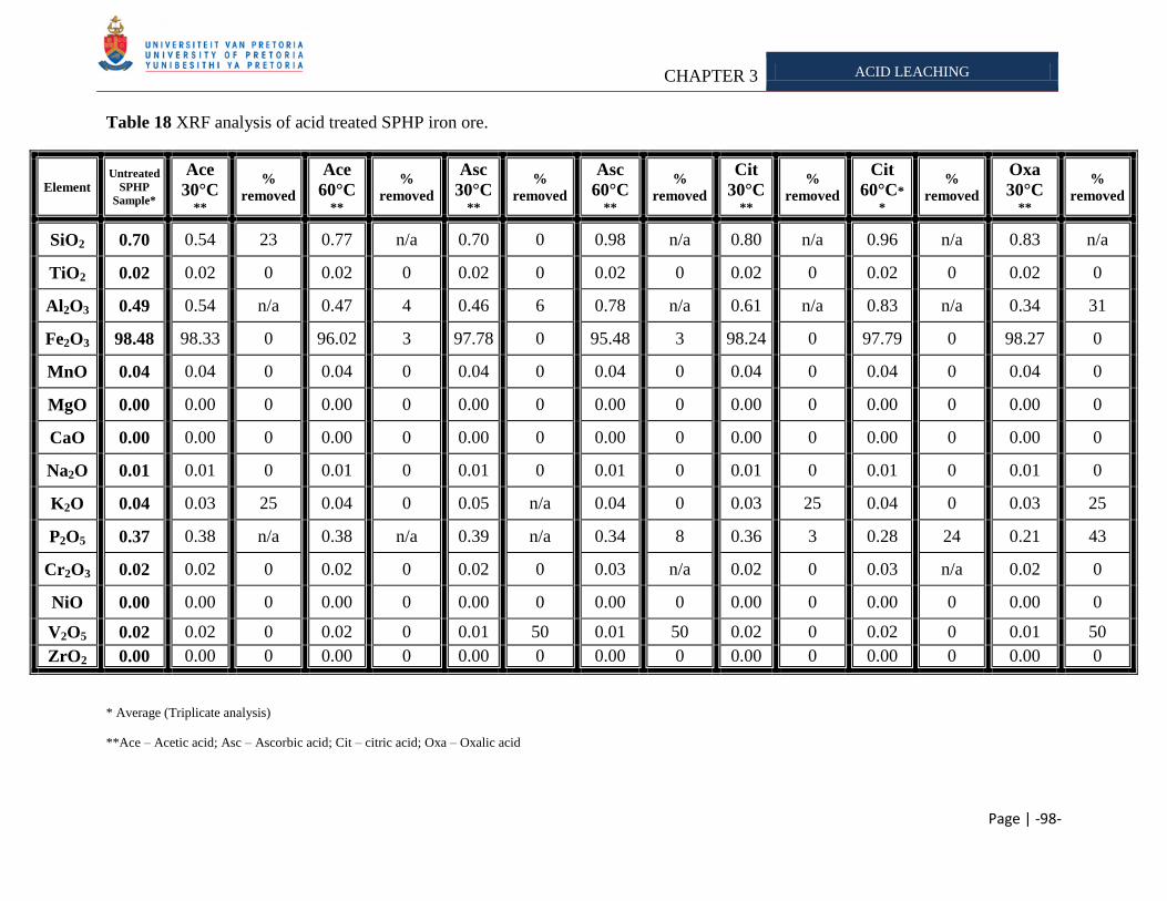
CHAPTER 3 ACID LEACHING
Page | -98-
Table 18 XRF analysis of acid treated SPHP iron ore.
Element Untreated
SPHP
Sample*
Ace
30°C **
%
removed
Ace
60°C **
%
removed
Asc
30°C**
%
removed
Asc
60°C **
%
removed
Cit
30°C**
%
removed
Cit
60°C*
*
%
removed
Oxa
30°C **
%
removed
SiO2 0.70 0.54 23 0.77 n/a 0.70 0 0.98 n/a 0.80 n/a 0.96 n/a 0.83 n/a
TiO2 0.02 0.02 0 0.02 0 0.02 0 0.02 0 0.02 0 0.02 0 0.02 0
Al2O3 0.49 0.54 n/a 0.47 4 0.46 6 0.78 n/a 0.61 n/a 0.83 n/a 0.34 31
Fe2O3 98.48 98.33 0 96.02 3 97.78 0 95.48 3 98.24 0 97.79 0 98.27 0
MnO 0.04 0.04 0 0.04 0 0.04 0 0.04 0 0.04 0 0.04 0 0.04 0
MgO 0.00 0.00 0 0.00 0 0.00 0 0.00 0 0.00 0 0.00 0 0.00 0
CaO 0.00 0.00 0 0.00 0 0.00 0 0.00 0 0.00 0 0.00 0 0.00 0
Na2O 0.01 0.01 0 0.01 0 0.01 0 0.01 0 0.01 0 0.01 0 0.01 0
K2O 0.04 0.03 25 0.04 0 0.05 n/a 0.04 0 0.03 25 0.04 0 0.03 25
P2O5 0.37 0.38 n/a 0.38 n/a 0.39 n/a 0.34 8 0.36 3 0.28 24 0.21 43
Cr2O3 0.02 0.02 0 0.02 0 0.02 0 0.03 n/a 0.02 0 0.03 n/a 0.02 0
NiO 0.00 0.00 0 0.00 0 0.00 0 0.00 0 0.00 0 0.00 0 0.00 0
V2O5 0.02 0.02 0 0.02 0 0.01 50 0.01 50 0.02 0 0.02 0 0.01 50
ZrO2 0.00 0.00 0 0.00 0 0.00 0 0.00 0 0.00 0 0.00 0 0.00 0
* Average (Triplicate analysis)
**Ace – Acetic acid; Asc – Ascorbic acid; Cit – citric acid; Oxa – Oxalic acid
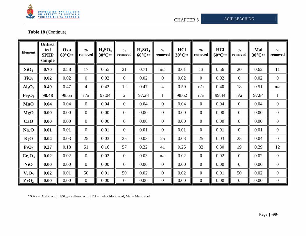
CHAPTER 3 ACID LEACHING
Page | -99-
Table 18 (Continue)
Element
Untrea
ted
SPHP
sample
Oxa
60°C** %
removed
H2SO4
30°C** %
removed H2SO4
60°C** %
removed HCl
30°C** %
removed HCl
60°C** %
removed Mal
30°C** %
removed
SiO2 0.70 0.58 17 0.55 21 0.71 n/a 0.61 13 0.56 20 0.62 11
TiO2 0.02 0.02 0 0.02 0 0.02 0 0.02 0 0.02 0 0.02 0
Al2O3 0.49 0.47 4 0.43 12 0.47 4 0.59 n/a 0.40 18 0.51 n/a
Fe2O3 98.48 98.65 n/a 97.04 2 97.28 1 98.62 n/a 99.44 n/a 97.84 1
MnO 0.04 0.04 0 0.04 0 0.04 0 0.04 0 0.04 0 0.04 0
MgO 0.00 0.00 0 0.00 0 0.00 0 0.00 0 0.00 0 0.00 0
CaO 0.00 0.00 0 0.00 0 0.00 0 0.00 0 0.00 0 0.00 0
Na2O 0.01 0.01 0 0.01 0 0.01 0 0.01 0 0.01 0 0.01 0
K2O 0.04 0.03 25 0.03 25 0.03 25 0.03 25 0.03 25 0.04 0
P2O5 0.37 0.18 51 0.16 57 0.22 41 0.25 32 0.30 19 0.29 12
Cr2O3 0.02 0.02 0 0.02 0 0.03 n/a 0.02 0 0.02 0 0.02 0
NiO 0.00 0.00 0 0.00 0 0.00 0 0.00 0 0.00 0 0.00 0
V2O5 0.02 0.01 50 0.01 50 0.02 0 0.02 0 0.01 50 0.02 0
ZrO2 0.00 0.00 0 0.00 0 0.00 0 0.00 0 0.00 0 0.00 0
**Oxa – Oxalic acid; H2SO4 – sulfuric acid; HCl – hydrochloric acid; Mal – Malic acid

CHAPTER 3 ACID LEACHING
Page | -100-
From XRF data obtained (Table 13 – Table 18), we observed that the acids were able to remove
varying amounts of minerals from each sample. Table 19 serves as a summary of the
composition of the various samples. Ballester et al. (1989) and Das et al. (1999) commented that
the intrinsic characteristics of a mineral can affect the rate of leaching. These factors include:
mineral type and composition, grain size, surface area, porosity, mineral dissemination,
formation of secondary minerals, etc (Table 10). Therefore it was expected that the different
samples (Export, KGT, SK and SPHP) obtained from Kumba Iron Ore, Ltd., would have varying
susceptibility to acid leaching, due to their intrinsic mineral composition. These preliminary
results are therefore important for the design of a pilot scale testing plant, as the type of ore
determines the amount of potassium and/or phosphorous that can be leached effectively.
Table 19 Quantitative mineralogy of iron ore sample supplied by Kumba Iron Ore, Ltd.
Export KGT SK SPHP
Mineral Quantity Quantity Quantity Quantity
SiO2 2.36 4.41 11.91 0.70
TiO2 0.08 0.10 0.61 0.02
Al2O3 1.32 2.02 9.93 0.49
Fe2O3 95.68 92.68 74.12 98.48
MnO 0.03 0.03 0.03 0.04
MgO 0.03 0.00 0.00 0.00
CaO 0.04 0.00 0.00 0.00
Na2O 0.01 0.01 0.13 0.01
K2O 0.17 0.52 2.37 0.04
P2O5 0.15 0.07 0.04 0.37
Cr2O3 0.02 0.02 0.03 0.02
NiO 0.01 0.00 0.00 0.00
V2O5 0.01 0.02 0.03 0.02
ZrO2 0.00 0.00 0.00 0.00
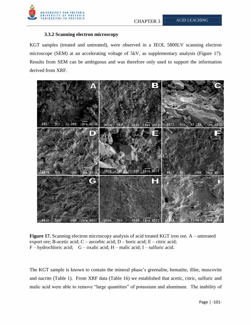
CHAPTER 3 ACID LEACHING
Page | -101-
3.3.2 Scanning electron microscopy
KGT samples (treated and untreated), were observed in a JEOL 5800LV scanning electron
microscope (SEM) at an accelerating voltage of 5kV, as supplementary analysis (Figure 17).
Results from SEM can be ambiguous and was therefore only used to support the information
derived from XRF.
Figure 17. Scanning electron microscopy analysis of acid treated KGT iron ore. A – untreated
export ore; B-acetic acid; C – ascorbic acid; D – boric acid; E – citric acid;
F – hydrochloric acid; G – oxalic acid; H – malic acid; I – sulfuric acid.
The KGT sample is known to contain the mineral phase‘s greenalite, hematite, illite, muscovite
and nacrite (Table 1). From XRF data (Table 16) we established that acetic, citric, sulfuric and
malic acid were able to remove ―large quantities‖ of potassium and aluminum. The inability of
CHAPTER 3 ACID LEACHING
Page | -102-
other acids tested to remove these elements was attributed to mineral dissemination within the
ore particles. The untreated iron ore particle (Figure 17 A) has an uneven surface due to sample
handling and processing at the Sishen mine, which makes surface comparison to treated samples
difficult and unreliable. However hydrochloric – and sulfuric acid was able to change the
surface of the particle (Figure 17 F and I) as smoothening of the surface was observed. From
Table 16 we found that sulfuric acid was able to remove potassium and aluminum, which lets us
deduce that the change in surface morphology is due to the removal of these elements
(Figure 17 – I). Although hydrochloric acid was unable to remove any of the potassium from the
KGT iron ore sample, we still saw a change in surface structure, which is ascribe to the removal
of other ore components (Figure 17 - F).
3.4 CONCLUSION
The Sishen Iron Ore Mine is one of the major suppliers of iron ore to both local and international
markets. The majority of the iron ore resource of the mine is made up of laminated and massive
ore bodies that belong to the Asbestos Hills Subgroup. These ore bodies are overlain by
conglomerate, shales, flagstone and quartzite. Potassium and phosphorous are common
constituents of iron ore, which have a deleterious effect on iron and steel manufacturing.
Therefore steel making companies charge penalties when purchasing iron ore concentrates with
an alkali concentration above predetermined limits. Conventional pyro- and hydrometallurgical
processes used to treat high alkali ore suffer from various limitations. Therefore the objective of
this thesis was to determine whether microorganisms could be applied to supplement
conventional methods. This study accessed the ability of microbially produced (in)organic acids
to solubilize potassium and phosphorous from different iron ore bodies mined at the Sishen Iron
Ore Mine. From the XRF data obtained, we observed that the acids tested were able to remove
varying amounts of potassium and phosphorous from the different iron ore samples. This was
expected as the mineral composition and distribution of the alkali in the ore particle affects
leaching efficiency.
Research has demonstrated that bidentate acids were more efficient at leaching than monodentate
acids, as they were able to form stronger complexes with minerals. In contrast, our results
CHAPTER 3 ACID LEACHING
Page | -103-
showed that a monodentate acid was as effective as a bidentate acid. This was however
attributed to mineral dissemination in the particle.
The SK sample analyzed contained illite, biotite and muscovite as potassium phases. It is known
that muscovite is sensitive to minute quantities of potassium in solution, and will therefore not
release this element if other phases such as biotite are present. The dissolution of biotite is
diffusion-controlled and also depends on the potassium concentration of the bulk solution.
Therefore only minute quantities of potassium could be released from this sample.
The KGT sample contained muscovite and illite. Here some of the potassium could be released.
Drever and Stillings (1997) stated that organic acids were able to affect the saturation state of a
solution by forming complexes with the minerals released. It was therefore expected that the
effective acids used in our study to remove potassium/phosphorous would be an organic acids.
Oxalic acid was able to leach of potassium from the KGT sample. However, sulfuric acid and
malic acid were also able to remove potassium from the KGT sample. These acids aid
weathering via a protonation mechanism (Table 13). Therefore the results obtained were
attributed to sample variation and mineral dissemination in the ore particle.
The SPHP sample analyzed contained a high phosphorous and low potassium concentration
compared to the other samples (Table 19). No potassium could be removed from the SPHP
sample, which was ascribed to the mineral composition and/or distribution of the alkali in the ore
particle. The acids tested were effective at removing phosphorous at higher temperatures, which
is in accordance with research done by Kim et al. (2003) which demonstrated that a higher
temperature favors more phosphorous release from apatite.
From this section of the study we therefore found that the composition of the ore body
determines its susceptibility to acid leaching. Furthermore, we found that sample variation
played a significant role in our data analysis and therefore more repitions needs to be completed.
However due to cost limitations this was not possible. However, these preliminary results can
still serve as a guideline for future acid leaching experiments on various iron ore bodies.
CHAPTER 4 IRON OXIDIZING BACTERIA
Page | -104-
CHAPTER 4
THE USE OF IRON OXIDIZING BACTERIA TO LEACH POTASSIUM AND PHOSPHOROUS FROM
SISHEN HEMATITE IRON ORE
Abstract
The Sishen Iron Ore Mine is situated in the Northern Cape, South Africa, and forms part
of the northern end of the Maremane anticline where the bulk of the hematite is buried
beneath younger cover lithologies. The iron ore bodies at the mine are overlain by
conglomerates, shales, flagstone and quartzite. Potassium and phosphorous are common
constituents of iron ore that have deleterious effect on iron and steel manufacturing,
therefore steel making companies charge penalties when purchasing iron ore with alkali
concentrations above predetermined limits. Conventional methods used to treat high alkali
ore have several drawbacks such as poor product recovery, high running cost and increase
pollution of water. Biohydrometallurgy is a natural alternative approach which could be
applied to supplement conventional methods. Our aim was to assess the ability of
commercially exploited iron oxidizing bacteria to remove potassium and phosphorous from
export iron ore samples mined at Sishen. We found that the organisms were able to
solubilize varying degrees of potassium and phosphorous, which was attributed to the
different metabolic capabilities of each organism tested. The Iron oxidizing bacteria were
more effective than the abiotic tests with sulfuric acids, which is ascribed to their ability to
form exopolysaccharides which influenced the saturation state of the minerals released in
solution. The iron oxidizing bacteria showed a synergistic effect on removing potassium
and phosphorous from export iron ore, which decreased the time required to efficiently
leach.
Keywords: Iron oxidizing bacteria, Sulfuric acid, Ferric iron, Bioleaching, Iron ore
______________________________________________________________________________
4.1 INTRODUCTION
CHAPTER 4 IRON OXIDIZING BACTERIA
Page | -105-
Leaching20
of sulfidic minerals using chemolithoautotrophic bacteria are the best studied and
commercially exploited of mineral biotechnology today (Jain and Sharma 2004). The
application of biohydrometallurgical processes have increased due to favorable process
economics and reduced environmental problems compared to conventional methods such as
pyro – and hydrometallurgical methods (Olson et al., 2003). The metabolic capabilities of these
organisms enable their use in industrial scale processes such as the BIOX®
(Van Aswegen et al., 1988), GEOCOATTM
(Harvey et al., 2002), GEOLEACHTM
(www.geobiotics.com/GeoL_Process.cfm), BIOCOPTM
(Batty and Rorke 2006) and the
BacTech/Mintek (Miller 1997) process (Table 5). The solubilization of the minerals in these
processes is considered to be largely a chemical process, with the microorganisms providing the
chemicals and the space where mineral dissolution reaction occurs (Gehrke et al., 1998,
Rawlings 2005) (Table 4). Iron oxidizing bacteria are able to aid mineral solubilization by
producing chemicals such as ferric iron and sulfuric acid from the ferrous iron and sulfur
contained in the mineral or solution (Rawlings et al., 2003). The ferric iron serves as an
oxidation attack on the mineral (Takai et al., 2001), whereas sulfuric acid is responsible for a
proton attack (Rawlings 2005). Bioleaching of non-sulfidic minerals occurs by acid attack
and/or complexation, while dissolution of sulfides requires a combined acid and/or oxidative
attack (Sand and Gehrke 2006). Bacteria are able to leach the minerals possibly via two
proposed systems namely ‗contact leaching‘ and ‗non-contact leaching‘ (Figure 8) (Sand and
Gehrke 2006). With contact leaching, biofilms are produced; that function in the following way:
the extracellular polymeric substances can mediate attachment to a (metal) sulfide surface and it
may concentrate the ferric ions by complexation through uronic acids or other residues at the
mineral surface, thus allowing an oxidative attack on the sulfide (Corzo et al., 1994; Comte et
al., 2006; Sand and Gehrke 2006). Kinzler et al. (2003) reported that Acidithiobacillus
ferrooxidans was able to accumulate ferric iron in its exopolysaccharides during biofilm
formation via a complexation mechanism. This feature modulated the colonization of the
bacteria on a pyrite surface.
20
Bioleaching refers to the conversion of an insoluble metal (typically a metal sulfide) into a soluble form (typically
a metal sulfate), via microbial activity (Rawlings 2002). When metals are extracted into solution, the process is
referred to as bioleaching, whereas if the metal remains in the mineral, it is referred to as biooxidation (Rawlings
2005).
CHAPTER 4 IRON OXIDIZING BACTERIA
Page | -106-
Kumba Iron Ore, Ltd. is the world‘s fourth largest supplier of sea-borne iron ore. The company
exports 73% of its 32Mtpa (million ton per annum) production to 30 international customers,
mainly in Europe and Asia. It currently operates two mines in South Africa namely the Sishen
Mine in the Northern Cape and Thabazimbi Mine in Limpopo. The iron ore mined at Sishen is
composed of several different minerals. Alkali (potassium and phosphorous) are a common
constituent of iron ore, which is known to have a deleterious effect on the manufacturing of iron
and steel (Delvasto et al., 2008). Therefore, steel making companies charge penalties when
purchasing iron ore concentrates with alkali concentrations above predetermined levels. The
limits allowed is determined by the steel making companies and ranges from 0.25% mass in
Japan to 0.55% mass in Switzerland for the alkali potassium. Kumba Iron Ore, Ltd., has an
industrial set limit of 0.24% potassium allowed in their export ore according to D. Krige
(Personal communication, 2006). To ensure that their export batches stay within this set limit,
the ores from different batches, at the Sishen Iron Ore Mine (with potassium >0.24% and
<0.24%) are mixed to produce an average potassium value of below 0.24%. However this
solution will soon become ineffective as the low potassium ore (< 0.24%) is progressively
depleted according to R. Grunewaldt (Personal communication, 2006). Certain pyro – and
hydrometallurgical methods can be applied to decrease the alkali concentration
(Cheng et al., 1999; Kokal et al., 2003), however there are several disadvantages when using
these methods such as: poor product recovery, involvement of high process and energy cost and
an increase in pollution load of water resources (Jain and Sharma 2004). Thus an alternative,
natural and economical feasible process is required aid conventional methods (such as pyro- and
hydrometallurgy processes) to remove unwanted alkalis from the ores. Biohydrometallurgy21
is
an option for the removal of the deleterious phosphate and potassium, as it is well established
that many microorganisms are capable of mobilizing these minerals, especially in nutrient
limited environments (Banfield et al., 1999; Nautiyal 1999),
The aim of our study was to determine whether the commercially exploited iron oxidizing
bacteria Acidithiobacillus ferrooxidans (ATCC 13598), Leptospirillum ferrooxidans
(ATCC 49879), L. ferriphillum (ATCC 49881) and Sulfobacillus thermosulfidooxidans (Kora)
21
Biohydrometallurgical processes include bioleaching and biobenefication (Ehrlich 1991).
CHAPTER 4 IRON OXIDIZING BACTERIA
Page | -107-
(Figure 4) can be employed in a biobenefication22
approach to remove potassium/phosphorous
from the iron ore mined at Sishen, as iron oxidizing bacteria have been shown to be able to
remove phosphorous from ore bodies (He and Wei 2000).
4.2 MATERIALS AND METHODS
4.2.1 Iron Ore
Export iron ore (alkali <0.24%), with a particle size range of 1mm - 5mm, from the Sishen Iron
Ore Mine, Northern Cape, South Africa was supplied by Kumba Iron Ore, Ltd., for iron
oxidizing bacteria leaching. Prior to leaching, the samples were rinsed with distilled water in a
sieve (< 1mm) to remove small debris which could hamper subsequent analysis, followed by
sterilization at 121°C for 15 min.
4.2.2 Culture
Acidithiobacillus ferrooxidans (ATCC 13598) was obtained from the American Type Culture
Collection (ATCC). Leptospirillum ferrooxidans (ATCC 49879) L. ferriphilum (ATCC 49881)
and Sulfobacillus thermosulfidooxidans (Kora) were obtained from the culture collection of the
University of Stellenbosch, Stellenbosch, South Africa. Iron oxidizing bacteria were grown in
iron-oxidizing medium, as described by Atlas (1993). Media prepared as follow. Solution A
contains: (NH4)2SO4 - 3.0g; K2HPO4 - 0.50g; MgSO4.7H2O - 0.50g; KCl - 0.10g; Ca(NO3)2 -
0.01g. Solution B contained 44.22g of FeSO4.7H2O. Distilled/deionized water was added to
solution A (700ml) and solution B (300ml). The solutions were autoclaved for 15min at 121°C
and then left to cool down to 25°C before adding them together. The pH was then adjusted to
2.20 by adding H2SO4 (V/V). The cultures were grown in 500ml Erlenmeyer to which 300ml of
the iron-oxidizing media was added. A. ferrooxidans, L. ferrooxidans and L. ferriphilum were
22
Biobenefication refers to a process in which microorganisms are used to selectively remove undesirable mineral components
from ores (Ehrlich 1991).
CHAPTER 4 IRON OXIDIZING BACTERIA
Page | -108-
incubated at 30°C and S. thermosulfidooxidans was incubated at 37°C, with shaking (150 rpm)
for 30 days before subsequent leaching experiments.
4.2.3 Leaching
Bioleaching experiments were carried out in 500ml Erlenmeyer flasks containing 150g export
ore, 200ml iron oxidizing media (Atlas 1993) and were each inoculated with 5 ml of a different
bacterial culture, followed by incubation at 30°C with agitation (150 rpm). Size of the
inoculums was not determined, as subsequent measuring would be inaccurate due to bacterial
attachment on the mineral surface (Kinzler et al., 2003). Therefore a decrease in solution pH
was used to determine bacterial activity.
The bacterial cultures were also added together, in equal volumes (2ml each), for a synergistic
effect on ore leaching.
4.2.4 DNA extraction
Total genomic DNA was extracted from the inoculated flasks using the cetyltrimethylammonium
bromide (CTAB method), as described by Ausubel et al. (2002), to ensure that no contaminants
were present in the samples. Cells from 1.5ml of the inoculated flasks were pelleted by
centrifugation at 13 400 rpm for 5 min and suspended in 567µl of 1 × TE buffer (10 mM Tris-
HCl, 1 mM EDTA; pH 8). The cells were lysed by adding sodium dodecyl phosphate (SDS) to a
final concentration of 0.5% (v/v) and the proteins were digested by adding Proteinase K to a final
concentration of 100 µg/ml in total volume of 600 µl. The suspension was then incubated
overnight at 37°C. Following incubation, 100 µl of 5M NaCl and 80µl of CTAB/NaCl solution
(10% [w/v] CTAB in 0.7 M NaCl) was added, mixed thoroughly and incubated at 65°C for 10
min. An equal volume of chloroform:isoamyl alcohol (24:1) was then added and followed by
centrifugation at 13 400 rpm for 5 min. The supernatant was then transferred to a clean
microfuge tube to which an equal volume of phenol:chloroform:isoamyl alcohol (25:24:1) was
added and centrifuged at 13 400 rpm for 5 min. The supernatant was then transferred to a new
microfuge tube, to which 0.6 volume isopropanol was added to precipitate the DNA. The
CHAPTER 4 IRON OXIDIZING BACTERIA
Page | -109-
precipitated DNA was pelleted by centrifugation at 13 400 rpm for 20 min, washed with 70%
ethanol, dried under vacuum and suspended in 20 µl of 1 × TE buffer. The suspension was then
incubated overnight at 37°C before analyzing an aliquot of the extracted genomic DNA was
analyzed by electrophoresis on a 1% (w/v) agarose gel.
4.2.5 Polymerase chain reaction (PCR)
Polymerase chain reaction (PCR) was used to amplify a section of the 16S rRNA of the DNA
extracted. The genes were amplified using the oligonucleotides PRUN518r (5'-ATT-ACC-GCG-
GCT-GCT-GG-3‘) (Siciliano et al., 2003) and pA8f-GC (5'-CGC-CCG-CCG-CGC-GCG-GCG-
GGC-GGG-GCG-GGG-GCACGG-GGG-GAG-AGT-TTG-ATC-CTG-GCT-CAG-3')
(Fjellbirkeland et al., 2001). Each PCR reaction mixture (20 μl) contained ~27ng/μl of genomic
DNA as template, 10.3 µl sterile distilled MilliQ water, 2.5μl PCR buffer with MgCl2 (10×), 2μl
dNTPs (2.5μM), 1μl primer PRUN518r (50μM), 1μl primer pA8f-GC (50μM) and 0.2μl
Supertherm Taq (5U/μl) (Southerm Cross Biotechnology). A negative control (all reagents and
no template DNA) was added to rule out contamination. The tubes were placed in a Perkin-
Elmer GeneAMP® 2700 thermal cycler. Following incubation at 95 °C for 10 min, the reaction
mixture were subjected to 35 cycles of denaturing at 94 °C for 30 s, annealing at 53°C for 30 s
and elongation at 72 °C for 1 min. Once the cycles were completed, a final elongation step was
performed at 72°C for 10 min to complete DNA synthesis. An aliquot of the amplified DNA
was analyzed by electrophoresis on a 1.5% (w/v) agarose gel in the presence of appropriate
molecular weight marker. The PCR products were stored at 4°C, until further analysis.
4.2.6 Denaturing gradient gel electrophoresis (DGGE)
PCR products were subjected to denaturing gradient gel electrophoresis (DGGE) according to
the method described by Muyzer et al. (1993), to ensure that no contamination were present in
our bioleaching experiments. 10µl of the each PCR product was loaded onto a 40-50%
denaturing gradient gels (Table 20). Gels were run at 70 V for 17 h at a constant temperature of
60 °C. Image analysis was performed using the Gel2K (Norland 2004) program. Selected bands
CHAPTER 4 IRON OXIDIZING BACTERIA
Page | -110-
were picked under blue light from the DGGE gels using a sterile scalpel and forceps. Each band
was assigned a number for sequence analysis. The gel fragments were place into 25µl filter-
sterilized deionized water and allowed to stand overnight to dissolve. The dissolved fragments
were then subjected to PCR as described in the previous section.
Table 20 Denaturing gradient table showing volumes in milliliters of DSSA (denaturing stock
solution A: 8% acrylamide in 0.5x TAE (40mM Tris, 20 mM acetic acid, 1nM EDTA (pH 8.3)
buffer) and DSSB (denaturing stock solution B: 8% acrylamide, 7M urea, 40% formamide in
0.5x TAE buffer) mixed to form a gradient within the gel.
Denaturing percentage DSSA(ml) DSSB (ml)
30 10.2 4.4
35 9.4 5.1
40 8.7 5.8
45 8.0 6.5
50 7.3 7.3
55 6.5 8.0
60 5.8 8.7
65 5.1 9.4
4.2.7 Sequencing
The amplified products from the DGGE bands were cleaned by adding 15ul sterile water,
transferring the entire volume to a 0.5ml Eppendorf sequencing tube, adding 2ul of sodium
acetate (3M) and 50ul ethanol (95%), and allowing it to stand on ice for 10min. The tubes were
then centrifuged at 10 000 rpm for 30 min. The ethanol solution was removed, the pellet rinsed
in 150μl ethanol (70%), and the tubes again centrifuged for 5 min at 10 000 rpm. The ethanol
was aspirated and the pellet dried under vacuum for approximately 10 min. These samples were
then submitted to Macrogen (U.S.A.) for sequencing using the oligonucleotides PRUN518r
(Siciliano et al., 2003). Nucleotide sequences were analyzed with BioEdit v.5.0.9.1 (Hall 2001)
CHAPTER 4 IRON OXIDIZING BACTERIA
Page | -111-
and their identity were verified by sequence match searches against the RDP database (available
at http://rdp.cme.msu.edu).
4.2.8 Quantitative mineralogy
The composition of iron samples were analyzed with X-ray fluorescence (XRF) at the X-ray
analytical facility of the University of Pretoria. Four samples of untreated export iron ore were
analyzed as controls. Samples were analyzed after 30 and 60 days of leaching.
The samples were prepared as follow for analysis: Iron ore samples were first ground to <75μm
in a tungsten carbide milling vessel. The loss of ignition (LOI) was determined by roasting the
milled samples at 1000ºC. 1g of a milled iron ore sample were added to 6g Li2B4O7 and fused
into a glass beads. The rest of the sample was then used to make a powder briquette for minor
element analysis (Loubser and Verryn 2008). Major element analyses were executed on the
fused bead using an ARL9400XP+ spectrometer.
4.2.9 Scanning electron microscopy (SEM)
Bacteria cells were fixed by placing the iron ore in a Greiner tube containing 20 ml fixing
solution (2.5% gluteraldehyde in 0.0075 M phosphate buffer) for 1 h. The ore was washed three
times for 15 min with 0.0375 M NaPO4 buffer, before dehydration by sequential treatment for 15
min in 50%, 70%, 90% and 100% ethanol. The 100% ethanol step was repeated twice more to
ensure complete dehydration. The iron ore samples were critical point-dried, scattered with gold
with a Polaron Equipment LTD SEM Autocoating unit E5200 and observed with a JEOL
5800LV scanning electron microscope at an accelerating voltage of 5kV.
4.3 RESULTS AND DISCUSSION
4.3.1 pH measurement
Iron oxidizing bacteria were grown in an iron-oxidizing media, as described by Atlas (1993).
The pH of the media was adjusted to 2.20 and measured every 5th
day as iron oxidizing bacteria
CHAPTER 4 IRON OXIDIZING BACTERIA
Page | -112-
are able produce chemicals such as ferric iron and sulfuric acid from the ferrous iron and sulfur
contained in the mineral or solution, which the latter product would influence pH
(Rawlings et al., 2003). Moreover, Štyriaková et al. (2004) stated that these organisms were also
able to produce negligible amounts of organic acids. Therefore the acids produced would
decrease the pH of the solution and make it inhospitable to other bacteria
(Mason and Rice 2002). Research indicated an increase in dissolution rate with increasing
acidity, which they ascribed in part to proton adsorption to oxygen sites on the mineral surface.
This weakens metal-oxygen bonds and therefore increases dissolution (Welch et al., 2002).
In the abiotic experiments (Chapter 3) these protons possibly decreased and therefore mineral
dissolution stopped/decreased. In contrast bacteria are able to regenerate these protons and
would hypothetically be able to solubilize more minerals. Therefore we assumed that a decrease
in pH would indirectly indicate bacterial activity. Although the solution pH was low, research
has demonstrated that the internal pH of the iron oxidizing bacteria is close to neutral, implying
that these organisms have to maintain a high proton gradient which would have a high ATP
(adenosine triphosphate) cost (White 1995). White (1995) has suggested that iron oxidizing
bacteria regulate their internal pH by consuming cytoplasmic protons during respiration rather
than by proton pumping across the membrane as with other aerobic microorganisms. A low
external pH is required for metabolic activity, because at a pH greater than 5, ferrous iron will
rapidly be oxidized to ferric iron, therefore the cells would struggle to compete with chemical
oxidation. However organisms have been characterized that are able to oxidize iron at different
pH than the norm. This includes organism that can oxidize iron near neutral pH, or archaea that
can oxidize iron at pH 0.5.
The lowest pH measured during this study was for A. ferrooxidans (Table 21), and was attributed
to sulfuric acid production during microbial oxidation (Rawlings et al., 2003). Research
demonstrated that this bacterium was able to grow optimally in a pH range of 1.5 – 2.5
(Niemelä et al., 1994; Razzell et al., 1962; Solisio et al., 2002). The low pH is essential for the
organism survival as Kupka and Kupsăvo (1999) demonstrated that the oxidation of ferrous ions
by A. ferrooxidans decreased when the pH exceeded 2.5. The pH measured for Leptospirilli was
1.86 – 1.91 (Table 21). These are known acid tolerant organisms, with the ability to grow in a
pH range of between 1.5-1.8. These organisms are only able to use ferrous iron as electron
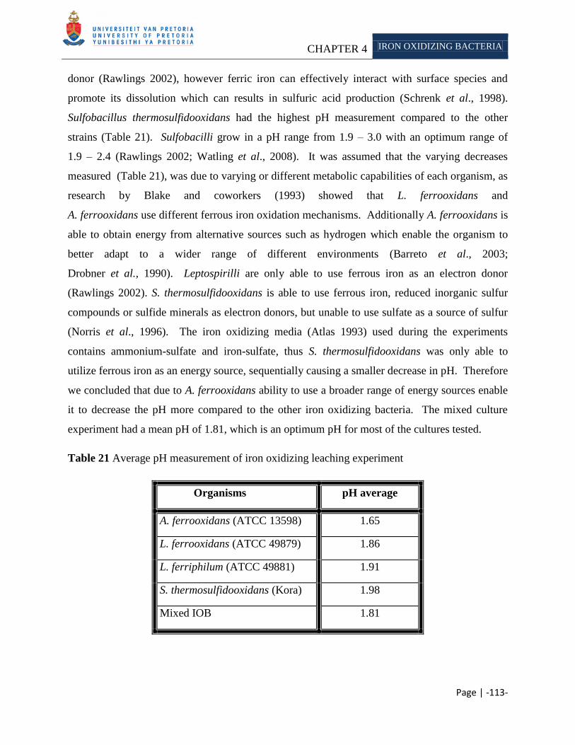
CHAPTER 4 IRON OXIDIZING BACTERIA
Page | -113-
donor (Rawlings 2002), however ferric iron can effectively interact with surface species and
promote its dissolution which can results in sulfuric acid production (Schrenk et al., 1998).
Sulfobacillus thermosulfidooxidans had the highest pH measurement compared to the other
strains (Table 21). Sulfobacilli grow in a pH range from 1.9 – 3.0 with an optimum range of
1.9 – 2.4 (Rawlings 2002; Watling et al., 2008). It was assumed that the varying decreases
measured (Table 21), was due to varying or different metabolic capabilities of each organism, as
research by Blake and coworkers (1993) showed that L. ferrooxidans and
A. ferrooxidans use different ferrous iron oxidation mechanisms. Additionally A. ferrooxidans is
able to obtain energy from alternative sources such as hydrogen which enable the organism to
better adapt to a wider range of different environments (Barreto et al., 2003;
Drobner et al., 1990). Leptospirilli are only able to use ferrous iron as an electron donor
(Rawlings 2002). S. thermosulfidooxidans is able to use ferrous iron, reduced inorganic sulfur
compounds or sulfide minerals as electron donors, but unable to use sulfate as a source of sulfur
(Norris et al., 1996). The iron oxidizing media (Atlas 1993) used during the experiments
contains ammonium-sulfate and iron-sulfate, thus S. thermosulfidooxidans was only able to
utilize ferrous iron as an energy source, sequentially causing a smaller decrease in pH. Therefore
we concluded that due to A. ferrooxidans ability to use a broader range of energy sources enable
it to decrease the pH more compared to the other iron oxidizing bacteria. The mixed culture
experiment had a mean pH of 1.81, which is an optimum pH for most of the cultures tested.
Table 21 Average pH measurement of iron oxidizing leaching experiment
Organisms pH average
A. ferrooxidans (ATCC 13598) 1.65
L. ferrooxidans (ATCC 49879) 1.86
L. ferriphilum (ATCC 49881) 1.91
S. thermosulfidooxidans (Kora) 1.98
Mixed IOB 1.81
CHAPTER 4 IRON OXIDIZING BACTERIA
Page | -114-
4.3.2 Quantitative mineralogy
Export ore (alkali <0.24%) was treated with different iron oxidizing bacteria (Table 22-25), to
determine their ability to remove alkali. Alkali removal was determined by measuring the
residual contained in the ore after treatment and comparing it to the untreated sample.
The alkali, aluminum and silicon concentrations were the main focus of the current research as
Mojallali and Weed (1978) stated that a loss of aluminum and/or silicon showed a structural
rearrangement of a mineral. Furthermore, Wilson and Jones (1983) found that a decrease in
magnesium and iron shows an even greater rearrangement of a mineral.
The mineral composition of the untreated iron ore sample is depicted in Table 19. X-ray
fluorescence is known to be a reasonably sensitive detection method, with detection limits23
for
most elements in the low ppm range (Table 14) (Jenkins 1988). Magnesium and sodium were
below these detection limits and will therefore be excluded from further analysis and not used as
an indication of structural rearrangement as stated by Wilson and Jones (1983). Results of
treated samples were expected to exceed the control sample due to sample variation; therefore a
range was determined which included the minimum and maximum amount of alkali measured in
the replicate untreated sample. From this data set we can possibly determine the amount of
alkali removed due to acid leaching and excluded data sets influenced by sample variation. From
XRF analysis the following ranges for the minerals were calculated as described in Chapter 3:
aluminum (1.15% - 1.56%), silicon (1.92% - 2.80%), potassium (0.16% - 0.18%) and
phosphorous (0.13% - 0.17%), as discussed by Mojallali and Weed (1978). Concentrations of
each mineral removed below these parameters will be reported as ―large quantities‖ removed.
Silicon was detected as quartz, muscovite, biotite, etc. in all of the samples analyzed (Table 1).
Quartz is known dissolved slowly, due to the high activation energy needed to break the
silicon-oxygen bond (Brehm et al., 2005 as cited in Wilson 2004). We did not determine the
source of silicon dissolution, as it was outside the scope of this study, however it is assumed that
the silicon released was from less resistant mineral. We accept the average value of the
untreated export sample as an acceptable representative. Mineral phases detected in the export
iron ore sample include apatite, hematite, muscovite and K-feldspar (Table 1).
23
Lower limit of detection is defined as that concentration equivalent to a certain number of standard deviations of
the background count rate.
CHAPTER 4 IRON OXIDIZING BACTERIA
Page | -115-
Acidithiobacillus ferrooxidans obtains energy by oxidizing ferrous iron and sulfur present in the
sample or solution. The iron ore analyzed did not contain sulfur (Table 22), therefore the
nutritional requirements of the organism was satisfied by the media (Atlas 1993).
A. ferrooxidans is able to obtain energy from alternative sources such as: hydrogen, with oxygen
as terminal electron acceptor under aerobic conditions (Barreto et al., 2003; Drobner et al., 1990)
or under anaerobic conditions by dissimilatory reduction of ferric iron (Ohmura et al., 1999;
Pronk et al., 1991) and formate with oxygen or ferric iron as terminal electron acceptor
(Pronk et al., 1991). The organism is known to produce sulfuric acid and ferric iron
(Ehrlich 1991; Rawlings 2005), which is possibly able to aid solubilization of metals.
Bioleaching of non-sulfidic minerals occurs by acid attack and/or complexation, while
dissolution of sulfides requires a combined acid and/or oxidative attack (Sand and Gehrke 2006).
A. ferrooxidans was able to remove ―large quantities‖ of potassium (35%), phosphorous (27%),
aluminum (55%) and silicon (42%) from the export iron ore. Increased solubilization of these
minerals (except phosphorous), occurred after 60 days (Table 22). This is ascribed to mineral
dissemination in the particle or possibly due to saturation of the alkali in solution. It was
demonstrated that a 0.1mg/l potassium concentration in solution would inhibit the exchange of
the alkali with other ions in solution (vermiculation) (Wilson 2004). The amount of silicon and
aluminum removed, illustrates a rearrangement of the minerals (Mojallali and Weed 1978). It
was not determined whether the organisms actively scavenged for the minerals, or whether the
solubilization of the minerals was indirectly due to the production of metabolites such as ferric
iron and/or sulfuric acid. There is however support for the former as Seeger and Jerez (1993)
found that when A. ferrooxidans was grown under phosphate-limited conditions, the organism
would adapt by reducing its growth rate, to be able to increase its capacity to oxidize ferrous iron
and to fix carbon dioxide. The researchers further discovered that the organism increased its
expression of certain proteins under nutrient limited conditions and they concluded that these
proteins might assist in scavenging for available phosphates. More recently work by Bennett
and coworkers (2001) demonstrated that microbial colonization of a mineral can be nutrient
driven. They using in situ and laboratory microcosms to determine this. The in situ (field
experiments) were conducted at two groundwater sites, which had similar characteristics:
abundant dissolved carbon and anoxic conditions. Here mineral chips were placed in permeable

CHAPTER 4 IRON OXIDIZING BACTERIA
Page | -116-
chambers and suspended in the groundwater for months. In the laboratory, mineral fragments
were inoculated with groundwater (from the two sites) and aquifer materials. They found that in
the groundwater, which was rich in carbon but contained limited amounts of phosphate,
microbial mediated weathering of minerals were sometimes determined by the nutritional
requirements of the microbial community. They further found that feldspar could be rapidly
dissolved in such nutrient limiting environments. Furthermore the authors argued that
microorganisms might be able to select minerals that contain beneficial elements, and leave
others intact.
Table 22 XRF analysis of A. ferrooxidans (ATCC 13598) treated export iron sample.
Element Untreated
Export Ore
A. ferrooxidans
30 days % removed 60 days % removed
SiO2 2.36 1.82 23 1.38 42
TiO2 0.08 0.07 13 0.06 25
Al2O3 1.32 1.17 11 0.60 55
Fe2O3 95.68 96.89 n/a 97.54 n/a
MnO 0.03 0.04 n/a 0.04 n/a
MgO 0.03 0.00 100 0.00 100
CaO 0.04 0.00 100 0.00 100
Na2O 0.11 0.03 73 0.01 91
K2O 0.17 0.14 18 0.11 35
P2O5 0.15 0.11 27 0.11 27
Cr2O3 0.02 0.02 0 0.02 0
NiO 0.01 0.00 100 0.00 100
V2O5 0.01 0.02 n/a 0.02 n/a
ZrO2 0.00 0.00 0 0.00 0
Potassium was detected as K-feldspar and muscovite in the export iron ore sample (Table 1).
Muscovite has considerable resistance toward leaching of interlayer potassium, compared to
trioctahedral micas (such as biotite). It was proposed that an inclined orientation of hydroxyl
ions in the dioctahedral micas results in stronger binding of potassium
(Kalinowski and Schweda 1996) resulting in its resistance. However, Scott and Smith (1966)
conducted experiments in which they determined the susceptibility of interlayer potassium in
micas to exchange with sodium. They found that essentially, all the potassium in muscovite,
CHAPTER 4 IRON OXIDIZING BACTERIA
Page | -117-
biotite, phlogopite and vermiculite were exchangeable when the concentration of the potassium
in the solution was kept low. Leaching was not extended beyond 60 days, as the implementation
of this approach into the mining schedule of the Sishen mine would not allow a longer time span.
Phosphorous was detected as apatite. It is relatively insoluble at neutral pH, although its
solubility and reactivity varies as a function of its composition (Anderson et al., 1985;
Jahnke 1984; LeGeros and Tung 1983; Valsami-Jones et al., 1998). Therefore the low pH
present in the bioleaching solution (Table 21) could possibly have aided the release of
phosphorous from the ore (Welch et al., 2002). Goldstein and Krishnaraj (2007) commented that
the most common mechanism used by microorganisms to solubilize phosphate from apatite,
seems to be by acidifying the media when organic acids were produced and released.
Moreover, biofilms bound to the surface (Figure 19), would also actively take up phosphate, due
to its importance in microbial cell processes (Delvasto et al., 2008; Tempest and Neijssel 1992).
Phosphate uptake by the biofilms is accomplished by the metals complexing with active moieties
(such as carboxyl groups) present in the exopolysaccharides or other cellular materials
(Comte et al., 2006; Corzo et al., 1994). Moreover, studies conducted with microbial
extracellular polymers and simple analogs of these compounds showed that acid polysaccharides
could increase dissolution of alumino-silicate minerals under mildly acidic conditions, by
complexing with the minerals solubilized into solution (Welch and Vandevivere 1994;
Welch et al., 1999) or inhibit dissolution at near neutral pH when bound on the mineral surface
(Poumier et al., 1999).
Microorganisms are able to produce other compounds that can affect mineral dissolution. For
example, when iron limitation occurs, microbes can produce siderophores, that have a high
affinity for iron, but can also bind to other cations. Moreover, organisms can also produce
phosphatases to degrade organophosphates and release orthophosphate. This led Welch and
coworkers (2002) to deduce that there could possibly be similar compounds that are able to
release phosphate from minerals such as apatite.
Sulfuric acid was able to remove 7% potassium and 29% phosphorous from the export ore
(Williams 2008), whereas the iron oxidizing bacteria could solubilize more potassium.
This could be due to a synergistic effect of all its metabolic capabilities, for example: it is able to
CHAPTER 4 IRON OXIDIZING BACTERIA
Page | -118-
produce sulfuric acid and ferric iron (oxidation and proton attack); able to produce biofilms
which can complex with minerals lowering the saturation state; actively take up phosphates as a
nutrient (lowers saturation state) and lastly it could produce compounds similar to
phosphatases/siderophores which further increase mineral dissolution (Welch et al., 1999).
The ability of sulfuric acid to remove more phosphorous was ascribed to sample variation24
, as
only a single repetition of the experiment was done.
Leptospirillum was first isolated in 1972 from a copper leach dump and coal spoil heap in
Armendia (Inoue et al., 2000). The bacteria of this genus are quite similar to that of
Acidithiobacillus. They are also acid tolerant, with the ability to grow in a pH range of between
1.5-1.8. However, based on 16S rRNA sequence analysis, the genus does not form part of the
division Proteobacteria as Acidithiobacilli, but rather Nitrospora (Lane et al., 1992). All
Leptospirilli are only able to use ferrous iron as an electron donor, resulting in higher affinity for
this compound (Rawlings 2002). This organism produced small quantities of hydrogen sulfide:
ferric ion reductase, which aids sulfur oxidation, but is inhibited by an intermediate product
(bisulfate) of sulfate oxidation (Sugio et al., 1994).
We calculated a range for the following minerals, as mentioned before: aluminum
(1.15% - 1.56%), silicon (1.92% - 2.80%), potassium (0.16% - 0.18%) and phosphorous
(0.13% - 0.17%). After 30 days leaching, no potassium could be removed using L. ferrooxidans
(Table 23) or L. ferriphilum (Table 24), however extended leaching enabled removal.
L. ferrooxidans and L. ferriphilum were able to remove ―large quantities‖ of phosphorous, silicon
and aluminum. The amount of aluminum and silicon demonstrates that mineral rearrangement
occurred (Mojallali and Weed 1978). Several factors such as intrinsic mineral characteristics,
processing, physiochemical and microbiological factors can influence leaching (Table 10).
A. ferrooxidans was able to remove 35% of the potassium and 27% of the phosphorous
(Table 22); compared to <30% for potassium and above 30% for phosphorous by Leptospirilli.
24
Sample contains an alkali concentration higher than the set parameter and therefore no apparent removal will be
reported.
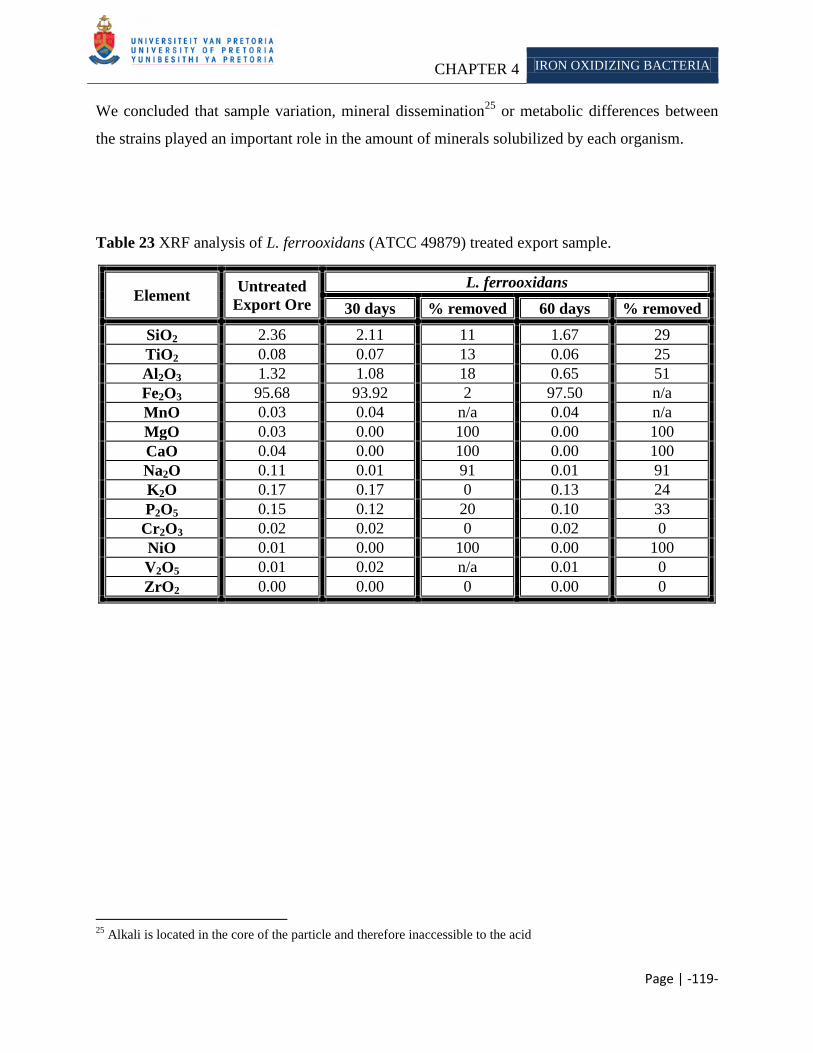
CHAPTER 4 IRON OXIDIZING BACTERIA
Page | -119-
We concluded that sample variation, mineral dissemination25
or metabolic differences between
the strains played an important role in the amount of minerals solubilized by each organism.
Table 23 XRF analysis of L. ferrooxidans (ATCC 49879) treated export sample.
Element Untreated
Export Ore
L. ferrooxidans
30 days % removed 60 days % removed
SiO2 2.36 2.11 11 1.67 29
TiO2 0.08 0.07 13 0.06 25
Al2O3 1.32 1.08 18 0.65 51
Fe2O3 95.68 93.92 2 97.50 n/a
MnO 0.03 0.04 n/a 0.04 n/a
MgO 0.03 0.00 100 0.00 100
CaO 0.04 0.00 100 0.00 100
Na2O 0.11 0.01 91 0.01 91
K2O 0.17 0.17 0 0.13 24
P2O5 0.15 0.12 20 0.10 33
Cr2O3 0.02 0.02 0 0.02 0
NiO 0.01 0.00 100 0.00 100
V2O5 0.01 0.02 n/a 0.01 0
ZrO2 0.00 0.00 0 0.00 0
25
Alkali is located in the core of the particle and therefore inaccessible to the acid

CHAPTER 4 IRON OXIDIZING BACTERIA
Page | -120-
Table 24 XRF analysis of L. ferriphilum (ATCC 49881) treated export sample.
Element Untreated
Export Ore
L. ferriphilum
30 days % removed 60 days % removed
SiO2 2.36 1.56 33 1.66 30
TiO2 0.08 0.07 13 0.06 25
Al2O3 1.32 0.99 25 0.72 46
Fe2O3 95.68 97.01 n/a 97.08 n/a
MnO 0.03 0.04 n/a 0.04 n/a
MgO 0.03 0.00 100 0.00 100
CaO 0.04 0.00 100 0.00 100
Na2O 0.11 0.01 91 0.01 91
K2O 0.17 0.16 6 0.12 29
P2O5 0.15 0.14 7 0.10 33
Cr2O3 0.02 0.02 0 0.02 0
NiO 0.01 0.00 100 0.00 100
V2O5 0.01 0.02 n/a 0.01 0
ZrO2 0.00 0.00 0 0.00 0
Sulfobacillus thermosulfidooxidans are able to grow autotrophically or heterotrophically. When
growing autotrophically they use ferrous iron, reduced inorganic sulfur compounds or sulfide
minerals as electron donors (Norris et al., 1996). Sulfobacilli are not able to efficiently fix
carbon dioxide, and therefore need elevated CO2 in the atmosphere or yeast extract can be added
to the media, furthermore they are not able to use sulfate as a source of sulfur and must therefore
be provided reduced sulfur (Rawlings 2002). When growing heterotrophically, glucose can
serve as a carbon and energy source. Furthermore these microorganisms are also able to grow in
the absence of oxygen, using ferric iron as an electron acceptor and either organic or inorganic
sulfur compounds as electron donor. During this study, S. thermosulfidooxidans were grow in an
iron oxidizing media described by Atlas (1993). Here the organisms could grow autotrophically
as the media supplied the sulfur source and the carbon dioxide in the air served as a carbon
source.
S. thermosulfidooxidans was able to remove a ―large quantity‖ of potassium after 30 days, but
was unable to remove more after extended leaching (Table 25). Furthermore phosphorous,
aluminum and silicon were removed in ―large quantities‖ after 30 days. The data obtained is
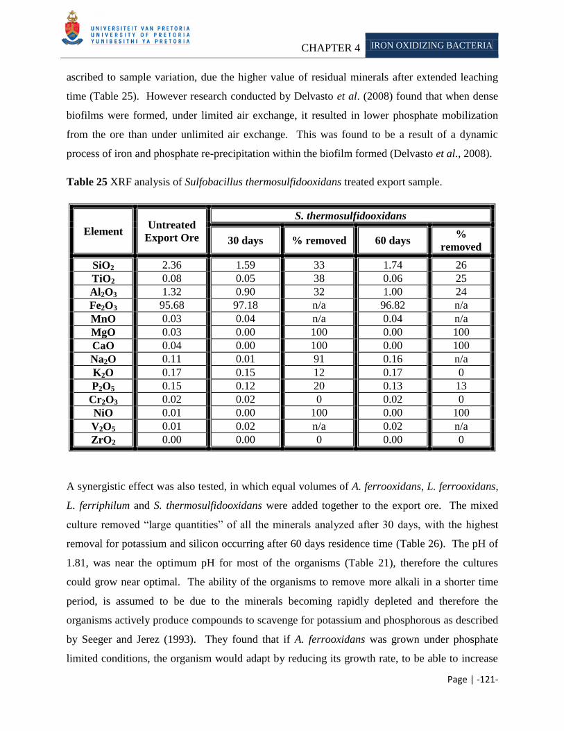
CHAPTER 4 IRON OXIDIZING BACTERIA
Page | -121-
ascribed to sample variation, due the higher value of residual minerals after extended leaching
time (Table 25). However research conducted by Delvasto et al. (2008) found that when dense
biofilms were formed, under limited air exchange, it resulted in lower phosphate mobilization
from the ore than under unlimited air exchange. This was found to be a result of a dynamic
process of iron and phosphate re-precipitation within the biofilm formed (Delvasto et al., 2008).
Table 25 XRF analysis of Sulfobacillus thermosulfidooxidans treated export sample.
Element Untreated
Export Ore
S. thermosulfidooxidans
30 days % removed 60 days %
removed
SiO2 2.36 1.59 33 1.74 26
TiO2 0.08 0.05 38 0.06 25
Al2O3 1.32 0.90 32 1.00 24
Fe2O3 95.68 97.18 n/a 96.82 n/a
MnO 0.03 0.04 n/a 0.04 n/a
MgO 0.03 0.00 100 0.00 100
CaO 0.04 0.00 100 0.00 100
Na2O 0.11 0.01 91 0.16 n/a
K2O 0.17 0.15 12 0.17 0
P2O5 0.15 0.12 20 0.13 13
Cr2O3 0.02 0.02 0 0.02 0
NiO 0.01 0.00 100 0.00 100
V2O5 0.01 0.02 n/a 0.02 n/a
ZrO2 0.00 0.00 0 0.00 0
A synergistic effect was also tested, in which equal volumes of A. ferrooxidans, L. ferrooxidans,
L. ferriphilum and S. thermosulfidooxidans were added together to the export ore. The mixed
culture removed ―large quantities‖ of all the minerals analyzed after 30 days, with the highest
removal for potassium and silicon occurring after 60 days residence time (Table 26). The pH of
1.81, was near the optimum pH for most of the organisms (Table 21), therefore the cultures
could grow near optimal. The ability of the organisms to remove more alkali in a shorter time
period, is assumed to be due to the minerals becoming rapidly depleted and therefore the
organisms actively produce compounds to scavenge for potassium and phosphorous as described
by Seeger and Jerez (1993). They found that if A. ferrooxidans was grown under phosphate
limited conditions, the organism would adapt by reducing its growth rate, to be able to increase

CHAPTER 4 IRON OXIDIZING BACTERIA
Page | -122-
its capacity to oxidize ferrous iron and to fix carbon dioxide. The researchers further discovered
that the organism increased its expression of certain proteins under the limiting conditions and
they concluded that these proteins might assist in scavenging for available phosphates.
Table 26 XRF analysis of mix iron oxidizing bacteria leaching of export sample.
Element Untreated
Export Ore
Mixed Iron oxidizing bacteria
30 days % removed 60 days %
removed
SiO2 2.36 1.48 37 1.51 36
TiO2 0.08 0.06 25 0.07 13
Al2O3 1.32 0.97 27 0.65 51
Fe2O3 95.68 97.00 n/a 96.66 n/a
MnO 0.03 0.04 n/a 0.04 n/a
MgO 0.03 0.00 100 0.00 100
CaO 0.04 0.00 100 0.00 100
Na2O 0.11 0.01 91 0.01 91
K2O 0.17 0.14 18 0.12 29
P2O5 0.15 0.11 27 0.11 27
Cr2O3 0.02 0.02 0 0.02 0
NiO 0.01 0.00 100 0.00 100
V2O5 0.01 0.02 n/a 0.02 n/a
ZrO2 0.00 0.00 0 0.00 0
We found that the iron oxidizing bacterial experiments were more variable than the abiotic tests
(Chapter 3), which is in accordance with other reports (Welch et al., 2004).
Welch and coworkers (2004) found that bacteria produce varying amounts of acids. We assume
that these varying amounts of acids are responsible for the different amounts of alkali removed.
A. ferrooxidans was able to remove greatest amount of potassium (Table 22) and the
Leptospirillum cultures were able to remove the largest concentration of phosphorous
(Table 22 and 23). S. thermosulfidooxidans was the most inefficient at removing potassium and
aluminum, which was ascribed to the metabolic capability of this organism. The mixed cultures
proved to be the most efficient for removing the minerals, which was ascribed to a synergistic
effect.
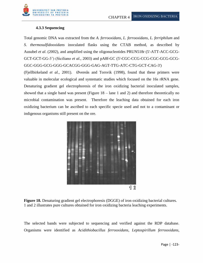
CHAPTER 4 IRON OXIDIZING BACTERIA
Page | -123-
4.3.3 Sequencing
Total genomic DNA was extracted from the A. ferrooxidans, L. ferrooxidans, L. ferriphilum and
S. thermosulfidooxidans inoculated flasks using the CTAB method, as described by
Ausubel et al. (2002), and amplified using the oligonucleotides PRUN518r (5'-ATT-ACC-GCG-
GCT-GCT-GG-3‘) (Siciliano et al., 2003) and pA8f-GC (5'-CGC-CCG-CCG-CGC-GCG-GCG-
GGC-GGG-GCG-GGG-GCACGG-GGG-GAG-AGT-TTG-ATC-CTG-GCT-CAG-3')
(Fjellbirkeland et al., 2001). Øvereås and Torsvik (1998), found that these primers were
valuable in molecular ecological and systematic studies which focused on the 16s rRNA gene.
Denaturing gradient gel electrophoresis of the iron oxidizing bacterial inoculated samples,
showed that a single band was present (Figure 18 – lane 1 and 2) and therefore theoretically no
microbial contamination was present. Therefore the leaching data obtained for each iron
oxidizing bacterium can be ascribed to each specific specie used and not to a contaminant or
indigenous organisms still present on the ore.
Figure 18. Denaturing gradient gel electrophoresis (DGGE) of iron oxidizing bacterial cultures.
1 and 2 illustrates pure cultures obtained for iron oxidizing bacteria leaching experiments.
The selected bands were subjected to sequencing and verified against the RDP database.
Organisms were identified as Acidithiobacillus ferrooxidans, Leptospirillum ferrooxidans,
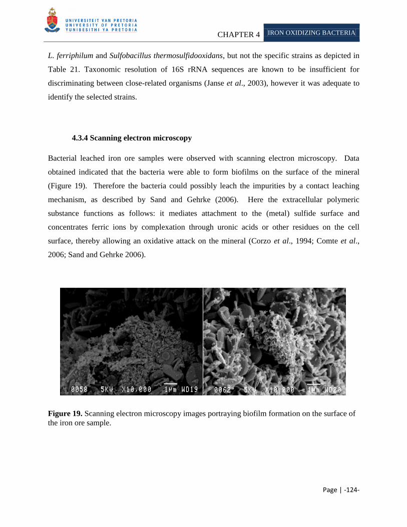
CHAPTER 4 IRON OXIDIZING BACTERIA
Page | -124-
L. ferriphilum and Sulfobacillus thermosulfidooxidans, but not the specific strains as depicted in
Table 21. Taxonomic resolution of 16S rRNA sequences are known to be insufficient for
discriminating between close-related organisms (Janse et al., 2003), however it was adequate to
identify the selected strains.
4.3.4 Scanning electron microscopy
Bacterial leached iron ore samples were observed with scanning electron microscopy. Data
obtained indicated that the bacteria were able to form biofilms on the surface of the mineral
(Figure 19). Therefore the bacteria could possibly leach the impurities by a contact leaching
mechanism, as described by Sand and Gehrke (2006). Here the extracellular polymeric
substance functions as follows: it mediates attachment to the (metal) sulfide surface and
concentrates ferric ions by complexation through uronic acids or other residues on the cell
surface, thereby allowing an oxidative attack on the mineral (Corzo et al., 1994; Comte et al.,
2006; Sand and Gehrke 2006).
Figure 19. Scanning electron microscopy images portraying biofilm formation on the surface of
the iron ore sample.
CHAPTER 4 IRON OXIDIZING BACTERIA
Page | -125-
Biofilm formation is a multistep process which is influenced by many factors, including the
specific mineralogy of the rock, solution chemistry (pH, ionic strength) and the characteristics of
the microorganism (hydrophobicity, surface charge) (Banfield and Hamers 1997;
Characklis 1989; Little et al., 1997). The attachment of cells to the surface can occur via random
processes such as diffusion and convection or more specifically via chemotaxis, which is the
active orientation of a bacterium to a chemical gradient (Jerez 2001). Several researchers studied
the chemotatic response of bacteria towards metal ions and other compounds, for example:
chemotaxis of A. ferrooxidans (Chakraborty and Roy 1992) and L. ferrooxidans
(Acuňa et al., 1986; Acuňa et al., 1992). The organisms can detect the site of mineral dissolution
(possibly potassium and phosphorous) by sensing a dissolution gradient. This allows the
bacterium to respond to the attractant/repellent by swimming away or towards the increased
concentration. When the bacterium reaches the surface, the cell can attach by mostly an
unknown process (Sand and Gehrke 2006). Blake et al. (2001) concluded that aporusticyanin
could possibly play an important role in the early attachment of A. ferrooxidans to mineral
surfaces. The exopolysaccharides produced by the cell, plays an important role in attachment of
the leaching bacterium. Research indicated that the exopolymers produced by A. ferrooxidans
and L. ferrooxidans, contain besides neutral sugars and lipids, some glucuronic acid residues and
complexed ferric ions (Gehrke et al., 1995; Gehrke et al., 1998, Gehrke et al., 2001).
The stoichiometry of glucuronic acid to ferric ions amounts to a molar ratio of about 2:1 for both
A. ferrooxidans and L. ferrooxidans tested thus far. Thus regardless of the phylogenetic class of
the bacterium, the physicochemical characteristics of the environment enforced a similar
adaption between the organisms. The exopolysaccharides formed by iron oxidizing bacteria has
a net positive charge and at acidic pH, pyrite has a negative charge (Devasia et al., 1993;
Hallman 1992; Ohmura et al., 1993). Thus, attachment of a positively charged bacterium to a
negatively charged surface may take place due to electrostatic interaction between the two
surfaces. Lazar (2004) found that both electrostatic and hydrophobic interactions could be
responsible for the tight adhesion of cells to mineral surfaces (as cited in Sand and Gehrke 2006).
The specificity of exopolysaccharides for various metal sulfides has not been termed.
Gehrke et al. (1998) has however indicated that the composition of the EPS of A. ferrooxidans
changed when the cells were grown in different substrates. Sulfur-grown cells had more lipids,
less neutral sugars and only trace glucuronic acid as compared to those of pyrite grown cells.
CHAPTER 4 IRON OXIDIZING BACTERIA
Page | -126-
4.4 CONCLUSION
Leaching of sulfidic minerals using chemolithoautotrophic bacteria are the best studied and
commercially exploited mineral biotechnology today (Jain and Sharma 2004). The solubilization
of the minerals is considered to be largely a chemical process, with the microorganisms
providing the chemicals and the space where the mineral dissolution reaction occurs.
Bioleaching of non-sulfidic ores, such as the export ore in this study, occurred by acid attack and
complexation. Iron oxidizing bacteria are able to provide ferric iron and/or sulfuric acid which
can serve as an oxidative attack in the former and a proton attack in the latter on mineral
samples. Moreover, EPS produced by the bacteria are able to form complexes with the minerals
which will influence the saturation state of the minerals in solution. The objective of this study
was to determine, whether the commercially exploited iron oxidizing bacteria, were able to
remove the alkali impurities from the export iron ore samples, provided by Kumba Iron Ore, Ltd.
We found that the various strains were indeed able to remove different amounts of potassium and
phosphorous from the export iron ore samples. Furthermore the organisms also demonstrated a
symbiotic/synergistic relationship, which enable mobilization of the minerals from the ore.
Scanning electron microscopy analysis demonstrated that biofilms were formed on the surface of
the iron ore, supporting a contact leaching mechanism.
The preliminary results suggest that iron oxidizing bacteria leaching could be a viable option for
treating the iron ore mined at Sishen. Bioleaching will however never completely replace the
conventional methods but rather supplement them, as bioleaching does not recover precious
metals from the ores which are often an essential component in the profitability of the operation;
also when ore bodies do not contain sufficient acid consuming minerals, the residual acids
generated have to be neutralized during the leaching process, thus increasing the operational cost
(Dreshner 2004).
CHAPTER 5 HETEROTROPHIC
BACTERIA
Page | -127-
CHAPTER 5
LEACHING OF POTASSIUM AND PHOSPHOROUS FROM SISHEN HEMATITE IRON ORE USING
HETEROTROPHIC BACTERIA
Abstract
The Sishen Iron Ore Mine is situated in the Northern Cape, South Africa, and forms part
of the northern end of the Maremane anticline where the bulk of the hematite is buried
beneath younger cover lithologies. The iron ore bodies minedare overlain by
conglomerates, shales, flagstone and quartzite. Potassium and phosphorous are common
constituents of the iron ore bodies and are known to have a deleterious effect on iron and
steel manufacturing. Therefore steel making companies charge penalties when purchasing
iron ore with alkali concentrations above predetermined limits. Conventional methods
used to treat high alkali ore have several drawbacks such as poor product recovery, high
running cost and increase pollution of water thus an alternative approach is needed to
supplement or replace these methods. Biohydrometallurgy is a natural alternative
approach which has been applied commercially to extract or treat various ore bodies. Here
we aim to asses the ability of the heterotrophic bacteria Bacillus megaterium, B. cereus,
B. subtilis and Pseudomonas putida to remove potassium and phosphorous from export
grade iron ore mined at Sishen. We found that the organisms were able to solubilize
varying degrees of potassium and phosphorous, which was attributed to their ability to
produce different organic. Furthermore we found that the biotic test with the
heterotrophic organims were more efficient at removing alkali than the abiotic test with
with acids (Chapter 3). This ability is ascribed to their ability to form EPS, which is able to
form complexes with minerals, thereby affecting the saturation state of the mineral in
solution.
Keywords: Heterotrophic bacteria, organic acids, muscovite, exopolysaccharides
______________________________________________________________________________

CHAPTER 5 HETEROTROPHIC
BACTERIA
Page | -128-
5.1 INTRODUCTION
Microbes are able to contribute to the biogeochemical cycles by transforming the metal phases
(soluble to volatile) and/or changing the oxidation state of the metal. They play a major role in
the: (i) cycling of elements and sorption of metals (Langley and Beveridge 1999);
(ii) the dissolution of minerals (Banfield and Hamers 1997; Hersman et al., 1996; Roden and
Zachara 1996; Schrenk et al., 1998), and (iii) mineral crystallization (Fortin et al., 1998).
Whereas, mineralogical processes affect the distribution, activity and diversity of microbes
(Schrenk et al., 1998); the expression of their genes (Gehrke et al., 1998), the structure and
development of communities (Kennedy and Gewin 1997; Torseth et al., 1995;
Wolfaardt et al., 1994) and the transfer of genetic material (Trevors and van Elsas 1997).
Kumba Iron Ore, Ltd. is the world‘s fourth largest supplier of sea-borne iron ore. The company
exports 73% of its 32Mtpa production to 30 international customers, mainly in Europe and Asia.
It currently operates two mines in South Africa namely the Sishen Mine in the Northern Cape
and Thabazimbi Mine in Limpopo. The iron ore mined at Sishen is composed of several
different minerals phases, with potassium and phosphorous as common constituents.
These alkalis have a deleterious effect on the manufacturing of iron and steel
(Delvasto et al., 2008). Therefore, steel making companies charge penalties when purchasing
iron ore concentrates with alkali concentrations above predetermined levels. The limits allowed
is determined by the steel making companies and range from 0.25% mass in Japan to 0.55%
mass in Switzerland for the alkali potassium. Kumba Iron Ore, Ltd., has an industrial set limit of
0.24% potassium allowed in their export ore D. Krige (Personal communication, 2006). To
ensure that their export batches stay within this set limit, the ores from different batches, at the
Sishen Iron Ore Mine (with potassium >0.24% and <0.24%) are mixed to produce an average
potassium value of below 0.24%. However this solution will soon become ineffective as the low
potassium ore (<0.24%) is progressively depleted according to D. Krige
(Personal communication, 2006). Certain pyro – and hydrometallurgical methods can be applied
to decrease the alkali concentration (Cheng et al., 1999; Kokal et al., 2003), however there are
several disadvantages when using these methods such as: poor product recovery, involvement of
high process and energy cost and an increase in pollution load of water resources
(Jain and Sharma 2004). Thus an alternative, natural and economical feasible process is required
CHAPTER 5 HETEROTROPHIC
BACTERIA
Page | -129-
aid conventional methods (such as pyro- and hydrometallurgy processes) in removing unwanted
alkalis from the ores. Biohydrometallurgy26
is an option for the removal of the deleterious
phosphate and potassium, as it is established that many microorganisms are capable of
mobilizing these minerals, especially in nutrient limited environments (Banfield et al., 1999;
Nautiyal 1999). Bosecker (1997) found that the dissolution of non-sulfidic ores can be achieved
by exploiting the metabolic capabilities of heterotrophic microorganisms. These organisms have
the potential to produce metabolites that are able to solubilize and/or aid in solubilizing oxide,
silicate, carbonate and hydroxide minerals via mechanisms such as reduction, acidolysis and
complexation (Burgstaller and Schinner 1993; Gadd and Sayer 2000; Jain and Sharma 2004;
Sayer and Gadd 1997). Metabolites include exopolysaccharides (Banfield et al., 1999;
Malinovskaya et al., 1990; Welch et al., 1999), amino acids, proteins (Bosecker 1997) and
organic acids (Agatzini and Tzeferis 1997; Barker et al., 1997; Castro et al., 2000; Natarajan and
Deo 2001; Štyriaková et al., 1999; Valsami-Jones and McEldowney 2000).
Among the heterotrophic bacteria, members of the genus Bacillus and Pseudomonas have been
shown to be effective in leaching non-sulfidic minerals (Karavaiko et al., 1988). Welch and
Ullman (1999) found that certain heterotrophic microorganisms were able to mobilize elements
such as Si, Al, Fe, K, Li, Ni, Zn, and Mg from the rock-forming minerals feldspar, pegmatite,
hornblende and spodumene.
Leaching with heterotrophic bacteria can occur at higher pH than with autotrophic
microorganisms, thus no equilibration of acids is required which would increase operational
costs. However, the higher solution pH makes the environment favorable for many microbes,
which again can contaminate the process and outcompete the leaching microbes (Jain and
Sharma 2004). The bacteria used in this study are reported to be capable of solubilizing
potassium and/or phosphates from various rocks and minerals. These include: Bacillus
megaterium (Han 2006; Jain and Sharma 2004), Bacillus cereus (Štyriaková et al., 2004),
Bacillus subtilis and Pseudomonas putida (Jiang et al., 2007).
Here we aim to asses the ability of the selected heterotrophic microorganisms to remove
potassium and phosphorous under nutrient limiting conditions for these minerals. It is
26
Biohydrometallurgical processes include bioleaching and biobenefication (Ehrlich 1991).
CHAPTER 5 HETEROTROPHIC
BACTERIA
Page | -130-
hypothesized that under potassium/phosphorous limiting conditions, certain genes will be
activated, that could enable the organism to actively scavenge for these minerals. This
hypothesis is supported by research research done by Bennet (2001), which found that silicate
weathering by bacteria is driven by nutrient requirement. Due to difficulty in establishing
selective growth conditions, heterotrophic microorganisms were not readily applied in industrial
scale bioleaching processes (Ehrlich 1991). Heterotrophs require one or more organic nutrient to
serve as an energy and carbon source, which increase operational costs. Furthermore the organic
nutrient can support growth of a great variety of different organisms, which could contaminate
the leaching processes and outcompete the organisms able to leach the ore (Ehrlich 1991).
5.2 MATERIALS AND METHODS
5.2.1 Iron Ore
Export iron ore (alkali <0.24%), with a particle range 1mm - 5mm, from the Sishen Iron Ore
Mine, Northern Cape, South Africa was supplied by Kumba Iron Ore, Ltd., for heterotrophic
bacterial leaching. Prior to leaching, the samples were rinsed with distilled water in a sieve
(< 1mm) to remove small debris which could hamper subsequent analysis, followed by
sterilization at 121°C for 15 min.
5.2.2 Cultures
Pseudomonas putida (ATCC 15070) were obtained from the American Type Culture Collection
(ATCC). Bacillus megaterium (J3-10DQ363436), B. subtilis (MG1-DQ228954) and B. cereus
(AMM202-AB092795) were obtained for the culture collection of the University of Pretoria,
Pretoria, South Africa.
CHAPTER 5 HETEROTROPHIC
BACTERIA
Page | -131-
5.2.3 Inoculum media
The cultures were grown in a media described by Štyriaková et al. (2004) at 30°C with shaking
(150rpm). The composition of the media per liter was as follow: 0.42g - NaH2PO4;
0.8g – (NH4)2SO4; 0.19g NaCl; 19.8g – glucose. The cultures were maintained on Nutrient Agar
(Biolab) at 4°C or as glycerol cultures at -70°C.
5.2.4 Leaching
Bioleaching experiments were carried out in 500ml Erlenmeyer flasks containing 150g export
ore, 300ml inoculum media (Štyriaková et al., 2004) and were inoculated with 5 ml of each
different bacterial culture, followed by incubation at 30°C with agitation (150 rpm) and a
duplicate without agitation (static). A decrease in solution pH was used to determine bacterial
activity (Kinzler et al., 2003).
The different bacterial cultures were also mixed, in equal volumes (2ml each), to evaluate a
potential synergistic effect on ore leaching.
5.2.5 pH calibration
pH of the inoculated samples were neutralized with 1M NaOH every 5th
day, as described by
Štyriaková et al. (2004).
5.2.6 DNA extraction
Total genomic DNA was extracted from the heterotrophic bacterial inoculated flasks using the
cetyltrimethylammonium bromide (CTAB method), as described by Ausubel et al. (2002), to
ensure that no contaminants were present in the samples. Cells from 1.5ml of the inoculated
flasks were pelleted by centrifugation at 13 400 rpm for 5 min and suspended in 567µl of
1 × TE buffer (10 mM Tris-HCl, 1 mM EDTA; pH 8). The cells were lysed by adding sodium
dodecyl phosphate (SDS) to a final concentration of 0.5% (v/v) and the proteins were digested by
CHAPTER 5 HETEROTROPHIC
BACTERIA
Page | -132-
adding Proteinase K to a final concentration of 100 µg/ml in total volume of 600 µl. The
suspension was then incubated overnight at 37°C. Following incubation, 100 µl of 5M NaCl and
80µl of CTAB/NaCl solution (10% [w/v] CTAB in 0.7 M NaCl) was added, mixed thoroughly
and incubated at 65°C for 10 min. An equal volume of chloroform:isoamyl alcohol (24:1) was
then added and followed by centrifugation at 13 400 rpm for 5 min. The supernatant was then
transferred to a clean microfuge tube to which an equal volume of phenol:chloroform:isoamyl
alcohol (25:24:1) was added and centrifuged at 13 400 rpm for 5 min. The supernatant was then
transferred to a new microfuge tube, to which 0.6 volume isopropanol was added to precipitate
the DNA. The precipitated DNA was pelleted by centrifugation at 13 400 rpm for 20 min,
washed with 70% ethanol, dried under vacuum and suspended in 20 µl of 1 × TE buffer. The
suspension was then incubated overnight at 37°C before analyzing an aliquot of the extracted
genomic DNA was analyzed by electrophoresis on a 1% (w/v) agarose gel.
5.2.7 Polymerase chain reaction
Polymerase chain reaction (PCR) was used to amplify a section of the 16S rRNA of the DNA
extracted. The genes were amplified using the oligonucleotides PRUN518r (5'-ATT-ACC-GCG-
GCT-GCT-GG-3‘) (Siciliano et al., 2003) and pA8f-GC (5'-CGC-CCG-CCG-CGC-GCG-GCG-
GGC-GGG-GCG-GGG-GCACGG-GGG-GAG-AGT-TTG-ATC-CTG-GCT-CAG-3')
(Fjellbirkeland et al., 2001). Each PCR reaction mixture (20 μl) contained ~27ng/μl of genomic
DNA as template, 10.3 µl sterile distilled MilliQ water, 2.5μl PCR buffer with MgCl2 (10×), 2μl
dNTPs (2.5μM), 1μl primer PRUN518r (50μM), 1μl primer pA8f-GC (50μM) and
0.2μl Supertherm Taq (5U/μl) (Southerm Cross Biotechnology). A negative control (all reagents
and no template DNA) was added to rule out contamination. The tubes were placed in a Perkin-
Elmer GeneAMP® 2700 thermal cycler. Following incubation at 95 °C for 10 min, the reaction
mixture were subjected to 35 cycles of denaturing at 94 °C for 30 s, annealing at 53°C for 30 s
and elongation at 72 °C for 1 min. Once the cycles were completed, a final elongation step was
performed at 72°C for 10 min to complete DNA synthesis. An aliquot of the amplified DNA
was analyzed by electrophoresis on a 1.5% (w/v) agarose gel in the presence of appropriate
molecular weight marker. The PCR products were stored at 4°C, until further analysis.
CHAPTER 5 HETEROTROPHIC
BACTERIA
Page | -133-
5.2.8 Denaturing gradient gel electrophoresis (DGGE)
The PCR products were subjected to denaturing gradient gel electrophoresis (DGGE) according
to the method described by Muyzer et al. (1993), to ensure that no contamination were present in
our bioleaching experiments. 10µl of the each PCR product was loaded onto a 40-50%
denaturing gradient gels (Table 20). Gels were run at 70 V for 17 h at a constant temperature of
60 °C. Image analysis was performed using the Gel2K (Norland 2004) program. Selected bands
were picked under blue light from the DGGE gels using a sterile scalpel and forceps. Each band
was assigned a number for sequence analysis. The gel fragments were place into 25µl filter-
sterilized deionized water and allowed to stand overnight to dissolve. The dissolved fragments
were then subjected to PCR as described in the previous section.
5.2.9 Sequencing
The amplified products from the DGGE bands were cleaned by adding 15µl sterile water,
transferring the entire volume to a 0.5ml Eppendorf sequencing tube, adding 2µl of sodium
acetate (3M) and 50ul ethanol (95%), and allowing it to stand on ice for 10min. The tubes were
then centrifuged at 10 000 rpm for 30 min. The ethanol solution was removed, the pellet rinsed
in 150μl ethanol (70%), and the tubes again centrifuged for 5 min at 10 000 rpm. The ethanol
was aspirated and the pellet dried under vacuum for approximately 10 min. These samples were
then submitted to Macrogen (U.S.A.) for sequencing using the oligonucleotides PRUN518r
(Siciliano et al., 2003). Nucleotide sequences were analyzed with BioEdit v.5.0.9.1 (Hall 2001)
and their identity were verified by sequence match searches against the RDP database (available
at http://rdp.cme.msu.edu).
5.2.10 Qualitative mineralogy
The composition of iron samples were analyzed with X-ray fluorescence (XRF) at the X-ray
analytical facility of the University of Pretoria. Four samples of untreated export iron ore were
analyzed as controls. Samples were analyzed after 30 and 60 days of bioleaching.
CHAPTER 5 HETEROTROPHIC
BACTERIA
Page | -134-
The samples were prepared as follows for analysis: Iron ore samples were first ground to <75μm
in a tungsten carbide milling vessel. The loss of ignition (LOI) was determined by roasting the
milled samples at 1000ºC. 1g of a milled iron ore sample were added to 6g Li2B4O7 and fused
into a glass beads. The rest of the sample was then used to make a powder briquette for minor
element analysis. Major element analyses were executed on the fused bead using an
ARL9400XP+ spectrometer (Loubser and Verryn 2008).
5.2.11 Scanning electron microscopy
Bacteria cells were fixed by placing the iron ore in a Greiner tube containing 20 ml fixing
solution (2.5% gluteraldehyde in 0.0075 M phosphate buffer) for 1 h. The ore was washed three
times for 15 min with 0.0375 M NaPO4 buffer, before dehydration by sequential treatment for 15
min in 50%, 70%, 90% and 100% ethanol. The 100% ethanol step was repeated twice more to
ensure complete dehydration. The iron ore samples were critical point-dried, scattered with gold
with a Polaron Equipment LTD SEM Autocoating unit E5200 and observed with a JEOL
5800LV scanning electron microscope at an accelerating voltage of 5kV.
5.3 RESULTS AND DISCUSSION
5.3.1 pH measurement
Heterotrophic bacteria were grown in a media described by Štyriaková et al. (2004), with an
initial pH under shaking (150 rpm) and static conditions. The media pH was neutralized every
5th
day, to ensure optimum conditions and to prohibit endospore formation as described by
Štyriaková et al. (2004). The selected organisms produce metabolites such as exopolysaccharides
(Banfield et al., 1999; Fortin 2004; Malinovskaya et al., 1990; Welch et al., 1999), amino acids,
proteins (Bosecker 1997) and organic acids (Agatzini and Tzeferis 1997; Barker et al., 1997;
Castro et al., 2000; Natarajan and Deo 2001; Valsami-Jones and McEldowney 2000; Štyriaková
et al., 1999). It is known that organic acid production constitutes an adaption strategy by which
bacteria and other microorganisms can extract limiting nutrients such as potassium, phosphorous
CHAPTER 5 HETEROTROPHIC
BACTERIA
Page | -135-
and calcium from insoluble mineral matrices, by chemical attack on the crystal structure of the
nutrient containing minerals (Banfield et al., 1999). The mineral dissolution rate as well as the
effectiveness of low/high molecular weight organic ligands on dissolution varies as a function of
pH (Stillings et al., 1996; Ullman and Welch 1998; Welch and Ullman 1996). Optimum mineral
dissolution should occur near the pH of the pKa of the acids produced. At the pKa of the acid
there will be both protons available to dissociate from the ligand and react with the mineral
surface and free ligands available to complex with metal ions (Ullman et al., 1997;
Ullman and Welch 1998). Welch (1999) found that at near neutral pH, the dissolution rate of
silicate minerals were relatively slow and independent of small changes in pH, however as the
acidity increased to below pH 4 to 5, the dissolution became more dependent on changes in pH.
With organic acid solutions, the rate of mineral dissolution increased with increasing acidity due
to proton-promoted dissolution and metal-ligand complexation. The average pH measured for
each culture used in this study is reported in Table 27. The decrease in pH may have been due
to: (i) the release of organic acid by the bacteria into the surrounding media (Krebs et al., 1997;
Štyriaková et al., 2004); (ii) hydrolysis of carbon dioxide to carbonic acid produced during
respiration and/or due to assimilation of ammonium sulfate present in the media
(Illmer and Schinner 1995; Illmer et al., 1995). Although the organic acids were not identified
and measured, we know from literature which acids these organisms might produce (Table 4)
and therefore we can compare these results to the finding in Chapter 3.
We found that the cultures that were grown under static conditions decreased the solution pH
more. This is in accordance with research from Štyriaková et al. (2004), which ascribed the
lower pH measured for static grown cultures to low dissolved oxygen present in solution.
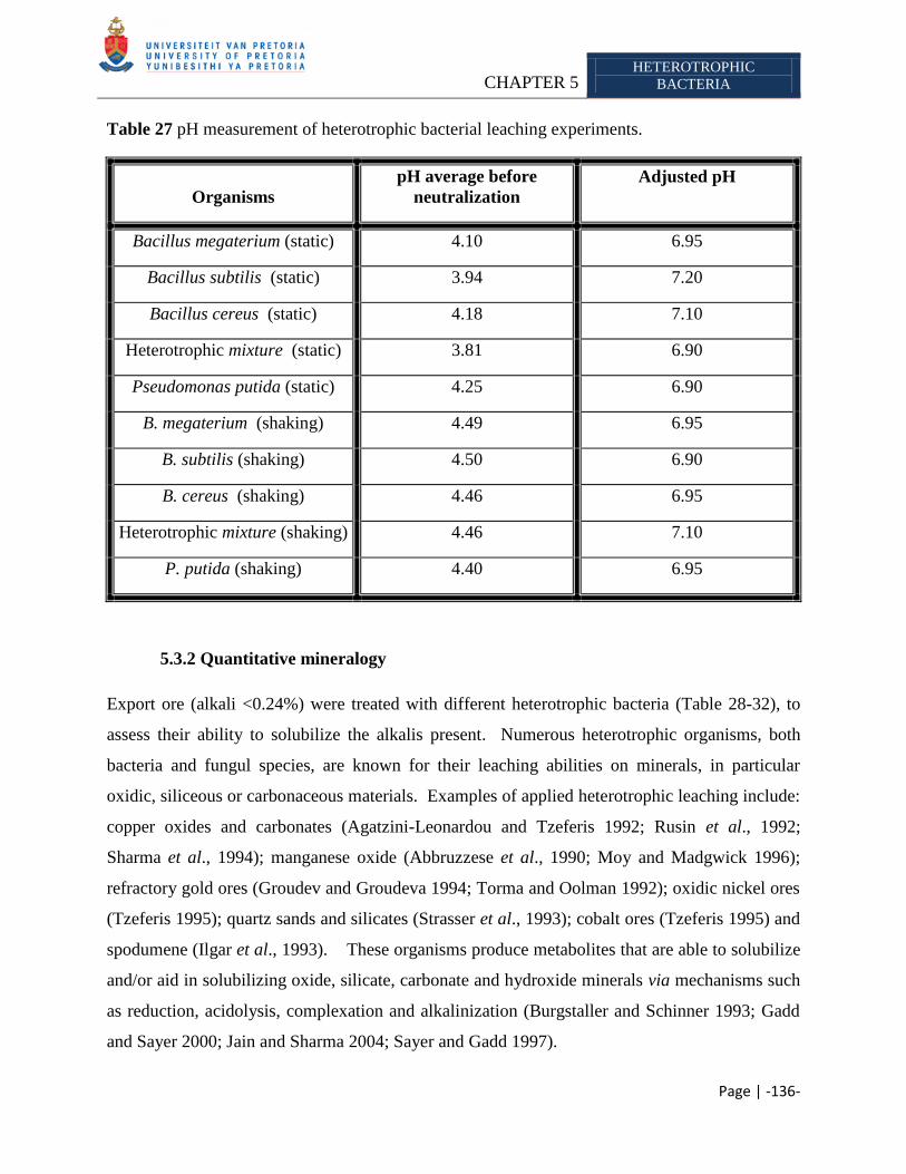
CHAPTER 5 HETEROTROPHIC
BACTERIA
Page | -136-
Table 27 pH measurement of heterotrophic bacterial leaching experiments.
Organisms
pH average before
neutralization
Adjusted pH
Bacillus megaterium (static) 4.10 6.95
Bacillus subtilis (static) 3.94 7.20
Bacillus cereus (static) 4.18 7.10
Heterotrophic mixture (static) 3.81 6.90
Pseudomonas putida (static) 4.25 6.90
B. megaterium (shaking) 4.49 6.95
B. subtilis (shaking) 4.50 6.90
B. cereus (shaking) 4.46 6.95
Heterotrophic mixture (shaking) 4.46 7.10
P. putida (shaking) 4.40 6.95
5.3.2 Quantitative mineralogy
Export ore (alkali <0.24%) were treated with different heterotrophic bacteria (Table 28-32), to
assess their ability to solubilize the alkalis present. Numerous heterotrophic organisms, both
bacteria and fungul species, are known for their leaching abilities on minerals, in particular
oxidic, siliceous or carbonaceous materials. Examples of applied heterotrophic leaching include:
copper oxides and carbonates (Agatzini-Leonardou and Tzeferis 1992; Rusin et al., 1992;
Sharma et al., 1994); manganese oxide (Abbruzzese et al., 1990; Moy and Madgwick 1996);
refractory gold ores (Groudev and Groudeva 1994; Torma and Oolman 1992); oxidic nickel ores
(Tzeferis 1995); quartz sands and silicates (Strasser et al., 1993); cobalt ores (Tzeferis 1995) and
spodumene (Ilgar et al., 1993). These organisms produce metabolites that are able to solubilize
and/or aid in solubilizing oxide, silicate, carbonate and hydroxide minerals via mechanisms such
as reduction, acidolysis, complexation and alkalinization (Burgstaller and Schinner 1993; Gadd
and Sayer 2000; Jain and Sharma 2004; Sayer and Gadd 1997).
CHAPTER 5 HETEROTROPHIC
BACTERIA
Page | -137-
The amount of alkali removed was determined by measuring the residual retained in the ore after
treatment and comparing it to the untreated sample (control) (Table 28-32). The alkali,
aluminum and silicon concentrations in the ore will be the main focus of the current research, as
Mojallali and Weed (1978) stated that a loss of aluminum and/or silicon shows a structural
rearrangement of the mineral. Furthermore we will also focus on iron and magnesium as Wilson
and Jones (1983) found that a decrease in these minerals also show a rearrangement of the
mineral.
Mineral composition of the untreated iron ore sample is depicted in Table 19.
X-ray fluorescence was used to determine the mineral composition of the export iron ore sample.
This method is reasonably sensitive, with detection limits27
for most elements in the low ppm
range as stated by Jenkins (1988) (Table 14). The magnesium and sodium concentrations in the
ore, were below the detection limit listed by Jenkins (1988) and will therefore be excluded from
further analysis. Results of treated samples were expected to exceed the control sample due to
sample variation; therefore a range was determined which included the minimum and maximum
amount of alkali measured in the quadruple analyzed untreated sample. From this data set we
can possibly determine the amount of alkali removed due to acid leaching and excluded data
influenced by sample variation. From XRF analysis of the untreated export iron ore, the
following ranges for the minerals were calculated: aluminum (1.15% - 1.56%), silicon
(1.92% - 2.80%), potassium (0.16% - 0.18%) and phosphorous (0.13% - 0.17). Concentrations
of each mineral removed below these parameters will be reported as ―large quantities28
‖
removed. We accept the average value of the untreated export sample as an acceptable
representative. The amount of magnesium present in the sample was excluded from analysis
(Table 14); therefore it could not be used as an indication of structural rearrangement of the
mineral (Wilson and Jones 1983).
Silicon was detected as quartz, muscovite, biotite, etc. in all of the samples analyzed (Table 1).
Quartz is known to dissolve slowly, due to the high activation energy required to break the
27
Lower limit of detection is defined as that concentration equivalent to a certain number of standard deviations of
the background count rate. 28
A maximum and minum concentration for each mineral present in the untreated sample was determined. Once the
ore was treated with the specific assay, we analyzed it again with XRF. If the residual amount of a specific mineral
was below the minimum concentration, we ascribe it to the assay tested.
CHAPTER 5 HETEROTROPHIC
BACTERIA
Page | -138-
silicon-oxygen bond (Brehm et al., 2005 as cited in Wilson 2004). We did not determine the
source of silicon dissolution, as it was outside the scope of this study; however it is assumed that
the silicon released was from less resistant mineral phases present in the export iron ore.
Mineral phases identified in the export sample using XRD, was apatite, hematite, muscovite and
K-feldspar (Table 1). Muscovite has considerable resistance toward leaching of interlayer
potassium, compared to trioctahedral micas (such as biotite). It has been proposed that an
inclined orientation of hydroxyl ions in the dioctahedral micas results in stronger binding of
potassium (Kalinowski and Schweda 1996) and therefore it has considerable resistance toward
leaching. However, Scott and Smith (1966) found that essentially all the potassium in
muscovite, biotite, phlogopite and vermiculite were exchangeable when the concentration of the
potassium in solution was kept low.
Bioleaching during this study was not extended beyond 60 days, as the implementation of this
approach into the mining schedule at Sishen would not allow such a time span.
Controversy exists in literature on the role of organic acids leaching in nature, due to its relative
low concentrations. However, recent experiments have demonstrated that naturally occurring
concentrations of organic acids, could increase mineral dissolution. The organic ligands can
directly or indirectly catalyze the reaction by forming complexes with either the metal ions in
solution (lowering the solution saturation state), or the metal ions on the mineral surface, thereby
weakening the metal oxygen bonds (Banfield et al., 1999; Welch et al., 1999). The effect of
organic ligands on mineral weathering reactions is affected by: (i) the ligand composition and
structure; (ii) metal-ligand stability and (iii) solution composition (pH, ligand concentration,
concentration of other metals) (Welch et al., 1999). Van Schöll and coworkers (2006) found that
mineral composition can also affect the efficiency of ligand weathering.
The bacteria selected for this section of the study includes: Bacillus megaterium, B. cereus and
B. subtilis. Bacillus megaterium is a gram positive, aerobic, spore-forming bacterium that
naturally occurs in soil, and has shown to effectively increase the mineral uptake of eggplants,
pepper and cucumber (Han et al., 2005). It is known to produce citric acid, which is a relatively
strong organic acid, and can assist mineral mobilization via acidulation and/or complexation
(Jain and Sharma 2004) (Table 13). From the acid leaching experiments (Chapter 3) we found
CHAPTER 5 HETEROTROPHIC
BACTERIA
Page | -139-
that citric acid was able to remove “large quantities‖ of potassium (24%) and phosphorous (13%)
(Table 15). We assume that the inability of the organisms to remove as much potassium as the
acid experiment is due to sample variation and/or mineral dissemination, as the iron oxidizing
bacterial results (Chapter 4) has an increased ability of the biotic experiments to remove these
alkalis.
Static grown cultures were able to remove silicon, aluminum and phosphorous after 60 days.
Shaken cultures were able to remove of silicon, aluminum, phosphorous and potassium after 60
days (Table 28). pH measured suggested that the static grown cultures were able to produce
more acids and was therefore thought to be more affective at solubilizing the alkalis (Table 27).
It is however likely that mineral dissemination29
and/or sample variation30
influenced these
XRF results and change the expected outcome.
Phosphorous was detected as apatite, which is known to be relatively insoluble at neutral pH,
although its solubility and reactivity varies as a function of its composition
(Anderson et al., 1985; Jahnke 1984; LeGeros and Tung 1983; Valsami-Jones et al., 1998).
Therefore the low pH present in the bioleaching solution (Table 28) could possibly have aided
the release of phosphorous from the ore (Welch et al., 2002). Recently, Goldstein and
Krishnaraj (2007) showed that the most common mechanism used by microorganisms to
solubilize phosphate from apatite, seems to be by acidifying the media when organic acids are
produced and released. Moreover, biofilms bound to the surface (Figure 22), would also actively
take up phosphate, because it is a key nutrient for cell processes (Delvasto et al., 2008; Tempest
and Neijssel 1992). This is accomplished by metals complexing with active moieties (such as
carboxyl groups) present in the exopolysaccharides or other cellular materials (Comte et al.,
2006; Corzo et al., 1994). Studies conducted with microbial extracellular polymers and simple
analogs of these compounds showed that acid polysaccharides could increase dissolution of
alumino-silicate minerals under mildly acidic conditions, by complexing with the minerals
solubilized into solution (Welch and Vandevivere 1994; Welch et al., 1999) or inhibit dissolution
at near neutral pH when bound on the minerals surface (Poumier et al., 1999).
29
Alkali is located in the core of the particle and therefore inaccessible to the acid 30
Sample contains an alkali concentration higher than the set parameter and therefore no apparent removal will be
reported.
CHAPTER 5 HETEROTROPHIC
BACTERIA
Page | -140-
The amount of silicon and aluminum removed, illustrates a structural rearrangement of the
minerals (Mojallali and Weed 1978). It was not determined whether the organisms actively
scavenged for the minerals, or whether the solubilization of the minerals was indirectly due to
the production of metabolites such as organic acid. There are however support for the former by
Bennett and coworkers (2001). They used in situ and laboratory microcosms to determine
whether colonization of minerals can be nutrient driven. The in situ (field experiments) were
conducted at two groundwater sites, which had similar characteristics: abundant dissolved carbon
and anoxic conditions. Here mineral chips were placed in permeable chambers and suspended in
the groundwater for months. In the laboratory, mineral fragments were inoculated with
groundwater (from the two sites) and aquifer materials. They found that in the groundwater,
which was rich in carbon but contained limited amounts of phosphate, microbial mediated
weathering of minerals were sometimes determined by the nutritional requirement of the
microbial community. They further found that feldspar could be rapidly dissolved in such
nutrient limiting environments. Furthermore the authors argued that microorganisms might be
able to select minerals that contain beneficial elements, and leave others intact.
Previously, Ullman et al. (1996) showed that bacteria are able to produce organic acids when
carbon sources are abundant but other nutrients scarce, therefore the organisms would still be
able to scavenge for nutrients. Furthermore, they showed that the excretion of organic acids can
also be achieved by fermentation under anoxic conditions. Therefore, the main conclusion of
their research study was that the dissolution rates of minerals should increase in organic-rich and
nutrient-poor environments, as well under anoxic conditions.
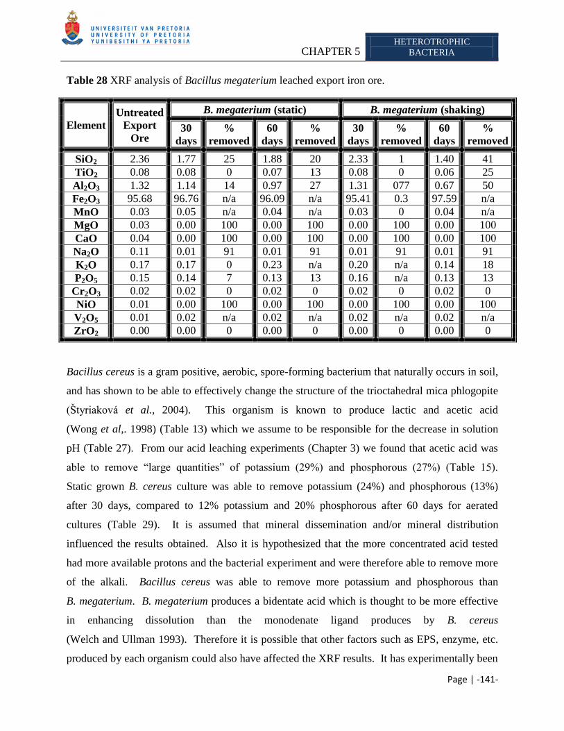
CHAPTER 5 HETEROTROPHIC
BACTERIA
Page | -141-
Table 28 XRF analysis of Bacillus megaterium leached export iron ore.
Element
Untreated
Export
Ore
B. megaterium (static) B. megaterium (shaking)
30
days
%
removed
60
days
%
removed
30
days
%
removed
60
days
%
removed
SiO2 2.36 1.77 25 1.88 20 2.33 1 1.40 41
TiO2 0.08 0.08 0 0.07 13 0.08 0 0.06 25
Al2O3 1.32 1.14 14 0.97 27 1.31 077 0.67 50
Fe2O3 95.68 96.76 n/a 96.09 n/a 95.41 0.3 97.59 n/a
MnO 0.03 0.05 n/a 0.04 n/a 0.03 0 0.04 n/a
MgO 0.03 0.00 100 0.00 100 0.00 100 0.00 100
CaO 0.04 0.00 100 0.00 100 0.00 100 0.00 100
Na2O 0.11 0.01 91 0.01 91 0.01 91 0.01 91
K2O 0.17 0.17 0 0.23 n/a 0.20 n/a 0.14 18
P2O5 0.15 0.14 7 0.13 13 0.16 n/a 0.13 13
Cr2O3 0.02 0.02 0 0.02 0 0.02 0 0.02 0
NiO 0.01 0.00 100 0.00 100 0.00 100 0.00 100
V2O5 0.01 0.02 n/a 0.02 n/a 0.02 n/a 0.02 n/a
ZrO2 0.00 0.00 0 0.00 0 0.00 0 0.00 0
Bacillus cereus is a gram positive, aerobic, spore-forming bacterium that naturally occurs in soil,
and has shown to be able to effectively change the structure of the trioctahedral mica phlogopite
(Štyriaková et al., 2004). This organism is known to produce lactic and acetic acid
(Wong et al,. 1998) (Table 13) which we assume to be responsible for the decrease in solution
pH (Table 27). From our acid leaching experiments (Chapter 3) we found that acetic acid was
able to remove ―large quantities‖ of potassium (29%) and phosphorous (27%) (Table 15).
Static grown B. cereus culture was able to remove potassium (24%) and phosphorous (13%)
after 30 days, compared to 12% potassium and 20% phosphorous after 60 days for aerated
cultures (Table 29). It is assumed that mineral dissemination and/or mineral distribution
influenced the results obtained. Also it is hypothesized that the more concentrated acid tested
had more available protons and the bacterial experiment and were therefore able to remove more
of the alkali. Bacillus cereus was able to remove more potassium and phosphorous than
B. megaterium. B. megaterium produces a bidentate acid which is thought to be more effective
in enhancing dissolution than the monodenate ligand produces by B. cereus
(Welch and Ullman 1993). Therefore it is possible that other factors such as EPS, enzyme, etc.
produced by each organism could also have affected the XRF results. It has experimentally been
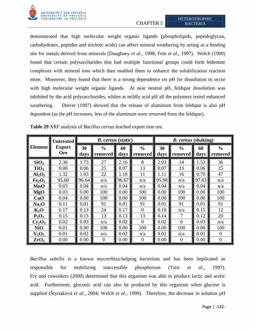
CHAPTER 5 HETEROTROPHIC
BACTERIA
Page | -142-
demonstrated that high molecular weight organic ligands (phospholipids, peptidoglycan,
carbohydrates, peptides and teichoic acids) can affect mineral weathering by acting as a binding
site for metals derived from minerals (Daughney et al., 1998; Fein et al., 1997). Welch (1999)
found that certain polysaccharides that had multiple functional groups could form bidentate
complexes with mineral ions which then enabled them to enhance the solubilization reaction
more. Moreover, they found that there is a strong dependence on pH for dissolution to occur
with high molecular weight organic ligands. At near neutral pH, feldspar dissolution was
inhibited by the acid polysaccharides, whiles at mildly acid pH all the polymers tested enhanced
weathering. Drever (1997) showed that the release of aluminum from feldspar is also pH
dependent (as the pH increases, less of the aluminum were removed from the feldspar).
Table 29 XRF analysis of Bacillus cereus leached export iron ore.
Element
Untreated
Export
Ore
B. cereus (static) B. cereus (shaking)
30
days
%
removed
60
days
%
removed
30
days
%
removed
60
days
%
removed
SiO2 2.36 1.73 27 2.16 8 2.03 14 1.52 36
TiO2 0.08 0.06 25 0.07 13 0.07 13 0.06 25
Al2O3 1.32 1.03 22 1.18 11 1.11 16 0.70 47
Fe2O3 95.68 96.64 n/a 96.67 n/a 95.98 n/a 97.63 n/a
MnO 0.03 0.04 n/a 0.04 n/a 0.04 n/a 0.04 n/a
MgO 0.03 0.00 100 0.00 100 0.00 100 0.00 100
CaO 0.04 0.00 100 0.00 100 0.00 100 0.00 100
Na2O 0.11 0.01 91 0.01 91 0.01 91 0.01 91
K2O 0.17 0.13 24 0.17 0 0.18 n/a 0.15 12
P2O5 0.15 0.13 13 0.13 13 0.14 7 0.12 20
Cr2O3 0.02 0.03 n/a 0.02 0 0.02 0 0.03 n/a
NiO 0.01 0.00 100 0.00 100 0.00 100 0.00 100
V2O5 0.01 0.02 n/a 0.02 n/a 0.02 n/a 0.01 0
ZrO2 0.00 0.00 0 0.00 0 0.00 0 0.00 0
Bacillus subtilis is a known mycorrhiza-helping bacterium and has been implicated as
responsible for mobilizing inaccessible phosphorous (Toro et al., 1997).
Fry and coworkers (2000) determined that this organism was able to produce lactic and acetic
acid. Furthermore, gluconic acid can also be produced by this organism when glucose is
supplied (Štyriaková et al., 2004; Welch et al., 1999). Therefore, the decrease in solution pH
CHAPTER 5 HETEROTROPHIC
BACTERIA
Page | -143-
was assumed to be due to acid production (Table 27). Leaching experiments with 1M acetic
acid, showed that it was able to solubilize 29% potassium and 27% phosphorous from the export
iron ore. 1M gluconic acid was able to solubilize 24% of the potassium and 27% of the
phosphorous present in the export iron ore (Table 15). Static grown cultures were able to
remove 6% potassium and 13% phosphorous after 60 days, however static grown cultures were
able to remove 29% potassium and 27% phosphorous. Moreover, the cultures were also able to
remove ―large quantities‖ of aluminum and silicon. Experiments conducted by
Delvasto et al. (2008) found that removal of phosphorous from iron ore appears not to be a
continuous process, which they attributed to the re-immobilization of the mineral due to the high
phosphate-binding capacity of iron oxides, and the ability of biofilms to readily take up this
mineral. Consequently, we conclude that the XRF data obtained for phosphorous
(aerated cultures) is possibly due to sample variation or re-immobilization of the mineral on the
surface. Therefore, Bacillus subtilis is able to remove a ―large quantity‖ of the alkalis compared
to acetic acid. The static grown cultures were able to decrease the solution pH more than the
aerated (shaken) cultures, which is in accordance Štyriaková et al. (2004). It was expected that
the lower pH present in the static grown cultures would favor more phosphorous release as
Welch et al. (2002) stated that a low solution pH increased apatite dissolution; however it is
known that its solubility and reactivity varies as a function of its composition
(LeGeros and Tung 1983; Jahnke 1984; Anderson et al., 1985; Valsami-Jones et al., 1998). We
found that the aerated cultures were able to solubilize more alkali, compared to the static
cultures. We presume that this is due to mineral dissemination, sample variation and/or the
difference in mineral composition. Furthermore it is also probable that the static grown cultures
produced more lactic acid, which might be less efficient than acetic acid, however due to
insufficient data we are unable to draw this conclusion.
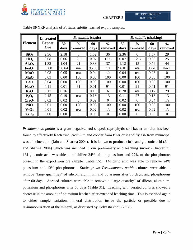
CHAPTER 5 HETEROTROPHIC
BACTERIA
Page | -144-
Table 30 XRF analysis of Bacillus subtilis leached export samples.
Element
Untreated
Export
Ore
B. subtilis (static) B. subtilis (shaking)
30
days
%
removed
60
days
%
removed
30
days
%
removed
60
days
%
removed
SiO2 2.36 2.18 8 1.52 36 2.36 0 1.67 29
TiO2 0.08 0.06 25 0.07 12.5 0.07 12.5 0.06 25
Al2O3 1.32 1.04 21 0.83 37 1.12 15 0.74 44
Fe2O3 95.68 96.64 n/a 95.95 n/a 96.01 n/a 96.70 n/a 1
MnO 0.03 0.05 n/a 0.04 n/a 0.04 n/a 0.03 0
MgO 0.03 0.00 100 0.00 100 0.00 100 0.00 100
CaO 0.04 0.00 100 0.00 100 0.00 100 0.00 100
Na2O 0.11 0.01 91 0.01 91 0.01 91 0.01 91
K2O 0.17 0.16 6 0.16 6 0.20 n/a 0.12 29
P2O5 0.15 0.19 n/a 0.13 13 0.11 27 0.13 13
Cr2O3 0.02 0.02 0 0.02 0 0.02 0 0.04 n/a
NiO 0.01 0.00 100 0.00 100 0.00 100 0.00 100
V2O5 0.01 0.02 n/a 0.02 n/a 0.02 n/a 0.02 n/a
ZrO2 0.00 0.00 0 0.00 0 0.00 0 0.00 0
Pseudomonas putida is a gram negative, rod shaped, saprophytic soil bacterium that has been
found to effectively leach zinc, cadmium and copper from filter dust and fly ash from municipal
waste incineration (Jain and Sharma 2004). It is known to produce citric and gluconic acid (Jain
and Sharma 2004) which was included in our preliminary acid leaching survey (Chapter 3).
1M gluconic acid was able to solubilize 24% of the potassium and 27% of the phosphorous
present in the export iron ore sample (Table 15). 1M citric acid was able to remove 24%
potassium and 13% phosphorous. Static grown Pseudomonas putida cultures were able to
remove ―large quantities‖ of silicon, aluminum and potassium after 30 days, and phosphorous
after 60 days. Aerated cultures were able to remove a ―large quantity‖ of silicon, aluminum,
potassium and phosphorous after 60 days (Table 31). Leaching with aerated cultures showed a
decrease in the amount of potassium leached after extended leaching time. This is ascribed again
to either sample variation, mineral distribution inside the particle or possible due to
re-immobilization of the mineral, as discussed by Delvasto et al. (2008).
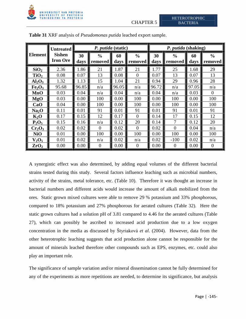
CHAPTER 5 HETEROTROPHIC
BACTERIA
Page | -145-
Table 31 XRF analysis of Pseudomonas putida leached export sample.
Element
Untreated
Sishen
Iron Ore
P. putida (static) P. putida (shaking)
30
days
%
removed
60
days
%
removed
30
days
%
removed
60
days
%
removed
SiO2 2.36 1.86 21 1.87 21 1.77 25 1.68 29
TiO2 0.08 0.07 13 0.08 0 0.07 13 0.07 13
Al2O3 1.32 1.13 15 1.04 21 0.94 29 0.96 28
Fe2O3 95.68 96.85 n/a 96.05 n/a 96.72 n/a 97.05 n/a
MnO 0.03 0.04 n/a 0.04 n/a 0.04 n/a 0.03 0
MgO 0.03 0.00 100 0.00 100 0.00 100 0.00 100
CaO 0.04 0.00 100 0.00 100 0.00 100 0.00 100
Na2O 0.11 0.01 91 0.01 91 0.01 91 0.01 91
K2O 0.17 0.15 12 0.17 0 0.14 17 0.15 12
P2O5 0.15 0.16 n/a 0.12 20 0.14 7 0.12 20
Cr2O3 0.02 0.02 0 0.02 0 0.02 0 0.04 n/a
NiO 0.01 0.00 100 0.00 100 0.00 100 0.00 100
V2O5 0.01 0.02 n/a 0.02 n/a 0.02 -100 0.02 n/a
ZrO2 0.00 0.00 0 0.00 0 0.00 0 0.00 0
A synergistic effect was also determined, by adding equal volumes of the different bacterial
strains tested during this study. Several factors influence leaching such as microbial numbers,
activity of the strains, metal tolerance, etc. (Table 10). Therefore it was thought an increase in
bacterial numbers and different acids would increase the amount of alkali mobilized from the
ores. Static grown mixed cultures were able to remove 29 % potassium and 33% phosphorous,
compared to 18% potassium and 27% phosphorous for aerated cultures (Table 32). Here the
static grown cultures had a solution pH of 3.81 compared to 4.46 for the aerated cultures (Table
27), which can possibly be ascribed to increased acid production due to a low oxygen
concentration in the media as discussed by Štyriaková et al. (2004). However, data from the
other heterotrophic leaching suggests that acid production alone cannot be responsible for the
amount of minerals leached therefore other compounds such as EPS, enzymes, etc. could also
play an important role.
The significance of sample variation and/or mineral dissemination cannot be fully determined for
any of the experiments as more repetitions are needed, to determine its significance, but analysis
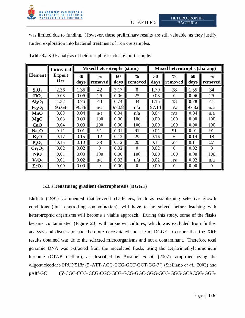
CHAPTER 5 HETEROTROPHIC
BACTERIA
Page | -146-
was limited due to funding. However, these preliminary results are still valuable, as they justify
further exploration into bacterial treatment of iron ore samples.
Table 32 XRF analysis of heterotrophic leached export sample.
Element
Untreated
Export
Ore
Mixed heterotrophs (static) Mixed heterotrophs (shaking)
30
days
%
removed
60
days
%
removed
30
days
%
removed
60
days
%
removed
SiO2 2.36 1.36 42 2.17 8 1.70 28 1.55 34
TiO2 0.08 0.06 25 0.06 25 0.08 0 0.06 25
Al2O3 1.32 0.76 43 0.74 44 1.15 13 0.78 41
Fe2O3 95.68 96.38 n/a 97.08 n/a 97.14 n/a 97.32 n/a
MnO 0.03 0.04 n/a 0.04 n/a 0.04 n/a 0.04 n/a
MgO 0.03 0.00 100 0.00 100 0.00 100 0.00 100
CaO 0.04 0.00 100 0.00 100 0.00 100 0.00 100
Na2O 0.11 0.01 91 0.01 91 0.01 91 0.01 91
K2O 0.17 0.15 12 0.12 29 0.16 6 0.14 18
P2O5 0.15 0.10 33 0.12 20 0.11 27 0.11 27
Cr2O3 0.02 0.02 0 0.02 0 0.02 0 0.02 0
NiO 0.01 0.00 100 0.00 100 0.00 100 0.00 100
V2O5 0.01 0.02 n/a 0.02 n/a 0.02 n/a 0.02 n/a
ZrO2 0.00 0.00 0 0.00 0 0.00 0 0.00 0
5.3.3 Denaturing gradient electrophoresis (DGGE)
Ehrlich (1991) commented that several challenges, such as establishing selective growth
conditions (thus controlling contamination), will have to be solved before leaching with
heterotrophic organisms will become a viable approach. During this study, some of the flasks
became contaminated (Figure 20) with unknown cultures, which was excluded from further
analysis and discussion and therefore necessitated the use of DGGE to ensure that the XRF
results obtained was de to the selected microorganisms and not a contaminant. Therefore total
genomic DNA was extracted from the inoculated flasks using the cetyltrimethylammonium
bromide (CTAB method), as described by Ausubel et al. (2002), amplified using the
oligonucleotides PRUN518r (5'-ATT-ACC-GCG-GCT-GCT-GG-3‘) (Siciliano et al., 2003) and
pA8f-GC (5'-CGC-CCG-CCG-CGC-GCG-GCG-GGC-GGG-GCG-GGG-GCACGG-GGG-
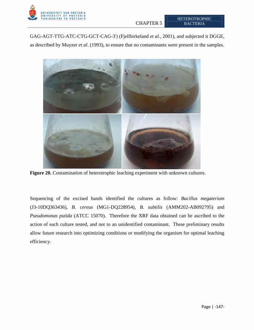
CHAPTER 5 HETEROTROPHIC
BACTERIA
Page | -147-
GAG-AGT-TTG-ATC-CTG-GCT-CAG-3') (Fjellbirkeland et al., 2001), and subjected it DGGE,
as described by Muyzer et al. (1993), to ensure that no contaminants were present in the samples.
Figure 20. Contamination of heterotrophic leaching experiment with unknown cultures.
Sequencing of the excised bands identified the cultures as follow: Bacillus megaterium
(J3-10DQ363436), B. cereus (MG1-DQ228954), B. subtilis (AMM202-AB092795) and
Pseudomonas putida (ATCC 15070). Therefore the XRF data obtained can be ascribed to the
action of each culture tested, and not to an unidentified contaminant. These preliminary results
allow future research into optimizing conditions or modifying the organism for optimal leaching
efficiency.
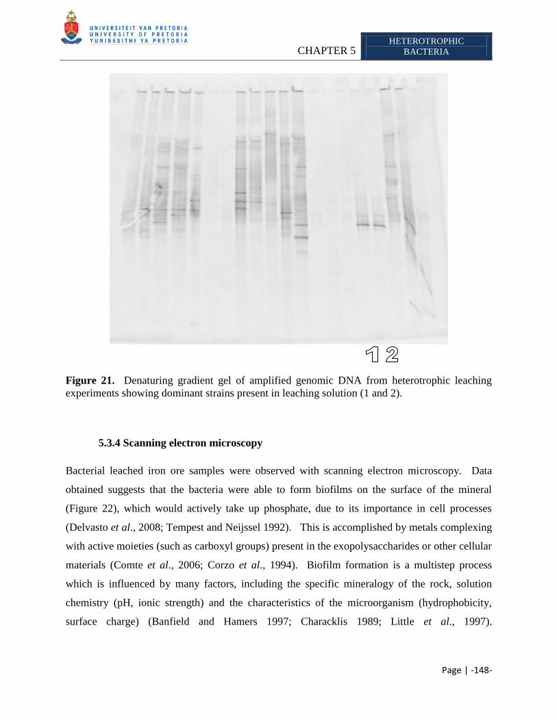
CHAPTER 5 HETEROTROPHIC
BACTERIA
Page | -148-
Figure 21. Denaturing gradient gel of amplified genomic DNA from heterotrophic leaching
experiments showing dominant strains present in leaching solution (1 and 2).
5.3.4 Scanning electron microscopy
Bacterial leached iron ore samples were observed with scanning electron microscopy. Data
obtained suggests that the bacteria were able to form biofilms on the surface of the mineral
(Figure 22), which would actively take up phosphate, due to its importance in cell processes
(Delvasto et al., 2008; Tempest and Neijssel 1992). This is accomplished by metals complexing
with active moieties (such as carboxyl groups) present in the exopolysaccharides or other cellular
materials (Comte et al., 2006; Corzo et al., 1994). Biofilm formation is a multistep process
which is influenced by many factors, including the specific mineralogy of the rock, solution
chemistry (pH, ionic strength) and the characteristics of the microorganism (hydrophobicity,
surface charge) (Banfield and Hamers 1997; Characklis 1989; Little et al., 1997).
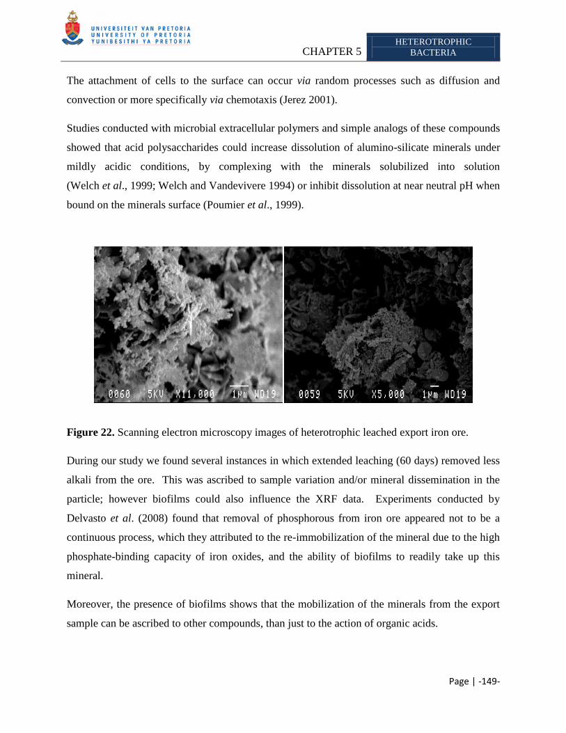
CHAPTER 5 HETEROTROPHIC
BACTERIA
Page | -149-
The attachment of cells to the surface can occur via random processes such as diffusion and
convection or more specifically via chemotaxis (Jerez 2001).
Studies conducted with microbial extracellular polymers and simple analogs of these compounds
showed that acid polysaccharides could increase dissolution of alumino-silicate minerals under
mildly acidic conditions, by complexing with the minerals solubilized into solution
(Welch et al., 1999; Welch and Vandevivere 1994) or inhibit dissolution at near neutral pH when
bound on the minerals surface (Poumier et al., 1999).
Figure 22. Scanning electron microscopy images of heterotrophic leached export iron ore.
During our study we found several instances in which extended leaching (60 days) removed less
alkali from the ore. This was ascribed to sample variation and/or mineral dissemination in the
particle; however biofilms could also influence the XRF data. Experiments conducted by
Delvasto et al. (2008) found that removal of phosphorous from iron ore appeared not to be a
continuous process, which they attributed to the re-immobilization of the mineral due to the high
phosphate-binding capacity of iron oxides, and the ability of biofilms to readily take up this
mineral.
Moreover, the presence of biofilms shows that the mobilization of the minerals from the export
sample can be ascribed to other compounds, than just to the action of organic acids.
CHAPTER 5 HETEROTROPHIC
BACTERIA
Page | -150-
5.4 CONCLUSION
Heterotrophic bacteria posses several mechanisms, which enable them to aid mineral
mobilization. These include exopolysaccharides (Banfield et al., 1999; Malinovskaya et al.,
1990; Welch et al., 1999), amino acids, proteins (Bosecker 1997) and organic acids (Agatzini
and Tzeferis 1997; Barker et al., 1997; Castro et al., 2000; Natarajan and Deo 2001; Štyriaková
et al., 1999; Valsami-Jones and McEldowney 2000). Heterotrophic organisms have been applied
to leach compounds such as: copper oxides and carbonates (Agatzini-Leonardo and Tzeferis
1992; Rusin et al., 1992; Sharma et al., 1994); manganese oxide (Abbruzzese et al., 1990; Moy
and Madgwick 1996); refractory gold ores (Groudev and Groudeva 1994; Torma and Oolman
1992); oxidic nickel ores (Tzeferis 1995); quartz sands and silicates (Strasser et al., 1993); cobalt
ores (Tzeferis 1995) and spodumene (Ilgar et al., 1993). However due to difficulty in
establishing selective growth conditions, heterotrophic leaching is not preferred for industrial
scale leaching as autotrophic leaching (Ehrlich 1991; Jain and Sharma 2004). The objective of
this study was to determine, whether known potassium/phosphorous solubilizing heterotrophic
bacteria, are able to remove the alkali impurities from the export iron ore samples, provided by
Kumba Iron Ore, Ltd. We found that the different strains were able to remove varying amounts
of potassium and phosphorous. Moreover, the organisms demonstrated a synergistic
relationship, which enable mobilization of the mineral. Scanning electron microscopy analysis
of the leached ore, demonstrated the formation of biofilms, which could also have affected the
leaching ability of the organisms.
The preliminary results suggest that heterotrophic bacteria leaching could possibly be applied to
remove some of the impurities from the ore. However due to difficulty in establishing selective
growth conditions, will make its introduction into the mining schedule more difficult than the
autotrophic bioleaching approach (Chapter 4). Furthermore bioleaching will never completely
replace the conventional methods but rather supplement them, as bioleaching does not recover
precious metals from the ores which are often an essential component in the profitability of the
operation; also when ore bodies do not contain sufficient acid consuming minerals, the residual
acids generated has to be neutralized during the leaching process, thereby increasing the
operational cost (Dreshner 2004).
CHAPTER 6 INDIGENOUS BACTERIA
Page | -151-
CHAPTER 6
MICROBIAL COMMUNITY STRUCTURE OF INDIGENOUS BACTERIA PRESENT ON THE SISHEN
HEMATITE IRON ORE DURING BIOBENEFICATION
Abstract
The Sishen Iron Ore Mine is situated in the Northern Cape, South Africa, and forms part
of the northern end of the Maremane anticline where the bulk of the hematite is buried
beneath younger cover lithologies. The iron ore bodies at the mine are overlain by
conglomerates, shales, flagstone and quartzite. Potassium and phosphorous are common
constituents of iron ore that have deleterious effect on iron and steel manufacturing,
therefore steel making companies charge penalties when purchasing iron ore with alkali
concentrations above predetermined limits. Conventional methods used to treat high alkali
ore have several drawbacks such as poor product recovery, high running cost and increase
pollution of water. Biohydrometallurgy is a natural alternative approach which could be
applied to supplement conventional methods. Our aim was to determine the microbial
community structure of the indigenous bacteria present on the iron ore and assess their
ability to remove potassium and phosphorous from the iron ore. We found that the
organisms were able to solubilize varying degrees of potassium and phosphorous from the
different iron ore samples, which is attributed to different metabolic capabilities of the
organisms and the composition of each ore body. Furthermore, from this section of the
study, we concluded that indigenous bacter are beter adapted than introduced species to
remove the alkalis from the ore.
Keywords: Indigenous bacteria, Iron ore, muscovite, exopolysaccharide, organic acid, muscovite
______________________________________________________________________________
CHAPTER 6 INDIGENOUS BACTERIA
Page | -152-
6.1 INTRODUCTION
Microorganisms play an important role in: (i) the cycling of elements and sorption of metals
(Langley and Beveridge 1999); (ii) the dissolution of minerals (Banfield and Hamers 1997;
Hersman et al., 1996; Roden and Zachara 1996; Schrenk et al., 1998) and
(iii) mineral crystallization (Fortin et al., 1997; Warren and Ferris 1998). They are able to
achieve this via: (1) assimilation/adsorption and mineralization; (2) precipitation and dissolution;
(3) oxidation and reduction and (4) methylation and dealkylation. For a full review on these
mechanisms please refer to Johnson (2006). Research has found that mineralogical process in
turn play a significant role in the distribution, activity and diversity of microbes
(Schrenk et al., 1998), the expression of their genes (Gehrke et al., 1998), development and
structure of communities (Kennedy and Gewin 1997; Thorseth et al., 1995; Wolfaardt et al.,
1994), and transfer of genetic material (Holben 1997; Trevors and van Elsas 1997).
Research by Johnson (1998) and Hallberg and Johnson (2001) indicated that there is a rich
diversity of microorganisms present in the mining environments. Several researchers have
reported that microorganisms are able to solubilize potassium from various minerals, for
example: Berthelin and Belgy (1979) demonstrated the complete removal of potassium and
titanium from biotite by a microbial consortium in a granitic sand, which resulted in brittle, white
micaceous particles. Leyval and Berthelin (1991) showed that rhizosphere microorganisms
associated with pine roots extracted potassium from phlogopite and this was attributed to organic
acid mediated solution. Buis (1995) and Parks et al. (1990) conducted work in which they used
fungal strains with high phosphate-solubilizing activity to treat phosphatic iron ore. The main
drawback of their investigation was that they used strains that were not associated with the ore
being treated. When artificially inoculated in an environment, the indigenous microorganisms as
a general rule compete better in terms of adaptation and cause fewer ecological distortions than
exogenous microorganisms (Delvasto et al., 2008b).
Iron ore mined at the Sishen, Northern Cape, South Africa contains several different mineral
phases (Table 1). Alkalis (potassium and phosphorous) are frequently found as constituents of
iron ore, which has a deleterious effect on the manufacturing of iron and steel
(Delvasto et al., 2008). Therefore, steel making companies charge penalties when purchasing
iron ore concentrates with alkali concentrations above certain levels. The limits allowed is
CHAPTER 6 INDIGENOUS BACTERIA
Page | -153-
determined by the steel making companies and ranges from 0.25% mass in Japan to 0.55% mass
in Switzerland for the alkali potassium. Kumba Iron Ore, Ltd., has an industrial set limit of
0.24% potassium allowed in their export ore according to D. Krige (Personal communication,
2006). To ensure that their export batches stay within this set limit, the ores from different
batches, at the Sishen Iron Ore Mine (with potassium >0.24% and <0.24%) are mixed to produce
an average potassium value of below 0.24%. However this solution will soon become
ineffective as the low potassium ore (< 0.24%) is progressively depleted according to D. Krige
(Personal communication, 2006). Certain pyro – and hydrometallurgical methods can be applied
to decrease the alkali concentration (Cheng et al., 1999; Kokal et al., 2003), however there are
several disadvantages when using these methods such as: poor product recovery, involvement of
high process and energy cost and an increase in pollution load of water resources
(Jain and Sharma 2004). Thus an alternative, natural and economical feasible process is required
aid conventional methods (such as pyro- and hydrometallurgy processes) to remove unwanted
alkalis from the ores. Biohydrometallurgy31
is an option for the removal of the deleterious
phosphate and potassium, as it is well established that many microorganisms are capable of
mobilizing these minerals, especially in nutrient limited environments (Banfield et al., 1999;
Nautiyal 1999),
It is thought that indigenous microorganisms occurring on the iron ore mined at Sishen might
possess the ability to leach the potassium and phosphorous from the iron, as these minerals might
have become limiting previously in their immediate environment. Experiments by
Bennett et al. (2001) provided evidence that silicate weathering by bacteria is sometimes driven
by the nutrient requirements of the microbial consortium. They used in situ and laboratory
microcosms to determine whether colonization of minerals can be nutrient driven. The in situ
(field experiments) were conducted at two groundwater sites, which had similar characteristics:
abundant dissolved carbon and anoxic conditions. Here mineral chips were placed in permeable
chambers and suspended in the groundwater for months. In the laboratory, mineral fragments
were inoculated with groundwater (from the two sites) and aquifer materials. They found that in
the groundwater, which was rich in carbon but contained limited amounts of phosphate,
microbial mediated weathering of minerals were sometimes determined by the nutritional
31
Biohydrometallurgical processes include bioleaching and biobenefication (Ehrlich 1991).
CHAPTER 6 INDIGENOUS BACTERIA
Page | -154-
requirement of the microbial community. They further found that feldspar could be rapidly
dissolved in such nutrient limiting environments. Furthermore the authors argued that
microorganisms might be able to select minerals that contain beneficial elements, and leave
others intact.
Therefore, the purpose of the study was to amplify the indigenous bacteria, using Aleksandrov
and Mikhauloskaya media previously described as able to select for potassium/phosphorous
solubilizing bacteria and assessing their ability to mobilize the detrimental minerals from the iron
ore mined at Sishen.
6.2 MATERIALS AND METHODS
6.2.1 Iron ore
Export (alkali <0.24%), KGT (conglomerate) and SK (shale) iron ore, as defined by supplier32
,
with a particle size range of 1mm - 5mm, from the Sishen Iron Ore Mine, Northern Cape, South
Africa was supplied by Kumba Iron Ore, Ltd., for indigenous bacterial leaching experiment.
Samples were not kept under sterile conditions; therefore an array of contaminant would be
present. However, due to the experimental setup, only those organisms with
potassium/phosphate-solubilizing abilities would survive.
6.2.2 Enrichment media
Bioleaching experiments were carried out in 500ml Erlenmeyer flasks containing 150g
unsterilized ore and 200ml media, followed by incubation at 30°C with agitation (150 rpm).
Indigenous bacteria were amplified from the export iron ore using Aleksandrov media (Hu et al.,
2006), Nutrient broth (Biolab) and a media described by Štyriaková et al. (2004), which will
hence be referred to as Štyriaková media (Table 33). Indigenous bacteria from the KGT sample
(Conglomerate) were amplified using Aleksandrov media (Hu et al., 2006), Nutrient broth
(Biolab) and a media described by Mikhailouskaya et al., 2005, which will consequently be
referred to as Mikhailouskaya media. The SK sample (Shale) was treated with Nutrient broth
(Biolab), Aleksandrov-, Mikhailouskaya- and Štyriaková media. The various iron ore samples
32
Bongi Ntsoelengoe, Kumba Iron Ore, Ltd., Rust Building, Frikkie Meyer Road, Pretoria West, South Africa
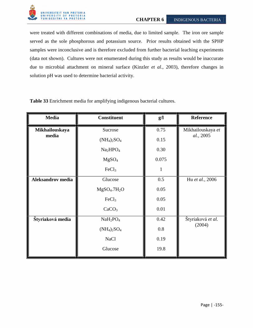
CHAPTER 6 INDIGENOUS BACTERIA
Page | -155-
were treated with different combinations of media, due to limited sample. The iron ore sample
served as the sole phosphorous and potassium source. Prior results obtained with the SPHP
samples were inconclusive and is therefore excluded from further bacterial leaching experiments
(data not shown). Cultures were not enumerated during this study as results would be inaccurate
due to microbial attachment on mineral surface (Kinzler et al., 2003), therefore changes in
solution pH was used to determine bacterial activity.
Table 33 Enrichment media for amplifying indigenous bacterial cultures.
Media Constituent g/l Reference
Mikhailouskaya
media
Sucrose
(NH4)2SO4
Na2HPO4
MgSO4
FeCl3
0.75
0.15
0.30
0.075
1
Mikhailouskaya et
al., 2005
Aleksandrov media
Glucose
MgSO4.7H2O
FeCl3
CaCO3
0.5
0.05
0.05
0.01
Hu et al., 2006
Štyriaková media NaH2PO4
(NH4)2SO4
NaCl
Glucose
0.42
0.8
0.19
19.8
Štyriaková et al.
(2004)
CHAPTER 6 INDIGENOUS BACTERIA
Page | -156-
6.2.3 pH measurements
pH of the inoculated samples were neutralized with 1M NaOH every 5th
day, as described by
Štyriaková et al. (2004).
6.2.4 DNA extraction
Total genomic DNA was extracted from the flasks using the cetyltrimethylammonium bromide
(CTAB method), as described by Ausubel et al. (2002), to ensure that no contaminants were
present in the samples. Cells from 1.5ml of the inoculated flasks were pelleted by centrifugation
at 13 400 rpm for 5 min and suspended in 567µl of 1 × TE buffer (10 mM Tris-HCl, 1 mM
EDTA; pH 8). The cells were lysed by adding sodium dodecyl phosphate (SDS) to a final
concentration of 0.5% (v/v) and the proteins were digested by adding Proteinase K to a final
concentration of 100 µg/ml in total volume of 600 µl. The suspension was then incubated
overnight at 37°C. Following incubation, 100 µl of 5M NaCl and 80µl of CTAB/NaCl solution
(10% [w/v] CTAB in 0.7 M NaCl) was added, mixed thoroughly and incubated at 65°C for 10
min. An equal volume of chloroform:isoamyl alcohol (24:1) was then added and followed by
centrifugation at 13 400 rpm for 5 min. The supernatant was then transferred to a clean
microfuge tube to which an equal volume of phenol:chloroform:isoamyl alcohol (25:24:1) was
added and centrifuged at 13 400 rpm for 5 min. The supernatant was then transferred to a new
microfuge tube, to which 0.6 volume isopropanol was added to precipitate the DNA. The
precipitated DNA was pelleted by centrifugation at 13 400 rpm for 20 min, washed with 70%
ethanol, dried under vacuum and suspended in 20 µl of 1 × TE buffer. The suspension was then
incubated overnight at 37°C before analyzing an aliquot of the extracted genomic DNA was
analyzed by electrophoresis on a 1% (w/v) agarose gel.
6.2.5 Polymerase chain reaction
Polymerase chain reaction (PCR) was used to amplify a section of the 16S rRNA of the DNA
extracted. The genes were amplified using the oligonucleotides PRUN518r (5'-ATT-ACC-GCG-
GCT-GCT-GG-3‘) (Siciliano et al., 2003) and pA8f-GC (5'-CGC-CCG-CCG-CGC-GCG-GCG-
CHAPTER 6 INDIGENOUS BACTERIA
Page | -157-
GGC-GGG-GCG-GGG-GCACGG-GGG-GAG-AGT-TTG-ATC-CTG-GCT-CAG-3')
(Fjellbirkeland et al., 2001). Each PCR reaction mixture (20 μl) contained ~27ng/μl of genomic
DNA as template, 10.3 µl sterile distilled MilliQ water, 2.5μl PCR buffer with MgCl2 (10×), 2μl
dNTPs (2.5μM), 1μl primer PRUN518r (50μM), 1μl primer pA8f-GC (50μM) and 0.2μl
Supertherm Taq (5U/μl) (Southerm Cross Biotechnology). A negative control (all reagents and
no template DNA) was added to rule out contamination. The tubes were placed in a Perkin-
Elmer GeneAMP® 2700 thermal cycler. Following incubation at 95 °C for 10 min, the reaction
mixture were subjected to 35 cycles of denaturing at 94 °C for 30 s, annealing at 53°C for 30 s
and elongation at 72 °C for 1 min. Once the cycles were completed, a final elongation step was
performed at 72°C for 10 min to complete DNA synthesis. An aliquot of the amplified DNA
was analyzed by electrophoresis on a 1.5% (w/v) agarose gel in the presence of appropriate
molecular weight marker. The PCR products were stored at 4°C, until further analysis.
6.2.6 Denaturing gradient gel electrophoresis (DGGE)
PCR products were subjected to denaturing gradient gel electrophoresis (DGGE) according to
the method described by Muyzer et al. (1993). 10µl of the each PCR product was loaded onto a
40-50% denaturing gradient gels (Table 20). Gels were run at 70 V for 17 h at a constant
temperature of 60 °C. Image analysis was performed using the Gel2K (Norland 2004) program.
Selected bands were picked under blue light from the DGGE gels using a sterile scalpel and
forceps. Each band was assigned a number for sequence analysis. The gel fragments were place
into 25µl filter-sterilized deionized water and allowed to stand overnight to dissolve. The
dissolved fragments were then subjected to PCR as described in the previous section.
6.2.7 Sequencing
The amplified products from the DGGE bands were cleaned by adding 15ul sterile water,
transferring the entire volume to a 0.5ml Eppendorf sequencing tube, adding 2ul of sodium
acetate (3M) and 50ul ethanol (95%), and allowing it to stand on ice for 10min. The tubes were
then centrifuged at 10 000 rpm for 30 min. The ethanol solution was removed, the pellet rinsed
in 150μl ethanol (70%), and the tubes again centrifuged for 5 min at 10 000 rpm. The ethanol
CHAPTER 6 INDIGENOUS BACTERIA
Page | -158-
was aspirated and the pellet dried under vacuum for approximately 10 min. These samples were
then submitted to Macrogen (U.S.A.) for sequencing using the oligonucleotides PRUN518r
(Siciliano et al., 2003). Nucleotide sequences were analyzed with BioEdit v.5.0.9.1 (Hall 2001)
and their identity were verified by sequence match searches against the RDP database (available
at http://rdp.cme.msu.edu).
6.2.8 Qualitative mineralogy
The composition of iron samples were analyzed with X-ray fluorescence (XRF) at the X-ray
analytical facility of the University of Pretoria. Four samples of untreated iron ore were
analyzed as controls
The samples were prepared as follow for analysis: Iron ore samples were first ground to <75μm
in a tungsten carbide milling vessel. The loss of ignition (LOI) was determined by roasting the
milled samples at 1000ºC. 1g of a milled iron ore sample were added to 6g Li2B4O7 and fused
into a glass beads. The rest of the sample was then used to make a powder briquette for minor
element analysis. Major element analyses were executed on the fused bead using an
ARL9400XP+ spectrometer (Loubser and Verryn 2008).
6.2.9 Scanning electron microscopy
Bacteria cells were fixed by placing the iron ore in a Greiner tube containing 20 ml fixing
solution (2.5% gluteraldehyde in 0.0075 M phosphate buffer) for 1 h. The ore was washed three
times for 15 min each with 0.0375 M NaPO4 buffer, before being dehydrated by sequential
treatment for 15 min each in 50%, 70%, 90% and 100% ethanol. The 100% ethanol step was
repeated twice more to ensure complete dehydration. The iron ore samples were critical point-
dried, scatter with gold with a Polaron Equipment LTD SEM Autocoating unit E5200 and
observed with a JEOL 5800LV scanning electron microscope at an accelerating voltage of 5kV.
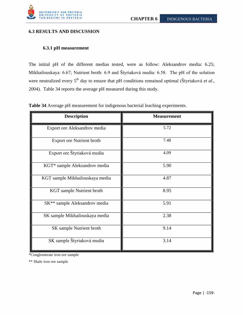
CHAPTER 6 INDIGENOUS BACTERIA
Page | -159-
6.3 RESULTS AND DISCUSSION
6.3.1 pH measurement
The initial pH of the different medias tested, were as follow: Aleksandrov media: 6.25;
Mikhailouskaya: 6.67; Nutrient broth: 6.9 and Štyriaková media: 6.58. The pH of the solution
were neutralized every 5th
day to ensure that pH conditions remained optimal (Štyriaková et al.,
2004). Table 34 reports the average pH measured during this study.
Table 34 Average pH measurement for indigenous bacterial leaching experiments.
Description Measurement
Export ore Aleksandrov media 5.72
Export ore Nutrient broth 7.48
Export ore Štyriaková media 4.09
KGT* sample Aleksandrov media 5.90
KGT sample Mikhailouskaya media 4.87
KGT sample Nutrient broth 8.95
SK** sample Aleksandrov media 5.91
SK sample Mikhailouskaya media 2.38
SK sample Nutrient broth 9.14
SK sample Štyriaková media 3.14
*Conglomerate iron ore sample
** Shale iron ore sample
CHAPTER 6 INDIGENOUS BACTERIA
Page | -160-
Microorganisms produce an array of metabolites which might assist in the mobilization of
minerals from the ore. These include exopolysaccharides (Banfield et al., 1999; Malinovskaya et
al., 1990; Welch et al., 1999), amino acids, proteins (Bosecker 1997) and organic acids (Agatzini
and Tzeferis 1997; Barker et al., 1997; Castro et al., 2000; Natarajan and Deo 2001; Valsami-
Jones and McEldowney 2000; Štyriaková et al., 1999). Mineral dissolution and the effectiveness
of low/high molecular weight organic ligands are known to be influenced by the solution pH
(Stillings et al., 1996; Welch and Ullman 1996; Ullman and Welch 1998). Most metals are
cationic and are therefore more soluble in acidic than in neutral pH and alkaline solutions,
however the reverse is true for oxyanionic metal species (Johnson 2006). For example, silicate
dissolution is reasonably slow at near neutral pH and independent on small pH changes, whereas
an increase in acidity to below pH 4 to 5, the dissolution becomes more dependent on the
changes in pH. With organic acid solutions, the rate of mineral dissolution increases with
increasing acidity due to proton-promoted dissolution and metal-ligand complexation
(Welch et al., 1999). The ore bodies used during this study were composed of an array of
minerals which included apatite and biotite (Table 1). Welch and coworkers (2002)
demonstrated that microorganisms are able to influence apatite and biotite dissolution by
producing organic acids, primarily pyruvate, fermentation products and oxalate. They found that
by lowering the pH between 3 to 5, the microorganisms were able to solubilize more of the
minerals. Moreover, they further found that some microorganisms were able to solubilize
phosphorous from the mineral bodies without lowering the solution pH by producing pyruvate.
Therefore we assumed that some of the potassium and phosphorous solubilized where from the
ore, were due to the low pH and complex forming ability of the acids.
We found that the microbes enriched for, using the Aleksandrov, Mikhailouskaya and Štyriaková
media, increased solution acidity, whereas the microbes amplified with Nutrient broth (Biolab)
increased alkalinity. The changes in pH measured during this study, was ascribed to the
following (Table 34). A decrease in pH may have been due to: the release of organic acid by the
bacteria into the surrounding media (Krebs et al., 1997); production of gluconic acid from the
carbon source (Štyriaková et al., 2004; Welch et al., 1999); hydrolysis of carbon dioxide to
carbonic acid produced during respiration and/or due to assimilation of ammonium sulfate
present in the media (Illmer and Schinner 1995; Illmer et al., 1995). Although the organic acids
CHAPTER 6 INDIGENOUS BACTERIA
Page | -161-
were not identified and measured, it is known that organic acid production constitutes an
adaption strategy by which bacteria and other microorganisms can extract limiting nutrients such
as potassium, phosphorous and calcium from insoluble mineral matrices, by chemical attack on
the crystal structure of the nutrient containing minerals (Banfield et al., 1999). Microbial
processed, such fermentation, nitrification and sulfur-oxidation generate acidity, whilst others
such as ammonification, denitrification, sulfate-reduction and methanogenesis generate
alkalinity, therefore it is assumed that the cultures enriched with nutrient broth possess such as
mechanism(s) (Johnson 2006). In addition, microorganisms are also able to produce other
compounds such as siderophores, which can actively scavenge for ferric iron and other trivalent
metals (such as aluminum and gallium), at neutral pH (Johnson 2006; White et al., 1995), which
could have aided mineral solubilization at near neutral pH (Table 34).
6.3.2 Quantitative mineralogy
Indigenous bacteria with possible potassium/phosphorous leaching abilities were enriched for by
inoculating Aleksandrov - (Hu et al., 2006), Mikhailouskaya – (Mikhailouskaya et al., 2005),
Štyriaková media and Nutrient broth (Biolab) with export iron ore. Bacteria with phosphate
solubilizing ability has been isolated from various environments such as eutrophic lakes
(Gen-Fu and Xue-Ping 2005), mangrove tree roots (Vazquez et al., 2000), the cactus rhizoplane
(Puente et al., 2004) and tree rhizospheres (Leyval and Berthelin 1991).
Mikhailouskaya et al. (2005) assessed the ability of two local strains to mobilize potassium from
biotite, muscovite-2, glauconite and hydromuscovite. They found that the mobility of potassium
in soil minerals and its availability for bacteria depend on factors such as the chemical structure
of the mineral, the degree of potassium dispersion, activity of the bacterial strain and the
ecological conditions. Hu et al. (2006) used a modified Aleksandrov media to isolate two
phosphate- and potassium-solubilizing bacteria from the Tianmu Mountain, Zhejang, China.
Preliminary results obtained with Aleksandrov media, justified repeating the experiment. Alkali
removal was determined by measuring the residual contained in the ore after treatment and
comparing it to the untreated sample (control). The alkali, aluminum and silicon concentrations
will be the main focus of the current research as Mojallali and Weed (1978) stated that a loss of
aluminum and/or silicon shows a structural rearrangement of the mineral structure. Furthermore,
CHAPTER 6 INDIGENOUS BACTERIA
Page | -162-
Wilson and Jones (1983) found that a decrease in magnesium and iron also shows a
rearrangement of the mineral structure.
The mineral composition of the untreated iron ore sample is depicted in Table 19. X-ray
fluorescence is known to be a reasonably sensitive detection method, with detection limits33
for
most elements in the low ppm range (Table 14) (Jenkins 1988). Therefore magnesium and
sodium will be excluded from further analysis. Focus is placed mainly on the residual alkali, as
these minerals have a deleterious effect in steel manufacturing (Delvasto et al., 2008). Results of
treated samples are expected to exceed the control sample due to sample variation; therefore
parameters were calculated to be able to determine the amount of alkali/mineral removed due to
microbial leaching and not sample variation. From XRF analysis the following ranges for the
minerals were calculated: aluminum (1.15% - 1.56%), silicon (1.92% - 2.80%), potassium
(0.16% - 0.18%) and phosphorous (0.13% - 0.17%), as discussed by Mojallali and Weed (1978).
The amount of magnesium present in the sample was excluded from analysis (Table 14),
therefore it could not be used as an indication of structural rearrangement of the mineral
(Wilson and Jones 1983). Silicon was detected as quartz, muscovite, biotite, etc. in all of the
samples analyzed (Table 1). Quartz is known dissolved slowly, due to the high activation energy
needed to break the silicon-oxygen bond (Brehm et al., 2005 as cited in Wilson 2004). We did
not determine the source of silicon dissolution, as it was outside the scope of this study, however
it is assumed that the silicon released was from less resistant mineral. Concentrations of each
mineral removed below these parameters will be reported as ―large quantities34
‖ removed. We
accept the average value of the untreated export sample as an acceptable representative. Mineral
phases detected in the sample include apatite, hematite, muscovite and K-feldspar (Table 1). We
accept the average value of the untreated export sample as an acceptable representative.
Potassium was detected as K-feldspar and muscovite (Table 1). Muscovite has considerable
resistance toward leaching of interlayer potassium, compared to trioctahedral micas (such as
biotite). It was proposed that an inclined orientation of hydroxyl ions in the dioctahedral micas
results in stronger binding of potassium (Kalinowski and Schweda 1996). Leaching was not
33
Lower limit of detection is defined as that concentration equivalent to a certain number of standard deviations of
the background count rate. 34
A maximum and minum concentration for each mineral present in the untreated sample was determined. Once the
ore was treated with the specific assay, we analyzed it again with XRF. If the residual amount of a specific mineral
was below the minimum concentration, we ascribe it to the assay tested.
CHAPTER 6 INDIGENOUS BACTERIA
Page | -163-
extended beyond 30 days, as the implementation of this approach into the mining schedule of the
Sishen mine would not allow such a time span. Scott and Smith (1966) conducted experiments
in which they determined the susceptibility of interlayer potassium in micas to exchange with
sodium. They found that essentially, all the potassium in muscovite, biotite, phlogopite and
vermiculite were exchangeable when concentration of the potassium in solution was kept low.
Phosphorous was detected as apatite. It is relatively insoluble at neutral pH, although its
solubility and reactivity varies as a function of its composition (Anderson et al., 1985;
Jahnke 1984; LeGeros and Tung 1983; Valsami-Jones et al., 1998). Therefore the low pH
present in some of the solution (Table 34) could possibly have aided the release of phosphorous
from the ore (Welch et al., 2002). Goldstein and Krishnaraj (2007) commented that the most
common mechanism used by microorganisms to solubilize phosphate from apatite, seems to be
by acidifying the media when organic acids are produced and released. Moreover, biofilms
bound to the surface (Figure 24), would also actively take up phosphate, because it is a key
nutrient for cell processes (Delvasto et al., 2008; Tempest and Neijssel 1992). This is
accomplished by metals complexing with active moieties (such as carboxyl groups) present in
the exopolysaccharides or other cellular materials (Comte et al., 2006; Corzo et al., 1994).
Studies conducted with microbial extracellular polymers and simple analogs of these compounds
showed that acid polysaccharides could increase dissolution of alumino-silicate minerals under
mildly acidic conditions, by complexing with the minerals solubilized into solution
(Welch and Vandevivere 1994; Welch et al., 1999) or inhibit dissolution at near neutral pH when
bound on the minerals surface (Poumier et al., 1999).
The indigenous bacteria amplified using the Aleksandrov media (Hu et al., 2006) were able to
solubilize ―large quantities‖ of aluminum, silicon, potassium and phosphorous. The range of
potassium solubilized was 0.13 to 0.15 and phosphorous was 0.11 to 0.15 (Table 35). The
amount of silicon and aluminum solubilized indicates a rearrangement of the minerals inside the
ore particle (Mojallali and Weed 1978). Here we observed a relationship between the amount of
aluminum and potassium leached. Greater amounts of potassium were released when large an
amount of aluminum was solubilized. We assume that this is due to the presence of aluminum
and potassium in the mineral phases muscovite and K-feldspar (Table 1). Cultures amplified
using Nutrient broth (Biolab) was unable to solubilize potassium but able to mobilize
CHAPTER 6 INDIGENOUS BACTERIA
Page | -164-
―large quantities‖ of phosphorous, aluminum and silicon (Table 35). Štyriaková media selected
for cultures, that were able to solubilize ―large quantities‖ of aluminum, silicon, potassium and
phosphorous. Štyriaková media had the lowest measured pH for the export iron ore inoculated
experiments, which could have aided in phosphorous solubilization (Welch et al., 2002). The
high pH measured for Nutrient broth is assumed to be due to the microbial action of the cultures
present in solution. The increase in pH could have affected the amount of alkali solubilized from
the export iron ore.
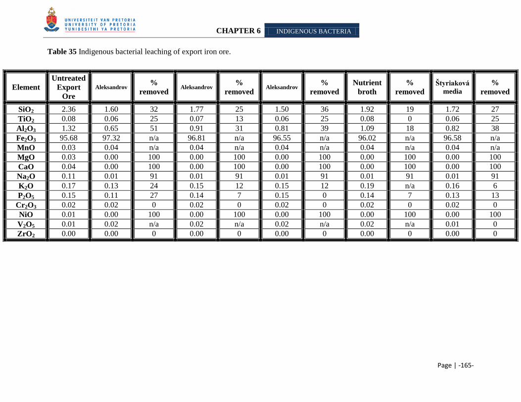
CHAPTER 6 INDIGENOUS BACTERIA
Page | -165-
Table 35 Indigenous bacterial leaching of export iron ore.
Element
Untreated
Export
Ore
Aleksandrov %
removed Aleksandrov
%
removed Aleksandrov
%
removed
Nutrient
broth
%
removed
Štyriaková
media
%
removed
SiO2 2.36 1.60 32 1.77 25 1.50 36 1.92 19 1.72 27
TiO2 0.08 0.06 25 0.07 13 0.06 25 0.08 0 0.06 25
Al2O3 1.32 0.65 51 0.91 31 0.81 39 1.09 18 0.82 38
Fe2O3 95.68 97.32 n/a 96.81 n/a 96.55 n/a 96.02 n/a 96.58 n/a
MnO 0.03 0.04 n/a 0.04 n/a 0.04 n/a 0.04 n/a 0.04 n/a
MgO 0.03 0.00 100 0.00 100 0.00 100 0.00 100 0.00 100
CaO 0.04 0.00 100 0.00 100 0.00 100 0.00 100 0.00 100
Na2O 0.11 0.01 91 0.01 91 0.01 91 0.01 91 0.01 91
K2O 0.17 0.13 24 0.15 12 0.15 12 0.19 n/a 0.16 6
P2O5 0.15 0.11 27 0.14 7 0.15 0 0.14 7 0.13 13
Cr2O3 0.02 0.02 0 0.02 0 0.02 0 0.02 0 0.02 0
NiO 0.01 0.00 100 0.00 100 0.00 100 0.00 100 0.00 100
V2O5 0.01 0.02 n/a 0.02 n/a 0.02 n/a 0.02 n/a 0.01 0
ZrO2 0.00 0.00 0 0.00 0 0.00 0 0.00 0 0.00 0
CHAPTER 6 INDIGENOUS BACTERIA
Page | -166-
KGT (conglomerate) samples were categorized as a high potassium/low phosphorous iron ore
samples according to B. Ntsoelengoe (Personal communication, 2006). Indigenous cultures from
the sample were enriched for by inoculating an unsterilized sample into Aleksandrov - (Hu et al.,
2006), Mikhailouskaya media and Nutrient broth (Biolab) (Table 36). Untreated KGT samples
(control) were analyzed in triplicate with XRF, to give a more accurate estimate of batch sample
(Table 19). Focus is placed mainly on the residual alkali, as these minerals have a deleterious
effect in steel manufacturing (Delvasto et al., 2008). Results of treated samples are expected to
exceed the control sample due to sample variation; therefore parameters were calculated to be
able to determine the amount of alkali/mineral removed due to microbial leaching and not
sample variation. Moreover, the effect on silicon and aluminum will also be investigated. Scott
and Amonette (1988) found that the micas biotite and muscovite undergo structural changes and
possible dissolution under low pH conditions or in the presence of an organic chelating agent.
Moreover, they also found that other cations are ejected along with potassium from the interlayer
of the mica.
From XRD analysis (Table 1), the minerals in which potassium occurs also includes aluminum,
therefore it is hypothesized that potassium or aluminum/silicon might be solubilized indirectly
due to the action of the acid on the other mineral, as discussed by Wilson (2004).
Mojallali and Weed (1978) stated that a loss of aluminum and/or silicon shows a structural
rearrangement of the mineral. Wilson and Jones (1983) found that a decrease in magnesium and
iron also shows a rearrangement of the mineral structure.
Therefore the following minerals and parameters for the export iron ore samples will be used
during this study: Al2O3 (1.90 - 2.14); SiO2 (3.90 – 4.90), K2O (0.48 – 0.56) and
P2O5 (0.06 – 0.07). Silicon was present in quartz, muscovite, biotite, etc. in all of the samples
analyzed (Table 1). Muscovite was detected as the main potassium bearing mineral, while no
phosphorous bearing mineral was identified. Concentrations of each mineral removed below
these parameters will be reported as ―large quantities‖ removed.
Untreated KGT samples (control) were subjected to X-ray powder diffraction (XRD) and X-Ray
fluorescence (XRF) to determine the quantitative and qualitative mineralogy of the iron ore
samples initially. Acid treated KGT samples were subjected to XRF analysis to determine the
residual mineral concentration after leaching (Table 16). The ―mineral phases‖ detected in the
CHAPTER 6 INDIGENOUS BACTERIA
Page | -167-
untreated sample included hematite, quartz and muscovite (Figure 14), however additional
phases, namely greenalite, illite, etc (Table 1) were reported by Kumba Iron Ore, Ltd. Jenkins
(1988) reported the lower detection limits of a wavelength dispersive spectrometer (Table 14),
thus sodium and magnesium will be excluded from further analysis. We accept the average
value of the untreated KGT sample as an acceptable representative (Table 16).
Potassium was detected as illite and muscovite in the KGT sample (Table 1). Illite is a non-
expanding, clay-sized phyllosilicate or layer alumino-silicate that commonly occurs in
sediments, soils and argillaceous sedimentary rocks. It is structurally similar to muscovite or
sericite [KAl2(OH)2(AlSi3O10)], with more silicon, magnesium, iron and water, with less
tetrahedral aluminum and interlayer potassium. Illite occurs as an alternation product of
muscovite and feldspar during weathering (Mengel and Uhlenbecker 1993).
XRF data obtained from the Aleksandrov media (Hu et al., 2006) were inconclusive, as some of
data suggests a removal of potassium and others not (Table 36). Rausell and coworkers (1965)
determined the sensitivity of muscovite to potassium concentration in solution. They discovered
that muscovite will not release potassium when it is placed in a dilute electrolyte solution.
Furthermore, Pal and coworkers (2001) demonstrated that if biotite and muscovite were both
present in soil, no potassium dissolution from muscovite would occur. It has been determined
that a 0.1mg/l potassium concentration in solution would inhibit the exchange of the alkali with
other ions in solution (vermiculation) (Wilson 2004). Therefore it is assumed that the presence
of illite and muscovite as potassium bearing minerals, inhibited/retarded the solubilization of the
alkali. Data obtained can also be ascribed to sample variation and/or mineral dissemination as
alkali was solubilized by the other experiments. With the former, there are varying amounts of
minerals in the sample which differs from the batch and therefore solubilization of some metals
from the sample would not apparent. In the latter, the minerals potassium/phosphorous might be
located mostly on the inside of the particle and therefore inaccessible for leaching. XRF analysis
of Mikhailouskaya media and Nutrient broth (Biolab), illustrated an inability of the microbial
cultures to remove potassium.
The KGT sample is a known low phosphorous ore, which is in accordance with our results. No
phosphorous bearing mineral could be determined. XRF data of the Nutrient broth (Biolab)
CHAPTER 6 INDIGENOUS BACTERIA
Page | -168-
experiment, reported that the cultures were able to remove a ―large quantity‖ of phosphorous.
Here the pH was more alkaline compared to the other media (Table 34). Microorganisms
possess mechanisms such as ammonification, denitrification, sulfate-reduction and
methanogenesis which can generate alkalinity (Johnson 2006), and therefore it is assumed that
the cultures enrich possess such a mechanism(s).
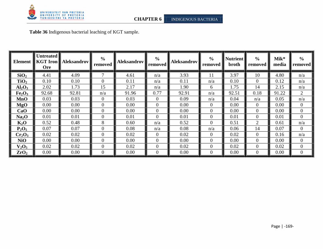
CHAPTER 6 INDIGENOUS BACTERIA
Page | -169-
Table 36 Indigenous bacterial leaching of KGT sample.
Element
Untreated
KGT Iron
Ore
Aleksandrov %
removed Aleksandrov
%
removed Aleksandrov
%
removed
Nutrient
broth
%
removed
Mik*
media
%
removed
SiO2 4.41 4.09 7 4.61 n/a 3.93 11 3.97 10 4.80 n/a
TiO2 0.10 0.10 0 0.11 n/a 0.11 n/a 0.10 0 0.12 n/a
Al2O3 2.02 1.73 15 2.17 n/a 1.90 6 1.75 14 2.15 n/a
Fe2O3 92.68 92.81 n/a 91.96 0.77 92.91 n/a 92.51 0.18 91.22 2
MnO 0.03 0.03 0 0.03 0 0.09 n/a 0.04 n/a 0.05 n/a
MgO 0.00 0.00 0 0.00 0 0.00 0 0.00 0 0.00 0
CaO 0.00 0.00 0 0.00 0 0.00 0 0.00 0 0.00 0
Na2O 0.01 0.01 0 0.01 0 0.01 0 0.01 0 0.01 0
K2O 0.52 0.48 8 0.60 n/a 0.52 0 0.51 2 0.61 n/a
P2O5 0.07 0.07 0 0.08 n/a 0.08 n/a 0.06 14 0.07 0
Cr2O3 0.02 0.02 0 0.02 0 0.02 0 0.02 0 0.16 n/a
NiO 0.00 0.00 0 0.00 0 0.00 0 0.00 0 0.00 0
V2O5 0.02 0.02 0 0.02 0 0.02 0 0.02 0 0.02 0
ZrO2 0.00 0.00 0 0.00 0 0.00 0 0.00 0 0.00 0
CHAPTER 6 INDIGENOUS BACTERIA
Page | -170-
SK samples (shale) are high potassium/low phosphorous iron ore. Indigenous bacteria with were
amplified by inoculating Aleksandrov -, Mikhailouskaya-, Štyriaková media and Nutrient broth
(Biolab) with unsterilized SK sample.
Mineral phases detected with XRD included muscovite, hematite and quartz (Figure 15).
Additional phases were reported by Kumba staff (Table 1). Potassium was present as biotite,
muscovite and illite. Biotite is a ferromagnesium mineral with compositional affinity to olivine
[(Mg,Fe)2SiO4], pyroxene [XY(Si,Al)2O6]35 and amphiboles36 (Deer et al., 2001). As with other
mica minerals, it has a highly perfect basal cleavage and consists of flexible sheets or lamellae37,
which can easily flake off. Calvaruso et al. (2006) stated that biotite can undergo two types of
weathering namely: congruent (destruction of the mineral surface) and incongruent
(transformation into vermiculite by release of interlayer potassium). The sheets in the crystal
structure of biotite are made up of iron, magnesium, aluminum, silicon, oxygen and hydrogen
ions that are weakly bound together by potassium. The potassium can be substituted in part by
sodium, calcium, barium. Magnesium can be completely replaced by ferrous iron and ferric iron
and in part by titanium and manganese. Biotite is often referred to as the iron mica because it
contains more iron than phlogopite (Hashemi-Nezhad 2005). No phosphorous phases were
detected.
The following parameters for the alkalis and mineral described by Mojallali and Weed (1978)
and Wilson and Jones (1983) were calculated from XRF data of untreated SK sample:
Al2O3 (9.69 – 10.17); SiO2 (11.56 – 12.26); K2O (2.31 – 2.43) and P2O5 (0.03 – 0.04). These
results are in accordance with classification by Kumba Iron Ore, Ltd. staff. Concentrations of
each mineral removed below these parameters will be reported as ―large quantities‖ removed.
Alkali removal from the ore was determined by measuring the residual contained in the ore after
treatment and comparing it to the untreated sample (control). Jenkins (1988) reported the lower
detection limits of a wavelength dispersive spectrometer (Table 14), therefore sodium will be
35
X represents calcium, sodium, ferrous iron, magnesium, zinc, manganese and lithium. Y represents ions such as
chromium, aluminium, ferric iron, magnesium, manganese, scandium, titanium, vanadium.
(http://en.wikipedia.org/wiki/Pyroxene). 36
The main differences between amphiboles and pyroxenes are that amphiboles contain essential hydroxyl (OH) or
halogene (F, Cl) and secondly the basic structure is a double chain of tetrahedra (as opposed to the single chain
structure of pyroxene). (http://en.wikipedia.org/wiki/Amphiboles). 37
A lamella is a gill-shaped structure: fine sheets of material held adjacent one another, with fluid in-between-(or
simply 'welded'-plates)[ http://en.wikipedia.org/wiki/Lamellae_(materials)].
CHAPTER 6 INDIGENOUS BACTERIA
Page | -171-
excluded from further analysis. We accept the average value of the untreated SK sample as an
acceptable representative (Table 37).
None of the potassium and phosphorous could be solubilized from the SK sample by the strains
amplified using the various media. This is ascribed to the intrinsic characteristic of the mineral
(Ballester et al., 1989; Das et al., 1999 as cited in Brandl 2001). The SK sample contains illite,
biotite and muscovite as potassium bearing minerals. Rausell and coworkers (1965)
demonstrated that the dissolution of muscovite is controlled by the concentration of potassium in
solution. Wilson (2004) determined that a 0.1mg/l potassium concentration in solution would
inhibit the exchange of the alkali with other cations in solution. The diffusion of biotite is
thought to be diffusion-controlled and therefore also depends on the potassium concentration in
the bulk solution (Wilson 2004). Pal et al. (2001) found that if biotite and muscovite were both
present in soil, no potassium dissolution from muscovite would occur. We assume that a
saturation state for potassium was reached in the solutions, and therefore none could be
mobilized.
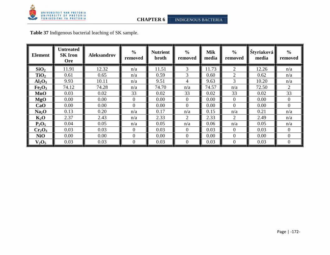
CHAPTER 6 INDIGENOUS BACTERIA
Page | -172-
Table 37 Indigenous bacterial leaching of SK sample.
Element
Untreated
SK Iron
Ore
Aleksandrov %
removed
Nutrient
broth
%
removed
Mik
media
%
removed
Štyriaková
media
%
removed
SiO2 11.91 12.32 n/a 11.51 3 11.73 2 12.26 n/a
TiO2 0.61 0.65 n/a 0.59 3 0.60 2 0.62 n/a
Al2O3 9.93 10.11 n/a 9.51 4 9.63 3 10.20 n/a
Fe2O3 74.12 74.28 n/a 74.70 n/a 74.57 n/a 72.50 2
MnO 0.03 0.02 33 0.02 33 0.02 33 0.02 33
MgO 0.00 0.00 0 0.00 0 0.00 0 0.00 0
CaO 0.00 0.00 0 0.00 0 0.00 0 0.00 0
Na2O 0.13 0.20 n/a 0.17 n/a 0.15 n/a 0.21 n/a
K2O 2.37 2.43 n/a 2.33 2 2.33 2 2.49 n/a
P2O5 0.04 0.05 n/a 0.05 n/a 0.06 n/a 0.05 n/a
Cr2O3 0.03 0.03 0 0.03 0 0.03 0 0.03 0
NiO 0.00 0.00 0 0.00 0 0.00 0 0.00 0
V2O5 0.03 0.03 0 0.03 0 0.03 0 0.03 0
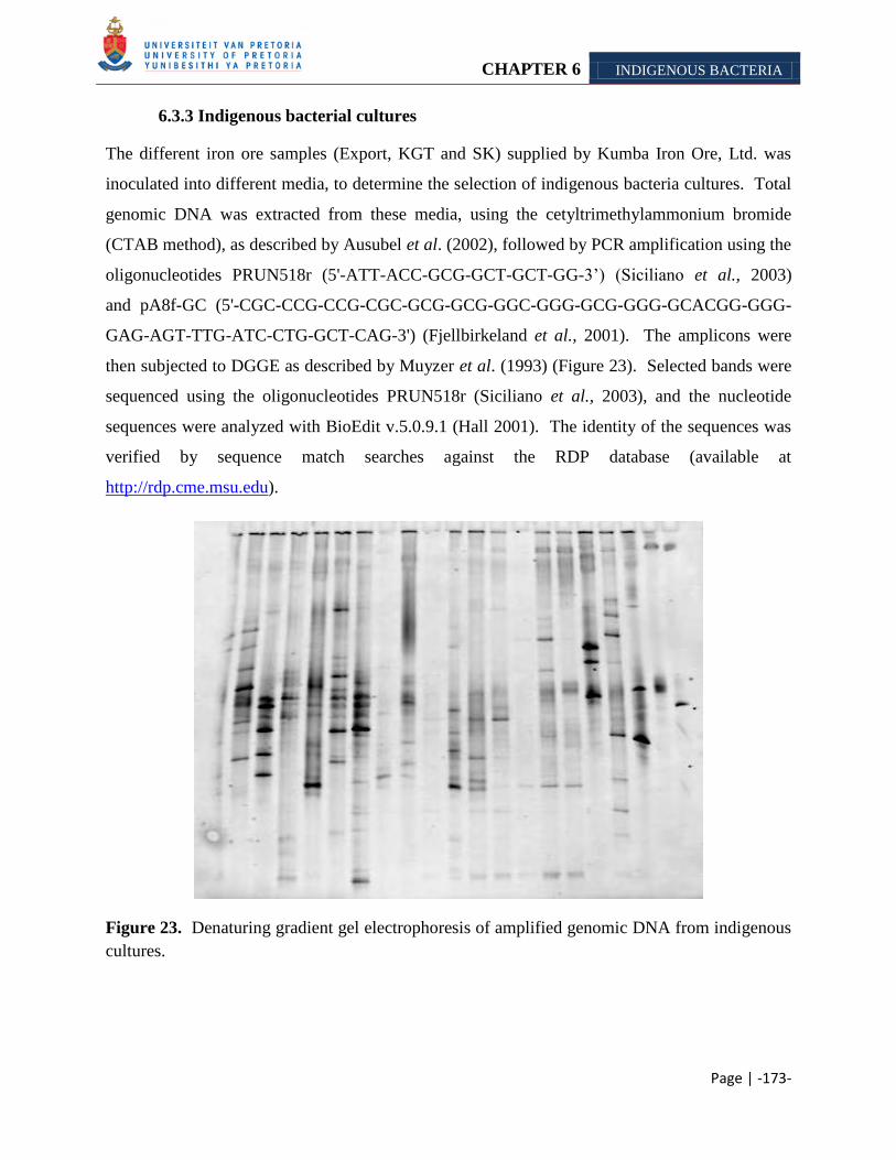
CHAPTER 6 INDIGENOUS BACTERIA
Page | -173-
6.3.3 Indigenous bacterial cultures
The different iron ore samples (Export, KGT and SK) supplied by Kumba Iron Ore, Ltd. was
inoculated into different media, to determine the selection of indigenous bacteria cultures. Total
genomic DNA was extracted from these media, using the cetyltrimethylammonium bromide
(CTAB method), as described by Ausubel et al. (2002), followed by PCR amplification using the
oligonucleotides PRUN518r (5'-ATT-ACC-GCG-GCT-GCT-GG-3‘) (Siciliano et al., 2003)
and pA8f-GC (5'-CGC-CCG-CCG-CGC-GCG-GCG-GGC-GGG-GCG-GGG-GCACGG-GGG-
GAG-AGT-TTG-ATC-CTG-GCT-CAG-3') (Fjellbirkeland et al., 2001). The amplicons were
then subjected to DGGE as described by Muyzer et al. (1993) (Figure 23). Selected bands were
sequenced using the oligonucleotides PRUN518r (Siciliano et al., 2003), and the nucleotide
sequences were analyzed with BioEdit v.5.0.9.1 (Hall 2001). The identity of the sequences was
verified by sequence match searches against the RDP database (available at
http://rdp.cme.msu.edu).
Figure 23. Denaturing gradient gel electrophoresis of amplified genomic DNA from indigenous
cultures.
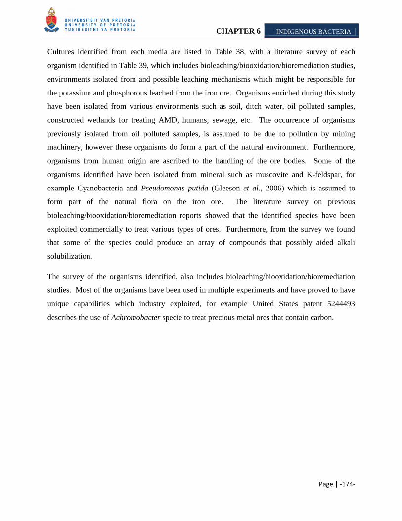
CHAPTER 6 INDIGENOUS BACTERIA
Page | -174-
Cultures identified from each media are listed in Table 38, with a literature survey of each
organism identified in Table 39, which includes bioleaching/biooxidation/bioremediation studies,
environments isolated from and possible leaching mechanisms which might be responsible for
the potassium and phosphorous leached from the iron ore. Organisms enriched during this study
have been isolated from various environments such as soil, ditch water, oil polluted samples,
constructed wetlands for treating AMD, humans, sewage, etc. The occurrence of organisms
previously isolated from oil polluted samples, is assumed to be due to pollution by mining
machinery, however these organisms do form a part of the natural environment. Furthermore,
organisms from human origin are ascribed to the handling of the ore bodies. Some of the
organisms identified have been isolated from mineral such as muscovite and K-feldspar, for
example Cyanobacteria and Pseudomonas putida (Gleeson et al., 2006) which is assumed to
form part of the natural flora on the iron ore. The literature survey on previous
bioleaching/biooxidation/bioremediation reports showed that the identified species have been
exploited commercially to treat various types of ores. Furthermore, from the survey we found
that some of the species could produce an array of compounds that possibly aided alkali
solubilization.
The survey of the organisms identified, also includes bioleaching/biooxidation/bioremediation
studies. Most of the organisms have been used in multiple experiments and have proved to have
unique capabilities which industry exploited, for example United States patent 5244493
describes the use of Achromobacter specie to treat precious metal ores that contain carbon.
CHAPTER 6 INDIGENOUS BACTERIA
Page | -175-
Table 38 Indigenous bacteria identified from study.
Sample Media Culture
Export Iron Ore Aleksandrov media (Hu et al,. 2006) Acetobacter specie
Achromobacter specie
Achromobacter xylosoxidans
Arthrobacter agilis
Citrobacter specie
Paenibacillus stellifer
Nutrient broth (Biolab) Acinetobacter junii
Comamonas specie
Pantoea agglomerans
Štyriaková media
(Štyriaková et al., 2004)
Bacillus specie
Burkholderia specie
Cyanobacterium
Pseudomonas putida
KGT sample Aleksandrov media (Hu et al., 2006) Acetobacter species
Burkholderia species
Paenibacillus stellifer
Nutrient broth (Biolab) Acinetobacter jejunni
Micrococcus luteus
Sphingomonas specie
Mikhailouskaya media
(Mikhailouskaya et al. 2005)
Alcaligenes faecalis
Citrobacter specie
Sphingomonas specie
SK sample Aleksandrov media (Hu et al., 2006) Arthrobacter specie
Citrobacter specie
Paenibacillus specie
Nutrient broth (Biolab) Acinetobacter junii
Burkholderia specie
Cyanobacterium
Stenotrophomonas specie
Mikhailouskaya media
(Mikhailouskaya et al. 2005)
Bacillus specie
Brevundi diminuta
Citrobacter specie
Paenibacillus specie
Štyriaková media (Štyriaková et al.,
2004)
Bacillus specie
Clostridium species
Stenotrophomonas
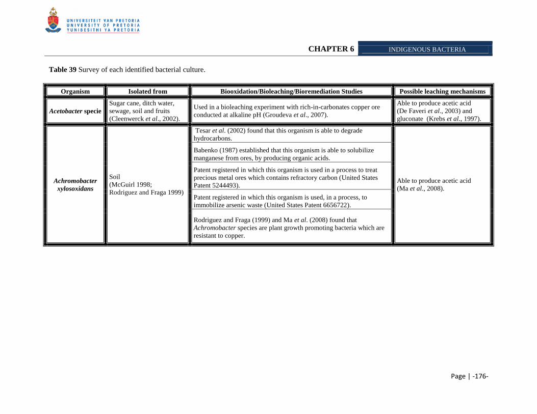
CHAPTER 6 INDIGENOUS BACTERIA
Page | -176-
Table 39 Survey of each identified bacterial culture.
Organism Isolated from Biooxidation/Bioleaching/Bioremediation Studies Possible leaching mechanisms
Acetobacter specie
Sugar cane, ditch water,
sewage, soil and fruits
(Cleenwerck et al., 2002).
Used in a bioleaching experiment with rich-in-carbonates copper ore
conducted at alkaline pH (Groudeva et al., 2007).
Able to produce acetic acid
(De Faveri et al., 2003) and
gluconate (Krebs et al., 1997).
Achromobacter
xylosoxidans
Soil
(McGuirl 1998;
Rodriguez and Fraga 1999)
Tesar et al. (2002) found that this organism is able to degrade
hydrocarbons.
Able to produce acetic acid
(Ma et al., 2008).
Babenko (1987) established that this organism is able to solubilize
manganese from ores, by producing organic acids.
Patent registered in which this organism is used in a process to treat
precious metal ores which contains refractory carbon (United States
Patent 5244493).
Patent registered in which this organism is used, in a process, to
immobilize arsenic waste (United States Patent 6656722).
Rodriguez and Fraga (1999) and Ma et al. (2008) found that
Achromobacter species are plant growth promoting bacteria which are
resistant to copper.
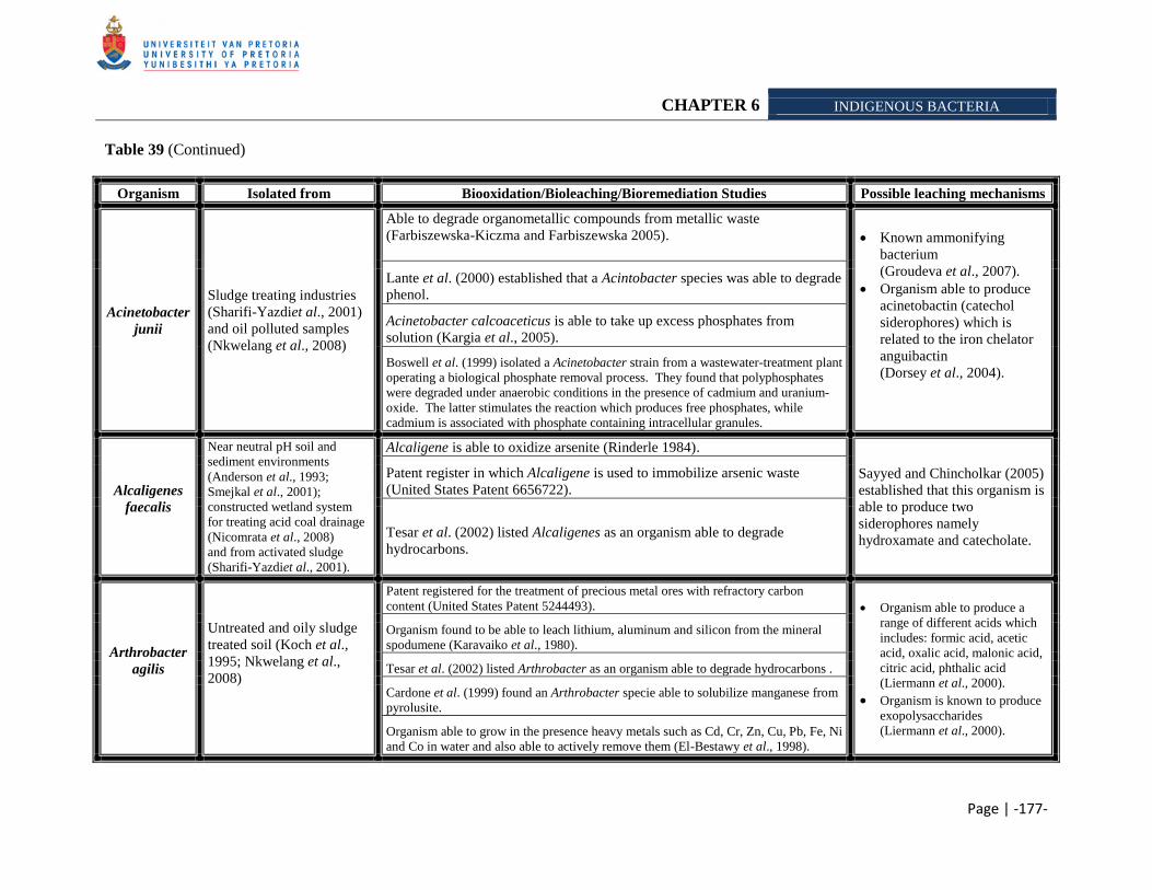
CHAPTER 6 INDIGENOUS BACTERIA
Page | -177-
Table 39 (Continued)
Organism Isolated from Biooxidation/Bioleaching/Bioremediation Studies Possible leaching mechanisms
Acinetobacter
junii
Sludge treating industries
(Sharifi-Yazdiet al., 2001)
and oil polluted samples
(Nkwelang et al., 2008)
Able to degrade organometallic compounds from metallic waste
(Farbiszewska-Kiczma and Farbiszewska 2005).
Known ammonifying
bacterium
(Groudeva et al., 2007).
Organism able to produce
acinetobactin (catechol
siderophores) which is
related to the iron chelator
anguibactin
(Dorsey et al., 2004).
Lante et al. (2000) established that a Acintobacter species was able to degrade
phenol.
Acinetobacter calcoaceticus is able to take up excess phosphates from
solution (Kargia et al., 2005).
Boswell et al. (1999) isolated a Acinetobacter strain from a wastewater-treatment plant
operating a biological phosphate removal process. They found that polyphosphates
were degraded under anaerobic conditions in the presence of cadmium and uranium-
oxide. The latter stimulates the reaction which produces free phosphates, while
cadmium is associated with phosphate containing intracellular granules.
Alcaligenes
faecalis
Near neutral pH soil and
sediment environments
(Anderson et al., 1993;
Smejkal et al., 2001);
constructed wetland system
for treating acid coal drainage
(Nicomrata et al., 2008)
and from activated sludge
(Sharifi-Yazdiet al., 2001).
Alcaligene is able to oxidize arsenite (Rinderle 1984).
Sayyed and Chincholkar (2005)
established that this organism is
able to produce two
siderophores namely
hydroxamate and catecholate.
Patent register in which Alcaligene is used to immobilize arsenic waste
(United States Patent 6656722).
Tesar et al. (2002) listed Alcaligenes as an organism able to degrade
hydrocarbons.
Arthrobacter
agilis
Untreated and oily sludge
treated soil (Koch et al.,
1995; Nkwelang et al.,
2008)
Patent registered for the treatment of precious metal ores with refractory carbon
content (United States Patent 5244493). Organism able to produce a
range of different acids which
includes: formic acid, acetic
acid, oxalic acid, malonic acid,
citric acid, phthalic acid
(Liermann et al., 2000).
Organism is known to produce
exopolysaccharides
(Liermann et al., 2000).
Organism found to be able to leach lithium, aluminum and silicon from the mineral
spodumene (Karavaiko et al., 1980).
Tesar et al. (2002) listed Arthrobacter as an organism able to degrade hydrocarbons .
Cardone et al. (1999) found an Arthrobacter specie able to solubilize manganese from
pyrolusite.
Organism able to grow in the presence heavy metals such as Cd, Cr, Zn, Cu, Pb, Fe, Ni
and Co in water and also able to actively remove them (El-Bestawy et al., 1998).
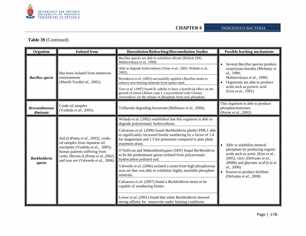
CHAPTER 6 INDIGENOUS BACTERIA
Page | -178-
Table 39 (Continued)
Organism Isolated from Biooxidation/Bioleaching/Bioremediation Studies Possible leaching mechanisms
Bacillus specie
Has been isolated from numerous
environments
(Sharifi-Yazdiet al., 2001).
Bacillus species are able to solubilize silicate (Ehrlich 1991;
Malinovskaya et al., 1990). Several Bacillus species produce
exopolysaccharides (Mohanty et
al., 1990;
Malinovskaya et al., 1990)
Organisms are able to produce
acids such as pyruvic acid
(Urzi et al., 1991).
Able to degrade hydrocarbons (Tesar et al., 2002; Widada et al.,
2002).
Štyriaková et al. (2003) successfully applied a Bacillus strain to
remove iron bearing minerals from quartz sand.
Toro et al. (1997) found B. subtilis to have a beneficial effect on the
growth of onion (Allium cepa L.) mycorrhized with Glomus
intraradices via the release of phosphate from rock phosphate.
Brevundimonas
diminuta
Crude oil samples
(Yoshida et al., 2005). Trifluralin degrading bacterium (Bellinaso et al., 2006).
This organism is able to produce
phosphotriesterases
(Horne et al., 2002).
Burkholderia
specie
Soil (LiPuma et al., 2002), crude-
oil samples from Japanese oil
stockpiles (Yoshida et al., 2005);
human patients suffering from
cystic fibrosis (LiPuma et al.,2002)
and iron ore (Valverde et al., 2006)
Widada et al. (2002) established that this organism is able to
degrade polyaromatic hydrocarbons.
Able to solubilize mineral
phosphate by producing organic
acids such as acetic (Kim et al.,
2005), citric (Delvasto et al.,
2008b) and gluconic acid (Lin et
al., 2006)
Known to produce biofilms
(Delvasto et al., 2008)
Calvaruso et al. (2006) found Burkholderia glathei PML1 able
to significantly increased biotite weathering by a factor of 1.4
for magnesium and 1.5 for potassium compared to pine plant
treatment alone.
O‘Sullivan and Mahenthiralingam (2005) found Burkholderia
to be the predominant genus isolated from polyaromatic
hydrocarbon polluted soil.
Valverde et al. (2006) isolated a strain from high phosphorous
iron ore that was able to solubilize highly insoluble phosphate
minerals.
Calvaruso et al. (2007) found a Burkholderia strain to be
capable of weathering biotite.
Lower et al. (2001) found that when Burkholderia showed
strong affinity for muscovite under limiting conditions.
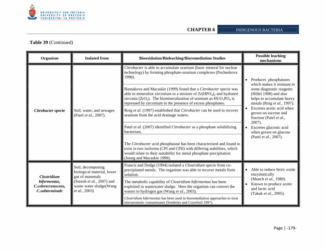
CHAPTER 6 INDIGENOUS BACTERIA
Page | -179-
Table 39 (Continued)
Organism Isolated from Biooxidation/Bioleaching/Bioremediation Studies Possible leaching
mechanisms
Citrobacter specie
Soil, water, and sewages
(Patel et al., 2007).
Citrobacter is able to accumulate uranium (basic mineral for nuclear
technology) by forming phosphate-uranium complexes (Puchenkova
1996).
Produces phosphatases
which makes it resistant to
some diagnostic reagents
(Hillel 1998) and also
helps to accumulate heavy
metals (Roig et al., 1997).
Excretes acetic acid when
grown on sucrose and
fructose (Patel et al.,
2007).
Excretes gluconic acid
when grown on glucose
(Patel et al., 2007).
Basnakova and Macaskie (1999) found that a Citrobacter specie was
able to mineralize zirconium to a mixture of Zr(HPO4)2 and hydrated
zirconia (ZrO2). The biomineralization of uranium as HUO2PO4 is
repressed by zirconium in the presence of excess phosphates.
Roig et al. (1997) established that Citrobacter can be used to recover
uranium from the acid drainage waters.
Patel et al. (2007) identified Citrobacter as a phosphate solubilizing
bacterium.
The Citrobacter acid phosphatase has been characterized and found to
exist in two isoforms (CPI and CPII) with differing stabilities, which
would relate to their suitability for metal phosphate precipitation
(Jeong and Macaskie 1999).
Clostridium
bifermentas,
C.celerecrenscens,
C.subterminale
Soil, decomposing
biological material, lower
gut of mammals
(Suresh et al., 2007) and
waste water sludge(Wang
et al., 2003)
Francis and Dodge (1994) isolated a Clostridium specie from co-
precipitated metals. The organism was able to recover metals from
solution.
Able to reduce ferric oxide
enzymatically
(Munch et al., 1980).
Known to produce acetic
and lactic acid
(Tabak et al., 2005).
The metabolic capability of Clostridium bifermentas has been
exploited in wastewater sludge. Here the organism can convert the
wastes to hydrogen gas (Wang et al., 2003).
Clostridium bifermentas has been used in bioremediation approaches to treat
nitroaromotic contaminants (Sembries and Crawford 1997).
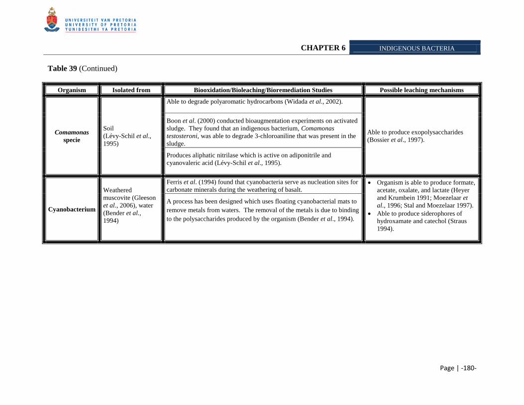
CHAPTER 6 INDIGENOUS BACTERIA
Page | -180-
Table 39 (Continued)
Organism Isolated from Biooxidation/Bioleaching/Bioremediation Studies Possible leaching mechanisms
Comamonas
specie
Soil
(Lévy-Schil et al.,
1995)
Able to degrade polyaromatic hydrocarbons (Widada et al., 2002).
Able to produce exopolysaccharides
(Bossier et al., 1997).
Boon et al. (2000) conducted bioaugmentation experiments on activated
sludge. They found that an indigenous bacterium, Comamonas
testosteroni, was able to degrade 3-chloroaniline that was present in the
sludge.
Produces aliphatic nitrilase which is active on adiponitrile and
cyanovaleric acid (Lévy-Schil et al., 1995).
Cyanobacterium
Weathered
muscovite (Gleeson
et al., 2006), water
(Bender et al.,
1994)
Ferris et al. (1994) found that cyanobacteria serve as nucleation sites for
carbonate minerals during the weathering of basalt. Organism is able to produce formate,
acetate, oxalate, and lactate (Heyer
and Krumbein 1991; Moezelaar et
al., 1996; Stal and Moezelaar 1997).
Able to produce siderophores of
hydroxamate and catechol (Straus
1994).
A process has been designed which uses floating cyanobacterial mats to
remove metals from waters. The removal of the metals is due to binding
to the polysaccharides produced by the organism (Bender et al., 1994).
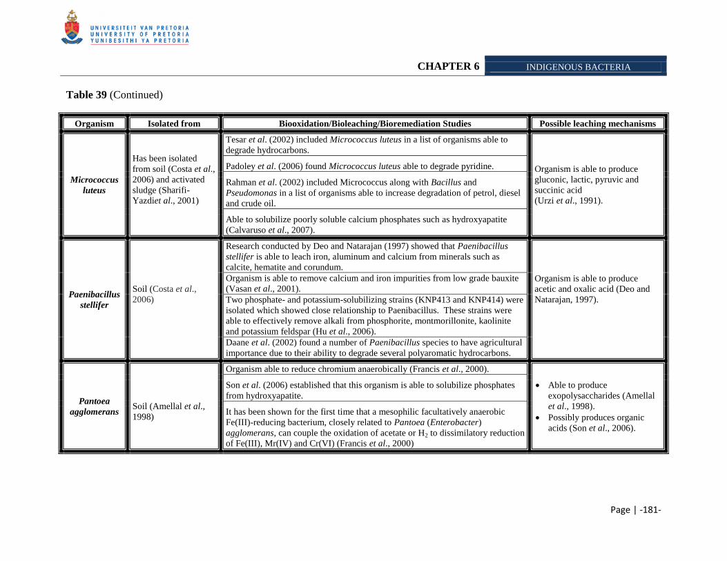
CHAPTER 6 INDIGENOUS BACTERIA
Page | -181-
Table 39 (Continued)
Organism Isolated from Biooxidation/Bioleaching/Bioremediation Studies Possible leaching mechanisms
Micrococcus
luteus
Has been isolated
from soil (Costa et al.,
2006) and activated
sludge (Sharifi-
Yazdiet al., 2001)
Tesar et al. (2002) included Micrococcus luteus in a list of organisms able to
degrade hydrocarbons.
Organism is able to produce
gluconic, lactic, pyruvic and
succinic acid
(Urzi et al., 1991).
Padoley et al. (2006) found Micrococcus luteus able to degrade pyridine.
Rahman et al. (2002) included Micrococcus along with Bacillus and
Pseudomonas in a list of organisms able to increase degradation of petrol, diesel
and crude oil.
Able to solubilize poorly soluble calcium phosphates such as hydroxyapatite
(Calvaruso et al., 2007).
Paenibacillus
stellifer
Soil (Costa et al.,
2006)
Research conducted by Deo and Natarajan (1997) showed that Paenibacillus
stellifer is able to leach iron, aluminum and calcium from minerals such as
calcite, hematite and corundum.
Organism is able to produce
acetic and oxalic acid (Deo and
Natarajan, 1997).
Organism is able to remove calcium and iron impurities from low grade bauxite
(Vasan et al., 2001).
Two phosphate- and potassium-solubilizing strains (KNP413 and KNP414) were
isolated which showed close relationship to Paenibacillus. These strains were
able to effectively remove alkali from phosphorite, montmorillonite, kaolinite
and potassium feldspar (Hu et al., 2006).
Daane et al. (2002) found a number of Paenibacillus species to have agricultural
importance due to their ability to degrade several polyaromatic hydrocarbons.
Pantoea
agglomerans
Soil (Amellal et al.,
1998)
Organism able to reduce chromium anaerobically (Francis et al., 2000).
Able to produce
exopolysaccharides (Amellal
et al., 1998).
Possibly produces organic
acids (Son et al., 2006).
Son et al. (2006) established that this organism is able to solubilize phosphates
from hydroxyapatite.
It has been shown for the first time that a mesophilic facultatively anaerobic
Fe(III)-reducing bacterium, closely related to Pantoea (Enterobacter)
agglomerans, can couple the oxidation of acetate or H2 to dissimilatory reduction
of Fe(III), Mr(IV) and Cr(VI) (Francis et al., 2000)
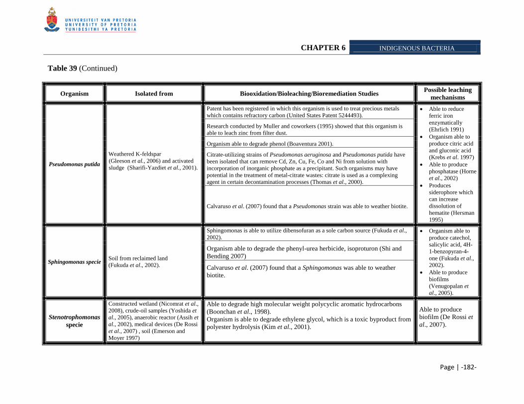
CHAPTER 6 INDIGENOUS BACTERIA
Page | -182-
Table 39 (Continued)
Organism Isolated from Biooxidation/Bioleaching/Bioremediation Studies Possible leaching
mechanisms
Pseudomonas putida
Weathered K-feldspar
(Gleeson et al., 2006) and activated
sludge (Sharifi-Yazdiet et al., 2001).
Patent has been registered in which this organism is used to treat precious metals
which contains refractory carbon (United States Patent 5244493). Able to reduce
ferric iron
enzymatically
(Ehrlich 1991)
Organism able to
produce citric acid
and gluconic acid
(Krebs et al. 1997)
Able to produce
phosphatase (Horne
et al., 2002)
Produces
siderophore which
can increase
dissolution of
hematite (Hersman
1995)
Research conducted by Muller and coworkers (1995) showed that this organism is
able to leach zinc from filter dust.
Organism able to degrade phenol (Boaventura 2001).
Citrate-utilizing strains of Pseudomonas aeruginosa and Pseudomonas putida have
been isolated that can remove Cd, Zn, Cu, Fe, Co and Ni from solution with
incorporation of inorganic phosphate as a precipitant. Such organisms may have
potential in the treatment of metal-citrate wastes: citrate is used as a complexing
agent in certain decontamination processes (Thomas et al., 2000).
Calvaruso et al. (2007) found that a Pseudomonas strain was able to weather biotite.
Sphingomonas specie Soil from reclaimed land
(Fukuda et al., 2002).
Sphingomonas is able to utilize dibensofuran as a sole carbon source (Fukuda et al.,
2002). Organism able to
produce catechol,
salicylic acid, 4H-
1-benzopyran-4-
one (Fukuda et al.,
2002).
Able to produce
biofilms
(Venugopalan et
al., 2005).
Organism able to degrade the phenyl-urea herbicide, isoproturon (Shi and
Bending 2007)
Calvaruso et al. (2007) found that a Sphingomonas was able to weather
biotite.
Stenotrophomonas
specie
Constructed wetland (Nicomrat et al.,
2008), crude-oil samples (Yoshida et
al., 2005), anaerobic reactor (Assih et
al., 2002), medical devices (De Rossi
et al., 2007) , soil (Emerson and
Moyer 1997)
Able to degrade high molecular weight polycyclic aromatic hydrocarbons
(Boonchan et al., 1998).
Organism is able to degrade ethylene glycol, which is a toxic byproduct from
polyester hydrolysis (Kim et al., 2001).
Able to produce
biofilm (De Rossi et
al., 2007).
CHAPTER 6 INDIGENOUS BACTERIA
Page | -183-
For each of the enrichment medias different organims were identified. From the literature survey
we identified several species, which is known to possess an alkali-solubilizing mechanism or
have shown to be effective in mineral solubilization, for example: Organisms from export iron
ore inoculated Aleksandrov media (Hu et al., 2006) included Acetobacter, Achromobacter,
Arthrobacter and Paenibacillus which could produce an array of organic acids such as acetic -,
oxalic -, formic -, malonic -, citric – and phthalic acid, which could leach the minerals via
protons and/or complexation mechanism. Moreover, Arthrobacter is able to produce
exopolysaccharides (Liermann et al., 2000), which could further aid solubilization of metals by
binding with them and therefore decrease the saturation state (Delvasto et al., 2008;
Tempest and Neijssel 1992). Citrobacter, a known phosphate solubilizing bacterium, was also
identified in the Aleksandrov media which could produce gluconic acid from the carbon source
supplied by the media (Patel et al., 2007). This organism was also isolated from
Mikhailouskaya media, where it was possibly able to produce acetic acid due to the carbon
source present (Patel et al., 2007). Furthermore, this organism could aid solubilization of
phosphorous by excreting phosphatases which can accumulate heavy metals (Roig et al., 1997).
Paenibacillus was also identified in the Aleksandrov media. Hu and coworkers (2006) identified
two potassium/phosphorous solubilizing bacteria which showed close relationship to
Paenibacillus. They further demonstrated that the organisms were able to remove alkali from
phosphorite, montmorillonite, kaolinite and K-feldspar. Therefore it is possible that this
organism played a vital role in the solubilization of alkali from the ore samples.
Acinetobacter junii were identified in the KGT and export iron ore inoculated nutrient broth.
This is an ammonifying bacterium. We found that the pH of the nutrient broth increased during
our study, therefore we assume that this is due to the presence of Acinetobacter junii
(Groudeva et al., 2007). The cocktail of organisms present in the Nutrient broth (Biolab) could
mobilize of phosphorous from export (Table 35) and KGT (Table 36) samples, possibly due to
Acinetobacter junii ability to take up excess phosphates in solution (Kargi et al., 2005), thereby
affecting the saturation state in solution.
Bacillus species were identified from export and SK inoculated Štyriaková media. Research has
shown that these organisms were able to solubilize silicates (Ehrlich 1991;
Malinovskaya et al., 1990). From research (Chapter 5), we found that these organisms are able
CHAPTER 6 INDIGENOUS BACTERIA
Page | -184-
to solubilize alkalis from the export ore (Table 28), but not from the SK sample (Table 37). This
is ascribed to the composition of the sample, as it contains illite, muscovite and biotite as
previously discussed. Clostridium and Stenotrophomonas species were also isolated from the
SK sample (Table 38). Clostridium is known to be a obligative anaerobe, and therefore we
assume that the organism produced biofilms to protect itself. Furthermore Tobak et al. (2005)
reported that these organisms are able to produce acetic – and lactic acid. Therefore the
measured decrease in pH and alkali solubilization is ascribed to the metabolic products of the
organism.
Burkholderia species were identified from Export iron ore inoculated Štyriaková media
(Table 35); KGT inoculated Aleksandrov media (Table 36) and SK inoculated Nutrient Broth
(Table 37). These organisms produce biofilms which could influence the saturation state in
solution (Delvasto et al., 2008). Reports indicated that these organisms are able to solubilize
phosphorous from biotite (Calvaruso et al., 2006) and iron ore (Valverde et al., 2006), possibly
by producing acids such as acetic - (Kim et al., 2005), citric – (Delvasto et al., 2008b) and
gluconic acid (Lin et al,. 2006). Therefore it is thought that this organism played an important
role in alkali solubilization for the export and KGT ore samples. Its inability to remove alkali
from the SK sample is ascribed to the intrinsic characteristics of the sample and to the presence
of Acinetobacter junii which increased the solution pH, which would allow the acids to only
form complexes with the metals.
Micrococcus luteus was isolated from KGT inoculated Nutrient broth (Table 38). Calvaruso et
al. (2007) demonstrated that this organism is able to solubilize poorly soluble calcium phosphate
such as hydroxylapatite. The pH of the nutrient broth was alkaline (Table 34), possibly due to
the presence of Acinetobacter junii, however some of the alkali could be solubilized from the ore
sample possibly due to the metabolic action of the organisms present (Table 36).
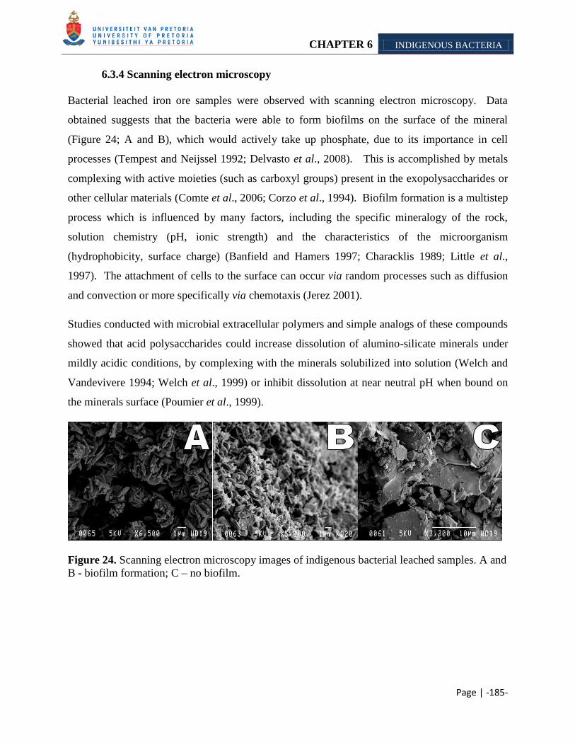
CHAPTER 6 INDIGENOUS BACTERIA
Page | -185-
6.3.4 Scanning electron microscopy
Bacterial leached iron ore samples were observed with scanning electron microscopy. Data
obtained suggests that the bacteria were able to form biofilms on the surface of the mineral
(Figure 24; A and B), which would actively take up phosphate, due to its importance in cell
processes (Tempest and Neijssel 1992; Delvasto et al., 2008). This is accomplished by metals
complexing with active moieties (such as carboxyl groups) present in the exopolysaccharides or
other cellular materials (Comte et al., 2006; Corzo et al., 1994). Biofilm formation is a multistep
process which is influenced by many factors, including the specific mineralogy of the rock,
solution chemistry (pH, ionic strength) and the characteristics of the microorganism
(hydrophobicity, surface charge) (Banfield and Hamers 1997; Characklis 1989; Little et al.,
1997). The attachment of cells to the surface can occur via random processes such as diffusion
and convection or more specifically via chemotaxis (Jerez 2001).
Studies conducted with microbial extracellular polymers and simple analogs of these compounds
showed that acid polysaccharides could increase dissolution of alumino-silicate minerals under
mildly acidic conditions, by complexing with the minerals solubilized into solution (Welch and
Vandevivere 1994; Welch et al., 1999) or inhibit dissolution at near neutral pH when bound on
the minerals surface (Poumier et al., 1999).
Figure 24. Scanning electron microscopy images of indigenous bacterial leached samples. A and
B - biofilm formation; C – no biofilm.
CHAPTER 6 INDIGENOUS BACTERIA
Page | -186-
Scanning electron microscopy of Nutrient broth (Biolab) samples revealed that no biofilms were
formed (Figure 24 C). Chan and Ting (1995) found that a decrease in pH leads to an increase in
negative charges on bacterial surfaces with subsequent attraction to the positively charged metal
ions. Therefore we assume that the opposite is true for the Nutrient broth (Biolab) cultures,
which would hinder attachment of cells on the mineral surface.
6.4 CONCLUSION
Microorganisms play an important role: in the cycling of elements and sorption of metals, the
dissolution of minerals and mineral crystallization. They are able to achieve this via mechanisms
such as: assimilation/adsorption and mineralization; precipitation and dissolution; oxidation and
reduction and methylation and dealkylation. On the other hand, research has found that
mineralogical process in turn play a significant role in the distribution, activity and diversity of
microbes, the expression of their genes, development and structure of communities, and transfer
of genetic material. The iron ore mined at Sishen, Northern Cape, South Africa contains several
different minerals, with potassium and phosphorous as common constituents. The latter minerals
have a deleterious effect on the manufacturing of steel. Experiments by Bennett et al. (2001)
provided evidence that silicate weathering by bacteria is sometimes driven by the nutrient
requirements of the microbial consortium. Therefore we hypothesized that natural occurring
bacteria on the surface of the ore, would have mineral solubilization capabilities, as these two
minerals are essential nutrients and might have become limiting in the environment.
From our research we found a rich consortium of indigenous microorganisms, with possible
leaching mechanisms which enabled them to remove mobilize some of the alkali from certain ore
bodies. Here we observed that the composition of the ore plays a important role in its
leachability and therefore the eventual application of indigenous bacterial leaching in the mining
schedule of Sishen. We found that the indigenous bacteria were beter adapted than the
introduced species (Chapter 4 and 5) to leach alkali. Furthermore, the indigenous organisms
were also able to solubilize more alkali than the abiotic tests (Chapter 3), which is ascribed to the
organisms ability to effect the saturation state of the minerals in solution.
Therefore our preliminary results justifies additional research into indigenous bacterial leaching
for the removal of alkali from the ore mined at Sishen.
Page | -187-
CHAPTER 7
GENERAL CONCLUSION
The alkalis potassium and phosphorous are common constituents of the various different
iron ore mined at Sishen, according to XRD analysis.
Effectivity of alkali removal from the different iron ore samples were possibly influenced
by mineral composition and – dissemination.
The presence of different potassium bearing minerals in each iron ore sample influenced
the amount of potassium that could be solubilized.
Low solution pH favored the release of phosphorous from the different iron ore samples.
Organic and inorganic acids were able to remove alkali from the export ore, but not from
KGT and SK sample.
Commercially used iron oxidizing bacteria were able to mobilize alkali from the export
iron ore sample. The iron oxidizing bacteria tested differed in their ability to mobilize
alkali from the export iron ore sample, which is ascribed to the metabolic capability of
each organism. However the effect of sample variation could not be disregarded.
Iron oxidizing bacteria were able to mobilize more alkali than 1M sulfur acid. This is
ascribed to biofilms formation which can bind metals and thereby decrease the solution
saturation state.
Heterotrophic bacteria were able to solubilize alkali, by decreasing the solution pH and
possibly by forming biofilms. The microorganisms tested produce several organic acids,
which were tested separately for their mobilization ability. We found that the acids
(abiotic) were less effective at solubilizing alkalis than the microorganisms (biotic)
tested, which is ascribed to the diverse metabolic capability of the organisms.
Page | -188-
Bacteria with possible potassium/phosphorous solubilizing ability were enriched for from
the different iron ore samples, using different medias. These bacteria were more
effective in mobilizing alkali from the ore than the introduced species. This is ascribed to
possible adaptation of the organisms to scavenge for limiting nutrients.
Acid production by the microorganisms do not account for all the alkali mobilized,
therefore it is assumed that the organisms possess alternative mechanisms which aids in
the solubilization process.
Aluminum and potassium were leached simultaneously from the ore particle, which is
ascribed to the presence of aluminum in the potassium bearing minerals.
Preliminary results suggest that a biotic approach could be used to remove the alkali from
the export iron ore mined at Sishen.
Page | -189-
CHAPTER 8
REFERENCES
1. Abbruzzese, C., Duarte, M.Y., Paponetti, B., Toro, L. 1990. Biological and chemical
processing of low grade manganese ores. Minerals Engineering, 3 (3– 4): 307–318.
2. Acuňa, J., Peirano, I., Jerez, C.A. 1986. In vivo and in vitro methylation of proteins
from chemolithotrophic microorganisms: a possible role in chemotaxis. Biotechnology
and applied biochemistry, 8: 309-317.
3. Acuňa, J., Rojas, J., Amaro, A., Toledo, H., Jerez, C.A. 1992. Chemotaxis of
Leptospirillum ferrooxidans and other acidophilic chemolithotrophs: comparison with the
Escherichia coli chemosensory system. FEMS Microbiology Letters, 96: 37-42.
4. Agatzini, S. and Tzeferis, P. 1997. Bioleaching of nickel-cobalt oxide ores. The
Australian Institute of Mining and Metallurgy, 1:9-15.
5. Agatzini-Leonardou, S.S. and Tzeferis, P.G. 1992. Nickel, cobalt and chromium
recovery from nickel laterite liquors by biosorption using immobilized algal biomass.
Orukt. Plout, 80: 7 – 24.
6. Amankwah, R.K., Yen, W.T., Ramsay, J.A. 2005. A two-stage bacterial pretreatment
process for double refractory gold ores. Minerals engineering, 18: 103-108.
7. Amellal, N., Burtin, G., Bartoli, F., Heulin, T. 1998. Colonization of wheat roots by
an exopolysaccharide-producing Pantoea agglomerans strain and its effect on
rhizosphere soil aggragation. Applied and Environmetal Microbiology, 64 (10): 3740-
3747.
8. Anderson, D., Krussow, W.R., Corey, R. 1985. Phosphate rock dissolution in soil:
implications from plant growth studies. Soil Science Society of American Journal, 49:
918–925.
9. Anderson, I.C., Poth, M., Homstead, J., Burdige, D. 1993. A comparison of NO and
N2O production by the autotrophic nitrifier Nitrosomonas eurpaea and the heterotrophic
nitrifier Alcaligenes faecalis. Applied and Environmental Microbiology, 59 (11): 3525-
3533.
Page | -190-
10. Appia-Ayme, C., Guiliani, N., Ratouchniak, J., Bonnefoy, V. 1999. Characterization
of an operon encoding two c-type cytochromes an aa3-type cytochrome oxidase and
rusticyanin in Acidithiobacillus ferrooxidans ATCC33020. Applied and Environmental
Microbiology, 65: 4781-4787.
11. Assih, E.A., Ouattara, A.S., Thierry, S., Cayol, J-L., Labat, M., Macarie, H. 2002.
Stenotrophomonas acidaminiphila sp. nov., strictly aerobic bacterium isolated from an
upfolw anaerobic sludge blanket (UASB) reactor. International Journal of Systematic
and Evolutionary Microbiology, 52: 559-568.
12. Atlas, R.M. 1993. Handbook of microbiological media ed. Parks, L.C. Florida: CRC
Press. 712pp.
13. Ausubel, F.M., Brent, R., Kingston, R.E., Moore, D.D., Seidman, J.G., Smith, I.A.,
Struhl, K. 2002. Short protocols in molecular biology (5th
Edition). A Compendium of
Methods from Current Protocols in Molecular Biology, 1: 2.
14. Avakyan, Z.A. 1994. The toxicity of heavy metals to microorganism. Qatar University
Science Journal, 14:3–65.
15. Babenko, Y.S., Serebryanaya, M.Z., Petrova, L.N., Dolgikh, L.M. 1987. Manganese
dioxide solubilization with the Krebs cycle metabolites. Prikladnya Biokhimiya I
Mikrobiologiya, 23: 245-252.
16. Banfield, J., Barker, W., Welch, S., Taunton, A. 1999. Biological impact on mineral
dissolution: application of the lichen model to understanding mineral weathering in the
rhizosphere. Proceedings of the National Academy of Sciences, 96: 3404–3411.
17. Banfield, J.F. and Hamers, R.J. 1997. Processes at minerals and surfaces with
relevance to microorganisms and prebiotic synthesis. Reviews in Mineralogy and
Geochemistry, 35:81–122.
18. Banks, M.K. and Alleman, J. 2002. Microbial indicators of bioremediation potential
and success. Hazardous substance research centres. Georgia Tech Research Corporation.
http://www.hsrc.org/mw-microbial.html.
19. Barker W. W., Welch S. A., Banfield, J. F. 1997. Geomicrobiology of silicate mineral
weathering. In: Geomicrobiology: Interactions between Microbes and Minerals (eds. J. F.
Banfield and K. H. Nealson). Reviews in Mineralogy, 35: 391–428.
Page | -191-
20. Barker, W. W. and Banfield, J. F. 1998. Zones of chemical and physical interaction at
interfaces between microbial communities and minerals: A model. Geomicrobiology
Journal, 15:223-244.
21. Barreto, M., Quatrini, R., Bueno, S., Arriagada, C., Valdes, J., Silver, S., Jedlicki,
E., Holmes, D.S. 2003. Aspects of the predicted physiology of Acidithiobacillus
ferrooxidans deduced from an analysis of its partial genome sequence. Hydrometallurgy,
71: 97-105.
22. Basnakova, G. and Macaskie, L.E. 1999. Accumulation of zirconium and nickel by
Citrobacter sp. Journal of Chemical Technology and Biotechnology, 74 (6): 509-514.
23. Batraglia-Brunet, F., d’Hugues, P., Cabral, T., Cezac, P., Garcia, J.L., Morin, D.
1998. The mutual effect of mixed Thiobacilli and Leptospirilli populations on pyrite
bioleaching. Minerals Engineering, 1 (2): 195-205.
24. Batty, J.D. and Rorke, G.V. 2006. Development and commercial demonstration of the
BioCOP™ thermophile process. Hydrometallurgy, 83: 83-89.
25. Belkova, N.L., Zakharova, J.R., Tazaki, K., Okrugin, V.M.,, Parfenova, V.V. 2004.
Fe-Si biominerals in the Vilyuchinskie hot springs, Kamchatka Peninsula, Russia.
International Microbiology, 7: 193-198.
26. Bender, J., Rodriguez-Eaton, S.U., Ekanemesang, M., Phillips, P. 1994.
Characterization of metal-binding bioflocculants produced by the cyanobacterial
component of mixed microbial mats. Applied and Environmental Microbiology, 60:
2311– 2315.
27. Bennett, J.W. and Ritchie, A.I.M. 2004. A proposed technique for measuring in situ
the oxidation rate in biooxidation and bioleach heaps. Hydrometallurgy, 72: 51-57.
28. Bennett, P.C., Rogers, J.R., Choi, W.J. 2001. Silicates, silicate weathering and
microbial ecology. Geomicrobiology Journal, 18: 3-19.
29. Berthelin, J. and Belgey, G. 1979. Microbial degradation of phyllosilicates during
simulated podzolization. Geoderma, 21: 297 – 310.
30. Blake II, R.C., Shute, E.A., Greenwood, M.M., Spencer, G.H., Ingledew, W.J. 1993.
Enzymes of aerobic respiration in iron. FEMS Microbiology Reviews, 11: 9-18.
31. Blake, R.C., Sasaki K., Ohmura N. 2001. Does aporusticyanin mediate the adhesion of
Thiobacillus ferrooxidans to pyrite? Hydrometallurgy, 59:357–72.
Page | -192-
32. Boon, N., Goris, J., De Vos, P., Verstraete, W., Top, E.M. 2000. Bioaugmentation of
activated sludge by an indigenous 3-chloroaniline-degrading Comamonas testosterone
strain I2gfp. Applied and Environmental Microbiology, 66 (7):2906-2913.
33. Boonchan, S., Britz, M.L., Stanley, G.A. 1998. Surfactant-enhanced biodegradation of
high molecular weight polycyclic aromatic hydrocarbons by Stenotrophomonas
maltophilia. Biotechnology and Bioengineering, 59 (4): 482-494.
34. Bosecker, K. 1997. Bioleaching: Metal solubilization by microorganisms. FEMS
Microbiology Reviews, 20, 591-604.
35. Bosecker, K. 2001. Microbial leaching in environmental clean-up program.
Hydrometallurgy, 59 (2-3), 24 – 248.
36. Boswell, C.D., Dick, R.E., Macaskie, L.E. 1999. The effect of heavy metals and other
environmental conditions on the anaerobic phosphate metabolism of Acinetobacter
johnsonii. Microbiology, 145: 1711-1720.
37. Brandl, H. 2004. Microbial leachoing of metals. In: Rehm H.J. (Ed.) Biotechnology,
10: 191-224.
38. Brantley, S.L. 2004. Reaction kinetics of primary rock-forming minerals under ambient
conditions. Editor James I. Drever. Executive editors: Heinrich D. Holland and Karl K.
Turekian. Treatise on Geochemistry, 5: 73-117.
39. Brantley, S.L. 2004. Reaction kinetics of primary rock-forming minerals at ambient
conditions. In: Drever, J.I. (Ed.), Surface and Ground Water, Weathering, and Soils.
Treatise on Geochemistry, 5: 73-117.
40. Brierley, C.L. and Briggs, A.P. 2002. Selection and sizing of biooxidation equipment
and circuits. In: Mular, A.L., Halbe, D.N., Barret, D.J. Mineral processing plant design,
practise and control. Society of Mining Engineers: 1540-1568.
41. Brierly, J.A. and Brierley, C.L. 2001. Present and future commercial application of
biohydrometallurgy. Hydrometallurgy, 59: 233-239.
42. Buis, P. 1995. Bioremediation techniques for the removal of phosphorus from iron ore.
PhD 291 Dissertation. Mining engineering. Michigan Technological University. U.S.A.
43. Burford, E.P., Fomina, M., Gadd, G.M. 2003. Fungal involvement in bioweathering
and biotransformation of rocks and minerals. Mineralogical Magazine, 67(6): 1127-
1155.
Page | -193-
44. Burgstaller, W. and Schinner, F. 1993. Leaching of metals with fungi. Journal of
Biotechnology, 27: 91-116.
45. Burgstaller, W., Strasser, H., Schinner, F. 1992. Solubilization ofzinc oxide from
filterdust with Penicillium simplicissimum: Bioreactor, leaching and stoichiometry.
Environmental Science and Technology, 26: 340-346.
46. Calvaruso, C., Turpault, M-P., Frey-Klett, P. 2006. Root-associated bacteria
contribute to mineral weathering and to mineral nutrition in Trees: a Budget analysis.
Applied and Environmental Microbiology, 72 (2): 1258-1266.
47. Cardonea, P., Ercolea, C., Brecciab, S., Lepidia, A. 1999. Fractal analysis to
discriminate between biotic and abiotic attacks on chalcopyrite and pyrolusite. Journal
of Microbiological Methods, 36: 11-19.
48. Carney, M.D. and Mienie, P.J. 2003. A geological comparison of the Sishen and
Sishen South (Welgevonden) iron ore deposits, Northern Cape Province, South Africa.
Applied Earth Science: IMM Transactions section B, 112 (1): 81-88.
49. Caruccio, F.T., Hossner, L.R., Geidel, G. 1988. Pyritic materials: acid drainage, soil
acidity, and liming. In: Reclamation of Surface-Mined Lands, CRC Press, Inc. Boca
Raton, FL, 1: 159-190.
50. Castro, I.M., Fietto, J.L.R., Vieira, R.X., Tropia, M.J.M., Campos, L.M., Paniago,
E.B., Brandao, R.L. 2000. Bioleaching of zinc and nickel from silicate using
Aspergillus niger culture, Hydrometallurgy, 57: 39-49.
51. Cavazza, C., Guidici-Orticoni, M.T., Nitschke, W., Appia, C., Bonnefoy, Bruschi,
M. 1996. Characterisation of a soluble cytochrome C4 isolated from Thiobacillus
ferrooxidans. European Journal of Biochemistry, 242: 308-314.
52. Cazalbou, S., Combes, C., Eichert, D., Rey, C. 2004. Adaptative physico-chemistry of
bio-related calcium phosphates. Journal of Materials and Chemistry, 14: 2148-2153.
53. Chakraborty, R. and Roy, P. 1992. Chemotaxis of chemolithotrophic Thiobacillus
ferrooxidans towards thiosulfate. FEMS Microbiology Letters, 98 (1-3): 9-12.
54. Chan, C.W. and Suzuki, I. 1993. Quantitative extraction and determination of
elemental sulfur and stoichiometic oxidation of sulfate to elemental sulfur by
Thiobacillus thiooxidans. Canadian Journal of Microbiology, 39: 1166-1168.
Page | -194-
55. Chan, L.C., Gu, X.Y., Wong, J.W.C. 2003. Comparison of bioleaching of heavy
metals from sewage sluge using iron- and sulfur-oxidizing bacteria. Advances in
Environmental Research, 7 (3): 603-607.
56. Chan, P. and Ting, Y.P. 1995. Effect of heavy metals uptake on the electrokinetic
properties of Saccharomyces cerevisiae. Biotechnology Letters, 17: 107-112.
57. Chapman, J.B., Weiss, D.J., Wilkinson, J.J. 2006. Stable Fe isotope variation within a
hematite ore-body and related Fe source rocks. Geophysical Research Abstract, 8: 06636
58. Characklis, W.G. 1989. Biofilm processes. In: Characklis, W.G., Marshall, K.C. Eds.,
Biofilms: 195–231.
59. Cheng, C. Y., Misra, V. N., Clough, J., Mun, R. 1999. Dephosphorisation of Western
Australian iron ore by hydrometallurgical process. Minerals Engineering 12: 1083–1092.
60. Cleenwerck, I., Vandermeulebroecke, K., Janssens, D., Swings, J. 2002.
Re-examination of the genus Acetobacter, with descriptions of Acetobacter cerevisiae sp.
nov. and Acetobacter malorum sp. nov. International Journal of Systematic and
Evolutionary Microbiology, 52: 1551-1558.
61. Climo, M., Watling, H.R., Van Bronswijk, W. 2000. Biooxidation as pretreatment for
telluride-rich refractory gold concentrate. Minerals Engineering, 13 (12): 1219-1229.
62. Coetser, S.E. and Cloete, T.E. 2005. Biofouling and biocorrosion in industrial water
systems. Critical Reviews in Microbiology, 31: 213-232.
63. Colmer, A.R. and Hinkle, M.E. 1947. The role of microorganisms in acid mine
drainage: a prelimary report. Science, 19 (2751): 253-256.
64. Colmer, A.R., Temple, K.L., Hinkle, M.E. 1950. An iron-oxidizing bacterium from
the acid drainage of some bituminous coal mines. Journal of Bacteriology, 59: 317-328.
65. Comte, S., Guibaud, G., Baudu, M. 2006. Relationship between protocols for
activated sludge extracellular polymeric substances (EPS) and EPS complexation
properties. Part I. Comparison of the efficiency of eight EPS extraction methods. Enzyme
and Microbial Technology., 38: 237-245.
66. Coram, N.J. and Rawlings D.E. 2002. Molecular relationships between two groups of
Leptospirillum and the finding that the world and the Leptospirillum ferriphilum sp. nov.
dominates South African commercial biooxidation tanks that operate at 40±°C. Applied
and Environmental Microbiology, 68: 838-845.
Page | -195-
67. Corzo, J., León-Barrios, M., Hernando-Rico, V., Gutiérrez-Navarro, A. 1994.
Precipitation of metallic cations by the acidic exopolysaccharides from Bradyrhizobium
(Chamaecytisus) strain BGA-1. Applied and Environmental Microbiology, 60: 4531–
4536.
68. Daane, L.L., Harjono, I., Barns, S.M., Laune, L.A., Palleroni, N.J., Häggblom, M.M.
2002. PAH-degradation by Paenibacillus spp. and description of Paenibacillus
naphtalenovorans sp. nov., a naphthalene-degrading bacterium from the rhizosphere of
salt marsh plants. International Journal of Systematic Bacteriology, 52: 131-139.
69. Das. T., Ayyappan, S., Chaudhury, G.R. 1999. Factors affecting bioleaching kinetics
of sulfide ores using acidophilic microorganisms. BioMetals, 12: 1-10.
70. Dave, S.R. 2008. Selection of Leptospirillum ferrooxidans SRPCBL and development
for enhanced ferric regeneration in stirred tank and airlift column reactor. Bioresource
Technology, 99: 7803–7806.
71. De Faveri, D., Torre, P., Molinari, F., Perego, P., Converti, A. 2003. Carbon
material balances and bioenergetics of 2,3-butanediol bio-oxidation by Acetobacter
hansenii. Enzyme and Microbial Technology, 33: 708-719.
72. de Jong, G.A.H., Hazeu, W., Bos, P., Kuenen, J.G. 1997. Polythionate degradation by
tetrathionate hydrolase of Thiobacillus ferrooxidans. Microbiology, 143: 499-514.
73. De Rossi, B.P., Calenda, M., Vay, C., Franco, M. 2007. Biofilm formation by
Stenotrophomonas maltophilia isolates from device-associated nosocomial infections.
Revista Argentina de Microbiologia, 39: 204-212.
74. de Wolf-Durand, P., Bryant, L.J., Sly, L.I. 1997. PCR-mediated detection for
acidophilic, bioleaching-associated bacteria. Applied and Environmental Microbiology,
63: 2944–2948.
75. Deer, W.A., Howie, R.A., Zussman, J. 2001. Rock-forming minerals: Framework
silicates feldspars, 4 (A). The Geological Society of London.
76. Delvasto, P., Valverde, A., Ballester, A., Muñoz, J.A., González, G., Blázquez, M.L.,
Igual, J.M., Garcia-Balboa, C. 2008. Diversity and activity of phosphate
bioleaching bacteria from a high-phosphorous iron ore. Hydrometallurgy.
Page | -196-
77. Denêvre, O., Garbaye, J., Botton, B. 1996. Release of complexing organic acids by
rhizosphere fungi as a factor in Norway spruce yellowing in acidic soils. Mycological
Research, 100: 1367-1374.
78. Deo, N. and Natarajan, K.A. 1997. Interaction of Bacillus polymyxa with some oxide
minerals with reference to mineral beneficiation and environmental control. Minerals
Engineering, 10:1339–1354.
79. Deo, N., Natarajan, K.A., Somasundaram, P. 2001. Mechanisms of adhesion of
Paenibacillus polymyxa onto hematite, corundum and quartz. International Journal of
Mineral Processing, 62 (1-4): 27-39.
80. Devasia, P., Natarajan, K.A., Sathyanarayana, D.N., Ramananda, R.G. 1993.
Surface chemistry of Thiobacillus ferrooxidans relevant to adhesion on mineral surfaces.
Applied and Environmental Microbiology, 59: 4051–4055.
81. Dew, D.W. and Miller, D.M. 1997. The BIONIC process, IBS Biomine ‘97 Conference
Proceedings, Australian Mineral Foundation, Adelaide, 1997, Chapter M7.1.1.
82. Dorsey, C.W., Tomaras, A.P., Connerly, P.L., Tolmasky, M.E., Crosa, J.H., Actis,
L.A. 2004. The siderophore-mediated iron acquisition systems of Acinetobacter
baumanni ATCC 19606 and Vibrio anguillarum 775 are structurally and functionally
related. Microbiology, 150: 3657-3667.
83. Douce, A.E.P. 1993. Titanium subsitution in biotite: an empirical model with
applications to thermometry, O2 and H2O barometries, and consequences for biotite
stability. Chemical Geology, 108 (1-4): 133.
84. Dreshner, W.H. 2004. Innovations in copper: Mining and extraction: Producing copper
nature‘s way: Bioleaching. www.copper.org/innovations/2004/may.
85. Drever, J.J and Stillings, L.L. 1997. The role of organic acids in mineral weathering.
Colloids and Surfaces A: Physicochemical and Engineering Aspects, 120: 167-181.
86. Drever, J.J. and Vance, G.F. 1994. Organic acids in geological processes. In: E.D.
Pittman and M.D. Lewan (Eds.). Springer-Verlag, New York: 138.
87. Drobner, E., Huber, H., Stetter, K.O. 1990. Thiobacillus ferroxidans, a facultative
hydrogen oxidizer. Applied and Environmental Microbiology, 56: 2922-2923.
Page | -197-
88. Dukino, R.D., England, B.M., Kneeshaw, M. 2000. Phosphorous distribution in BIF-
derived iron ores of Hamersley Province, Western Australia. Appled Earth Science, 109
(3): 168-176.
89. Dwivedy, K.K. and Mathur, A.K. 1999. Bioleaching – our experience.
Hydrometallurgy, 38: 99-109.
90. Edwards, K.J., Goebel, B.M., Rodgers, T.M., Schrenk, M.O., Gihring, T.M.,
Cardona, M.M., Hu, B., McGuire, M.M., Hamers, R.J., Pace, N.R., Banfield, J.F.
1998. Geomicrobiology of pyrite (FeS2) dissolution: a case study at Iron Mountain,
California. Geomicrobiology, 16: 1–25.
91. Edwards, K.J., Hu, B., Hamers, R.J., Banfield, J.F. 2001. A new look at microbial
leaching patterns on sulfide minerals. FEMS Microbiology Ecology, 34:197–206.
92. Ehrlich, H.L. 1991. Microbes for biohydrometallurgy. Mineral Bioprocessing. (Edited
by W.Smith and Manoranjan Misra). The Minerals, Metals and Material Society.
93. Ehrlich, H.L. 1996. Geomicrobiology, New York, Dekker: 719pp
94. El-Bestawy, E., El-Kheir, A., El-Fatah, H.I.Abd., Hassouna, S.M. 1998.
Enhancement of bacterial efficiency for metal removal using mutation techniques. World
Journal of Microbiology and Biotechnology, 14 (6): 853-856.
95. Emerson, D. and Moyer, C. 1997. Isolation and characterization of novel iron-
oxidizing bacteria that grow at circumneutral pH. Applied and Environmental
Microbiology, 63 (12): 4784-4792.
96. Emerson, D., Weiss, J.V., Megonigal, J.P. 1999. Iron-oxidizing bacteria are associated
with ferric hydroxide precipitates (Feplaque) on the roots of wetland plants. Applied and
Environmental Microbiology, 65: 2758–2761.
97. Farbiszewska-Kiczma, J. and Farbiszewska, T. 2005. Isolation of bacteria that
degrade organometallic compounds from metallic wastes. Physicochemical Problems of
Mineral Processing, 39: 263-267.
98. Ferris, F.G.,Wiese, R.G., Fyfe, W.S. 1994. Precipitation of carbonate minerals by
microorganisms: Implications for silicate weathering and the global carbon dioxide
budget. Geomicrobiology Journal, 12: 1–13.
Page | -198-
99. Fjellbirkeland A., Torsvik V., Øvreås L. 2001. Methanotrophic diversity in an
agricultural soil as evaluated by denaturing gradient gel electrophoresis profiles of pmoA,
mxaF and 16S rDNA sequences. Antonie van Leewenhoek, 79: 209-217.
100. Fortin, D., Beveridge, T.J. 1997. Role of the bacterium, Thiobacillus, in the formation
of silicates in acidic mine tailings. Chemical Geology, 141: 235-250.
101. Fournier, D., Lemieux, R., Couillard, D. 1998. Essential interactions between
Thiobacillus ferrooxidans and heterotrophic microorganisms during a wastewater sludge
bioleaching process. Environmental Pollution, 101: 303–309.
102. Fowler, T.A., Holmes, P.R., Crundwell, F.K. 1999. Mechanism of pyrite dissolution
in the presence of Thiobacillus ferrooxidans. Applied and Environmental Microbiology,
65: 2987–29 93.
103. Francis, A.J. and Dodge, C.J. 1994. Anaerobic microbial remobilization of
coprecipitated metals. U.S. Patent 5,354,688.
104. Francis, C.A., Obraztsova, A.Y., Tebo, B.M. 2000. Dissimilatory metal reduction by
the facultative anaerobe Pantoea agglomerans SP1. Applied and Environmental
Microbiology, 66: 543–548.
105. Franzmann, P.D. and Williams, T.L. 1997. In: In P. Delvasto, A. Ballester, J.A.
Muñoz, F. González, M.L. Blázquez, J.M. Igual, A. Valverde, C. García-Balboa. 2008.
Mobilization of phosphorus from iron ore by the bacterium Burkholderia caribensis
FeGL03. Minerals Engineering xxx (2008) xxx–xxx.
106. Fukuda, K., Nagata, S., Taniguchi, H. 2002. Isolation and characterization of
dibenzofuran-degrading bacteria. FEMS Microbiology Letters, 208: 179-185.
107. Furrer, G. and Stumm, W. 1986. The coordination chemistry of weathering: I.
Dissolution kinetics of Al2O3 and BeO. Geochim. Cosmochim Acta, 50: 1847-1860.
108. Gadd GM. 1999. Fungal production of citric and oxalic acid: importance in metal
speciation, physiology and biogeochemical processes. Advances in Microbial
Physiology, 41: 47-92.
109. Gadd, G.M., and Sayer, J.A. 2000. Fungal transformations of metals and metalloids. In
D.R. Lovley (ed.), Environmental Microbe-Metal Interactions: 237-256.
Page | -199-
110. Gehrke, T., Hallmann, R., Kinzler, K., Sand, W. 2001. The EPS of Acidithiobacillus
ferrooxidans—a model for structure-function relationship of attached bacteria and their
physiology, Water Science and Technology. 43: 159–167.
111. Gehrke, T., Hallmann, R., Kinzler, K., Sand, W. 2001. The EPS of Acidithiobacillus
ferrooxidans — a model for structure–function relationships of attached bacteria and
their physiology. Water Science and Technology. 43, 159–167.
112. Gehrke, T., R. Hallmann, and W. Sand. 1995. Importance of exopolymers from
Thiobacillus ferrooxidans and Leptospirillum ferrooxidans for bioleaching. In T. Vargas,
C. A. Jerez, J. V. Wiertz, and H. Toledo (ed.), Biohydrometallurgical processing.
University of Chile, Santiago: 1-11.
113. Gehrke, T., Telegdi, J., Thierry, D., Sand, W. 1998. Importance of extracellular
polymeric substances from Thiobacillus ferrooxidans for bioleaching. Applied and
Environmental Microbiology, 64: 2743–2747.
114. Gehrke, T., Telegdi, J., Thierry, D., Sand, W. 1998. Importance of extracellular
polymeric substances from Thiobacillus ferrooxidans for bioleaching. Applied and
Environmental Microbiology,. 54: 2737–2747.
115. Gen-Fu, W., and Z. Xue-Ping. 2005. Characterization of phosphorus-releasing bacteria
in a small eutrophic shallow lake, Eastern China. Water Research, 39: 4623–4632.
116. Gleeson, D.B., Kennedy, N.M., Clipson, N., Melville, K., Gadd, G.M., McDermott, P.
2006. Characterization of bacterial community structure on a weathered pegmatitic
granite. Microbial Ecology, 51: 526-534.
117. Goebel, B.M., Stackebrandt E. 1994. Cultural and phylogenetic analysis of mixed
microbial populations found in natural and commercial bioleaching environments.
Applied and Environmental Microbiology, 60: 1614-1621.
118. Goldstein, A.H. 1995. Recent progress in understanding the molecular genetics and
biochemistry of calcium phosphate solubilization by gram negative bacteria. Biological
Agriculture and Horticulture, 12: 185–193.
119. Goldstein, A.H. and Krishnaraj, P.U. 2007. Phosphate solubilizing microorganisms vs.
phosphate mobilizing microorganisms: what separates a phenotype from a trait? In:
Velázquez, E., Rodríguez-Barrueco, C. (Eds.), First International Meeting on Microbial
Phosphate Solubilization. Springer, Dordrecht, The Netherlands. 203–213.
Page | -200-
120. Golyshina, O.V., Pivovarova, T.A., Karavaiko, G.I., Kondrateva, T.F., Moore, E.R.,
Abraham, W.R., Lunsdorf, H., Timmis, K.N. 2000. Ferroplasma acidiphilum gen.
nov., sp. Nov. an acidophilic, autotrophic, ferrous-iron-oxidizing, cell-wall-lacking,
mesophilic member of the Ferroplasmaceae fam. Nov. comprising a distinct lineage.
International Journal Journal of Systematic and Evolutionary Microbiology. 50: 997-
1006.
121. Groudev, S.N. and Groudeva, V.I. 1994. Biohydrometallurgy of gold: present-day
status and future prospects. Inustire Minerale Mines ET Carrieres Les Techniques.
(3– 4): 118– 121.
122. Groudeva, V., Krumova, K., Groudev, S. 2007. Bioleaching of a rich-in-carbonates
copper ore at alkaline pH. Advance Materials Research, 20-21: 103-106.
123. Gu, X.Y. and Wong, J.W.C. 2007. Degradation of inhibitory substances by
heterotrophic microorganisms during bioleaching of heavy metals from anaerobically
digested sewage sludge. Chemosphere, 69: 311-318.
124. Guo, W. and Cravotta, C.A. 1996. Oxygen transport and pyrite oxidation in
unsaturated coal-mine spoil. p.3-14. In: Proceedings, Thirteenth American Society for
Surface Mining and Reclamation Meeting, May 1996, Knoxville, TN.
125. Guo, W., Parizek, R.R., Rose, A.W. 1994. The role of thermal convection in
resupplying O2 to strip coal mine spoil. Soil Science, 158: 47-55.
126. Schnell, H.A. 1997. In: Biomining: Theory, Microbes and Industrial Processes,
D. E. Rawlings (Ed.): 21–43.
127. Hall, T. 2001. BioEdit, v 5.0.9.1. North Carolina State University Raleigh, NC.
128. Hallbeck, L., Ståhl, F., Pedersen, K. 1993. Phylogeny and phenotypic characterization
of the stalk-forming and iron oxidizing bacterium Gallionella ferruginea. Journal of
General Microbiology, 139: 1531-1535.
129. Hallberg, K.B. and Johnson, D.B. 2001. Biodiversity of acidophilic prokaryotes.
Advances in Applied Microbiology, 49: 37–84.
130. Hallberg, K.B. and Lindstrőm, E.B. 1994. Characterization of Thiobacillus caldus sp.
nov., a moderately thermophilic acidophile. Microbiology, 140: 3451–3456.
131. Hallberg, K.B., Dopson, M., Lindström, E.B. 1996. Reduced sulfur compound
oxidation by Thiobacillus caldus. Journal of Bacteriology, 178: 6–11.
Page | -201-
132. Hallmann, R., Friedrich, A., Koops, H-P., Pommerening-Roeser, A., Rohde, K.,
Zenneck, C., Sand, W. 1992. Physiological characteristics of Thiobacillus ferrooxidans
and Leptospirillum ferrooxidans and physicochemical factors influence microbial metal
leaching. Geomicrobiology Journal, 10: 193–206.
133. Han, H.S. and Lee, K.D. 2005. Phosphate and potassium solubilizing bacteria effect on
mineral uptake, soil availability and growth of eggplant. Research Journal of Agriculture
and Biological Science, 1 (2): 176-180.
134. Hancock, P.J. and Peacey, J.G. 1994. Review of pyrometallurgical processes for
treating non-ferrous complex materials and wastes. Pyrometallurgy for Complex
Materials and Wastes Edited by M.Nilmani, T. Lehner and W.J. Ranklin. The Minerals,
Metals and Materials Society.
135. Hanert, H.H. 1992. The Genus Gallionella. In A. Balows, H.G. Trűper, M. Dworkin,
W. Harder, and K.H. Scleifer (ed.), The Prokaryotes 2nd
edition, 4: 4082-4088.
136. Harrison, R.J., Stone, H.J., Redfern, S.A.T. 2006. Pressure dependence of Fe-Ti order
in the ilmenite-hematite solid solution: Implications for the origin of lower crustal
magnetization. Physics of the Earth and Planetary Interiors, 154 (3-4): 266-275.
137. Hashemi-Nezhad, S.R. 2005. The geometry of etched heavy ion tracks in phlogopite
mica; a clear dependence on energy deposited. Nuclear instruments and Methods in
Physics Reseach Section B: Beam Interactions with Materials and Atoms, 234 (4): 533-
547.
138. He, L.J. and Wei, D.Z. 2000. Research on biodephosphorization of Meishan high
phosphorous iron ore. Mineral Metallurgy (China), 9: 31-35.
139. Hersman, L., Lloyd, T., Sposito, G. 1995. Siderophore promoted dissolution of
hematite. Geochem. Cosmochim Acta, 59 (16): 3327-3330.
140. Hersman, L., Maurice, P.A., Sposito, G. 1996. Iron acquisition from hydrous Fe(III)-
oxides by an aerobic Pseudomonas sp. Chemical Geology, 132: 25–31.
141. Heyer, H. and Krumbein, W. E. 1991. Excretion of fermentation products in dark and
anaerobically incubated cyanobacteria. Archives of Microbiology, 155: 284-287.
142. Hinsinger, P., Gilkes, R.J. 1997. Dissolution of phosphate rock in the rhizosphere of
five plant species grown in an acid, P fixing mineral substrate. Geoderma, 75: 231-249.
Page | -202-
143. Horne, I., Harcourt, R.L., Sutherland, T.D., Russell, R.J., Oakeshott, J.G. 2002.
Isolation of a Pseudomonas monteilli strain with a novel phosphotriesterase. FEMS
Microbiology Letters, 206: 51-55.
144. Huang, W.H. and Longo, J.M. 1992. The effect of organics on feldspar dissolution
and the development of secondary porosity. Chemical Geology, 98: 271-292.
145. Huber, H. and Stetter, K.O. 1990. Thiobacillus cuprinus sp. nov., a novel facultatively
organotrophic metal mobilizing bacterium. Applied and Environmental Microbiology,
56: 315-322.
146. Ilgar, E., Guay, R., Torma, A.E. 1993. Kinetics of lithium extraction from spodumene
by Aspergillus niger. In: Torma, A.E., Wey, J.E., Lakshmanan, V.I. (Eds.).
Biohydrometallurgical Technologies, 1: 293– 301.
147. Illmer, H.. and Schinner, F. 1995. Solubilization of inorganic calcium phosphates –
solubilization mechanisms. Soil Biology and Biochemistry, 27: 257-263.
148. Illmer, P., Barbato, A., Schinner, F. 1995. Solubilization of hardly soluble AlPO4 with
P-solubilizing microorganisms. Soil Biology and Biochemistry, 27 (3): 265-270.
149. Ishigaki, T., Nakanishi, A., Tateda, M., Ike, M., Fujita, M. 2005. Bioleaching of
metal from municipal waste incineration fly ash using a mixed culture of sulfur-oxidizing
and iron-oxidizing bacteria. Chemosphere, 60: 1087-1094.
150. Ivarson, K.C., Ross, G.J., Miles, N.M. 1981. Formation of rubidium jarosite during the
microbiological oxidation of ferrous iron at room temperature. Canadian Mineralogy, 19:
429– 434.
151. Jahnke, R.A. 1984. The synthesis and solubility of carbonate fluoroapatite. American
Journal of Science, 284: 58–78.
152. Jain, N. and Sharma, D.K. 2004. Biohydrometallurgy for nonsulfidic minerals – A
review. Geomicrobiology Journal, 21: 135-144.
153. Janse, I., Meima, M., Kardinaal, W.E.A., Zwart G. 2003. High-resolution
differentiation of cyanobacteria by using rRNA-internal transcribed spacer denaturing
gradient gel electrophoresis. Applied and Environmental Microbiology, 69: 6634-6643.
154. Jenkins, R. 1988. X-Ray fluorescence spectroscopy. Wiley, New York, 256pp.
155. Jennings, D.H. 1995. The physiology of fungal nutrition. Cambridge, University Press,
Cambridge.
Page | -203-
156. Jeong, B.C. and Macaskie, L.E. 1999. Production of two phosphatases by Citrobacter
sp. grown in batch and continuous culture. Enzyme and Microbial Technology, 24:
218-224.
157. Jerez, C.A. 2001. Chemotactic transduction in biomining microorganisms.
Hydrometallurgy, 59: 347-356.
158. Jiang, D., Huang, Q., Cai, P., Rong, X., Chen, W. 2007. Adsorption of Pseudomonas
putida on clay minerals and iron oxide. Colloids and Surfaces B: Biointerfaces, 54 (2):
217-221.
159. Johnson DB. 1995. Selective solid media for isolating and enumerating acidophilic
bacteria. Journal of Microbiological Methods, 23:205– 18.
160. Johnson, D.B. 1998. Biodiversity and ecology of acidophilic microorganisms. FEMS
Microbiology Ecology, 27:307–317.
161. Johnson, D.B. 2006. Biohydrometallurgy and the environment: Intimate and important
interplay. Hydrometallurgy, 83: 153-166.
162. Johnson, D.B. and Hallberg, K.B. 2005. Acid mine draingae remediation options: a
review. Science of the Total Environment, 338: 3-14.
163. Johnson, D.B., Ghauri, M.A., Said, M.F. 1992. Isolation and characterization of an
acidophilic, heterotrophic bacterium capable of oxidizing ferrous iron. Applied and
Environmental Microbiology, 58 (5): 1423-1428.
164. Johnson, D.B., Liu, Y., Okibe, N., Coupland, K., Hallber, K.B. 2007. Concentrate
mineralogy dictates the composition of bioleaching microbial consortia. Advanced
Material Research, 20-21: 403-404.
165. Jones, D.L., Dennis, P.G., Owen, A.G., Van Hees, P.A.W. 2003. Organic acid
behavior in soils – misconceptions and knowledge gaps. Plant and Soil, 248: 31-41.
166. Jorgenson, J.D. and Kirk, W.S. 2003. Iron ore. U.S. Geological Survey Minerals
Yearbook: 40: 1-40.22.
167. Kai, T., Suenaga, Y., Migita, A., Takahashi, T. 2000. Kinetics model for
simultaneous leaching of zinc sulfide and manganese dioxide in the presence of iron-
oxidizing bacteria. Chemical Engineering Science, 55: 3429-3436.
Page | -204-
168. Kalinowski, B.E. and Schweda, P. 1996. Kinetics of muscovite, phlogopite and biotite
dissolution and alteration at pH 1-4, room temperature. Geochimica Cosmochimica Acta,
60 (3): 367 –385.
169. Kargia, F., Uygurb, A., Savas, H., Kayab, S. 2005. Phosphate uptake and release rates
with different carbon sources in biological nutrient removal using a SBR. Journal of
Environmental Management, 76:71–75.
170. Kawai F, Zhang D, Sugimoto M. 2000. Isolation and characterization of acid and Al-
tolerant microorganisms. FEMS Microbiology Letters, 189: 143–147.
171. Kelly, D.P. and Wood, A.P. 2000. Re-classification of some species of Thiobacillus to
the newly designated genera Acidithiobacillus gen. nov., Halothiobacillus gen. nov. and
Thermithiobacillus gen. nov. International Journal of Systematic and Evolutionary
Microbiology, 50: 511–516.
172. Kennedy, A.C., Gewin, V.L. 1997. Soil microbial diversity: Present and future
considerations. Soil Science, 162: 607–617.
173. Khalid, A.M., Anwar, M.A., Shemsi, A.M., Niazi, G., Akhtar, K. 1993.
Biohydrometallurgy of low-grade, carbonate bearing sandstone uranium ore. In: Torma
AE, Wey JE, Lakshmanan VI, editors. Biohydrometallurgical Technologies: 285–292.
174. Kim, C. H., Han, S. H., Kim, K. Y., Cho, B. H., Kim, Y. H., Koo, B. S., Kim, Y. C.
2003. Cloning and expression of pyrroloquinoline quinone (PQQ) genes from a
phosphate solubilizing bacterium Enterobacter intermedium. Current Microbiology, 47:
457-461.
175. Kinzler, K., Gehrke, T., Telegdi, J., Sand, W. 2003. Bioleaching – a result of
interfacial processes caused by extracellular polymeric substances (EPS).
Hydrometallurgy, 71: 83-88.
176. Klein, C. 2005. Some Precambrian banded iron-formation (BIFs) from around the
world: Their age, geologic setting, mineralogy, metamorphism, geochemistry, and
origins. American Mineralogist, 90 (10): 1473-1499.
177. Koch, C., Schumann, P., Stackebrandt, E. 1995. Reclassification of Micrococcus
agilis (Mi-Choen 1889) to the genus Arthrobacter as Arthrobacter agilis comb. nov. and
emendation of the genus Arthrobacter. International Journal of Systematic Bacteriology,
4.5 (4): 837-839.
Page | -205-
178. Koizumi, Y., Kelly, J.J., Nakagawa, T., Urakawa, H., El-Fantroussi, S., Al-Muzaini,
S., Fukui, M., Urushigawa, Y., Stahl, D.A. 2002. Parallel characterisation of anaerobic
toluene- and ethylbenzene-degrading microbial consortia by PCR denaturing gradient gel
electrophoresis, RNA-DNA membrane hybridisation, and DNA microarray technology.
Applied and Environmental Microbiology, 68: 3215-3225.
179. Kokal, H.R., Singh, M.P., Naydyonov, V.A. 2003. Removal of phosphorous from
Lisakovsky iron ore by roast-leach process. In: Young, A., et al. (Eds.): 1517-1530.
180. Krebs, W., Brombacher, C., Bosshard, P.P., Bachofen, R., Brandl, H. 1997.
Microbial recovery of metals from solid. FEMS Microbiology Reviews, 20: 605-617.
181. Kupka, D. and Kupsăkova, I. 1999. Iron (II) oxidation kinetics in Thiobacillus
ferrooxidans in the presence of heavy metals. In: Amils, R., Ballester, A. (Eds).
Biohydrometallurgy and the environment toward the mining of the 21st century, Part A,
Elsevier Press, Amsterdam, 387-396
182. Kusano, T., Takeshima, T., K., Inoue, C., Shiratori, T., Yano, T., Fukumori, Y.,
Yamanaka, T. 1992. Molecular cloning of the gene encoding Thiobacillus ferrooxidans
Fe(II) oxidase. Journal of Biological Chemistry, 267: 11242-11247.
183. Lacey, D.Y. and Lawson, F. 1970. Biotechnology Bioengineering, 12: 29
184. Landeweert, R., Hoffland, E., Finlay, R.D., Kuyper, T.W., van Breemen, N. 2001.
Linking plants to rocks: ectomycorrhizal fungi mobilize nutrients from minerals.
TRENDS in Ecology and Evolution, 16 (5): 248-253.
185. Lane, D.J., Harrison, A.P., Stahl, D., Pace, B., Giovanoni, S.J., Olsen, G.J. Pace,
N.R. 1992. Evolutionary relationship among sulfur and iron oxidizing eubacteria.
Journal of Bacteriology, 174: 269 – 278.
186. Langley, S. and Beveridge, T.J. 1999. Effect of O-side-chain-lipopolysaccharide
chemistry on metal binding. Applied Environmental Microbiology, 65: 489-498.
187. Lante, A. Crapisi, A. Krastanov, A. Spettoli, P. 2000. Biodegradation of phenols by
laccase immobilised in a membrane reactor. Process Biochemistry, 36 (1–2): 51–58.
188. Lavalle, L., Chiacchiarini, P., Pogliani, C., Donati, E. 2005. Isolation and
characterization of acidophilic bacteria from Patagonia, Argentina. Process
Biochemistry, 40 (3-4): 1095-1099.
Page | -206-
189. LeGeros, R.Z. and Tung, M.S. 1983. Chemical stability of carbonate and fluoride
containing apatites. Caries Research, 17: 419–429.
190. Levicán, G., Bruscella, P., Guacunano, M., Inostroza, C., Bonnefoy, V., Holmes,
D.S., Jedlicki, E. 2002. Characterization of the petl and res operons of Acidithiobacillus
ferrooxidans. Journal of Bacteriology, 184 (5): 1498-1501.
191. Levinson, H. and Mahler, I. 1998. Phosphatase activity and lead resistance in
Citrobacter freundii and Staphyllococcus aureus. FEMS Microbiology Letters, 161 (1):
135-138.
192. Lévy-Schil, S., Soubrier, F., Coq, A-M.C-L., Faucher, D., Crouzet, J., Pétré, D.
1995. Aliphatic nitrilase from a soil-isolated Comamonas testosteroni sp.: gene cloning
and overexpression, purification and primary structure. Gene, 161 (1): 15-20.
193. Leyval, C. and Berthelin, J. 1991. Weathering of a mica by roots and rhizospheric
microorganisms of pine. Soil Science Society of America Journal, 55: 1009-1016.
194. Liermann, L.J., Barnes, A.S., Kalinowski, B.E., Zhou, X., Brantley, S.L. 2000.
Microenvironments of pH in biofilms grown on dissolving silicate surfaces. Chemical
Geology, 171: 1-16.
195. Liermann, L.J., Kalinowski, B.E., Ferry, B.J. 2000. Role of bacterial siderophores in
dissolution of hornblende. Geochim Cosmochim Acta, 64 (4):587–602.
196. LiPuma, J.J., Spilker, T., Coenye, T., LiPuma, C.F.G.J.J., Spilker, T., Coenye, T.,
Gonzalez, C.F. 2002. An epidemic Burkholderia cepacia complex strain identified in
soil. The Lancet, 359 (9322): 2002-20030.
197. Little B. J., Wagner P. A., Lewandowski, Z. 1997. Spatial relationships between
bacteria and mineral surfaces. In Geomicrobiology: Interactions between Microbes and
Minerals (J. F. Banfield and K. H. Nealson, Eds.). Reviews in Mineralogy, 35: 123–159.
198. Little, D.A., Field, J.B., Welch, S.A. 2005. Metal dissolution from rhizosphere and
non-rhizosphere soils using low molecular weight organic acids. In: Roach I.C. ed 2005.
Regolith 2005 – Ten years of CRC Leme, CRC Leme, 197-201.
199. Loubser, M. and Verryn, S. 2008. Combining XRF and XRD analyses and sample
preparation to solve mineralogical problems.
Page | -207-
200. Lower, S.K., Tadanier, C.J., Hochella, Jr. M.F. 2001. Dynamics of the mineral-
microbe interface: Use of biological force microscopy in biogeochemistry and
geomicrobiology. Geomicrobiology Journal, 18: 63-76.
201. Dopson, M. and Lindstrőm, E.B. 2004. Analysis of community composition during
moderately thermophilic bioleaching of pyrite, arsenical pyrite and chalcopyrite.
Microbial Ecology, 48 (1): 19-28.
202. Ma, Y., Rajkumar, M., Freitas, H. 2008. Inoculation of plant growth promoting
bacterium Achromobacter xylosoxidans strain Ax10 for the improvement of copper
phytoextraction by Brassica juncea. Journal of Environmental Management, 90 (2):
831-837.
203. Madigan, M.T., Martinko, J.M., Parker, J. 2002. Brock Biology of Microorganisms,
10th ed. Prentice-Hall, Englewood Cliffs. 1104 pp.
204. Maeda, T., Negishi, A., Komoto, H., Oshima, Y., Kamimura, K., Sugio, T. 1999.
Isolation of iron-oxidizing bacteria from corroded concretes of sewage treatment of
plants. Journal of Bioscience and Bioengineering, 88 (3): 300-305.
205. Makino, K., Amemura, M., Kim, S., Nakata, A., Shingawa, H. 1994. Mechanism of
transcriptional activation of the phosphate regulon in Escherichia coli. In: Torriani-
Gorini, A., Yagil, E., Silver, S. (Eds.), Phosphate in Microorganisms. ASM Press,
Washington, D.C.: 5 –12.
206. Malinovskaya, I.M., Kosenko, L.V., Votseko, S.K., Podgorskii, V.S. 1990. Role of
Bacillus mucilaginosus polysaccharide in degradation of silicate minerals.
Mikrobiologiya, 59:70–78 (Engl. Transl. pp 49–55).
207. Manning H.L. 1975. Mew medium for isolating iron-oxidising and heterotrophic
acidophilic bacteria from acid mine drainage. Applied Microbiology, 30: 1010-1016.
208. Marchand, E.A. 2003. The role of enhanced heterotrophic bacterial growth on iron
oxidation by Acidithiobacillus ferrooxidans. Geomicrobiology Journal, 20: 231-244.
209. Margolis, H.C., Moreno, E.C. 1992. Kinetics of hydroxyapatite dissolution in acetic,
lactic and phosphoric acid solution. Calcified Tissue International, 50: 137-143.
210. Martell, A.E. and Calvin, M. 1952. Chemistry of the metal chelate compounds. Prentice
Hall, Inc., New York.
Page | -208-
211. Mason, L.J. and Rice, N.M. 2002. The adaptation of Thiobacillus ferrooxidans for the
treatment of nickel-iron sulphide concentrates. Minerals Engineering, 15: 795-808.
212. Matin, A. 1991. The molecular basis of carbon-starvation-induced general resistance in
Escherichia coli. Molecular Microbiology. 5: 3 – 10.
213. Mazzucato, E., Artioli, G., Gualtieri, A. 1998. Dehydroxylation kinetics of
muscovite-2M. Material Science Forum, 278-281: 424-429.
214. McGuirl, M.A., Nelson, L.K., Bollinger, J.A., Chan, Y-K., Dooley, D.M. 1998. The
nos (nitrous oxide reductase) gene cluster from the soil bacterium Achromobacter
cycloclastes: Cloning, sequence analysis, and expression. Journal of Inorganic
Biochemistry, 70: 155-169.
215. Mckeown, D. A., Bell, M.I., Edgar, S. 1999. Vibrational analysis of the dioctahedral
mica: 2M muscovite. American Mineralogist, 84: 1041-1048.
216. Medvedev, D., Stuchebrukhov, A.A. 2001. DNA repair mechanism by photolyase:
Eletron transfer path from the photolyase cofactor FADH- to DNA thymine dimmer.
Journal of Theoretical Biology, 210: 237-248.
217. Mengel, K. and Rahmatullah. 1994. Exploitation of potassium by various crop species
from primary minerals in soils richin micas. Biology and Fetility of Soils, 57: 761-766.
218. Mengel, L. and Uhlenbecker, K. 1993. Determination of available interlayer
potassium and its uptake by ryegrass. Soil Science Society of American Journal, 57: 761-
766.
219. Mignone, C.F. and Donati, E.R. 2004. ATP requirements for growth and
maintenanace of iron-oxidizing bacteria. Biochemical Engineering Journal, 18: 211-216.
220. Moezelaar, R., Bijvank, S. M., Stal, L. J. 1996. Fermentation and sulfur reduction in
the mat-building cyanobacterium Microcoleus chthonoplastes. Applied and
Environmental Microbiology, 62: 1752-1758.
221. Mohanty, B.K., Ghosh, S., Mishra, A.K. 1990. The role of silica in Bacillus
licheniformis. Journal of Applied Bacteriology, 68: 55-60.
222. Mojallali, M. and Weed, S.B. 1978. Weathering of micas by mycorrhizal soybean
plants. Journal of the American Society of Soil Science, 42: 367-372.
223. Moskalyk, R.R. and Alfantazi, A.M. 2003. Processing of vanadium: A review.
Minerals Engineering, 16: 793 – 805.
Page | -209-
224. Moy, Y.P. and Madgwick, J.C. 1996. The effect of manganous ion on carbohydrate use
during heterotrophic bacterial leaching of manganese dioxide tailings. Hydrometallurgy,
43 (1–3): 257– 264.
225. Muller, B., Burgstaller, W., Strasser, H., Zanella, A., Schinner, F. 1995. Leaching of
zinc from industrial filter dust with Penicillium, Pseudomonas and Corynebacterium-
citric acid is the leaching agent rather than amino acids. Journal of Industrial
Microbiology, 14: 208–212.
226. Munch, J.C., Ottow, J.C., Ottow, J.C.G. 1980. Preferential reduction of amorphous to
crystalline iron oxides by bacterial activity. Soil Science, 129: 15-21.
227. Muyzer, G., De Waal, E.C., Uitterlinden, A.G. 1993. Profiling of complex microbial
communities by denaturing gradient gel electrophoresis analysis of polymerase chain
reaction-amplified genes coding for 16S rRNA. Applied and Environmental
Microbiology, 59: 695-700.
228. Natarajan, K.A. and Deo, N. 2001. Role of bacterial interaction and bioreagents in iron
ore flotation. International Journal of Mineral Process, 62:143–157.
229. Nautiyal, C. 1999. An efficient microbiological growth medium for screening
phosphate solubilizing microorganisms. FEMS Microbiology Letters, 170: 265 – 270.
230. Neaman, A., Chorover, J., Brantley, S.L. 2005. Implications of the evolution of
organic acid moieties for basalt weathering over geological time. American Journal of
Science, 305: 147-185.
231. Nemati, M., Harrison, S.T.L., Hansford, G.S., Webb, C. 1998. Biological oxidation
of ferrous sulphate by Thiobacillus ferrooxidans: a review on the kinetics aspects.
Biochemical Engineering Journal, 1: 171-190.
232. Nicomrata, D., Dicka, W.A., Dopsonc, M., Tuovinen, O.H. 2008. Bacterial
phylogenetic diversity in a constructed wetland system treating acid coal mine drainage.
Soil Biology & Biochemistry, 40: 312-321.
233. Niemelä, S., Riekkola-Vanhanen, Marja, Sivelä, C., Viguera, F., Tuovinen, O.H.
1994. Nutrient effect on the biological leaching of a Black-Schist Ore. Applied and
Environmental Microbiology, 60 (4): 1287-1291.
234. Nies, D.H. and Silver, S. 1995. Ion efflux systems involved in bacterial metal
resistances. Journal of Industrial Microbiology,. 14: 186– 199.
Page | -210-
235. Nkwelang, G., Kamga, H.F.L., Nkeng, G.E., Antai, S.P. 2008. Studies on the
diversity, abundance and succession of hydrocarbon utilizing microorganisms in tropical
soil polluted with oily sludge. African Journal of Biotechnology, 7 (8): 1075-1080.
236. Norland S. 2004. Gel2K gel analysis software. University of Bergen, Norway.
http://www.im.uib.no/~nimsn/program.
237. Norris, P.R. 1997. Thermophiles and bioleaching. Biomining: Theory, Microbes and
Industrial Processes: 247–258.
238. Norris, P.R., Burton, N.P., Foulis, N.A.M. 2000. Acidophiles in bioreactor mineral
processing. Extremophiles 4: 71–76
239. Norris, P.R., Clark, D.A., Owen, J.P., Waterhouse, S. 1996. Characteristics of
Sulfobacillus acidophilus sp. nov. and other moderately thermophilic mineral-sulphide-
oxidizing bacteria. Microbiology 142: 775–783
240. O’Sullivan, L.A. and Mahenthiralingam, E. 2005. Biotechnological potential within
the genus Burkholderia. Letters in Applied Microbiology, 41: 8-11.
241. Ohmura, N., Kitamura, K., Saiki, H. 1993. Selective adhesion of Thiobacillus
ferrooxidans to pyrite. Applied and Environmental Microbiology, 59: 4044–4050.
242. Ohmura, N., Matsumoto, N., Sasaki, K., Nagaoka, T., Saiki, H. 1999. In: Amils, R.,
Ballester, A. (Eds.), Biohydrometallurgy and the Environment: Toward the Mining of the
21st Century. Elsevier, Amsterdam, 767-775
243. Ohmura, N., Tsugita, K., Koizumi, J.I., Saiki, H. 1996. Sulfur binding protein of
flagella of Thiobacillus ferrooxidans. Journal of Bacteriology, 178: 5776-5780
244. Olson, G.J. 1994. Microbial oxidation of gold ores and gold bioleaching. FEMS
Microbiology Letters, 119: 1–6.
245. Olson, G.J., Brierley, J.A., Brierley, C.L. 2003. Bioleaching review part B: Progress
in bioleaching: application of microbial processes by the minerals industries. Applied
Microbiology and Biotechnology, 63: 249-257.
246. Øvereås L. and Torsvik V. 1998. Microbial diversity and community structure in two
different agricultural soil communities. Microbial Ecology 36: 303-315.
247. Miller, P.C. 1997. The design and operation practice of bacterial oxidation plant using
moderate thermophiles (the BactTech process). P81-102. In Biomining; Theory, Microbes
and Industrial Processes, D. E. Rawlings (Ed.), RG Landes and Springer-Verlag, Berlin.
Page | -211-
248. Padoley, K.V., Rajvaidya, A.S., Subbarao, T.V., Pandey, R.A. 2006. Biodegradation
of pyridine in a completely mixed activated sludge process. Bioresource Technology, 97:
1225 – 1236.
249. Pal, D.K., Srivastava, P., Durge, S.L., Bhattacharyya, T. 2001. Role of weathering of
fine-grained micas in potassium management of Indian soils. Applied Clay Science, 20:
39-52.
250. Paris, F., Botton, B., Lapeyrie, F. 1996. In vitro weathering of phlogopite by
ectomycorrhizal fungi. 2. Effect of K+ and Mg
+ deficiency and N source on accumulation
of oxalate and H+. Plant and Soil, 179: 141-150.
251. Park, D., Lee, D.S., Joung, J.Y., Park, J.M. 2005. Comparison of different bioreactor
systems for indirect H2S removal using iron-oxidizing bacteria. Process Biochemistry,
40: 1461-1467.
252. Parks, E., Olson, G., Brinckman, F., Baldi, F. 1990. Characterization by high
performance liquid chromatography (HPLC) of the solubilization of phosphorus in iron
ore by a fungus. Journal of Industrial Microbiology, 5: 183-190.
253. Patel, K.P., Archana, G., Kumar, G.N. 2008. Variation in the of organic acid
secretion and mineral phosphate solubilization by Citrobacter sp. DHRSS in the presence
of different sugars. Current Microbiology, 56 (2): 168-174.
254. Pinches, A., Neale, J.W., Kruger, P.P., Van Staden, P.F., Miller, P.C. 1997. The
Mintek/BacTech bioleach process for copper sulfide concentrates, Proceedings of the
copper Hydromet Roundtable‘97 Conference, Vancouver, British Columbia, 2–5
November.
255. Porro S, Ramı´rez S, Reche C, Curutchet G, Alonso S, Donati E. 1997. Bacterial
attachment: its role in bioleaching processes. Process Biochemistry, 32: 573–578.
256. Poumier, F., Schaad, P., Haikel, Y., Voegel, J.C., Gramain, P. 1999. Dissolution of
synthetic hydroxyapatite in the presence of acidic polypeptides. Journal of Biomedical
Materials Research, 45: 92–99.
257. Pronk, J.T., Liem, K., Bos, P., Keunen, J.G. 1991. Energy transduction by anaerobic
ferric iron respiration in Thiobacillus ferrooxidans. Applied and Environmental
Microbiology, 57: 2063-2068.
Page | -212-
258. Pronk, J.T., Meulenberg, R., Hazeu, W., Bos, P., Kuenen, J.G. 1990. Oxidation of
reduced inorganic sulfur compounds by acidophilic Thiobacilli. FEMS Microbiology
Reviews, 75:293-306.
259. Puchenkova, S.G. 1996. Enterobacteria in areas of water along the Crimean Coast.
Mikrobiolohichnyĭ zhurnal; 58 (2): 3-7.
260. Puente, M. E., Y. Bashan, C. Y. Li, and V. K. Lebsky. 2004. Microbial populations
and activities in the rhizoplane of rock-weathering desert plants. I. Root colonization and
weathering of igneous rocks. Plant Biology, 6: 629–642.
261. Qiu, M., Xiong, S., Zhang, W., Wang, G. 2005. A comparison of bioleaching of
chalcopyrite using pure or a mixed culture. Minerals Engineering, 18: 987-990.
262. Ramírez, P., Guiliani, N., Valenzuela, L., Beard, S., Jerez, C.A. 2004. Differential
protein expression during growth of Acidithiobacillus ferrooxidans on ferrous iron, sulfur
compounds or metal sulphides. Applied and Environmental Microbiology, 70: 4491-
4498.
263. Rausell-Colom, J.A., Sweatman, T.R., Wells, C.B., Norrish, K. 1965. Studies in the
artificial weathering of mica. In: Hallsworth, E.G. Crawfor, D.V. (Eds). Experimental
Pedology. Butterwenth. London, 40-72.
264. Rawlings, D.E. 2002. Heavy metal mining using microbes. Annual review of
Microbiology, 56: 65-91.
265. Rawlings, D.E. 2004. Microbially assisted dissolution of minerals and its use in the
mining industry. Pure and Applied Chemistry, 76 (4): 847-859.
266. Rawlings, D.E. 2005. Characteristics and adaptability of iron and sulfur oxidizing
microorganisms used for the recovery of metals from minerals and their concentrates.
Microbial Cell Factory, 4: 13.
267. Rawlings, D.E. and Johnson, D.B. 2007. Review: The microbiology of biomining:
development and optimization of mineral-oxidizing microbial consortia. Microbiology,
153: 315-324.
268. Rawlings, D.E., Dew, D., du Plessis, C. 2003. Biomineralization of metal-containing
ores and concentrates. Trends in Biotechnology, 21 (1): 38-44.
Page | -213-
269. Rawlings, D.E., Tributsch, H., Hansford, G.S. 1999. Reasons why ‗Leptospirillum‘-
like species rather than Thiobacillus ferrooxidans are the dominant iron-oxidizing
bacteria in many commercial processes for the biooxidation of pyrite and related ores.
Microbiology, 145: 5–13.
270. Reichenbach, H.G and Rich C.I. 1969. Potassium release from muscovite as
influenced by particle size. Clays and Clay Minerals, 17: 23-29.
271. Rahman, K.S.M., Thahira-Rahman, J., Lakshmanaperualsamy, P., Banat, I.M.
2002. Towards efficient crude oil degradation by a mixed bacterial consortium.
Bioresource Technology, 85 (3): 257-261.
272. Rinderle, S.J., Schrier, J., Williams, J.W. 1984. Purification and properties of arsenite
oxidase (abstract). Federation Proceedings, 43: 2060.
273. Roden, E.E., Zachara, J.M. 1996. Microbial reduction of crystalline iron (III) oxides:
influence of oxide surface area and potential for cell growth. Environmental Science and
Technology, 30: 1618–1628.
274. Rodriguez, H. and Fraga, R. 1999. Phosphate solubilizing bacteria and their role in
plant growth promotion. Biotechnology Advances, 17:319–339.
275. Rohwerder, T. and Sand, W. 2003. The sulfane sulfur of persulfides is the actual
substrate of the sulfur-oxidizing enzymes from Acidihiobacillus and Acidiphillum spp.
Microbiology, 149: 1699-1710.
276. Rogers, J.R. and Bennett, P.C. 2004. Mineral stimulation of subsurface
microorganisms: release of limiting nutrients from silicates. Chemical Geology, 203: 91-
108.
277. Rogers, J.R., Bennett, P.C., Choi, W.J. 1998. Feldspars as a source of nutrients for
microorganisms. American Mineralogist. 83: 1532– 1540.
278. Roig, M.G., Manzano, T., Diaz, M. 1997. Biochemical process for the removal of
uranium from acid mine drainages. Water Research, 31 (8): 2073-2083.
279. Rojas, J., Giersig, M., Tributsch, H. 1995. Sulfur colloids as temporary energy
reservoirs for Thiobacillus ferrooxidans during pyrite oxidation. Archives of
Microbiolgoy, 163: 352–56.
280. Romero, J., Yanf ez, C., Va´ squez, M., Moore, E.R.B., Espejo, R.T. 2003.
Characterization and identification of an iron-oxidizing, Leptospirillum-like bacterium,
Page | -214-
present in the high sulfate leaching solution of a commercial bioleaching plant. Research
in Microbiology, 154: 353–359.
281. Rossi, G. 1990. Biohydrometallurgy, McGraw-Hill, Hamburg.
282. Rusin, P.A. 1992. Biological processes for recovering heavy metals. International Patent
Application WO 92/14848.
283. Să, C.S.A. and Boaventura, R.A.R. 2001. Biodegradation of phenol by Pseudomonas
putida DSM 548 in a trickling bed reactor. Biochemical Engineering Journal, 9 (3): 211-
219.
284. Sand, W. and Gehrke, T. 2006. Extracellular polymeric substances mediate
bioleaching/biocorrosion via interfacial processes involving iron (III) ions and
acidophilic bacteria. Research in Microbiology, 157: 49-56.
285. Sand, W., Gehrke, T., Hallmann, R., Rhode, K., Sobotke, B., Wentzien, S. 1993. In
situ bioleaching of metal sulfides: The importance of Leptospirillum ferrooxidans. In:
Biohydrometallurgical Technologies, 1: 15-27.
286. Sand, W., Gehrke, T., Hallmann, R., Schippers, A. 1995. Sulfur chemistry, biofilm,
and the (in)direct attack mechanism—critical evaluation of bacterial leaching. Applied
Microbiology and Biotechnology. 43: 961–66.
287. Sand, W., Gehrke, T., Jozsa, P.G., Schippers, A. 2001. Biochemistry of bacterial
leaching direct vs. indirect bioleaching. Hydrometallurgy, 59: 159–175.
288. Sand, W., Rohde, K., Sobotke, B., Zenneck, C. 1992. Evaluation of Leptospirillum
ferrooxidans for leaching. Applied and Environmental Micorbiology, 58: 85-92.
289. Sayer, J.A. and G.M. Gadd. 1997. Solubilization and transformation of insoluble
inorganic metal compounds to insoluble metal oxalates by Aspergillus niger. Mycol. Res.
101: 653-661.
290. Sayer, J.A. and Gadd, G.M. 2001. Binding of cobalt and zinc by organic acids and
culture filtrates of Aspergillus niger grown in the absence or presence of insoluble cobalt
or zinc phosphate. Mycology Research, 105: 1261-1267.
291. Sayyed, R.Z. and Chincholkar, S.B. 2006. Purification of siderophores of Alcaligenes
faecalis on Amberlite XAD. Bioresource Technology, 97: 1026-1029.
Page | -215-
292. Scaife, P.H. 1994. Environmental issues – Problems and opportunities. Pyrometallurgy
for Complex Materials and Wastes Edited by M.Nilmani, T.Lehner and W.J. Rankin.
The Minerals, Metals and Material Society.
293. Scheckel, K.G. and Impellitteri, C.A. 2005. Dissolution processes kinetics.
Encyclopedia of Soils in the Environment, Elsevier Ltd: 400-409.
294. Schippers, A and Sand, A. 1999. Bacterial leaching of metal sulfides proceeds by two indirect
mechanisms via thiosulfate or via polysulfides and sulfur. Applied and Environmental
Microbiology, 65: 319-321.
295. Schippers, A. and Sand, W. 1999. Bacterial leaching of metal sulfides proceeds by two
indirect mechanisms via thiosulfate or via polysulfides and sulfur. Applied and
Environmental Microbiology, 65 (1): 319-321.
296. Schnitzer, M. and Kodama, H. 1976. The dissolution of micas by fulvic acid.
Geoderma, 15: 381-391.
297. Schrenk, M.O., Edwards, K.J., Goodman, R.M., Hamers, R.J., Banfield, J.F. 1998.
Distribution of Thiobacillus ferrooxidans and Leptospirillum ferrooxidans: implications
for generation of acidic mine drainage. Science, 279:1519–1522.
298. Scott, A.D. and Amonette, J. 1988. Role of iron in mica weathering. In: Stucki, J.W.,
Goodman, B.A., Schwertmann, U. (Eds). Iron in Soils and Clay minerals, Reidel Boston,
537-624.
299. Scott, A.D. and Smith, S.J. 1966. Susceptibility of interlayer potassium in micas to
exchange with sodium. Fourteen National Conceference on Clays and clay minerals, 69-
81, Journal paper No. J-5216 of the Iowa Agricultural and Home Economics Experiment
Station, Ames, Iowa, Project No. 1234.
300. Seeger, M. and Jerez, C.A. 1993. Response of Thiobacillus ferrooxidans to phosphate
limitation. FEMS Microbiology Reviews, 11: 37-42.
301. Sembries, S. and Crawford, R.L. 1997. Production of Clostridium bifermentas spores
as inoculum for bioremediation of nitroaromatic contaminants. Applied and
Environmental Microbiology, 63 (5): 2100-214.
302. Sharifi, M.K., Azimi, C., Khalili, M.B. 2001. Isolation and identification of bacteria
present in the activated sludge unit, in the treatment of industrial waste water Iranian.
Journal of Public Health, 30: 91-94.
Page | -216-
303. Sharma, A., Patlak, R., Lal, M. 1994. Leaching of copper from its sulfide and oxide ore
by two indigenous bacteria. Indian J. Exp. Biol., 32 (10): 702– 705.
304. Shi, S. and Bending, G.D. 2007. Changes to the structure of Sphingomonas spp.
associated with biodegradation of the herbicide isoproturon in soil. FEMS Microbiology
Letters, 269: 110-116.
305. Sililiano, S.D., Germida, J.J., Banks, K., Greer, C.W. 2003. Changes in microbial
community composition and function during a polyaromatic hydrocarbon
phytoremediation field triel. Applied and Environmental Microbiology, 69: 483-489.
306. Silverman, M.P. and Ehrlich, H.L. 1964. Microbial formatin and degradation of
minerals. Advanced Applied Microbiology, 6: 153-206.
307. Skousen, J., Rose, A., Geidel, G., Foreman, J., Evans, R., Hellier, W. 1998.
Handbook of technologies for avoidance and remediation of acid mine drainage. The
National Mine Land Reclamation Center located at West Virginia University in
Morgantown, West Virginia.
308. Skousen, J.G. and Ziemkiewicz, P.F. 1996. Acid mine drainage control and treatment.
2nd Edition. National Research Center for Coal and Energy, West Virginia University,
Morgantown, WV. 356 pp.
309. Smejkal, C.W., Vallaeys, T., Seymour, F.A., Burton, S.K., Lappin-Scott, H.M. 2001.
Characterization of (R/S)-mecoprop [2-(2-methyl-4-chlorophenoxy) propionic acid]-
degrading Alcaligenes sp. CS1 and Ralstonia sp. CS2 isolated from agricultural soils.
Environmental Microbiology, 3: 288–293.
310. Sobek, A.A., Schuller, W.A., Freeman, J.R., Smith, R.M. 1978. Field and laboratory
methods applicable to overburdens and minesoils. US EPA 600/2-78-054. Washington,
DC.
311. Sobek, A.A., Schuller, W.A., Freeman, J.R., Smith, R.M. 1987. US EPS Report
600/2-78-054, 203.
312. Solisio, C., Lodi, A., Veglio, F. 2002. Bioleaching of zinc and aluminum from
industrial waste sludge by means of Thiobacillus ferrooxidans. Waste Management, 22:
667-675.
Page | -217-
313. Son, H-J., Park, G-T., Cha, M-S., Heo, M-S. 2006. Solubilization of insoluble
inorganic phosphates by a novel salt- and pH-tolerant Pantoea agglomerans R-42
isolated from soybean rhizosphere. Bioresource Technology, 97: 204–210.
314. Stal, L. J. and Moezelaar, R. 1997. Fermentation in cyanobacteria. FEMS
Microbiology Reviews, 21: 179-211.
315. Steudel, R. 1996. Mechanism for the formation of elemental sulfur from aqueous sulfide
in chemical and microbiological desulfurization processes. Industrial and Engineering
Chemical Research. 35:1417–1423.
316. Steudel, R., Holdt, G., Gőbel, T., Hazeu, W. 1987. Chromatographic separation of
higher polythionates SnO2-6 (n = 3…22) and their detection in cultures of Thiobacillus
ferrooxidans; molecular compostion of bacterial sulfur excretions, Industrial and
Engineering Chemical Research., 26: 151-153.
317. Stillings L. L., Drever J. I., Brantley S. L., Sun Y., Oxburgh, R. 1996. Rates of
feldspar dissolution at pH 3-7 in 0-8 mM oxalic acid. Chemical Geology, 132: 79–90.
318. Stone, A.T. 1997. Reactions of extracellular organic ligands with dissolved metal ions
and mineral surface. Reviews in Mineralogy, 35: 309–341.
319. Strasser, H., Puempel, T., Brunner, H., Schinner, F. 1993. Improvement of the
quality of quartz sand by microbial leaching of iron oxide. Arch. Lagersta¨ttenforsch.
Geol. Bundesanst, 16: 103– 107.
320. Straus, N. A. 1994. Iron deprivation: Physiology and gene regulation, in Bryant, D. A.,
editor, The Molecular Biology of Cyanobacteria: Dordrecht, Netherlands, Kluwer
Academic Publishers, p. 731-750.
321. Stumm, W. and J.J. Morgan. 1996. Aquatic chemistry: an introduction emphasizing
chemical equilibria in natural waters. 3rd Ed. John Wiley and Sons. New York.
322. Štyriaková, I., Bhatti, T.M., Bigham J.M., Štyriak, I., Vuorinen, A., Tuovinen, O.H.
2004. Weathering of phlogopite by Bacillus cereus and Acidithiobacillus ferrooxidans.
Canandian Journal of Microbiology, 50: 213-219.
323. Štyriaková, I., Styriak, I., Galko, I., Hradil, D., Bezdicka, P. 2003. The release of
iron-bearing minerals and dissolution of feldspar by heterotrophic bacteria of Bacillus
species. Ceramics Silicaty, 47 (1): 20-26.
Page | -218-
324. Štyriaková, I., Štyriak, I., Kraus, I., Hradil, D., Grygar, T., Bezdička. 2003.
Biodestruction and deferritization of quartz sand by Bacillus species. Minerals
Engineering, 16: 709-713.
325. Sugio, T., Akhter, F. 1996. Solubilization of Cu2+
from copper ore by iron oxidizing
bacteria isolated from the natural environment and identification of the enzyme that
dtermines Cu2+
solubilization activity. Journal of Fermentation and Bioengineering, 82:
346-350.
326. Sugio, T., Fujioka, A., Tsuchiya, M., Shibusawa, N., Iwahori, K., Kamimura, K.
1998. Isolation and some properties of a strain of the iron oxidizing bacterium
Thiobacillus ferrooxidans resistant to 2,4-Dinitrophenol. Journal of Fermentation and
Bioengineering, 86 (1): 134-137.
327. Sugio, T., Hirayama, K., Inagaki, K. 1992. Molybdenum oxidation by Thiobacillus
ferrooxidans. Applied and Environmental Microbiology, 58:1768-1771.
328. Sugio, T., Kanao, T., Furukawa, H., Nagasawa, T., Blake II, R.C. 1996. Isolation
and identification of an iron-oxidizing bacterium which can grow on tetrathionate
medium and the properties of a tetrathionate-decomposing enzyme isolated from the
bacterium. Journal of Fermentation and Bioengineering, 82: 233– 238.
329. Sugio, T., Uemura, S., Makino, I., Iwahori, K., Tano, T., Blake II, R.C. 1994.
Sensitiviy of iron oxidizing bacteria, Thiobacillus ferrooxidans and Leptospirillum
ferrooxidans, to bisulfite ion. Applied and Environmental Microbiology, 60 (2): 722-725.
330. Suresh, K., Prakash, D., Rastogi, N., Jain, R.K. 2007. Clostridium nitrophenolicum
sp. nov., a novel anaerobic p-nitrophenol-degrading bacterium, isolated from a subsurface
soil sample. International Journal of Systematic and Evolutionary Microbiology, 57:
1886–1890.
331. Tabak, H.H., Lens, P., van Hullebusch E.D., Dejonghe, W. 2005. Developments in
bioremediation of soils and sediments polluted with metals and radionuclides – 1.
Microbial processes and mechanisms affecting bioremediation of metal contamination
and influencing metal toxicity and transport. Reviews in Environmental Science and
Bio/Technology, 4: 115-156.
Page | -219-
332. Takai, M., Kamimura, K., Sugio, T. 2001. A new iron oxidase from a moderately
thermophilic iron oxidizing bacterium Tl-1. European Journal of Biochemistry, 268:
1653-1658.
333. Tano, T., Kitaguchi, H., Harada, M., Nagasawa, T., Sugio, T. 1996. Purification and
some properties of a tetrathionate decomposing enzyme from Thiobacillus thiooxidans.
Bioscience, Biotechnology and Biochemistry, 60: 224–227.
334. Tempest, D.W. and Neijssel, O.M. 1992. Physiological and energetic aspects of
bacterial metabolite overproduction. FEMS Microbiology Letters, 100: 169–176.
335. Tesar, M., Reichenauer, T.G., Sessitsch, A. 2002. Bacterial rhizosphere communities of
black poplar and herbal plants to be used for phytoremediation of diesel fuel. Soil
Biology and Biochemistry, 34: 1883- 1892.
336. Thomas, R.A.P., Beswick, A.J., Basnakova, G., Moller, R., Macaskie, L.E. 2000.
Growth of naturally occurring microbial isolates in metal-citrate medium and
bioremediation of metal-citrate wastes. Journal of Chemical Technology and
Biotechnology, 75: 187-195
337. Thorseth, I. H., Torsvik, T., Furnes, H., Muehlenbachs, K. 1995. Microbes play an
important role in the alteration of oceanic crust. Chemical Geology. 126: 137–146.
338. Harvey, T.J., Holder, N., Stanek, T. 2002. Thermophilic bioleaching of chalcopyrite
concentrates with GEOCOAT® Process. Presented at Alta 2002 Nickel/Cobalt 8 -
Copper 7 Conference, Perth, Australia.
339. Torma, A.E., Oolman, T. 1992. Bioliberation of gold. International Materaterials
Review, 37 (4): 187–193.
340. Toro, M., Azcón, R., Barea, J.M. 1997. Improvement of arbuscular mycorrhiza
development by inoculation of soils with phosphate solubilizing rhizobacteria to improve
rock phosphate bioavailability (32
P) and nutrient cycling. Applied and Environmental
Microbiology, 63 (1): 4408-4412.
341. Trevors, J.T. and van Elsas, J.D. 1997. Quantification of gene transfer in soil and the
rhizosphere. In: Hurst CJ, editor. Manual of environmental microbiology. Washington,
DC: American Society for Microbiology: 500–508.
342. Tributsch, H. 2001. Direct vs. indirect bioleaching. Hydrometallurgy, 59: 177–185.

Page | -220-
343. Tuffin, I.M., Hector, S.B., Deane, S.M., Rawlings, D.E. 2006. Resistance
determinants of a highly arsenic-resistant strain of Leptospirillum ferriphilum isolated
from a commercial biooxidation tank. Applied and Environmental Microbiology, 72 (3):
2247-2253.
344. Tuffin, M., de Groot, P., Deane, S.M., Rawlings, D.E. 2005. An unusual Tn21-like
transposon containing an ars operon is present in highly arsenic-resistant strains of the
biominig bacterium Acidithiobacillus caldus. Microbiology, 151: 3027-3039.
345. Tuovinen, O. 1990. Biological fundamentals of mineral leaching processes. In:
Ehrlich, H., Brierley, C. (Eds.), Microbial Mineral Recovery. McGraw-Hill, New York:
55-77.
346. Tuovinen, O. 1990. Biological fundamentals of mineral leaching processes. In: Ehrlich,
H., Brierley, C. (Eds.), Microbial Mineral Recovery: 55-77.
347. Tyagi, R.D., Blais, J.F., Auclair, J.C. 1993. Bacterial leaching of metals from sewage
sludge by indigenous iron-oxidizing bacteria. Environmental Pollution, 82: 9–12.
348. Tzeferis, P.G. 1994. Leaching of a low-grade hematitic laterite ore using fungi and
biologically produced acid metabolite. International Journal of Mineral Processing, 42:
267–283.
349. Tzeferis, P.G. 1995. Use of molasses in heterotrophic laterite leaching. Erzmetall
48 (10): 726– 738.
350. Ullman, W.J. and Welch, S.A. 1998. Competitive adsorption at surface aluminum
sites: An approach to determining feldspar dissolution rates in mixed proton ligand
system. Abstracts of the Eighth Annual V.M. Goldschmidt Conference, Toulouse,
France. Mineralogical Magazine, 62A: 1559-1560.
351. Ullman, W.J. Welch, S.A., Bennett, P.C. 1997. Microbially mediated, proton-
promoted feldspar dissolution by organic acids excretion under unbuffered and nutrient
limited conditions in oxic aquifers. Seventh Annual V.M. Goldschmidt Conference,
Tuscon AZ June 2-6.
352. Urzi, C., Lisi, S., Criseo, G., Pernice, A. 1991. Adhesion to and degradation of marble
by a Micrococcus strain isolated from it. Geomicrobiology Journal. 9: 81–90.
Page | -221-
353. Valenzuela, L., Chi, A., Beard, S., Orell, A., Guiliani, N., Shabanowitz, J., Hunt,
D.F., Jerez, C.A. 2005. Genomics, metagenomics and proteomics in biomining
microorganisms. Biotechnology Advances: 1-15.
354. Valix, M., Tang, J.Y., Malik, R. 2001a. Heavy metal tolerance of fungi. Mineral
Engineering, 14(5): 499–505.
355. Valix, M., Usai, F., Malik, R. 2001b. Fungal bioleaching of low-grade laterite ores.
Mineral Engineering, 14(2): 197–203.
356. Valsami-Jones, E. and McElowney, S. 2000. Mineral dissolution by hetertrophic
bacteria: principles and methodologies. In: Environmental mineralogy: Microbial
interactions, anthropogenic influences, contaminated land and waste management: Editd
by J.D. Cotter-Howell, L.S. Campbell, E. Valsami-Jones and M.Batchelder. The
Mineralogical Society, London, UK, 27-55.
357. Valsami-Jones, E., McLean, J., McEldowney, S., Hinrichs, H., Pili, A. 1998.
Mechanism of transcriptional activation of the phosphate regulon in Escherichia coli.
Mineralogy Magazine. 62A (3), 1563– 1564.
358. Valsami-Jones, E., Ragnarsdottir, K.V., Putnis, A., Bosbach, D., Kemp, A.J.,
Cressey, G. 1998. The dissolution of apatite in the presence of aqueous metal cations at
pH 2–7. Chemical Geology, 151: 215–233.
359. Valverde, A., Delvasto, P., Peix, A., Velázquez, Santa-Regina, I., Ballester, A.,
Rodrĭgiez-Barrueco, C., Garcĭa-Balboa, C., Igual, J.M. 2006. Burkholderia ferrariae
sp. nov. isolated from an iron ore in Brazil. Interational Journal of Systematic and
Evolutionary Microbiology, 56: 2421-2425.
360. Van Aswegen, P.C. 1993. Bio-oxidation of refractory gold ores – the GEMIN
experience, BIOMINE ‘93, Application of bioleaching of the mineral industry, Australian
mineral foundation, Adelaide, 1994, Chapter 10.
361. Van Aswegen, P.C., Haines, A.K., Marais, H.J. 1988. Design and operation of a
commercial bacterial oxidation plant at Fairview, in Pert Int. Gold Conf., Rand of
International, Golden, CO, 144.
362. Van Aswegen, P.C., Van Niekerk, J., Olivier, W. 2006. The BIOXTM
process for the
treatment of refractory gold concentrates. Biomining ed. by Douglas E. Rawlings and D.
Barrie Johnson. Springer-Verlag Berlin Heidelberg.
Page | -222-
363. Van Schöll, L., Hoffland, E., Van Breemen, N. 2006. Organic anion exudation by
ectomycorrhizal fungi and Pinus sylvestris seedlings in response to nutrient deficiencies.
New Phytologist, 170: 153-163.
364. Vasan, S.S., Modak, J.M., Natarajan, K.A. 2001. Some recent advances in the
bioprocessing of bauxite. International Journal of Mineral Processing, 62:173–186.
365. Vazquez, P., G. Holguin, M. E. Puente, A. Lopez-Cortes, and Y. Bashan. 2000.
Phosphate-solubilizing microorganisms associated with the rhizosphere of mangroves in
a semiarid coastal lagoon. Biology and Fertility Soils, 30: 460– 468.
366. Venugopalan, V.P., Kuehn, M., Hausner, M., Springael, D., Wilderer, P.A., Wuertz,
S. 2005. Architecture of a nascent Sphingomonas sp. biofilm under varied hydrodynamic
conditions. Applied and Environmental Microbiology, 71 (5): 2677-2686.
367. Wang, C.C., Change, C.W., Chu, C.P., Lee, D.J., Change, B.V., Liao, C.S. 2003.
Producing hydrogen from wastewater sludge by Clostridium bifermentans. Journal of
Biotechnology, 102: 83-92.
368. Warren, B. 1996. Phosphorous assimilation and control of the phosphate regulon. In:
Neidhardt, R., et al., (Eds.), Escherichia coli and Salmonella. Molecular and Cellular
Biology. ASM Press, New York: 1357-1381.
369. Warren, L.A., Ferris, F.G. 1998. Continuum between sorption and precipitation of Fe
(III) on microbial surfaces. Environmental Science and Technology, 32: 2331–2337.
370. Watling, H.R., Perrot, F.A., Shiers, D.W. 2008. Comparison of selected
characteristics of Sulfobacillus species and review their occurrence in acidic and
bioleaching environments. Hydrometallurgy, 93: 57-65.
371. Welch, S.A., Barker, W.W., Banfield, J.E. 1999. Microbial extracellular
polysaccharides and plagioclase dissolution. Geochimica et Cosmochimica Acta, 63:
1405-1419.
372. Welch S. A. and Ullman W. J. 1996. Feldspar dissolution in acidic and organic
solutions: compositional and pH dependence of dissolution rate. Geochim Cosmochim.
Acta, 60: 2939–2948.
373. Welch, S.A. and Ullman, W.J. 1999. The effect of microbial glucose metabolism on
bytownite feldspar dissolution rates between 5°C and 35°C. Geochim Cosmochim Acta
63 (19/20):3247–3259.
Page | -223-
374. Welch, S.A. and Vandevivere, P. 1994. Effect of microbial and other naturally
occurring polymers on mineral dissolution. Geomicrobiology Journal. 12: 227–238.
375. Welch, S.A., Barker, W.W., Banfield, W.W. 1999. Microbial extracellular
polysaccharides and plagioclase dissolution. Geochim Cosmochim Acta 63 (9): 1405–
1419.
376. Welch, S.A., Taunton, A.E., Banfield, J.F. 2002. Effect of microorganisms and
microbial metabolites on apatite dissolution. Geomicrobiology Journal, 19: 343-367.
377. White, D. 1995. The Physiology and Biochemistry of Prokaryotes, Oxford University
Press, New York.
378. Widada J., Nojiri H., Kasuga K., Yoshida T., Habe H., Omori T. 2002. Molecular
detection and diversity of polycyclic aromatic hydrocarbon-degrading bacteria isolated
from geographically diverse sites. Applied Microbiology and Biotechnology, 58: 202-209.
379. Williams, P.J.J. 2008. Use of Aspergillus niger for the removal of potassium and
phosphorous from the Sishen Hematite iron ore. Phd. Thesis dissertation, University of
Pretoria, Pretoria, South Africa.
380. Wilson, M.J. 2004. Weathering of the primary rock-forming minerals: processes,
products and rates. Clay Minerals, 39: 233-266.
381. Wilson, M.J. and Jones, D. 1983. Lichen weathering of minerals: Implications for
pedogenesis. In: Residual deposits: Surface related weathering processes and materials.
Journal of the Geological Society: 5-12.
382. Wogelius R. A. and Walther J. V. 1991. Olivine dissolution at 25°C: Effects of pH,
CO2, and organic acids. Geochim. Cosmochim Acta, 55: 943–954.
383. Wolfaardt, G.M., Lawrence, J.R., Roberts, R.D., Caldwell, S.J., Caldwell, D.E.
1994. Multicellular organization in a degradative biomining community. Applied and
Environmental Microbiology, 60: 434-446.
384. Yarzábal, A., Brasseur, G., Appia-Ayme, C., Ratchouchniak, J., Lund, K., Lemesle-
Meunier, D., DeMoss, J.A., Bonnefoy, V. 2002. The high molecular weight
cytochrome c Cyc2 of Acidithiobacillus ferrooxidans is an outer membrane protein.
Journal of Bacteriology, 184:313-317.
Page | -224-
385. Yoshida, N., Yagi, K., Sato, D., Watanabe, N., Kuroishi, T., Nishimoto, K.,
Yanagida, A., Katsuragi, T., Kanagawa, T., Kurane, R., Tani, Y. 2005. Bacterial
communities in petroleum oil in stockpiles. Journal of Bioscience and Bioengineering,
99 (2): 143-149.
386. Yusfin, Y.S., Chernousov, P.I., Garten, V., Karpov, Y.A., Petelin, A.L. 1999. The
role of alkalis and conserving resources in blast-furnace smelting. Metallurgist, 43 (2):
54-58.