Embed Size (px)
Citation preview
PowerPoint® Lecture Slide Presentation
by Patty Bostwick-Taylor,
Florence-Darlington Technical College
Copyright © 2009 Pearson Education, Inc., publishing as Benjamin Cummings
PART A15
The Urinary
System
Copyright © 2009 Pearson Education, Inc., publishing as Benjamin Cummings
Functions of the Urinary System
Elimination of waste products
Nitrogenous wastes
Toxins
Drugs
Copyright © 2009 Pearson Education, Inc., publishing as Benjamin Cummings
Functions of the Urinary System
Regulate aspects of homeostasis
Water balance
Electrolytes
Acid-base balance in the blood
Blood pressure
Red blood cell production
Activation of vitamin D
Copyright © 2009 Pearson Education, Inc., publishing as Benjamin Cummings
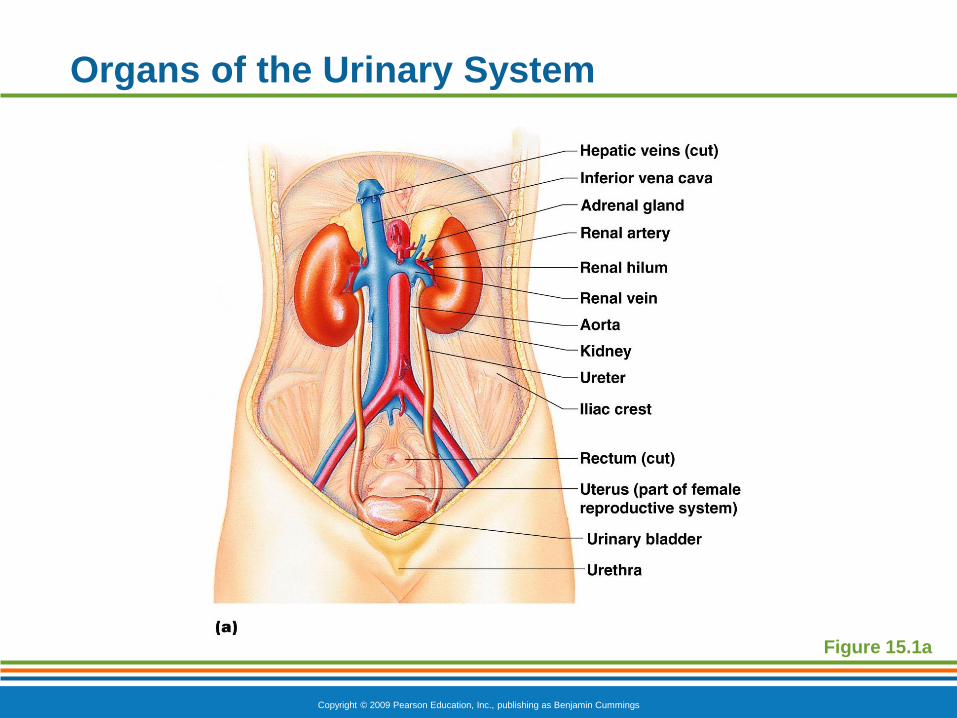
Organs of the Urinary System
Kidneys
Ureters
Urinary bladder
Urethra

Copyright © 2009 Pearson Education, Inc., publishing as Benjamin Cummings
Organs of the Urinary System
Figure 15.1a
Copyright © 2009 Pearson Education, Inc., publishing as Benjamin Cummings
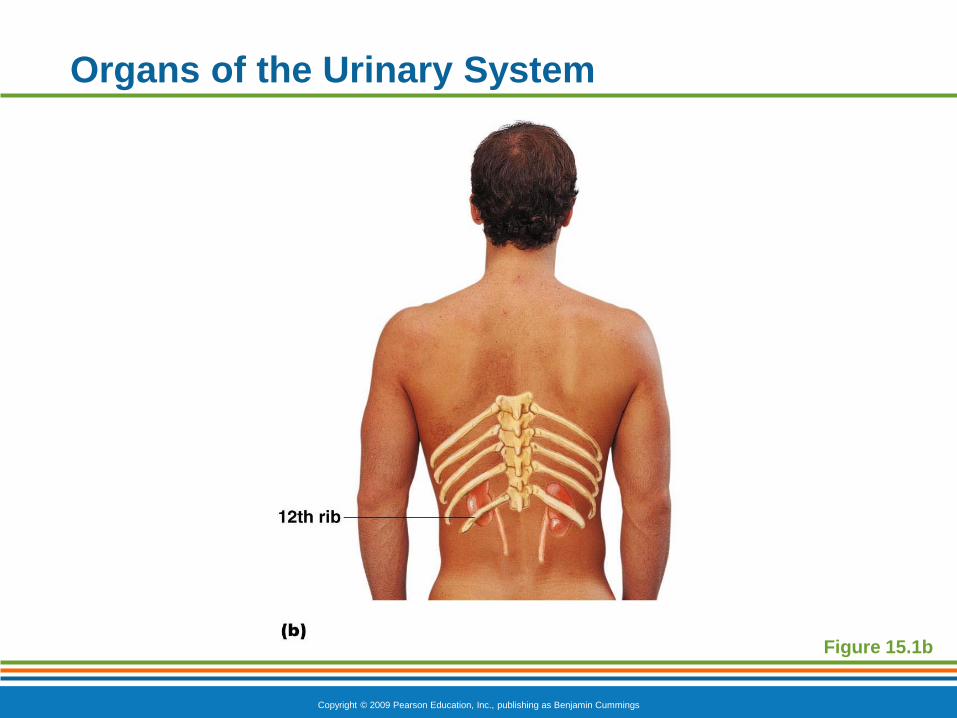
Organs of the Urinary System
Figure 15.1b
Copyright © 2009 Pearson Education, Inc., publishing as Benjamin Cummings
Location of the Kidneys
Against the dorsal body wall
At the level of the T12 to L3 vertebrae
The right kidney is slightly lower than the left (due
to position of the liver)
Copyright © 2009 Pearson Education, Inc., publishing as Benjamin Cummings
Kidney Features
Renal hilum
A medial indentation where several structures
enter or exit the kidney (ureters, renal blood
vessels, and nerves)
An adrenal gland sits atop each kidney
Copyright © 2009 Pearson Education, Inc., publishing as Benjamin Cummings
Organs of the Urinary System
Figure 15.1a
Copyright © 2009 Pearson Education, Inc., publishing as Benjamin Cummings
Coverings of the Kidneys
Fibrous capsule
Surrounds each kidney
Perirenal fat capsule
Surrounds the kidney and cushions against
blows
Renal fascia
Outermost capsule that helps hold the kidney
in place against the muscles of the trunk wall
Copyright © 2009 Pearson Education, Inc., publishing as Benjamin Cummings
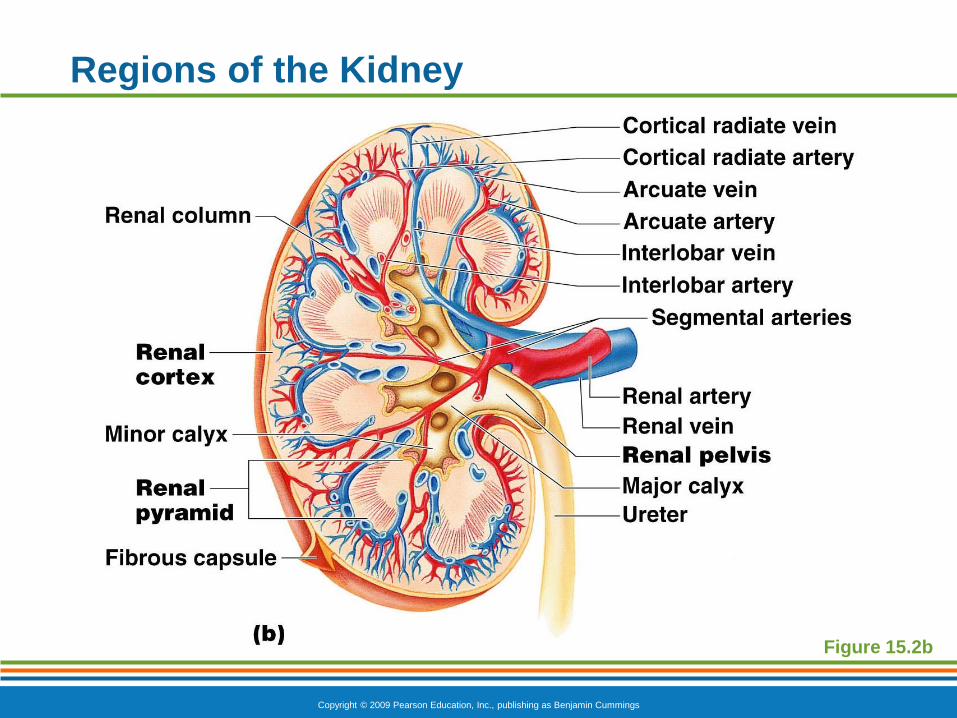
Regions of the Kidney
Renal cortex—outer region
Renal medulla—inside the cortex
Renal pelvis—inner collecting tube
Copyright © 2009 Pearson Education, Inc., publishing as Benjamin Cummings
Regions of the Kidney
Figure 15.2b
Copyright © 2009 Pearson Education, Inc., publishing as Benjamin Cummings
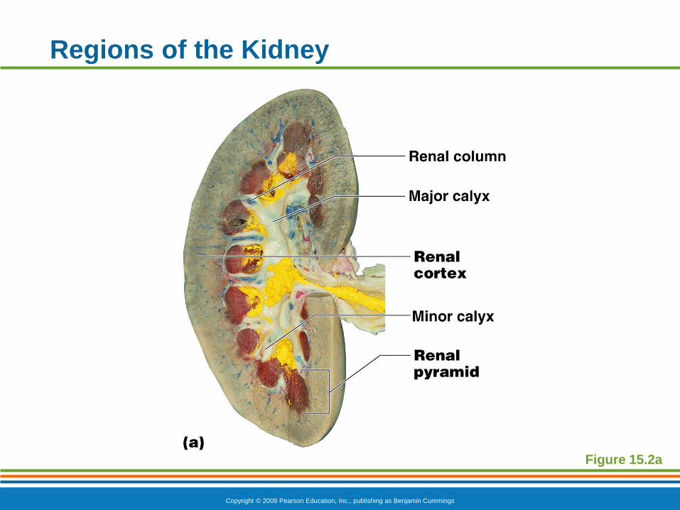
Kidney Structures
Renal or medullary pyramids—triangular regions
of tissue in the medulla
Renal columns—extensions of cortex-like
material inward that separate the pyramids
Calyces—cup-shaped structures that funnel urine
towards the renal pelvis
Copyright © 2009 Pearson Education, Inc., publishing as Benjamin Cummings
Regions of the Kidney
Figure 15.2a
Copyright © 2009 Pearson Education, Inc., publishing as Benjamin Cummings
Blood Supply
One-quarter of the total blood supply of the body
passes through the kidneys each minute
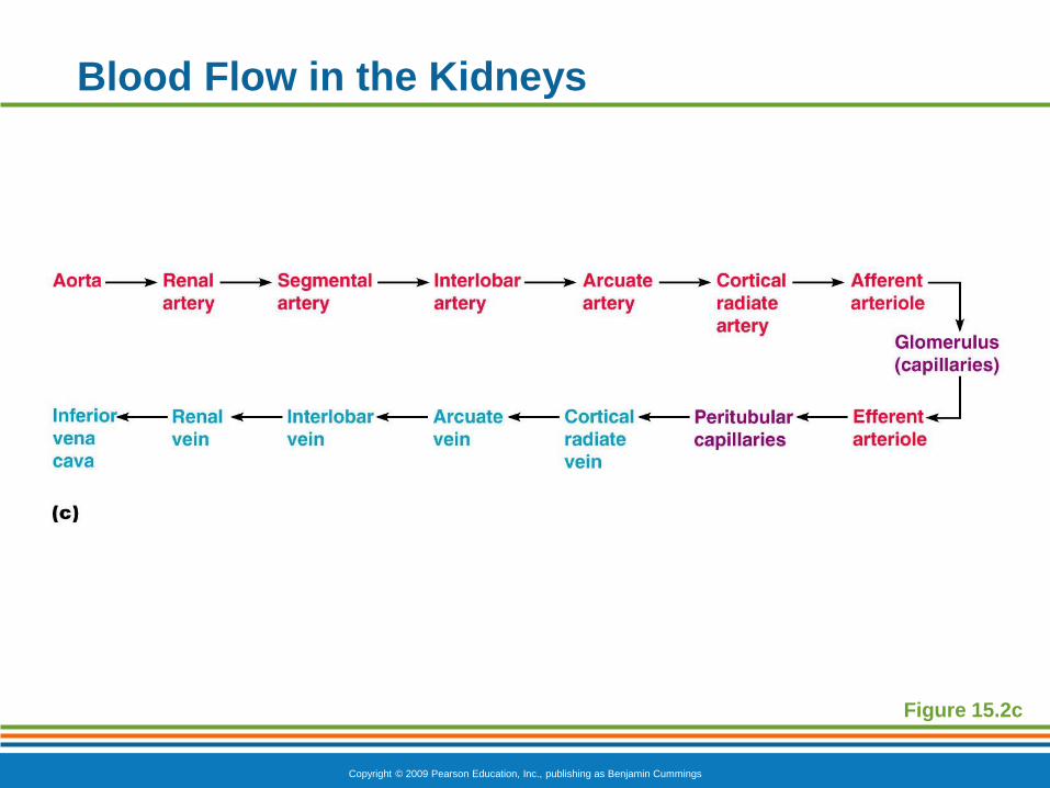
Renal artery provides each kidney with arterial
blood supply
Renal artery divides into segmental arteries
interlobar arteries arcuate arteries cortical
radiate arteries
Copyright © 2009 Pearson Education, Inc., publishing as Benjamin Cummings
Blood Supply
Venous blood flow
Cortical radiate veins arcuate veins
interlobar veins renal vein
There are no segmental veins
Copyright © 2009 Pearson Education, Inc., publishing as Benjamin Cummings
Blood Flow in the Kidneys
Figure 15.2c
Copyright © 2009 Pearson Education, Inc., publishing as Benjamin Cummings
Nephron Anatomy and Physiology
The structural and functional units of the kidneys
Responsible for forming urine
Main structures of the nephrons
Glomerulus
Renal tubule
Copyright © 2009 Pearson Education, Inc., publishing as Benjamin Cummings
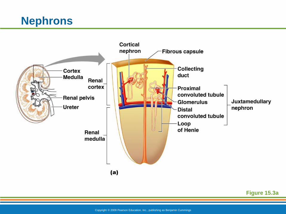
Nephrons
Figure 15.3a
Copyright © 2009 Pearson Education, Inc., publishing as Benjamin Cummings
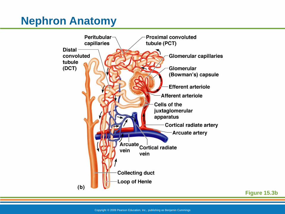
Nephron Anatomy
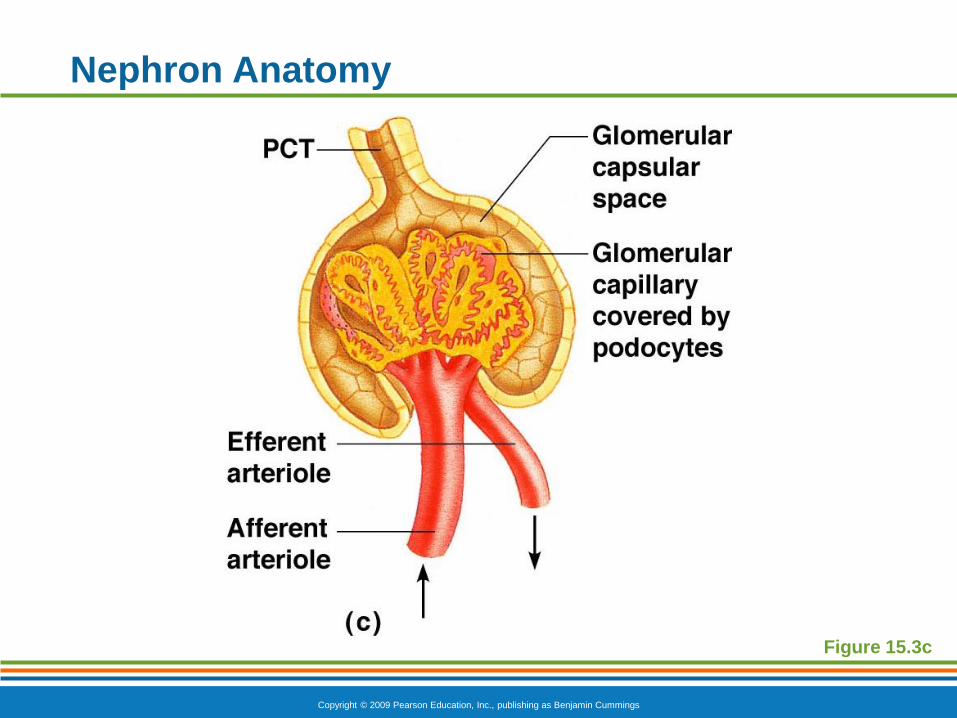
Glomerulus
Knot of capillaries
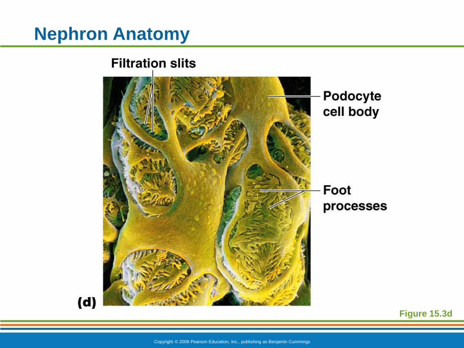
Capillaries are covered with podocytes from
the renal tubule
Glomerulus sits within a glomerular
(Bowman’s) capsule (the first part of the renal
tubule)
Copyright © 2009 Pearson Education, Inc., publishing as Benjamin Cummings
Nephron Anatomy
Figure 15.3c
Copyright © 2009 Pearson Education, Inc., publishing as Benjamin Cummings
Nephron Anatomy
Figure 15.3d
Copyright © 2009 Pearson Education, Inc., publishing as Benjamin Cummings
Nephron Anatomy
Renal tubule extends from glomerular capsule
and ends at the collecting duct
Glomerular (Bowman’s) capsule
Proximal convoluted tubule (PCT)
Loop of Henle
Distal convoluted tubule (DCT)
Copyright © 2009 Pearson Education, Inc., publishing as Benjamin Cummings
Nephron Anatomy
Figure 15.3b
Copyright © 2009 Pearson Education, Inc., publishing as Benjamin Cummings
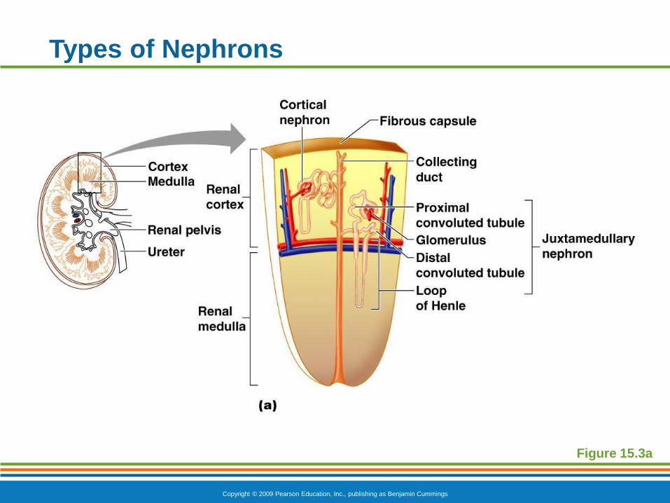
Types of Nephrons
Cortical nephrons
Located entirely in the cortex
Includes most nephrons
Juxtamedullary nephrons
Found at the boundary of the cortex and
medulla
Copyright © 2009 Pearson Education, Inc., publishing as Benjamin Cummings
Types of Nephrons
Figure 15.3a
Copyright © 2009 Pearson Education, Inc., publishing as Benjamin Cummings
Collecting Duct
Receives urine from many nephrons
Run through the medullary pyramids
Deliver urine into the calyces and renal pelvis

Copyright © 2009 Pearson Education, Inc., publishing as Benjamin Cummings
Nephron Anatomy
Figure 15.3b
Copyright © 2009 Pearson Education, Inc., publishing as Benjamin Cummings
Nephron Anatomy
Nephrons are associated with two capillary beds
Glomerulus
Peritubular capillary bed
Copyright © 2009 Pearson Education, Inc., publishing as Benjamin Cummings
Glomerulus
Fed and drained by arterioles
Afferent arteriole—arises from a cortical
radiate artery and feeds the glomerulus
Efferent arteriole—receives blood that has
passed through the glomerulus
Specialized for filtration
High pressure forces fluid and solutes out of
blood and into the glomerular capsule

Copyright © 2009 Pearson Education, Inc., publishing as Benjamin Cummings
Nephron Anatomy
Figure 15.3c
Copyright © 2009 Pearson Education, Inc., publishing as Benjamin Cummings
Nephron Anatomy
Figure 15.4
Copyright © 2009 Pearson Education, Inc., publishing as Benjamin Cummings
Peritubular Capillary Beds
Arise from efferent arteriole of the glomerulus
Normal, low pressure capillaries
Adapted for absorption instead of filtration
Cling close to the renal tubule to reabsorb
(reclaim) some substances from collecting tubes

Copyright © 2009 Pearson Education, Inc., publishing as Benjamin Cummings
Nephron Anatomy
Figure 15.3b
Copyright © 2009 Pearson Education, Inc., publishing as Benjamin Cummings
Urinary System Review
Copyright © 2009 Pearson Education, Inc., publishing as Benjamin Cummings
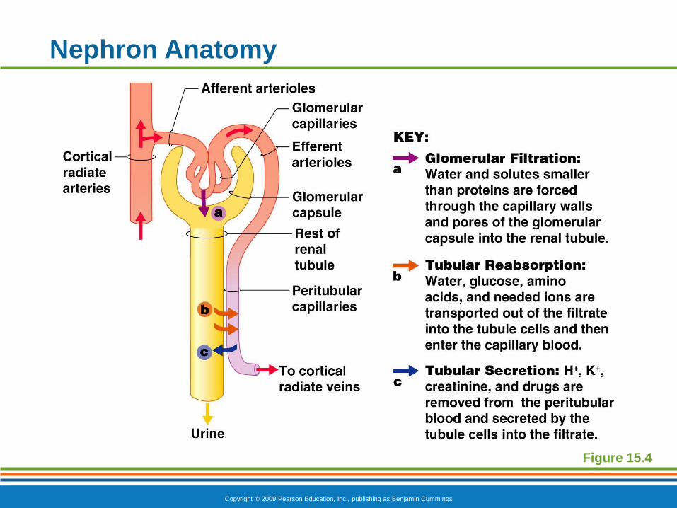
Urine Formation
Glomerular filtration
Tubular reabsorption
Tubular secretion
Copyright © 2009 Pearson Education, Inc., publishing as Benjamin Cummings
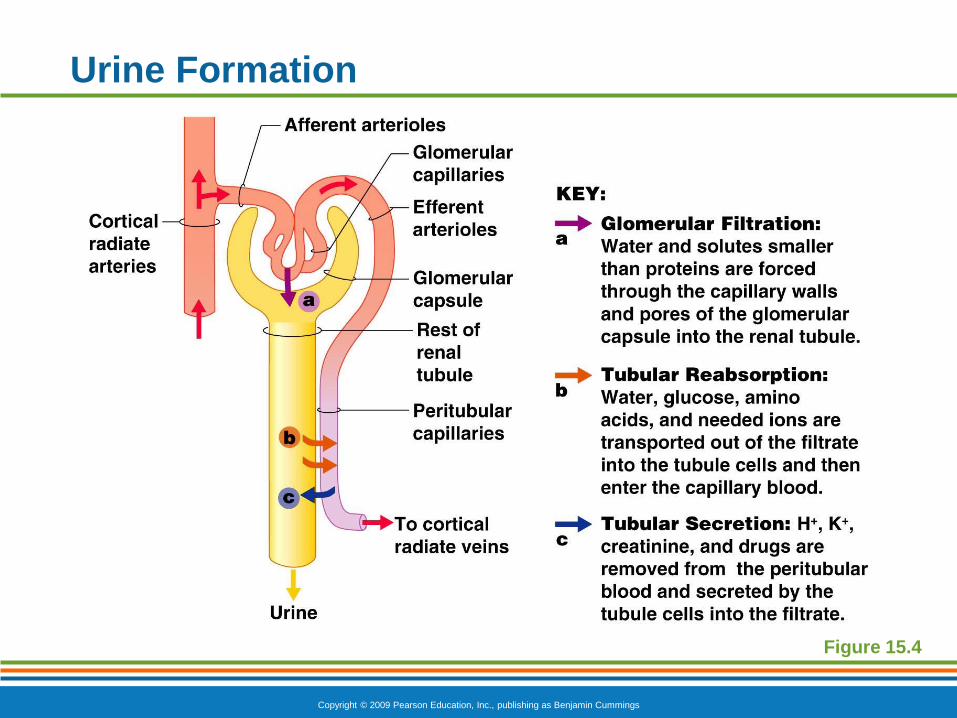
Urine Formation
Figure 15.4
Copyright © 2009 Pearson Education, Inc., publishing as Benjamin Cummings
Glomerular Filtration
Nonselective passive process
Water and solutes smaller than proteins are
forced through capillary walls
Proteins and blood cells are normally too large to
pass through the filtration membrane
Filtrate is collected in the glomerular capsule and
leaves via the renal tubule
Copyright © 2009 Pearson Education, Inc., publishing as Benjamin Cummings
Tubular Reabsorption
The peritubular capillaries reabsorb useful
substances
Water
Glucose
Amino acids
Ions
Some reabsorption is passive, most is active
Most reabsorption occurs in the proximal
convoluted tubule
Copyright © 2009 Pearson Education, Inc., publishing as Benjamin Cummings
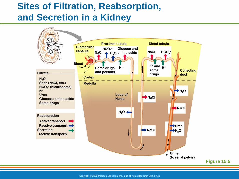
Sites of Filtration, Reabsorption,
and Secretion in a Kidney
Figure 15.5
Copyright © 2009 Pearson Education, Inc., publishing as Benjamin Cummings
Tubular Reabsorption
Materials not reabsorbed
Nitrogenous waste products
Urea—protein breakdown
Uric acid—nucleic acid breakdown
Creatinine—associated with creatine
metabolism in muscles
Copyright © 2009 Pearson Education, Inc., publishing as Benjamin Cummings
Tubular Secretion: Reabsorption in Reverse
Some materials move from the peritubular
capillaries into the renal tubules
Hydrogen and potassium ions
Creatinine
Process is important for getting rid of substances
not already in the filtrate
Materials left in the renal tubule move toward the
ureter
Copyright © 2009 Pearson Education, Inc., publishing as Benjamin Cummings
Review: Nephron Function
Copyright © 2009 Pearson Education, Inc., publishing as Benjamin Cummings
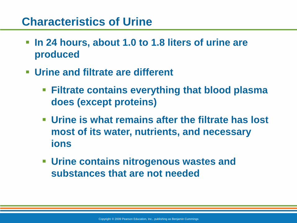
Characteristics of Urine
In 24 hours, about 1.0 to 1.8 liters of urine are
produced
Urine and filtrate are different
Filtrate contains everything that blood plasma
does (except proteins)
Urine is what remains after the filtrate has lost
most of its water, nutrients, and necessary
ions
Urine contains nitrogenous wastes and
substances that are not needed
Copyright © 2009 Pearson Education, Inc., publishing as Benjamin Cummings
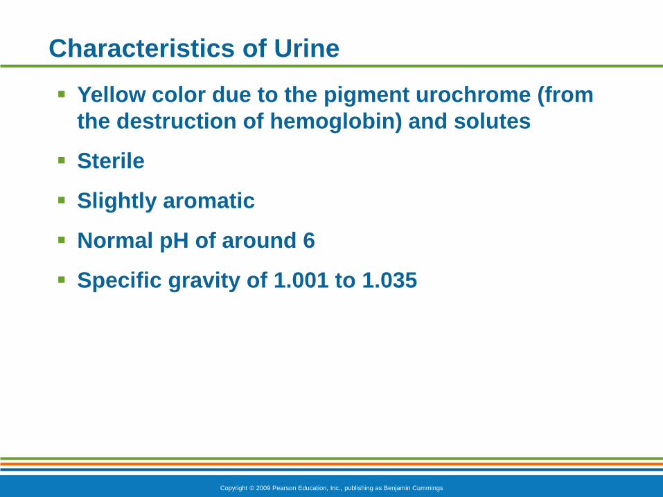
Characteristics of Urine
Yellow color due to the pigment urochrome (from
the destruction of hemoglobin) and solutes
Sterile
Slightly aromatic
Normal pH of around 6
Specific gravity of 1.001 to 1.035
Copyright © 2009 Pearson Education, Inc., publishing as Benjamin Cummings
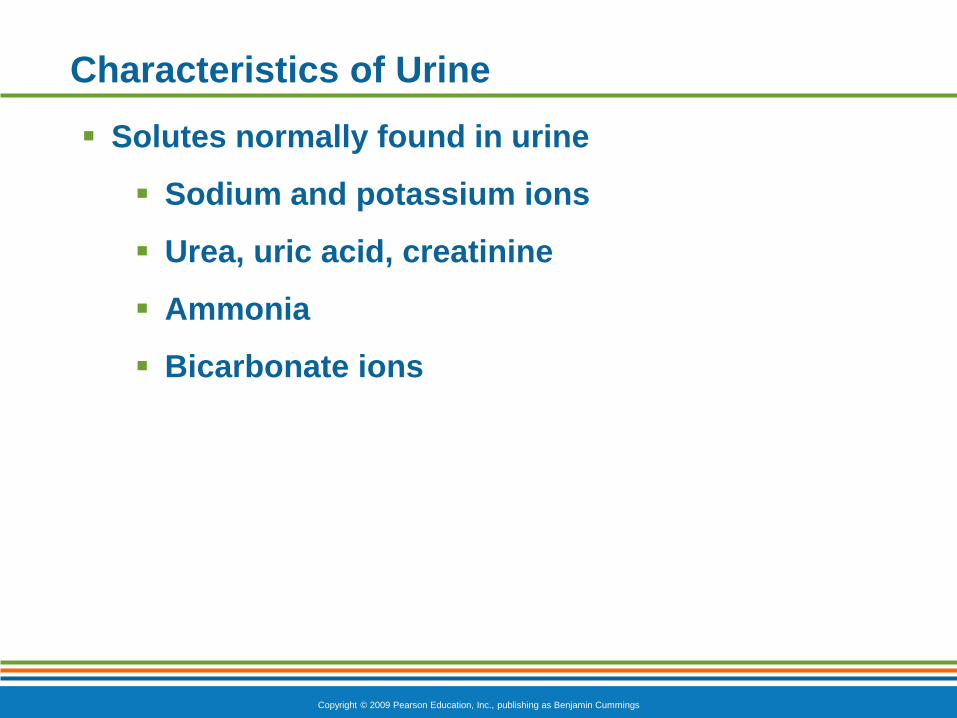
Characteristics of Urine
Solutes normally found in urine
Sodium and potassium ions
Urea, uric acid, creatinine
Ammonia
Bicarbonate ions
Copyright © 2009 Pearson Education, Inc., publishing as Benjamin Cummings
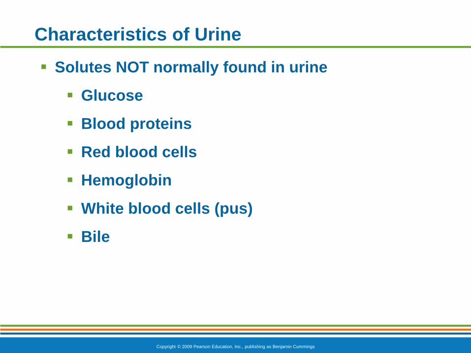
Characteristics of Urine
Solutes NOT normally found in urine
Glucose
Blood proteins
Red blood cells
Hemoglobin
White blood cells (pus)
Bile
Copyright © 2009 Pearson Education, Inc., publishing as Benjamin Cummings
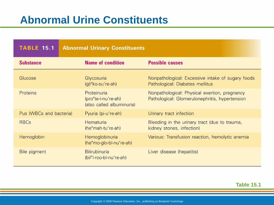
Abnormal Urine Constituents
Table 15.1