Embed Size (px)
Citation preview
CASE STUDY – ABNORMAL APPEARING GANGLION IN THE DORSAL WRIST
Paige Fabre
PATIENT PRESENTATION
34 year old female patient Working as cleaner on the mines – FIFO
worker History
Several months dorsal wrist pain Lump appearing on the dorsal aspect of the
wrist, increasing in size while at work, decreasing while at home
No previous imaging, investigations or interventional treatment
ULTRASOUND REQUEST
The examination was requested by the patient’s GP
A “wrist ultrasound +/- aspiration and injection” was requested
“Lump on dorsal wrist ? Ganglion” was the clinical indication given for the exam
ULTRASOUND PROTOCOL
No exact protocol is require for a wrist ultrasounds
Images taken and areas examined are determined by the sonographer and reporting radiologist
PROTOCOL FOR THE EXAMINATION
As I am still in training, my supervisor suggested that I examine all compartments of the dorsal wrist in longitudinal and transverse to gain a better understanding of the anatomy.
If these areas were normal then I was to image them in transverse to demonstrate that these areas have viewed. An additional image was to be taken of Compartment 3 as it crosses over Compartment 2.
ROOM PREPARATION
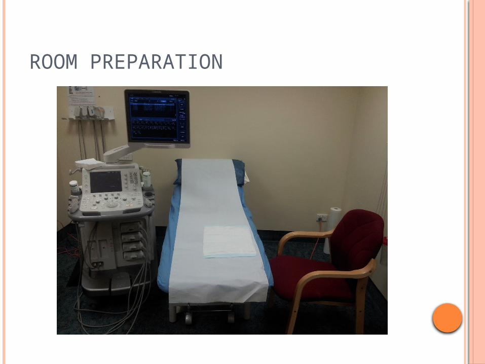
The room was prepared before the patient entered. A chair was placed on the side of the bed
(opposite the machine) facing the towards the bed.
A bluey was positioned on the table where the patient was to place their arms.
The ultrasound machine was moved to the foot end of the bed to allow for more ergonomic scanning.
Appropriate high frequency transducers were attached to the machine or within close proximity.
A pre-prepared injection trolley was brought into the room.
ROOM PREPARATION

PATIENT PREPARATION
At admission to the practice the patient read and completed a consent form for aspiration and injection of the wrist in preparation for this being carried out.
The patient was wearing an elbow length shirt and was therefore not required to change.
The patient’s identity was confirmed and they were invited into the room.
PATIENT PREPARATION
The examination was discussed with the patient before any scanning was attempted. The patient was made aware that I was in
training and that my supervisor would be assisting me with the scan. The patient was asked if this would be acceptable, and their consent was gained.
The patient was also made aware that we would first perform the diagnostic scan and from that the radiologist would decide the best course of action for the procedure.
ULTRASOUND EXAMINATION
The examination was performed on a Toshiba Aplio 500 using a variable frequency 18mHz linear transducer.
Thick ultrasound gel was applied to the area of examination to allow for light pressure and sufficient contact.
ULTRASOUND IMAGES OF THE COMPARTMENTS
COMPARTMENT 1
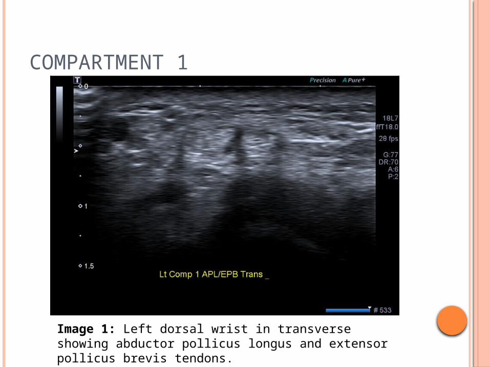
Image 1: Left dorsal wrist in transverse showing abductor pollicus longus and extensor pollicus brevis tendons.
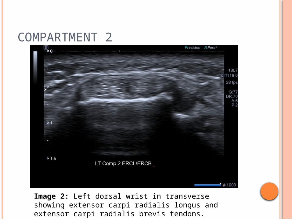
COMPARTMENT 2
Image 2: Left dorsal wrist in transverse showing extensor carpi radialis longus and extensor carpi radialis brevis tendons.
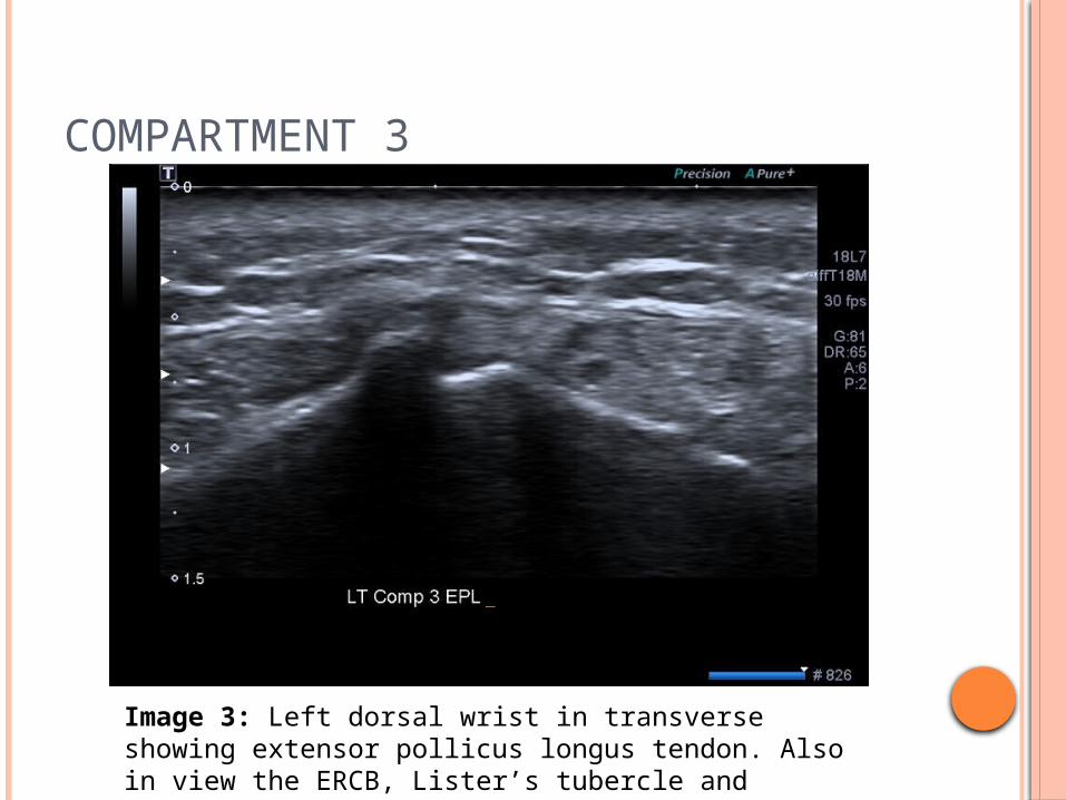
COMPARTMENT 3
Image 3: Left dorsal wrist in transverse showing extensor pollicus longus tendon. Also in view the ERCB, Lister’s tubercle and Compartment 4.
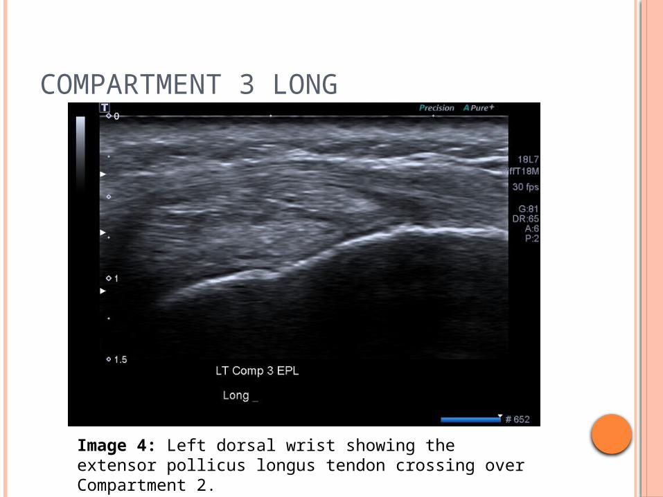
COMPARTMENT 3 LONG
Image 4: Left dorsal wrist showing the extensor pollicus longus tendon crossing over Compartment 2.
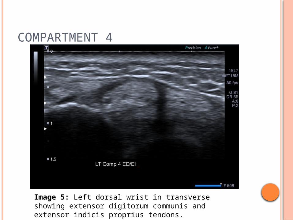
COMPARTMENT 4
Image 5: Left dorsal wrist in transverse showing extensor digitorum communis and extensor indicis proprius tendons.
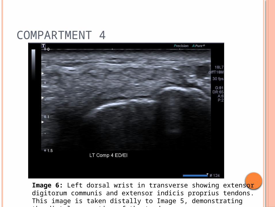
COMPARTMENT 4
Image 6: Left dorsal wrist in transverse showing extensor digitorum communis and extensor indicis proprius tendons. This image is taken distally to Image 5, demonstrating the distal separation of the tendons.
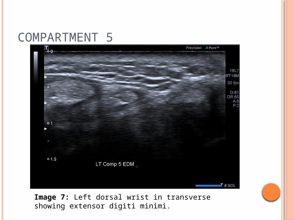
COMPARTMENT 5
Image 7: Left dorsal wrist in transverse showing extensor digiti minimi.
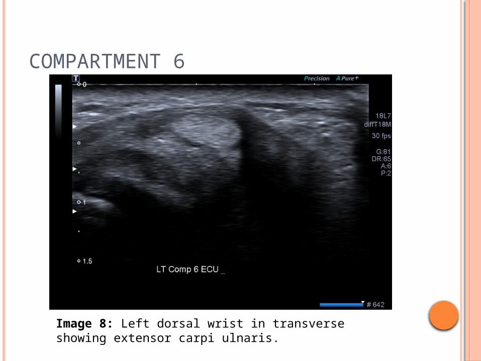
COMPARTMENT 6
Image 8: Left dorsal wrist in transverse showing extensor carpi ulnaris.
EXTENSION OF THE EXAMINATIONThe compartments of the wrist were examined in both longitudinal and transverse planes. The compartments were all deemed normal and the examination was extended to allow for better visualisation of the lump as described by the patient.
EXTENSION OF THE EXAMINATION
The following images were taken over the lump as identified by the patient as the region of interest.
ROI IMAGES
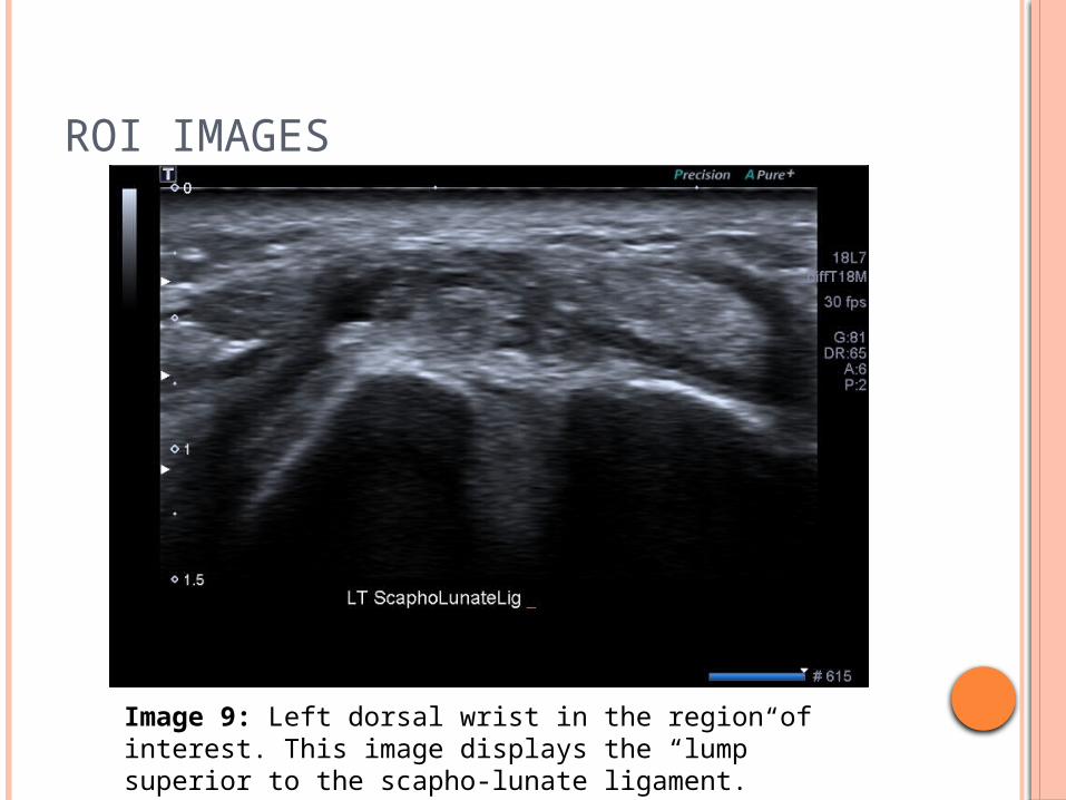
Image 9: Left dorsal wrist in the region of interest. This image displays the “lump” superior to the scapho-lunate ligament.
ROI IMAGES
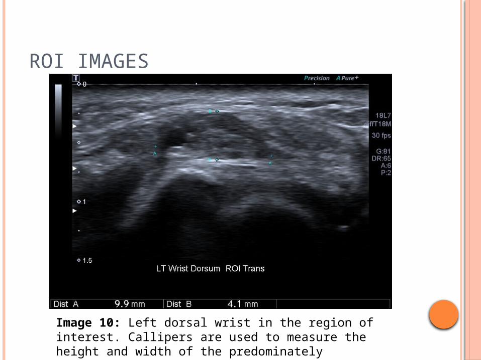
Image 10: Left dorsal wrist in the region of interest. Callipers are used to measure the height and width of the predominately hypoechoic area present.
ROI IMAGES
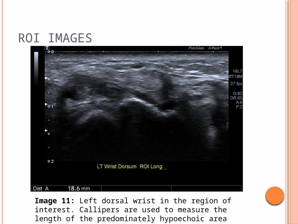
Image 11: Left dorsal wrist in the region of interest. Callipers are used to measure the length of the predominately hypoechoic area present.
ROI IMAGES
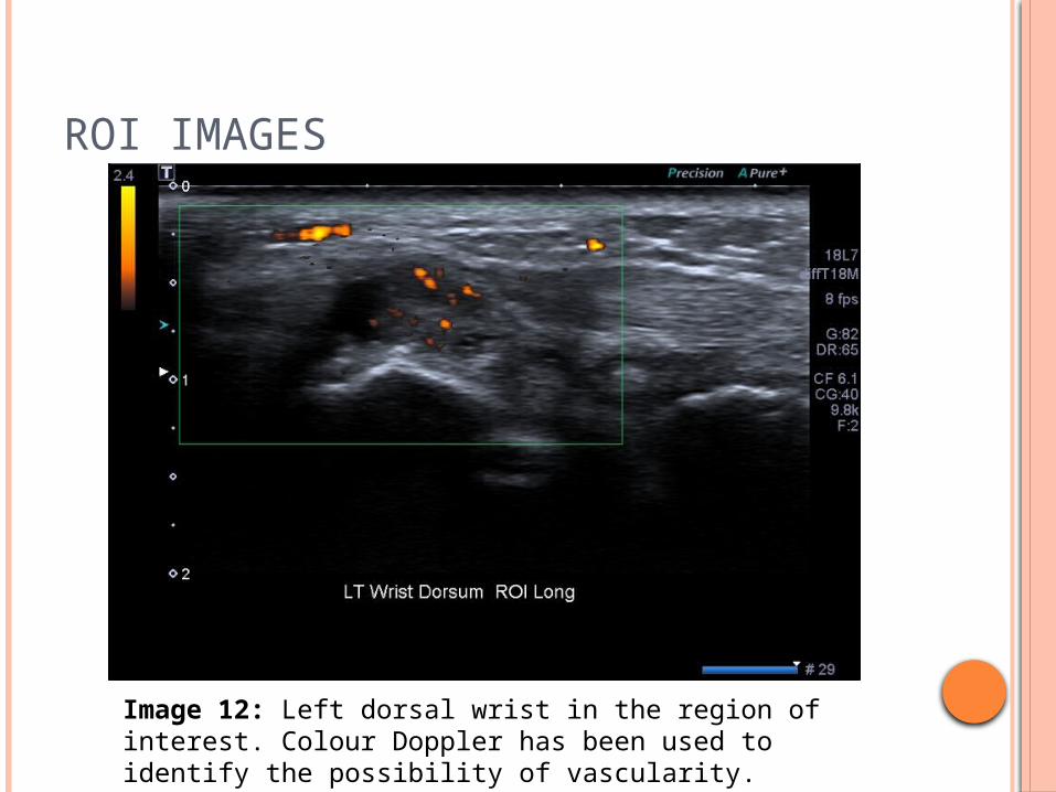
Image 12: Left dorsal wrist in the region of interest. Colour Doppler has been used to identify the possibility of vascularity.
ROI IMAGES
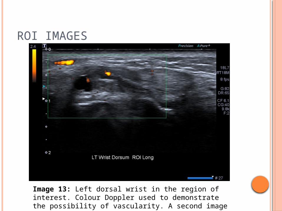
Image 13: Left dorsal wrist in the region of interest. Colour Doppler used to demonstrate the possibility of vascularity. A second image was done to highlight the anechoic region.
ROI IMAGES
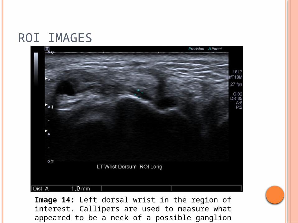
Image 14: Left dorsal wrist in the region of interest. Callipers are used to measure what appeared to be a neck of a possible ganglion extending from the radio-carpal joint.
EXTENSION OF EXAMINATION
In an attempt to better understand the origin of the possible ganglion/mass dynamic visualisation was attempted.
For these additional images a thick gel layer was applied to the wrist to maintain contact with light pressure.
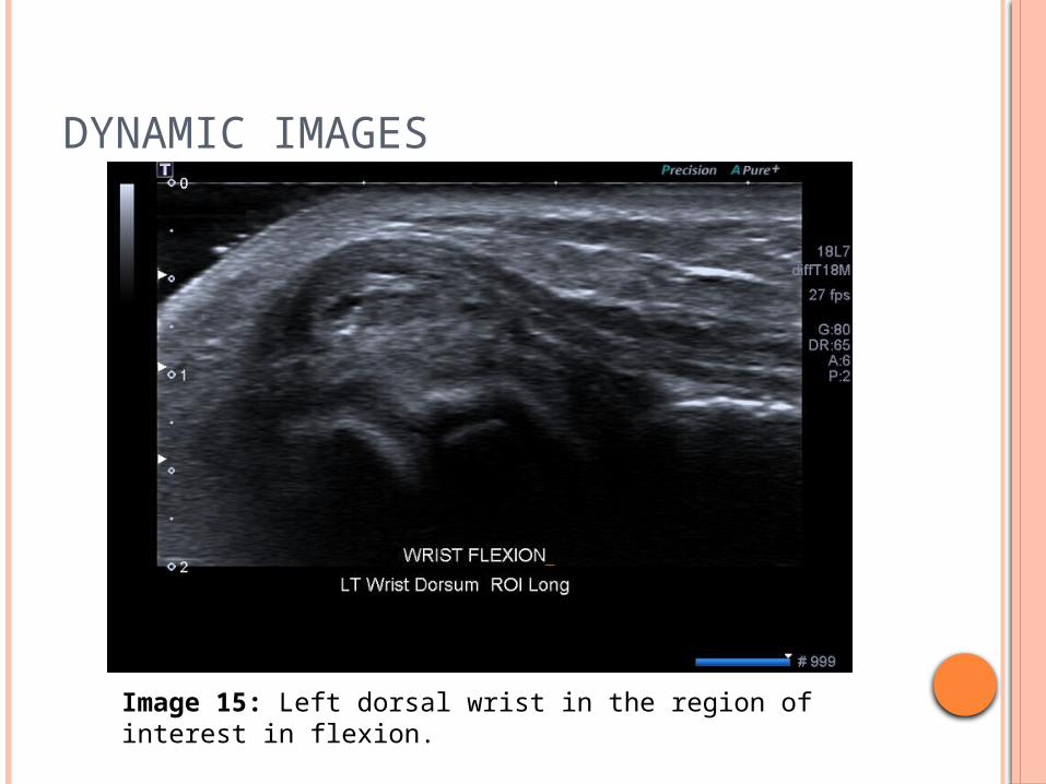
DYNAMIC IMAGES
Image 15: Left dorsal wrist in the region of interest in flexion.
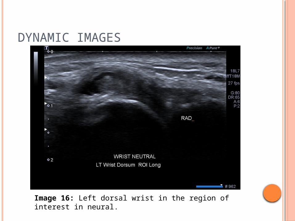
DYNAMIC IMAGES
Image 16: Left dorsal wrist in the region of interest in neural.
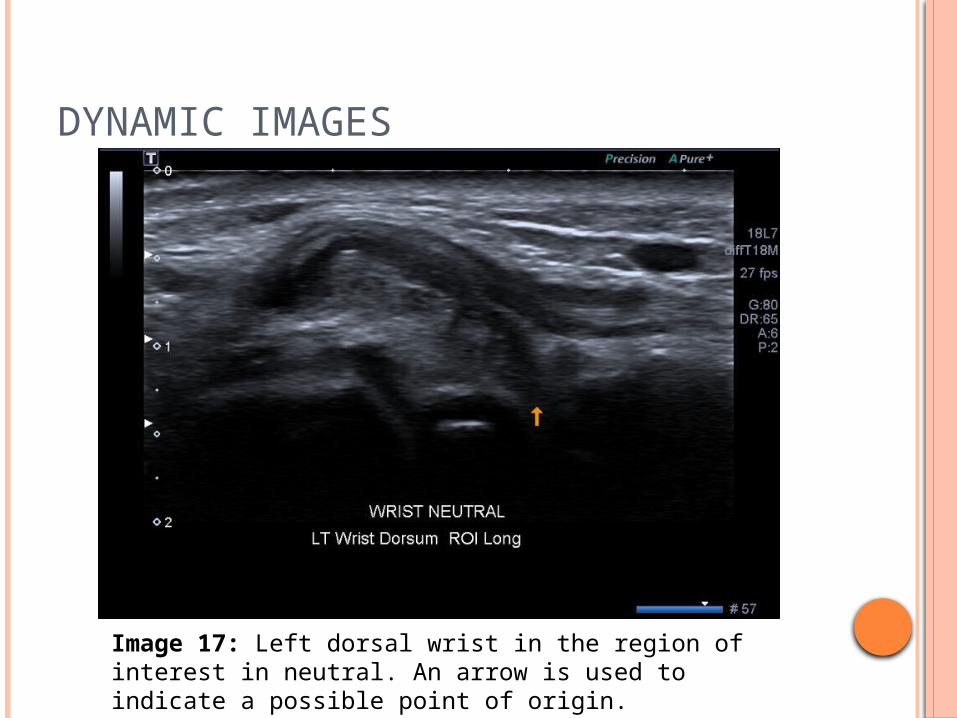
DYNAMIC IMAGES
Image 17: Left dorsal wrist in the region of interest in neutral. An arrow is used to indicate a possible point of origin.
ULTRASOUND FINDINGS
ULTRASOUND FINDINGS
Solitary well defined encapsulated lesion seen in region of interest
Predominately hypoechoic to surrounding tissues but somewhat heterogenous
Slight increased vascularity Possible neck extending to the scapholunate
ligament
DIFFERENTIAL DIAGNOSIS
Ganglion or Mass
GANGLIONS IN THE WRIST
GANGLION
Definition Mucin filled soft tissue cyst (Pal and Wallman
2014, 500) (Ahuja 2007, 13:114) (Tsou and Khoo 2012,450)
May either be from the synovial lining of the joint or the tendon sheath (Pal and Wallman 2014, 500)
Represent 80% of all soft tissue tumours in the hand and wrist (Ahuja 2007, 13:116)
AETIOLOGY
Mucoid degeneration Synovial herniation Trauma to the joint capsule Trauma to the ligaments Repetitive actions
(Pal and Wallman 2014, 500)(Tsou and Khoo 2012,450)
PATIENT PRESENTATION
Lump on the wrist Usually on the dorsal surface Usually painless however some patients exhibit
pain on dorsiflexion Changing in size with time and activity May be slow growing or spontaneously appear
Patients at risk Women are more affected than men Teenagers through to those in their middle adult
hood more affected Patients with laxity in their ligaments Previous history of trauma or injury to the wrist(Pal and Wallman 2014, 500)(Ahuja 2007, 13:116)
DIFFERENTIAL DIAGNOSIS
Tenosynovitis Giant Cell Tumour Vascular Anomaly
(Ahuja 2007, 13: 116)
ULTRASOUND APPEARANCE OF GANGLION
Hypoechoic fluid filled sac Stalk may be seen extending to the joint
from which it arises Non compressible Non vascular unless there has been a recent
leakage. Surrounding tissues may be oedematous and with a slight increase in vascularity
(Ahuja 2007, 13:114)
ALTERNATIVE IMAGING MODALITIES
Ultrasound is the preferred method of imaging however MRI may also be useful in identifying non symptomatic ganglia
INTERVENTIONAL TECHNIQUES
Non Invasive Aspiration and injection
60% chance of reoccurrence (Pal and Wallman 2014, 502)
Rest Bible/ Manual rupture (not recommended)
Invasive Surgery to excise either open or arthroscopic
Most affective however longer healing time
(Pal and Wallman 2014, 502) (Ahuja 2007, 13:116)
GANGLION IN THE PRESENCE OF A SLL TEAR
A ganglion extending from the scaphoid lunate joint space is often indicative of a scapholunate ligament tear (Harish et.al. 2009, 118)
It is believed that this was the case in this patient.
ASPIRATION AND INJECTIONAfter a discussion with the radiologist and the patient it was decided that aspiration of the ganglion was to go ahead followed by an injection of Xylocaine, Ropivacaine and Celestone.
PROCEDURE PREPARATION
A sterile trolley was assembled. The following was included
Sterile dressing back 2 x 3ml luerlock syringes Extension tube Drawing-up needle 21G needle
Sterile gloves were made available for the radiologist.
PROCEDURE
A clean environment was established.
The patient’s wrist was positioned in slight flexion. This was achieved with a gel bottle being placed beneath the wrist covered with bluey.
PROCEDURE
The skin was prepped with an alcohol solution as per practice protocol.
Appropriate volumes of the drugs to be administered were decanted.
Local anaesthetic was first injected into the site and allowed to take effect.
An attempt to aspirate the suspected ganglion was made.
ASPIRATION
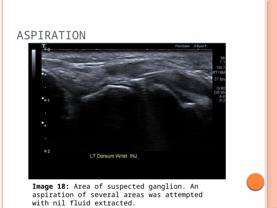
Image 18: Area of suspected ganglion. An aspiration of several areas was attempted with nil fluid extracted.
PROCEDURE
As no fluid was extracted, an injection of Ropivacaine and Celestone was administered
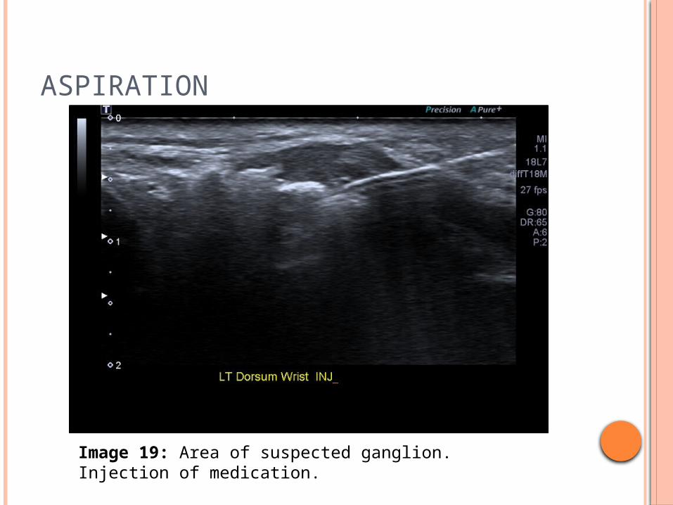
ASPIRATION
Image 19: Area of suspected ganglion. Injection of medication.
POST PROCEDURE

POST PROCEDURE
The patient appeared to tolerate the procedure well.
They were informed to rest the wrist for 24-48 hours and were given post procedure care instructions as per practice protocol.
A “pain chart” was also given to the patient to record the effectiveness of the procedure.
REFERENCES
Ahuja, Anil T. 2007. Diagnostic Imaging: Ultrasound. Salt Lake City: Amyirsys.
Harish, Srinivasan, John O'Neill, Karen Finlay, Erik Jurriaans, and Lawrence Friedman. 2009. Current Problems in Diagnostic Radiology 38(3): 111-125. DOI 10.1067/j.cpradiol.2008.02.001
Jacobson, Jon A. 2007. Fundementals of Musculoskeletal Ultrasound. Philadelphia: Saunders Elsevier.
Pal, Julie, Jackie Wallman. 2014.Fundamentals of Hand Therapy. 2nd ed. Online. DOI 10.1016/B978-0-323-09104-6.00036-5
Tsou, Ian Y.Y., and Jenn Nee Khoo. 2012. “Ultrasound of the wrist and hand”. Ultrasound Clinics 7(4): 439-455. DOI 10.1016/j.cult.2012.08.001