Embed Size (px)
Citation preview
259www.i-mri.org
Calcifying Aponeurotic Fibroma of the Knee: a Case Report with Radiographic and MRI Finding
INTRODUCTION
Calcifying aponeurotic fibroma (CAF) is an uncommon benign tumor and a locally aggressive fibroblastic lesion that was first described by Keasbey in 1953 as juvenile aponeurotic fibroma (1). In most cases, CAF is commonly seen or occurs in young patients with a peak incidence between the ages of 8 and 14 years; although, there are documented cases that ranged from birth to 64 years old (2). It typically occurs in the distal extremities, most commonly on the palmar side of the hand and fingers, and the soles of the feet; however, it can also affect other less common sites, such as the neck, elbow, forearm, thigh, knee and lumbosacral region (3, 4). These areas are closely related to aponeuroses, tendons or fascia (3, 4). To our knowledge, only two cases of CAF have been reported in the knee region. This report provides the radiographic and MRI finding of CAF arising in an uncommon site, the knee region, of a 19-year-old male who presented with a painful and palpable mass.
This is an Open Access article distributed under the terms of the Creative Commons Attribution Non-Commercial License (http://creativecommons.org/licenses/by-nc/3.0/) which permits unrestricted non-commercial use, distribution, and reproduction in any medium, provided the original work is properly cited.
Received: July 3, 2017Revised: September 4, 2017Accepted: September 26, 2017
Correspondence to: In Sook Lee, M.D.Department of Radiology, Pusan National University School of Medicine, 179, Gudeok-ro, Seo-gu, Busan 602-739, Korea.Tel. +82-51-240-7354Fax. +82-51-244-7534E-mail: [email protected]
Copyright © 2017 Korean Society of Magnetic Resonance in Medicine (KSMRM)
iMRI 2017;21:259-263 https://doi.org/10.13104/imri.2017.21.4.259
Case ReportCalcifying aponeurotic fibroma (CAF) is an uncommon benign tumor and a locally aggressive fibroblastic lesion. It commonly affects the palmar side of the hand and fingers, and the soles of the feet. The typical clinical manifestations are known as a poorly circumscribed, slow-growing, and asymptomatic firm mass. Most CAFs usually reveal low to intermediate or isointensity on T1-weighted images, and strong heterogeneous enhancement. However, various signal intensities on T2-weighted images have been reported depending on the degree of hypocellularity or the amount of calcification or collagen within the tumor. This report provides the radiographic and MRI finding of CAF arising in uncommon site, the knee region, of a 19-year-old male who presented with a painful and palpable mass.
Keywords: Calcifying aponeurotic fibroma; Knee; Magnetic resonance imaging
pISSN 2384-1095eISSN 2384-1109
Seung Hyun Lee1,2, In Sook Lee1,2, You Seon Song1,2, Kyung Un Choi3, Jeung Il Kim4, Jong Woon Song5
1Department of Radiology, Pusan National University Hospital, Busan, Korea2Pusan National University School of Medicine, Busan, Korea3Department of Pathology, Pusan National University Hospital, Busan, Korea4Department of Orthopedic Surgery, Pusan National University Hospital, Busan, Korea5Department of Radiology, Inje University Haeundae Paik Hospital, Busan, Korea
www.i-mri.org260
Calcifying Aponeurotic Fibroma of the Knee | Seung Hyun Lee, et al.
CASE REPORT
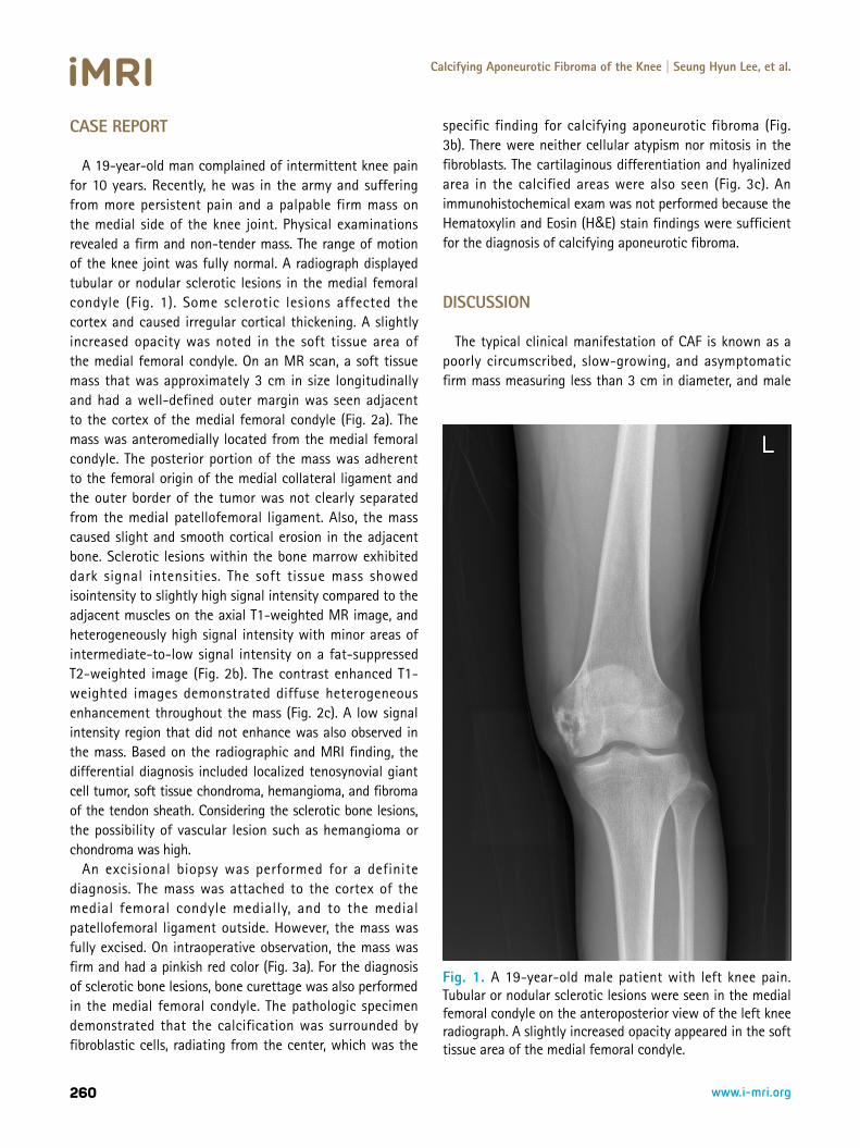
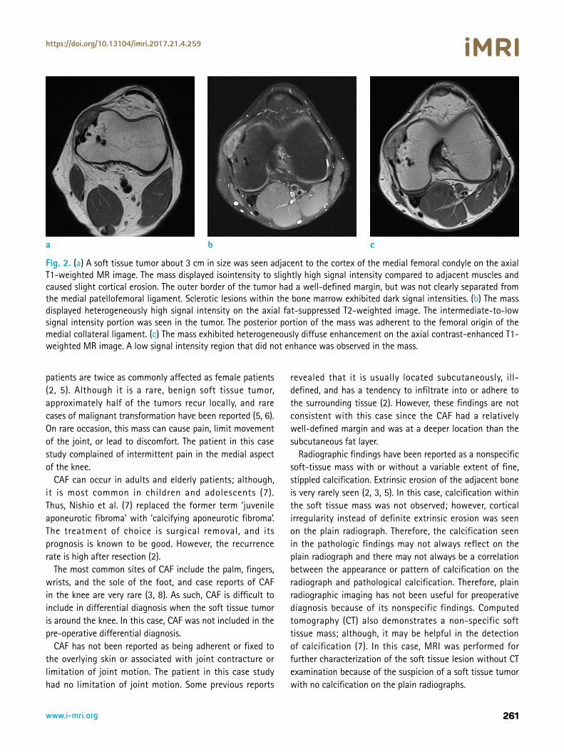
A 19-year-old man complained of intermittent knee pain for 10 years. Recently, he was in the army and suffering from more persistent pain and a palpable firm mass on the medial side of the knee joint. Physical examinations revealed a firm and non-tender mass. The range of motion of the knee joint was fully normal. A radiograph displayed tubular or nodular sclerotic lesions in the medial femoral condyle (Fig. 1). Some sclerotic lesions affected the cortex and caused irregular cortical thickening. A slightly increased opacity was noted in the soft tissue area of the medial femoral condyle. On an MR scan, a soft tissue mass that was approximately 3 cm in size longitudinally and had a well-defined outer margin was seen adjacent to the cortex of the medial femoral condyle (Fig. 2a). The mass was anteromedially located from the medial femoral condyle. The posterior portion of the mass was adherent to the femoral origin of the medial collateral ligament and the outer border of the tumor was not clearly separated from the medial patellofemoral ligament. Also, the mass caused slight and smooth cortical erosion in the adjacent bone. Sclerotic lesions within the bone marrow exhibited dark signal intensities. The soft tissue mass showed isointensity to slightly high signal intensity compared to the adjacent muscles on the axial T1-weighted MR image, and heterogeneously high signal intensity with minor areas of intermediate-to-low signal intensity on a fat-suppressed T2-weighted image (Fig. 2b). The contrast enhanced T1-weighted images demonstrated diffuse heterogeneous enhancement throughout the mass (Fig. 2c). A low signal intensity region that did not enhance was also observed in the mass. Based on the radiographic and MRI finding, the differential diagnosis included localized tenosynovial giant cell tumor, soft tissue chondroma, hemangioma, and fibroma of the tendon sheath. Considering the sclerotic bone lesions, the possibility of vascular lesion such as hemangioma or chondroma was high.
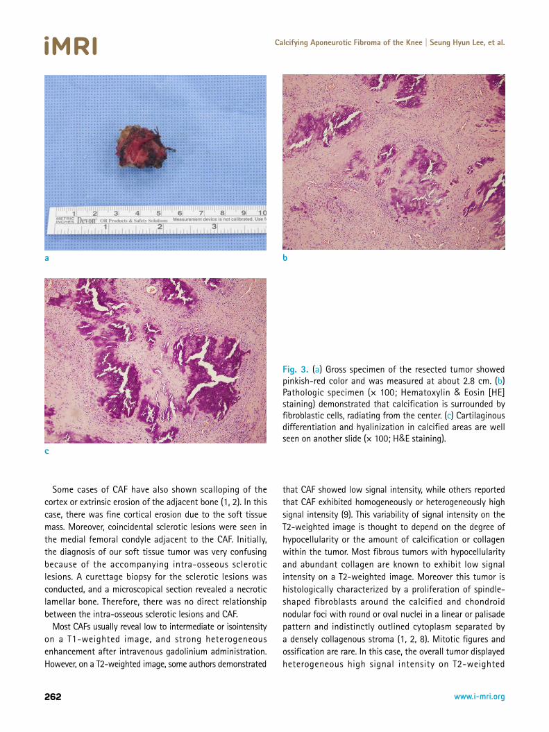
An excisional biopsy was performed for a definite diagnosis. The mass was attached to the cortex of the medial femoral condyle medially, and to the medial patellofemoral ligament outside. However, the mass was fully excised. On intraoperative observation, the mass was firm and had a pinkish red color (Fig. 3a). For the diagnosis of sclerotic bone lesions, bone curettage was also performed in the medial femoral condyle. The pathologic specimen demonstrated that the calcification was surrounded by fibroblastic cells, radiating from the center, which was the
specific finding for calcifying aponeurotic fibroma (Fig. 3b). There were neither cellular atypism nor mitosis in the fibroblasts. The cartilaginous differentiation and hyalinized area in the calcified areas were also seen (Fig. 3c). An immunohistochemical exam was not performed because the Hematoxylin and Eosin (H&E) stain findings were sufficient for the diagnosis of calcifying aponeurotic fibroma.
DISCUSSION
The typical clinical manifestation of CAF is known as a poorly circumscribed, slow-growing, and asymptomatic firm mass measuring less than 3 cm in diameter, and male
Fig. 1. A 19-year-old male patient with left knee pain. Tubular or nodular sclerotic lesions were seen in the medial femoral condyle on the anteroposterior view of the left knee radiograph. A slightly increased opacity appeared in the soft tissue area of the medial femoral condyle.
261www.i-mri.org
https://doi.org/10.13104/imri.2017.21.4.259
patients are twice as commonly affected as female patients (2, 5). Although it is a rare, benign soft tissue tumor, approximately half of the tumors recur locally, and rare cases of malignant transformation have been reported (5, 6). On rare occasion, this mass can cause pain, limit movement of the joint, or lead to discomfort. The patient in this case study complained of intermittent pain in the medial aspect of the knee.
CAF can occur in adults and elderly patients; although, it is most common in children and adolescents (7). Thus, Nishio et al. (7) replaced the former term ‘juvenile aponeurotic fibroma’ with ‘calcifying aponeurotic fibroma’. The treatment of choice is surgical removal, and its prognosis is known to be good. However, the recurrence rate is high after resection (2).
The most common sites of CAF include the palm, fingers, wrists, and the sole of the foot, and case reports of CAF in the knee are very rare (3, 8). As such, CAF is difficult to include in differential diagnosis when the soft tissue tumor is around the knee. In this case, CAF was not included in the pre-operative differential diagnosis.
CAF has not been reported as being adherent or fixed to the overlying skin or associated with joint contracture or limitation of joint motion. The patient in this case study had no limitation of joint motion. Some previous reports
revealed that it is usually located subcutaneously, ill-defined, and has a tendency to infiltrate into or adhere to the surrounding tissue (2). However, these findings are not consistent with this case since the CAF had a relatively well-defined margin and was at a deeper location than the subcutaneous fat layer.
Radiographic findings have been reported as a nonspecific soft-tissue mass with or without a variable extent of fine, stippled calcification. Extrinsic erosion of the adjacent bone is very rarely seen (2, 3, 5). In this case, calcification within the soft tissue mass was not observed; however, cortical irregularity instead of definite extrinsic erosion was seen on the plain radiograph. Therefore, the calcification seen in the pathologic findings may not always reflect on the plain radiograph and there may not always be a correlation between the appearance or pattern of calcification on the radiograph and pathological calcification. Therefore, plain radiographic imaging has not been useful for preoperative diagnosis because of its nonspecific findings. Computed tomography (CT) also demonstrates a non-specific soft tissue mass; although, it may be helpful in the detection of calcification (7). In this case, MRI was performed for further characterization of the soft tissue lesion without CT examination because of the suspicion of a soft tissue tumor with no calcification on the plain radiographs.
Fig. 2. (a) A soft tissue tumor about 3 cm in size was seen adjacent to the cortex of the medial femoral condyle on the axial T1-weighted MR image. The mass displayed isointensity to slightly high signal intensity compared to adjacent muscles and caused slight cortical erosion. The outer border of the tumor had a well-defined margin, but was not clearly separated from the medial patellofemoral ligament. Sclerotic lesions within the bone marrow exhibited dark signal intensities. (b) The mass displayed heterogeneously high signal intensity on the axial fat-suppressed T2-weighted image. The intermediate-to-low signal intensity portion was seen in the tumor. The posterior portion of the mass was adherent to the femoral origin of the medial collateral ligament. (c) The mass exhibited heterogeneously diffuse enhancement on the axial contrast-enhanced T1-weighted MR image. A low signal intensity region that did not enhance was observed in the mass.
a b c
www.i-mri.org262
Calcifying Aponeurotic Fibroma of the Knee | Seung Hyun Lee, et al.
Some cases of CAF have also shown scalloping of the cortex or extrinsic erosion of the adjacent bone (1, 2). In this case, there was fine cortical erosion due to the soft tissue mass. Moreover, coincidental sclerotic lesions were seen in the medial femoral condyle adjacent to the CAF. Initially, the diagnosis of our soft tissue tumor was very confusing because of the accompanying intra-osseous sclerotic lesions. A curettage biopsy for the sclerotic lesions was conducted, and a microscopical section revealed a necrotic lamellar bone. Therefore, there was no direct relationship between the intra-osseous sclerotic lesions and CAF.
Most CAFs usually reveal low to intermediate or isointensity on a T1-weighted image, and strong heterogeneous enhancement after intravenous gadolinium administration. However, on a T2-weighted image, some authors demonstrated
that CAF showed low signal intensity, while others reported that CAF exhibited homogeneously or heterogeneously high signal intensity (9). This variability of signal intensity on the T2-weighted image is thought to depend on the degree of hypocellularity or the amount of calcification or collagen within the tumor. Most fibrous tumors with hypocellularity and abundant collagen are known to exhibit low signal intensity on a T2-weighted image. Moreover this tumor is histologically characterized by a proliferation of spindle-shaped fibroblasts around the calcified and chondroid nodular foci with round or oval nuclei in a linear or palisade pattern and indistinctly outlined cytoplasm separated by a densely collagenous stroma (1, 2, 8). Mitotic figures and ossification are rare. In this case, the overall tumor displayed heterogeneous high signal intensity on T2-weighted
Fig. 3. (a) Gross specimen of the resected tumor showed pinkish-red color and was measured at about 2.8 cm. (b) Pathologic specimen (× 100; Hematoxylin & Eosin [HE] staining) demonstrated that calcification is surrounded by fibroblastic cells, radiating from the center. (c) Cartilaginous differentiation and hyalinization in calcified areas are well seen on another slide (× 100; H&E staining).
a
c
b
263www.i-mri.org
https://doi.org/10.13104/imri.2017.21.4.259
images; although, there was a difference in signal intensity depending on the presence or absence of fat-suppression application. These signal intensity characteristics might be affected by the degrees of calcification, fibrous component, and cellularity influence (4). Typical MRI finding of fibroma or fibromatosis including prominent low to intermediate signal intensity and bands of low signal intensity representing highly collagenized tissue on the T1-weighted image were not observed in this case. However, fibroma or fibromatosis also have variable MRI finding. On the other hand, globular low signal intensity may be seen on all MR pulse sequences, corresponding with the presence of calcification (7). However, in this case, low signal intensities reflecting calcification were not evident on all sequences.
Many authors who reported CAF included hemangioma, fibroma or fibromatosis, localized tenosynovial giant-cell tumor (GCT), soft-tissue chondroma, and less commonly soft-tissue sarcoma such as synovial sarcoma in differential diagnosis (9, 10). Especially, localized tenosynovial GCT is associated intimately with the tendon sheath and may erode bone. It has relatively characteristic MRI finding with well-defined, lobulated margins, uniform enhancement, heterogeneous hypointense signals associated with hemosiderin deposition and pressure erosion, but very rare calcifications. Also, it rarely occurs in children (9, 10). A soft-tissue chondroma has a well-defined, lobulated, and diffuse calcification. Bands of low-signal intensity in MRI finding are common in desmoid-type fibromatosis. Heterogeneity caused by areas of necrosis, calcification, cysts and hemorrhage within a tumor can be helpful for suspicious of synovial sarcoma.
Biphasic features in the development of CAF have been described by some authors (1). In the early or initial phase, the tumor has a diffuse and infiltrative growth with a lack of calcification; and in the later phase, the tumor is more compact and nodular with calcification and cartilage formation. The former is common in infants and young children, and the latter is frequent in adolescents and adults. This case was considered to clinically or pathologically correspond to the later phase, but radiologically correspond to the early phase.
In both previous reports and this case, imaging findings which included radiographic, CT and MR images were nonspecific. Therefore, histopathologic examination is necessary for definite diagnosis of these lesions and to allow differentiation from malignant tumors such as
synovial sarcoma. Although a CAF is usually located in the subcutaneous
fat layer of the palm, finger or sole, it may occur in deeper areas than subcutaneous fat layer of the knee region, as in this case. Also, the presence of calcification in the tumor may or may not be helpful in differential diagnosis, because there are many soft-tissue tumors with calcifications. In addition, the clinical manifestations of a CAF can be variable, and the onset age can have a wide range. Therefore, clinicians and radiologists should keep in mind that a CAF without definite calcifications can occur in the knee region during late adolescence.
REFERENCES 1. Keasbey LE. Juvenile aponeurotic fibroma (calcifying
fibroma); a distinctive tumor arising in the palms and soles of young children. Cancer 1953;6:338-346
2. Kim OH, Kim YM. Calcifying aponeurotic fibroma: case report with radiographic and MR features. Korean J Radiol 2014;15:134-139
3. Fetsch JF, Miettinen M. Calcifying aponeurotic fibroma: a clinicopathologic study of 22 cases arising in uncommon sites. Hum Pathol 1998;29:1504-1510
4. Kwak HS, Lee SY, Kim JR, Lee KB. MR imaging of calcifying aponeurotic fibroma of the thigh. Pediatr Radiol 2004;34:438-440
5. Enzinger FM, Weiss SW. Fibrous tumors of infancy and childhood: soft tissue tumors. 4th ed. St. Louis: Mosby-Year Book, 2001:388-395
6. Amaravati R. Rare malignant transformation of a calcifying aponeurotic fibroma. J Bone Joint Surg Am 2002;84-A:1889; author reply 1889
7. Nishio J, Inamitsu H, Iwasaki H, Hayashi H, Naito M. Calcifying aponeurotic fibroma of the finger in an elderly patient: CT and MRI findings with pathologic correlation. Exp Ther Med 2014;8:841-843
8. Hasegawa HK, Park S, Hamazaki M. Calcifying aponeurotic fibroma of the knee: a case report with radiological findings. J Dermatol 2006;33:169-173
9. Parker WL, Beckenbaugh RR, Amrami KK. Calcifying aponeurotic fibroma of the hand: radiologic differentiation from giant cell tumors of the tendon sheath. J Hand Surg Am 2006;31:1024-1028
10. Sekiguchi T, Nakagawa M, Miwa S, et al. Calcifying aponeurotic fibroma in a girl: MRI findings and their chronological changes. Radiol Case Rep 2017;12:620-623