Embed Size (px)
Citation preview
Empty
Fig 1. A) Implant with proximal
gap packed with
B) Post-harvest radiograph.
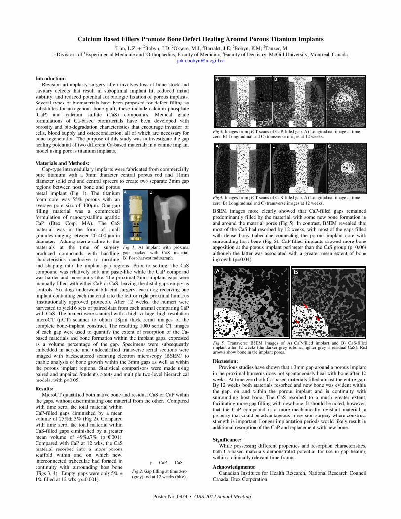
Fig 2. Gap f
(grey) and
Calcium Based Fillers Promote1Lim, L Z; +1,2Bobyn
+Divisions of 1Experimental Medicine and 2Orthopaedics
Introduction:
Revision arthroplasty surgery often involves loss of bone stock and
cavitary defects that result in suboptimal implant fit, reduced initial
stability, and reduced potential for biologic fixation of porous implants.
Several types of biomaterials have been proposed for defect filling as
substitutes for autogenous bone graft; these include calcium
(CaP) and calcium sulfate (CaS) compounds. Medical grade
formulations of Ca-based biomaterials have been developed with
porosity and bio-degradation characteristics that encourage
cells, blood supply and osteoconduction, all of which are necessary
bone regeneration. The purpose of this study was to investigate the
healing potential of two different Ca-based materials in a canine implant
model using porous titanium implants.
Materials and Methods: Gap-type intramedullary implants were fabricated from commercially
pure titanium with a 5mm diameter central porous rod and 11mm
diameter solid end and central spacers to create two separate 3mm gap
regions between host bone and porous
metal implant (Fig 1). The titanium
foam core was 55% porous with an
average pore size of 400µm. One gap
filling material was a commercial
formulation of nanocrystalline apatitic
CaP (Etex Corp, MA). The CaS
material was in the form of small
granules ranging between 20-400 µm in
diameter. Adding sterile saline to the
materials at the time of surgery
produced compounds with handling
characteristics conducive to molding
and shaping into the implant gap regions. Prior to setting, t
compound was relatively soft and paste-like while the
was harder and more putty-like. The proximal 3mm implant gaps were
manually filled with either CaP or CaS, leaving the distal gaps empty as
controls. Six dogs underwent bilateral surgery, each dog receiving one
implant containing each material into the left or right proximal humerus
(institutionally approved protocol). After 12 weeks,
harvested to yield 6 sets of paired data from each animal
with CaS. The humeri were scanned with a high voltage, high resolution
microCT (µCT) scanner to obtain 18µm thick serial images of the
complete bone-implant construct. The resulting 1000 serial CT images
of each gap were used to quantify the extent of resorption of the
based materials and bone formation within the implant
as a volume percentage of the gap. Specimens were subsequently
embedded in acrylic and undecalcified transverse serial sections were
imaged with backscattered scanning electron microscopy
enable analysis of bone growth within the 3mm gaps a
the porous implant regions. Statistical comparisons were made using
paired and unpaired Student's t-tests and multiple two
models, with p≤0.05.
Results:
MicroCT quantified both native bone and residual CaS or CaP within
the gaps, without discriminating one material from the other.
with time zero, the total material within
CaP-filled gaps diminished by a mean
volume of 25%±13% (Fig 2). Compared
with time zero, the total material within
CaS-filled gaps diminished by a greater
mean volume of 49%±7% (p=0.001).
Compared with CaP at 12 wks, the CaS
material resorbed into a more porous
scaffold within and on which new,
interconnected trabeculae had formed in
continuity with surrounding host bone
(Figs 3, 4). Empty gaps were only 5% ±
1% filled at 12 wks (p=0.001).
Empty CaP CaS
mplant with proximal
gap packed with CaS material.
harvest radiograph.
Gap filling at time zero
and at 12 weeks (blue).
Promote Bone Defect Healing Around Porous Titanium Implants
Bobyn, J D; 2Okyere, M J; 3Barralet, J E; 2Bobyn, K M; 2Tanzer, M
Orthopaedics, Faculty of Medicine, 3Faculty of Dentistry, McGill University
arthroplasty surgery often involves loss of bone stock and
defects that result in suboptimal implant fit, reduced initial
stability, and reduced potential for biologic fixation of porous implants.
Several types of biomaterials have been proposed for defect filling as
calcium phosphate
. Medical grade
based biomaterials have been developed with
courage invasion of
n, all of which are necessary for
to investigate the gap
based materials in a canine implant
implants were fabricated from commercially
pure titanium with a 5mm diameter central porous rod and 11mm
diameter solid end and central spacers to create two separate 3mm gap
Prior to setting, the CaS
like while the CaP compound
The proximal 3mm implant gaps were
, leaving the distal gaps empty as
Six dogs underwent bilateral surgery, each dog receiving one
left or right proximal humerus
the humeri were
paired data from each animal comparing CaP
The humeri were scanned with a high voltage, high resolution
scanner to obtain 18µm thick serial images of the
000 serial CT images
ent of resorption of the Ca-
implant gaps, expressed
. Specimens were subsequently
embedded in acrylic and undecalcified transverse serial sections were
imaged with backscattered scanning electron microscopy (BSEM) to
3mm gaps as well as within
porous implant regions. Statistical comparisons were made using
and multiple two-level hierarchical
MicroCT quantified both native bone and residual CaS or CaP within
from the other. Compared
Fig 3. Images from µCT scans of CaP-filled gap. A) Longitudinal image at zero. B) Longitudinal and C) transverse images
Fig 4. Images from µCT scans of CaS-filled gap. A) Longitudinal image at
zero. B) Longitudinal and C) transverse images
BSEM images more clearly showed that
predominantly filled by the material, with
and around the material pores (Fig 5). In contrast, BSEM revealed that
most of the CaS had resorbed by 12 weeks, with most of the gaps filled
with dense bony trabeculae connecting the porous implant c
surrounding host bone (Fig 5). CaP-filled implants
apposition at the porous implant perimeter
although the latter was associated with a greater mean extent of bone
ingrowth (p=0.04).
Discussion: Previous studies have shown that a 3mm gap around a porous implant
in the proximal humerus does not spontaneously heal with bone after 12
weeks. At time zero both Ca-based materials filled almost the entire gap.
By 12 weeks both materials resorbed and
the gap, on and within the porous implant and in continuity with
surrounding host bone. The CaS resorbed to a much greater extent,
facilitating more gap filling with new bone. It should be noted, however,
that the CaP compound is a more mechanic
property that could be advantageous in revision surgery where construct
strength is important. Longer implantation periods would likely result in
additional resorption of the CaP and replacement with new
Significance:
While possessing different properties and resorption characteristics,
both Ca-based materials demonstrated potential for use in gap healing
within a clinically relevant time frame.
Acknowledgments:
Canadian Institutes for Health Research, Na
Canada, Etex Corporation.
Fig 5. Transverse BSEM images of A) CaPimplant after 12 weeks (the darker grey is bone, lighter grey is residualarrows show bone in the implant pores.
round Porous Titanium Implants
McGill University, Montreal, Canada
ed gap. A) Longitudinal image at time at 12 weeks.
ed gap. A) Longitudinal image at time
s at 12 weeks.
that CaP-filled gaps remained
by the material, with some new bone formation in
In contrast, BSEM revealed that
had resorbed by 12 weeks, with most of the gaps filled
with dense bony trabeculae connecting the porous implant core with
filled implants showed more bone
apposition at the porous implant perimeter than the CaS group (p=0.06)
although the latter was associated with a greater mean extent of bone
that a 3mm gap around a porous implant
in the proximal humerus does not spontaneously heal with bone after 12
based materials filled almost the entire gap.
and new bone was evident within
the gap, on and within the porous implant and in continuity with
resorbed to a much greater extent,
facilitating more gap filling with new bone. It should be noted, however,
ompound is a more mechanically resistant material, a
property that could be advantageous in revision surgery where construct
Longer implantation periods would likely result in
and replacement with new bone.
While possessing different properties and resorption characteristics,
based materials demonstrated potential for use in gap healing
ealth Research, National Research Council
P-filled implant and B) CaS-filled (the darker grey is bone, lighter grey is residual CaS). Red
Poster No. 0979 • ORS 2012 Annual Meeting