Embed Size (px)
Citation preview
EUKARYOTIC CELL, Aug. 2005, p. 1493–1502 Vol. 4, No. 81535-9778/05/$08.00�0 doi:10.1128/EC.4.8.1493–1502.2005Copyright © 2005, American Society for Microbiology. All Rights Reserved.
Candida albicans Biofilm-Defective MutantsMathias L. Richard,1† Clarissa J. Nobile,1,2 Vincent M. Bruno,1,3 and Aaron P. Mitchell1*
Department of Microbiology, Columbia University, New York, New York 100321; Biological Sciences Program,Department of Biological Sciences, Columbia University, New York, New York 100272; and
Program in Cellular, Molecular, and Biophysical Studies, Columbia University,New York, New York 100323
Received 4 June 2004/Accepted 17 June 2005
Biofilm formation plays a key role in the life cycles and subsistence of many microorganisms. For the humanfungal pathogen Candida albicans, biofilm development is arguably a virulence trait, because medical implantsthat serve as biofilm substrates are significant risk factors for infection. The development of C. albicansbiofilms in vitro proceeds through an early phase, in which yeast cells populate a substrate, an intermediatephase, in which pseudohyphal and hyphal cell types are produced, and a maturation phase, in which continuedcell growth is accompanied by accumulation of an extracellular matrix. Here we report the results of a screenfor C. albicans biofilm-defective mutants, in which homozygous insertions in NUP85, MDS3, KEM1, and SUV3were found to block biofilm development. Confocal microscopic examination suggests that nup85, suv3, andmds3 mutations cause early-phase arrest, whereas the kem1 mutation causes intermediate-phase arrest. All ofthe mutants are defective in hypha production in several media. Analysis of mixed-biofilm developmentindicates that all of the mutants are defective in the production of hyphae in the context of a biofilm. Becauseall of the mutants are defective in the retention of cells in the biofilm, we infer that hyphae provide an adherentscaffold that stabilizes the biofilm structure.
Microorganisms are often studied as a mass of cells sus-pended in a liquid medium, but in nature these organismsroutinely interact with surfaces. There are two general types ofcell-surface interaction: population and penetration. Popula-tion is the phenomenon of surface colonization to produce abiofilm (9, 50); penetration is the phenomenon of surface in-vasion (5, 14, 27, 38, 46, 52). Both interactions often involvedifferentiation, as illustrated by the unique phenotypes mani-fested by biofilm cells and the specific structures, such as hy-phae and appressoria, that are elaborated for surface disrup-tion. The interactions are not mutually exclusive, and surfacesmay be modified during biofilm development to facilitate in-vasion.
The human fungal pathogen Candida albicans can populateand penetrate surfaces. The ability to penetrate a surface haslong been appreciated as a key virulence trait (14, 38), becausethe commensal C. albicans cells of mucosal and intestinal sur-faces can serve as the source for invasive infection (45, 53).However, the ability of C. albicans to populate a surface andproduce a biofilm is also a virulence trait (9, 29). Medicalimplants such as vascular catheters are significant risk factorsfor C. albicans infection, and biofilms have been observed ondevice surfaces after removal from patients (29). Indeed, oneform of Candida infection, called thrush or oropharyngeal can-didiasis, is a dense fungal mat adhering to the oral mucosalsurface. Therefore, thrush may be considered an infecting bio-
film. Thus, the ability of C. albicans to form a biofilm has aprofound impact on its ability to cause disease.
Biofilms of C. albicans have been characterized in consider-able detail. They are composed of a mixture of cell types,including yeast, pseudohyphal, and hyphal cells, and include anextracellular matrix comprising polysaccharide and protein (9,31). Like bacterial biofilms, C. albicans biofilms are much moreresistant than free-living planktonic cells to many antimicrobialdrugs (18, 42, 43). This biofilm-specific cell property provides avery clear link to virulence and has prompted much recentinterest in C. albicans biofilm structure, physiology, and regu-lation.
C. albicans biofilm development occurs in three phases, asobserved with in vitro models (4, 9). During the early phase,yeast cells adhere to the surface of the support and begin todivide and form a layer of microcolonies. During the interme-diate phase, continued yeast cell growth is accompanied byextracellular material production and initial differentiation toproduce elongated pseudohyphae and hyphae. Finally, a mat-uration phase occurs, in which the amount of extracellularmaterial increases, and the network of pseudohyphae and hy-phae embedded in this matrix grows in parallel to assemble abiofilm of at least 200 �m in depth. The precise kinetics andsequence of events depend upon the substrate and biofilmgrowth medium (4, 17, 42), as well as the C. albicans strain (22,33, 47).
A few genes that govern biofilm formation have been char-acterized. Ramage et al. have shown that the hyphal regulatorygene EFG1 is required for normal biofilm growth (1, 44).Reduced hypha production by the efg1/efg1 mutant and themore extreme efg1/efg1 cph1/cph1 mutant (34, 36) certainlycontributes to the biofilm defect, but the mutants also ap-peared to adhere poorly to the substrate (1, 44). Similarly, thepresence of the quorum-sensing molecule farnesol, an inhibi-
* Corresponding author. Mailing address: Department of Microbi-ology, Columbia University, Hammer Building, Room no. 906, 701West 168th Street, New York, NY 10032. Phone: (212) 305-8251. Fax:(212) 342-4070. E-mail: [email protected].
† Present address: Laboratoire de Microbiologie et Genetique Mo-leculaire, INA P-G, UMR-INRA216, URA-CNRS1925, BP01, 78850Thiverval-Grignon, France.
1493
on July 8, 2018 by guesthttp://ec.asm
.org/D
ownloaded from
tor of hypha formation, inhibits biofilm formation (30). Thegeneral amino acid control regulatory gene GCN4 is also re-quired for full biofilm biomass production, though the gcn4/gcn4 mutant does not display a qualitative developmental de-fect (11). GCN4 is required for hypha formation under somegrowth conditions (51) but apparently not in the context of abiofilm. Finally, a contact-activated protein kinase, Mkc1p, isrequired for biofilm development (32), thus suggesting that C.albicans may respond uniquely to surface contact during bio-film formation.
The understanding of bacterial biofilms has advanced enor-mously in recent years (6, 16, 41). A substantial contribution tothis understanding has come from the identification and anal-ysis of biofilm-defective mutants. Such studies have definedroles for adherence, motility, and extracellular matrix materialsin bacterial biofilm development (6, 41). We describe here theresults of a screen for C. albicans mutants defective in biofilmformation. Genetic characterization of random mutants hasbeen a challenge with this organism because it is an asexualdiploid. However, the C. albicans genomic sequence has pro-vided a platform for molecular gene disruption technologiesthat cause partial or complete gene function defects (2). Herewe used a collection of insertion mutations in 197 differentopen reading frames (ORF) (3, 8, 40) to identify biofilm-defective mutants. Our results permit the genetic definition oftwo different blocks in biofilm development, which (as we dis-cuss) might correspond to two temporal stages in the develop-ment of wild-type biofilms. Analysis of mixed biofilms compris-ing both wild-type and mutant cells indicates that the mutantsdescribed here have defects in hypha formation in the contextof a biofilm.
MATERIALS AND METHODS
Yeast strains and media. All C. albicans strains (Table 1) were derivatives ofstrain BWP17 (genotype, ura3::�imm434/ura3::�imm434 arg4::hisG/arg4::hisGhis1::hisG/his1::hisG [54]). The His� homozygous insertion mutant strains, cre-ated in this lab by random transposon mutagenesis with the UAU1 cassette, havebeen described previously (8). We used two different reference strains (Table 1)for comparison to mutants. DAY286 is an Arg� Ura� His� derivative of strainBWP17 and was used for comparison to Arg� Ura� His� mutant strains.DAY185 is an Arg� Ura� His� derivative of strain BWP17 and was used forcomparison to Arg� Ura� His� mutant strains.
C. albicans strains were grown in yeast extract-peptone-dextrose plus uridine(2% dextrose, 2% Bacto peptone, 1% yeast extract, and 80 �g/ml uridine) at30°C. Following transformation, selection was accomplished on synthetic me-dium (2% dextrose, 6.7% yeast nitrogen base plus ammonium sulfate, and thenecessary auxotrophic supplements).
These media followed standard recipes (7). Biofilms were cultured in SDmedium plus 50 mM glucose (50 mM dextrose, 6.7% yeast nitrogen base plusammonium sulfate, and the necessary auxotrophic supplements [17]) or, whennoted, in Spider medium (1% Difco nutrient broth, 1% mannitol, 0.2% dibasicpotassium phosphate, and auxotrophic supplements if necessary [35]).
Biofilm growth. Squares of silicone (1.5 cm by 1.5 cm) were cut from siliconesheets (Cardiovascular Instrument Corp.), washed in water, and autoclaved.Prior to inoculation, the squares were incubated with bovine serum (B-9433;Sigma) overnight and then washed once in phosphate-buffered saline (PBS)immediately before inoculation. Strains were grown overnight in yeast extract-peptone-dextrose at 37°C and diluted in SD medium plus 50 mM glucose to anoptical density at 600 nm (OD600) of 1.0 or in Spider medium to an OD600 of 0.5.Inoculation was accomplished by adding 2 ml of this cell suspension to a siliconesquare in a 12-well plate and incubating at 37°C for 90 min with gentle agitation(�150 rpm). After this adherence step, each square was washed with PBS and 2ml of fresh medium was added. Biofilms were grown for 60 h at 37°C with gentleagitation.
For the mutant screen, two independent isolates of each insertion mutant were
tested. For biomass dry mass determinations, each biofilm was removed from thesubstrate by vortexing the silicone square in PBS and then filtering the cellsuspension on preweighed filter paper. The filtrate and filter were dried at 75°Covernight and then weighed. The average dry biomass was calculated from sixindependent samples.
Reconstitution of wild-type alleles. The ORF affected by each insertion wasidentified through sequence determination and BLASTN searches (8) to theCandida albicans genome database (http://www-sequence.stanford.edu/group/candida/). Insertion sites were localized to codon 572 out of 774 for nup85::Tn7,codon 81 out of 686 for mds3::Tn7, codon 770 out of 1,469 for kem1::Tn7, andcodon 365 out of 720 for suv3::Tn7.
The construction of reconstituted strains for SUV3 (orf19.4519) and MDS3(orf19.6759) has been described previously (8, 40). Reconstituting the NUP85(orf19.5887) and KEM1 (orf19.4969) plasmids was done as follows. PCR wasused to produce a fragment for NUP85 from approximately 1,000 bp upstream ofthe ATG to approximately 300 bp downstream of the stop codon of the NUP85ORF. Because of the size of the KEM1 ORF, the strategy used was to integratethe complementing plasmid at the KEM1 locus by using a PCR fragment of a partof the KEM1 ORF and thus reconstitute a functional KEM1 allele. The PCRproduct for KEM1 begins 600 bp upstream of the site of integration (BstEIIrestriction site) and ends 250 bp downstream of the stop codon. NUP85 andKEM1 PCR fragments were inserted into the pGEMT-Easy vector (Promega),which contains NotI sites flanking the insertion. The inserts were releasedthrough NotI digestion and ligated into NotI-digested dephosphorylatedpDDB78, a HIS1 vector (49), to generate plasmids pMLR2 (containing theNUP85 insert) and pMLR8 (containing the KEM1 insert). Strain MLR12 wasconstructed by transforming GKO814, the nup85/nup85 homozygous insertionmutant, with the NruI-digested plasmid pMLR2 to histidine prototrophy. Theunique NruI site in this plasmid lies in HIS1 sequences, and NruI digestion thusdirects integration to the HIS1 locus. Strain MLR28 was constructed by trans-forming GKO798, the kem1/kem1 homozygous insertion mutant, with theBstEII-digested plasmid pMLR8 to histidine prototrophy, directing the integra-tion to the KEM1 locus.
GFP expression in mutant and reference strains. The green fluorescent pro-tein (GFP) ORF and ADH1 terminator were amplified by PCR with pGFP-HIS1as a template (12). Primers were designed to add EcoRI and SpeI restriction sitesupstream and downstream, respectively, of the GFP gene. The fragment wasligated to EcoRI- and SpeI-digested vector pTEF1 to yield plasmid pMLR31.pTEF1 is a vector derived from plasmid pDDB78 (49) that harbors the strong C.albicans TEF1 promoter. A unique NruI site in this plasmid lies in HIS1 se-quences, and NruI digestion thus directs integration to the HIS1 locus. EachHis� insertion mutant strain was transformed with NruI-digested pMLR31 toproduce a strain that expresses GFP.
KEM1 deletion construction. We created kem1�::ARG4 and kem1�::URA3DNA fragments by PCR product-directed gene deletion (54), using 80-meroligonucleotides kem1-3DR (5�-CAATAATGCATTACTGATAACTGTAAGAGAATGGAATATCTGACATATTCATATGGGTGGAATTGTGAGCGGATA) and kem1-5DR (5�-GAGATAGACATATATCATGGGTATGTTATTTGTTGTTAGATTCTGCTAAATCCCTAGTGTTTCCCAGTCACGACGTT).The kem1�/kem1� mutant, strain MLR74, was derived from strain BWP17through two successive transformations and lacks the entire KEM1 ORF.
CSLM images. Confocal scanning laser microscopy (CSLM) images wereobtained in the Columbia University Optical Microscopy Facility by using a ZeissLSM510 NLO multiphoton confocal microscope. This system is composed of aZeiss Axioskop 2 FS MOT upright microscope and a laser with multiphotoncapability. Biofilms were stained in a 2-ml solution with either 0.2 mg/ml cal-cofluor white (fluorescent brightener 28, F3543; Sigma) or 50 to 100 �g/ml Alexaconjugate of concanavalin A (Alexa Fluor 594 nm, C-11253; Molecular Probes)for 1 h in the dark and observed without washing. The CSLM imaging uses twovisible-light lasers: (i) a 25-mW argon laser exciting at 458 nm (for calcofluor)and 488 nm (for GFP) and (ii) a 1-mW helium-neon laser exciting at 543 nm (forAlexa-concanavalin A). We used a 40� water immersion objective. Image anal-ysis and three-dimensional reconstruction were conducted using Zeiss LSM5Image browser software. Depth views are translucent images with an artificialcolor gradient indicating cells closest to (blue) and furthest from (red) thesilicone.
RESULTS
Identification of biofilm-defective mutants. To identifygenes required for biofilm development, we screened a set ofC. albicans homozygous insertion mutants for biofilm forma-
1494 RICHARD ET AL. EUKARYOT. CELL
on July 8, 2018 by guesthttp://ec.asm
.org/D
ownloaded from
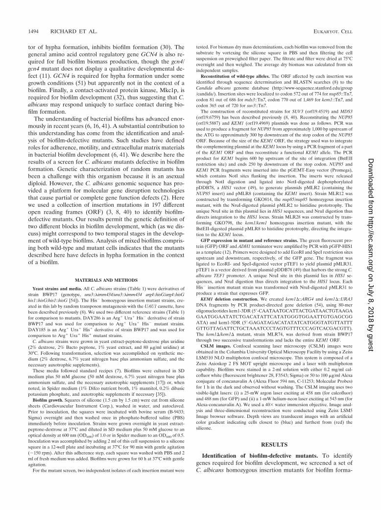
tion ability on slices of silicone catheter material. Screeningwas accomplished by visual inspection of silicone slices in 12-well plates after 60 h of culture. The wild-type reference strainand most insertion mutants formed a thick biofilm that madethe silicone slice appear opaque (Fig. 1A) compared to thetranslucent appearance of an uninoculated control (Fig. 1C).Growth was mainly restricted to the silicone surface; removalof the silicone slice revealed medium with little turbidity,though some biofilm fragments were dislodged (Fig. 1D). Weidentified biofilm-defective strains that met four criteria. First,they caused a less opaque appearance of the silicone surfacethan the reference strain (Fig. 1B). Second, they caused sub-stantial turbidity of the growth medium (Fig. 1E) and grew asrapidly as the reference strain in planktonic culture conditions
(Table 2). Third, biofilm formation defects were evident for atleast two independently created insertion mutants for eachgene. Fourth, reconstitution of a wild-type allele for each in-sertion-bearing gene restored a biofilm formation ability com-parable to that of the reference strain (see below). Insertionsassociated with a defect in biofilm formation lay in four genes:SUV3, NUP85, MDS3, and KEM1.
There is no single unifying theme among the functions ofthese genes, as inferred from studies of their Saccharomycescerevisiae homologs. ScSuv3p is a mitochondrial RNA helicasethat is required for respiratory competence (37). It has beenrecently reported that C. albicans suv3 insertion mutants arealso defective in chlamydospore formation (40). ScNup85p is acomponent of the Nup84 nuclear pore complex that is required
TABLE 1. C. albicans strains
Strain Genotype Description Reference or source
BWP17 ura3::�imm434 arg4::hisG his1::hisG Parent strain 54ura3::�imm434 arg4::hisG his1::hisG
DAY185 ura3::�imm434 ARG4:URA3::arg4::hisG his1::hisG::pHIS1 Arg� Ura� His� 7ura3::�imm434 arg4::hisG his1::hisG reference strain
DAY286 ura3::�imm434 ARG4:URA3::arg4::hisG his1::hisG Arg� Ura� His� 8ura3::�imm434 arg4::hisG his1::hisG reference strain
GKO443 ura3::�imm434 arg4::hisG his1::hisG suv3::Tn7-UAU1 suv3 GKO 8ura3::�imm434 arg4::hisG his1::hisG suv3::Tn7-URA3
GKO798 ura3::�imm434 arg4::hisG his1::hisG kem1::Tn7-UAU1 kem1 GKO 8ura3::�imm434 arg4::hisG his1::hisG kem1::Tn7-URA3
GKO814 ura3::�imm434 arg4::hisG his1::hisG nup85::Tn7-UAU1 nup85 GKO 8ura3::�imm434 arg4::hisG his1::hisG nup85::Tn7-URA3
GKO9 ura3::�imm434 arg4::hisG his1::hisG mds3::Tn7-UAU1 mds3 GKO 8ura3::�imm434 arg4::hisG his1::hisG mds3::Tn7-URA3
MLR12 ura3::�imm434 arg4::hisG his1::hisG::pHIS1-NUP85 nup85::Tn7-UAU1 NUP85 reconstituted strain This studyura3::�imm434 arg4::hisG his1::hisG nup85::Tn7-URA3
MLR28 ura3::�imm434 arg4::hisG his1::hisG kem1::Tn7-UAU1::pHIS1-KEM1 KEM1 reconstituted strain This studyura3::�imm434 arg4::hisG his1::hisG kem1::Tn7-URA3
MLR3 ura3::�imm434 arg4::hisG his1::hisG::pHIS1-SUV3 suv3::Tn7-UAU1 SUV3 reconstituted strain 40ura3::�imm434 arg4::hisG his1::hisG suv3::Tn7-URA3
MLR56 ura3::�imm434 arg4::hisG his1::hisG::pTEF1-GFP suv3::Tn7-UAU1 suv3 GKO tagged with GFP This studyura3::�imm434 arg4::hisG his1::hisG suv3::Tn7-URA3
MLR57 ura3::�imm434 arg4::hisG his1::hisG::pTEF1-GFP kem1::Tn7-UAU1 kem1 GKO tagged with GFP This studyura3::�imm434 arg4::hisG his1::hisG kem1::Tn7-URA3
MLR59 ura3::�imm434 arg4::hisG his1::hisG::pTEF1-GFP nup85::Tn7-UAU1 nup85 GKO tagged with GFP This studyura3::�imm434 arg4::hisG his1::hisG nup85::Tn7-URA3
MLR61 ura3::�imm434 arg4::hisG his1::hisG::pTEF1-GFP mds3::Tn7-UAU1 mds3 GKO tagged with GFP This studyura3::�imm434 arg4::hisG his1::hisG mds3::Tn7-URA3
MLR74 ura3::�imm434 arg4::hisG his1::hisG kem1�::ARG4 kem1 deletion mutant This studyura3::�imm434 arg4::hisG his1::hisG kem1�::URA3
MLR91 ura3::�imm434 arg4::hisG his1::hisG::pHIS1-TEF1 kem1�::ARG4 His� kem1 deletion mutant This studyura3::�imm434 arg4::hisG his1::hisG kem1�::URA3
VIC21 ura3::�imm434 arg4::hisG his1::hisG mds3::Tn7-UAU1::pHIS1-MDS3 MDS3 reconstituted strain 8ura3::�imm434 arg4::hisG his1::hisG mds3::Tn7-URA3
VOL. 4, 2005 BIOFILM FORMATION IN CANDIDA ALBICANS 1495
on July 8, 2018 by guesthttp://ec.asm
.org/D
ownloaded from
for poly(A) plus RNA export to the cytoplasm (13). Mds3pfunctions in C. albicans and S. cerevisiae to promote responsesto alkaline growth conditions (8). In that context, we note thatRim101p, which functions independently of Mds3p to promotealkaline pH responses (8), is not required for biofilm formation(data not shown). ScKem1p, which has exoribonuclease activ-ity, has pleiotropic roles in growth, mRNA turnover, nuclearfusion during mating, and filamentation (21, 23, 25, 26). Giventhat we identified 4 genes from among the 197 represented bythis mutant collection and that there are approximately 5,600nonessential genes in the C. albicans genome (80% nonessen-tial gene fraction multiplied by 7,000 total genes [24]), weestimate that there are roughly 114 genes required for C.albicans biofilm formation.
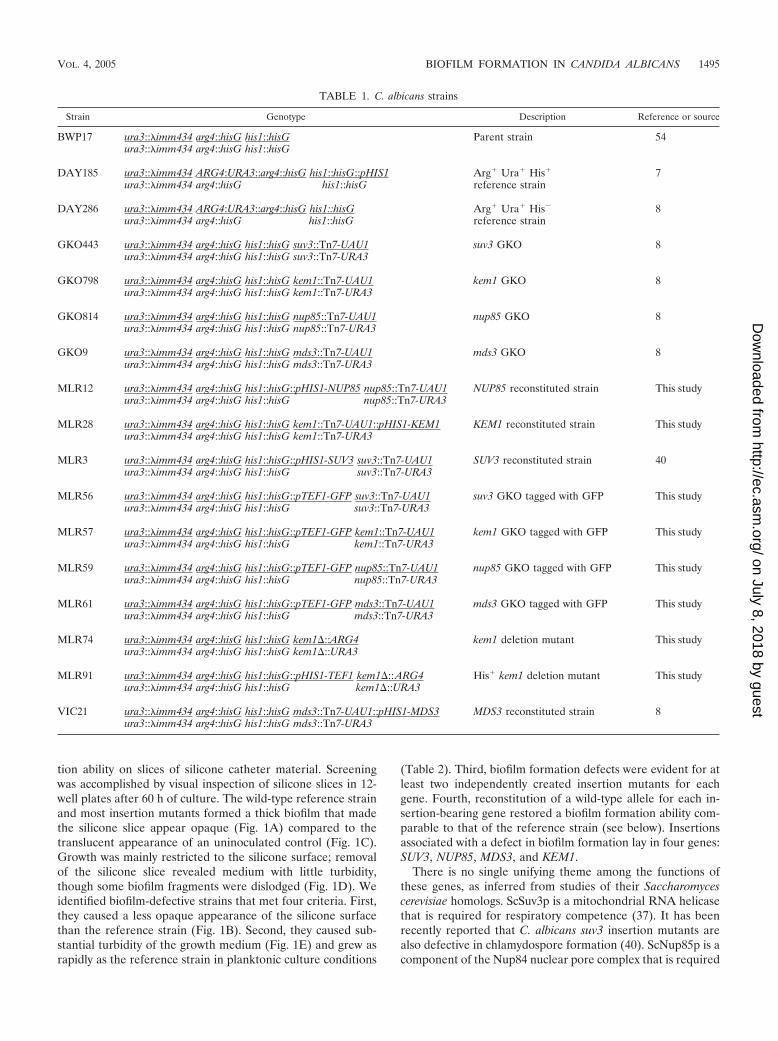
The biofilm defect was quantified by measurement of thebiomass dry weight retrieved from the silicone after 60 h ofculture (Table 2). The mutants showed a dramatic reduction ofbiofilm mass. These findings argue that the mutants are capa-ble of adherence to the silicone surface to produce a rudimen-tary biofilm but are defective in the production of a maturebiofilm.
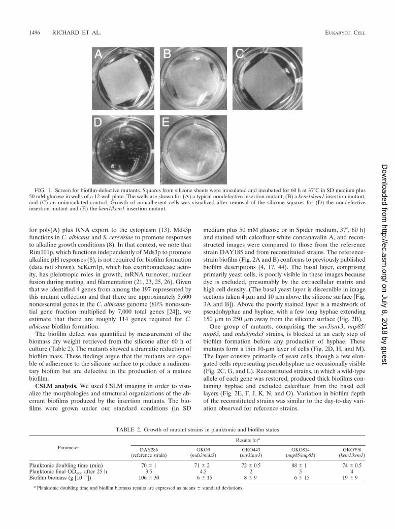
CSLM analysis. We used CSLM imaging in order to visu-alize the morphologies and structural organizations of the ab-errant biofilms produced by the insertion mutants. The bio-films were grown under our standard conditions (in SD
medium plus 50 mM glucose or in Spider medium, 37°, 60 h)and stained with calcofluor white concanavalin A, and recon-structed images were compared to those from the referencestrain DAY185 and from reconstituted strains. The reference-strain biofilm (Fig. 2A and B) conforms to previously publishedbiofilm descriptions (4, 17, 44). The basal layer, comprisingprimarily yeast cells, is poorly visible in these images becausedye is excluded, presumably by the extracellular matrix andhigh cell density. (The basal yeast layer is discernible in imagesections taken 4 �m and 10 �m above the silicone surface [Fig.3A and B]). Above the poorly stained layer is a meshwork ofpseudohyphae and hyphae, with a few long hyphae extending150 �m to 250 �m away from the silicone surface (Fig. 2B).
One group of mutants, comprising the suv3/suv3, nup85/nup85, and mds3/mds3 strains, is blocked at an early step ofbiofilm formation before any production of hyphae. Thesemutants form a thin 10-�m layer of cells (Fig. 2D, H, and M).The layer consists primarily of yeast cells, though a few elon-gated cells representing pseudohyphae are occasionally visible(Fig. 2C, G, and L). Reconstituted strains, in which a wild-typeallele of each gene was restored, produced thick biofilms con-taining hyphae and excluded calcofluor from the basal celllayers (Fig. 2E, F, J, K, N, and O). Variation in biofilm depthof the reconstituted strains was similar to the day-to-day vari-ation observed for reference strains.
FIG. 1. Screen for biofilm-defective mutants. Squares from silicone sheets were inoculated and incubated for 60 h at 37°C in SD medium plus50 mM glucose in wells of a 12-well plate. The wells are shown for (A) a typical nondefective insertion mutant, (B) a kem1/kem1 insertion mutant,and (C) an uninoculated control. Growth of nonadherent cells was visualized after removal of the silicone squares for (D) the nondefectiveinsertion mutant and (E) the kem1/kem1 insertion mutant.
TABLE 2. Growth of mutant strains in planktonic and biofilm states
Parameter
Results fora
DAY286(reference strain)
GKO9(mds3/mds3)
GKO443(suv3/suv3)
GKO814(nup85/nup85)
GKO798(kem1/kem1)
Planktonic doubling time (min) 70 1 71 2 72 0.5 88 1 74 0.5Planktonic final OD600 after 25 h 3.5 4.5 2 5 4Biofilm biomass (g [10�5]) 106 30 6 15 8 9 6 15 19 9
a Planktonic doubling time and biofilm biomass results are expressed as means standard deviations.
1496 RICHARD ET AL. EUKARYOT. CELL
on July 8, 2018 by guesthttp://ec.asm
.org/D
ownloaded from
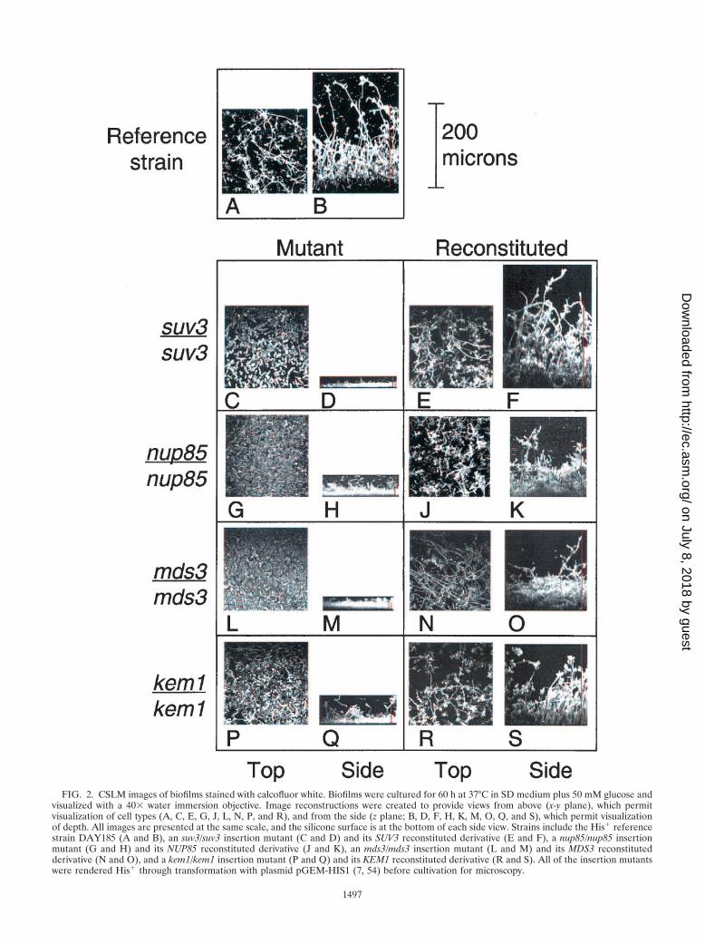
FIG. 2. CSLM images of biofilms stained with calcofluor white. Biofilms were cultured for 60 h at 37°C in SD medium plus 50 mM glucose andvisualized with a 40� water immersion objective. Image reconstructions were created to provide views from above (x-y plane), which permitvisualization of cell types (A, C, E, G, J, L, N, P, and R), and from the side (z plane; B, D, F, H, K, M, O, Q, and S), which permit visualizationof depth. All images are presented at the same scale, and the silicone surface is at the bottom of each side view. Strains include the His� referencestrain DAY185 (A and B), an suv3/suv3 insertion mutant (C and D) and its SUV3 reconstituted derivative (E and F), a nup85/nup85 insertionmutant (G and H) and its NUP85 reconstituted derivative (J and K), an mds3/mds3 insertion mutant (L and M) and its MDS3 reconstitutedderivative (N and O), and a kem1/kem1 insertion mutant (P and Q) and its KEM1 reconstituted derivative (R and S). All of the insertion mutantswere rendered His� through transformation with plasmid pGEM-HIS1 (7, 54) before cultivation for microscopy.
1497
on July 8, 2018 by guesthttp://ec.asm
.org/D
ownloaded from
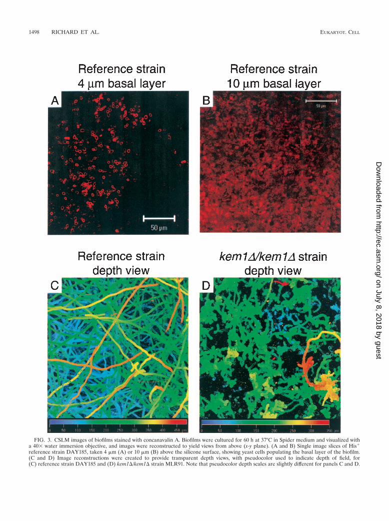
FIG. 3. CSLM images of biofilms stained with concanavalin A. Biofilms were cultured for 60 h at 37°C in Spider medium and visualized witha 40� water immersion objective, and images were reconstructed to yield views from above (x-y plane). (A and B) Single image slices of His�
reference strain DAY185, taken 4 �m (A) or 10 �m (B) above the silicone surface, showing yeast cells populating the basal layer of the biofilm.(C and D) Image reconstructions were created to provide transparent depth views, with pseudocolor used to indicate depth of field, for(C) reference strain DAY185 and (D) kem1�/kem1� strain MLR91. Note that pseudocolor depth scales are slightly different for panels C and D.
1498 RICHARD ET AL. EUKARYOT. CELL
on July 8, 2018 by guesthttp://ec.asm
.org/D
ownloaded from
The kem1/kem1 mutant has a different phenotype than theother mutants, forming a 30-�m biofilm that includes yeastcells, pseudohyphae, and some rare hyphae (Fig. 2P and Q).Reconstitution of a KEM1 allele restored the ability to form anormal biofilm (Fig. 2R and S), and a kem1�/kem1� strainlacking the entire KEM1 ORF produced a 30-�m biofilm sim-ilar in appearance to the insertion mutant biofilm (data notshown). Therefore, a loss of Kem1p function causes a biofilmformation defect. The kem1/kem1 insertion and deletion mu-tant strains both produced thick (�200-�m) biofilms in Spidermedium that were similar in depth to reference-strain biofilms.However, examination of a CSLM depth view revealed a mesh-work comprising almost exclusively long hyphae for the refer-ence strain and primarily pseudohyphae for the kem1�/kem1�strain (Fig. 3C and D, respectively).
We conclude that there are two distinct phenotypic classesof mutants. In addition, restoration of biofilm formation abilityin the reconstituted strains argues that the insertion mutations,rather than secondary mutations in the strains, cause the phe-notypic defect in biofilm formation.
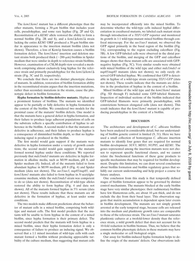
Hypha formation by biofilm-defective mutants. Hyphae area prominent feature of biofilms. The mutants we identifiedappear to be partially or fully defective in hypha formation inthe context of the biofilm. We considered two models for theproximal cause of the mutants’ biofilm defects. One model isthat the mutants have a general defect in hypha formation, andtheir failure to produce large adherent populations of cells onthe substrate reflects a specific contribution of hyphae to ad-herence in the biofilm. A second model is that the mutants aredefective in adherence, and their failure to produce hyphae isa consequence of diminished biofilm depth, so that no hypha-inducing signal is produced in the biofilm.
The first model would gain support if the mutants weredefective in hypha formation under a variety of growth condi-tions; the second model would gain support if the mutantsformed normal hyphae under these conditions. We reportedpreviously that mds3/mds3 mutants are defective in hypha for-mation in alkaline media, such as M199 medium, pH 8, andSpider medium (8). Indeed, all of the mutants failed to formabundant hyphae in M199 medium, pH 8 (Fig. 4) and Spidermedium (data not shown). The suv3/suv3, nup85/nup85, andkem1/kem1 mutants also failed to form hyphae in N-acetylglu-cosamine medium, while the mds3/mds3 strain was competentto do so (data not shown). Reconstitution of wild-type allelesrestored the ability to form hyphae (Fig. 4 and data notshown). All of the mutants formed hyphae in 5% serum (datanot shown). These results indicate that these mutants are alldefective in the formation of hyphae, at least under someconditions.
The two models make different predictions about the behav-ior of mutant cells in a mixed biofilm, comprising both wild-type and mutant cells. The first model predicts that the mu-tants will be unable to form hyphae in the context of a mixedbiofilm, since hypha formation is their primary defect. Thesecond model predicts that the mutants will form hyphae in amixed biofilm, since their hypha formation defect is only aconsequence of failure to produce an inducing signal. We ob-served that a 1:1 mixed inoculum of wild-type cells with eachmutant formed a biofilm without producing appreciable tur-bidity of the culture medium, thus suggesting that mutant cells
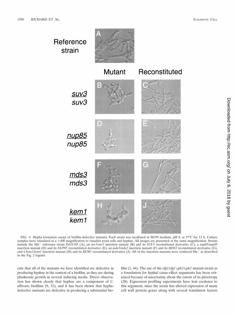
may be incorporated efficiently into the mixed biofilm. Todetermine whether the wild-type cells induced hyphal differ-entiation in cocultured mutants, we labeled each mutant strainthrough introduction of a TEF1-GFP reporter and monitoredits growth in 1:1 wild-type:mutant mixed biofilms through con-focal microscopy. For the suv3/suv3 mutant, we observed theGFP signal primarily in the basal region of the biofilm (Fig.5A), corresponding to the region excluding calcofluor (Fig.5B). A few GFP-labeled cells were observed in the distal por-tions of the biofilm, and merging of the GFP and calcofluorimages shows that these mutant cells are associated with GFP-negative hyphae (Fig. 5C). Very similar results were obtainedwith mixed biofilms of the wild type and either mds3/mds3 ornup85/nup85 mutants (data not shown); we only rarely ob-served GFP-labeled hyphae. We confirmed that GFP is detect-able in hyphae of a wild-type strain carrying TEF1-GFP (datanot shown). These results indicate that the mutants are defec-tive in the production of hyphae in the context of a biofilm.
Mixed biofilms of the wild type and the kem1/kem1 mutant(Fig. 5D through F) included some GFP-labeled filaments.Inspection of individual sections of the biofilm indicated thatGFP-labeled filaments were primarily pseudohyphae, withconstrictions between elongated cells (data not shown). Thisresult indicates that the kem1/kem1 mutant is capable of pro-ducing pseudohyphae in the context of a biofilm.
DISCUSSION
The architecture and development of C. albicans biofilmshave been analyzed in considerable detail, but our understand-ing of biofilm genetic control is limited (9, 31). Here we haveused a small collection of homozygous C. albicans insertionmutants to identify four genes that are required for normalbiofilm development: SUV3, MDS3, NUP85, and KEM1. Thegenes represented among the insertion mutants were not cho-sen to represent specific functional classes, and we cannotrelate the biochemical functions of these gene products tospecific mechanisms that may be required for biofilm develop-ment. Despite this limitation, we can draw several conclusionsabout biofilm formation and biofilm regulatory genes that so-lidify our current understanding and help project a course forfuture analyses.
One conclusion from this study is that temporally definedstages of biofilm formation appear to be governed by distinctcontrol mechanisms. The mutants blocked at the early biofilmstage have very similar phenotypes: their rudimentary biofilmshave few filamentous cells, are about 10 �m thick, and do notexclude the dye from their basal layer. This observation sug-gests that matrix accumulation is dependent upon later eventsin biofilm development. The mutants are not simply growtharrested at the early temporal stage, because cells are releasedinto the medium and planktonic growth rates are comparableto those of the reference strain. The suv3/suv3 mutant saturatesplanktonic cultures at a twofold-lower density than the refer-ence strain, a mild growth defect that does not account for its10-fold reduction in biofilm biomass. Thus, we believe that thecommon biofilm phenotypic defects in these mutants may havea single molecular or cell biological origin.
Our assay for biofilm-induced hypha formation helps to de-fine the origin of the mutants’ defects. Our observations indi-
VOL. 4, 2005 BIOFILM FORMATION IN CANDIDA ALBICANS 1499
on July 8, 2018 by guesthttp://ec.asm
.org/D
ownloaded from
cate that all of the mutants we have identified are defective inproducing hyphae in the context of a biofilm, as they are duringplanktonic growth in several inducing media. Direct observa-tion has shown clearly that hyphae are a component of C.albicans biofilms (9, 31), and it has been shown that hypha-defective mutants are defective in producing a substantial bio-
film (1, 44). The use of the efg1/efg1 cph1/cph1 mutant strain asa foundation for hyphal cause-effect arguments has been crit-icized because of uncertainty about the extent of its pleiotropy(28). Expression profiling experiments have lent credence tothis argument, since the strain has altered expression of manycell wall protein genes along with several translation factors
FIG. 4. Hypha formation assays of biofilm-defective mutants. Each strain was incubated in M199 medium, pH 8, at 37°C for 12 h. Culturesamples were visualized at a �400 magnification to visualize yeast cells and hyphae. All images are presented at the same magnification. Strainsinclude the His� reference strain DAY185 (A), an suv3/suv3 insertion mutant (B) and its SUV3 reconstituted derivative (C), a nup85/nup85insertion mutant (D) and its NUP85 reconstituted derivative (E), an mds3/mds3 insertion mutant (F) and its MDS3 reconstituted derivative (G),and a kem1/kem1 insertion mutant (H) and its KEM1 reconstituted derivative (J). All of the insertion mutants were rendered His� as describedin the Fig. 2 legend.
1500 RICHARD ET AL. EUKARYOT. CELL
on July 8, 2018 by guesthttp://ec.asm
.org/D
ownloaded from
and transport modulators (39, 48). These gene expressionchanges may account for the altered adherence properties ofefg1/efg1 cph1/cph1 yeast cells when grown under biofilm-in-ducing conditions (11). Our suv3/suv3, mds3/mds3, and nup85/nup85 mutants have more subtle hypha formation defects; forexample, they produce hyphae in response to serum. There-fore, their biofilm-deficient phenotype strengthens the argu-ment that mature biofilm formation depends upon hyphae,because these mutants clearly adhere to the silicone substrateand can populate the basal biofilm layer. Given that the mu-tants can be incorporated efficiently into mixed biofilms, weconclude that their adherence defect can be remedied by thepresence of hyphae. Thus, we infer that the mutants’ primarydefect is in hypha formation and that the failure of mutant cellsto adhere to their 10-�m biofilms is a consequence of the lackof hyphae.
If these mutants’ biofilm defects arise from a hypha forma-tion defect, it follows then that hyphae provide unique adher-ent surfaces that facilitate the formation of thick biofilms. Thisidea is in keeping with the increased expression of several ALSgenes, which specify a surface adhesin family (19), in biofilmscompared to planktonic cultures (4, 11, 15) and with the factthat several ALS genes are preferentially expressed duringhyphal growth (10, 19, 20). The recent finding that adherenceregulator Bcr1p is required for biofilm formation (40a) alsosupports this view.
The kem1/kem1 mutant is clearly blocked at a different stageof biofilm formation than are the other mutants describedhere, based on the mass and depth of its rudimentary biofilm.In addition, its biofilms include substantial numbers ofpseudohyphal cells, distinguishable by their elongated mor-phology and growth in filamentous chains. The extent of itsbiofilm defect is dependent on the medium, a feature com-monly found among bacterial biofilm mutants (16, 41). How-ever, kem1/kem1 biofilms remain deficient in hyphae in bothSD medium plus 50 mM glucose and Spider medium. Theproperties of the kem1/kem1 mutant raise a type of questionthat is commonly encountered in analyses of developmentalpathways. The question is whether the kem1/kem1 terminalbiofilm phenotype represents a true intermediate stage in nor-mal biofilm development or whether it is an aberrant responseto the primary mutant defect. The temporal analyses of Chan-dra et al. and of Hawser and Douglas did not reveal a stage inwhich only yeast cells and pseudohyphae were present (4, 17).In addition, we have not seen a distinct pseudohyphal layer inoptical sections of biofilms. Thus, we believe that the prepon-derance of pseudohyphal cells produced by the kem1/kem1mutant does not reflect an intermediate stage in biofilm for-mation.
ACKNOWLEDGMENTS
We thank Theresa Swayne, Sudhindra Swamy, and Peter Carroll fortheir help with the use of the confocal microscope at the ColumbiaUniversity Optical Microscopy Facility. We gratefully acknowledge theavailability of the C. albicans genome sequence provided by the Stan-ford DNA Sequencing and Technology Center supported by theNIDR, NIH, and Burroughs Wellcome Fund, without which this workwould not have been possible.
This work was supported by Public Health Services grants R01AI50931, T32 AI07161 (V.M.B.), and T32 DK007786 (C.J.N.), forwhich we are very grateful. The Optical Microscopy Facility was es-
FIG. 5. Confocal images of mixed biofilms. Biofilms were culturedfor 60 h at 37°C in SD medium plus 50 mM glucose and visualized witha 40� water immersion objective. The images are oriented with sili-cone at the bottom of each panel. Biofilms were created from a 1:1inoculum of reference strain DAY185 with (A, B, and C) GFP-taggedsuv3/suv3 strain MLR56 or (D, E, and F) GFP-tagged kem1/kem1strain MLR57. Images show (A and D) GFP alone, (B and E) cal-cofluor alone, or (C and F) both GFP and calcofluor signals merged.
VOL. 4, 2005 BIOFILM FORMATION IN CANDIDA ALBICANS 1501
on July 8, 2018 by guesthttp://ec.asm
.org/D
ownloaded from
tablished by NIH Shared Instrumentation Grants S10 RR10506 andS10 RR13701 and by the Lieber Foundation. It is supported by NIHGrant P30 CA13696 as part of the Herbert Irving ComprehensiveCancer Center at Columbia University.
REFERENCES
1. Baillie, G. S., and L. J. Douglas. 1999. Role of dimorphism in the develop-ment of Candida albicans biofilms. J. Med. Microbiol. 48:671–679.
2. Bruno, V. M., and A. P. Mitchell. 2004. Large-scale gene function analysis inCandida albicans. Trends Microbiol. 12:157–161.
3. Bruno, V. M., and A. P. Mitchell. 2005. Regulation of azole drug suscepti-bility by Candida albicans protein kinase CK2. Mol. Microbiol. 56:559–573.
4. Chandra, J., D. M. Kuhn, P. K. Mukherjee, L. L. Hoyer, T. McCormick, andM. A. Ghannoum. 2001. Biofilm formation by the fungal pathogen Candidaalbicans: development, architecture, and drug resistance. J. Bacteriol. 183:5385–5394.
5. Cossart, P., and P. J. Sansonetti. 2004. Bacterial invasion: the paradigms ofenteroinvasive pathogens. Science 304:242–248.
6. Davey, M. E., and G. A. O’Toole. 2000. Microbial biofilms: from ecology tomolecular genetics. Microbiol. Mol. Biol. Rev. 64:847–867.
7. Davis, D., J. E. Edwards, Jr., A. P. Mitchell, and A. S. Ibrahim. 2000.Candida albicans RIM101 pH response pathway is required for host-patho-gen interactions. Infect. Immun. 68:5953–5959.
8. Davis, D. A., V. M. Bruno, L. Loza, S. G. Filler, and A. P. Mitchell. 2002.Candida albicans Mds3p, a conserved regulator of pH responses and viru-lence identified through insertional mutagenesis. Genetics 162:1573–1581.
9. Douglas, L. J. 2003. Candida biofilms and their role in infection. TrendsMicrobiol. 11:30–36.
10. Fu, Y., A. S. Ibrahim, D. C. Sheppard, Y. C. Chen, S. W. French, J. E. Cutler,S. G. Filler, and J. E. Edwards, Jr. 2002. Candida albicans Als1p: an adhesinthat is a downstream effector of the EFG1 filamentation pathway. Mol.Microbiol. 44:61–72.
11. Garcia-Sanchez, S., S. Aubert, I. Iraqui, G. Janbon, J. M. Ghigo, and C.d’Enfert. 2004. Candida albicans biofilms: a developmental state associatedwith specific and stable gene expression patterns. Eukaryot. Cell 3:536–545.
12. Gerami-Nejad, M., J. Berman, and C. A. Gale. 2001. Cassettes for PCR-mediated construction of green, yellow, and cyan fluorescent protein fusionsin Candida albicans. Yeast 18:859–864.
13. Goldstein, A. L., C. A. Snay, C. V. Heath, and C. N. Cole. 1996. Pleiotropicnuclear defects associated with a conditional allele of the novel nucleoporinRat9p/Nup85p. Mol. Biol. Cell 7:917–934.
14. Gow, N. A., A. J. Brown, and F. C. Odds. 2002. Fungal morphogenesis andhost invasion. Curr. Opin. Microbiol. 5:366–371.
15. Green, C. B., G. Cheng, J. Chandra, P. Mukherjee, M. A. Ghannoum, andL. L. Hoyer. 2004. RT-PCR detection of Candida albicans ALS gene expres-sion in the reconstituted human epithelium (RHE) model of oral candidiasisand in model biofilms. Microbiology 150:267–275.
16. Hall-Stoodley, L., J. W. Costerton, and P. Stoodley. 2004. Bacterial biofilms:from the natural environment to infectious diseases. Nat. Rev. Microbiol.2:95–108.
17. Hawser, S. P., and L. J. Douglas. 1994. Biofilm formation by Candida specieson the surface of catheter materials in vitro. Infect. Immun. 62:915–921.
18. Hawser, S. P., and L. J. Douglas. 1995. Resistance of Candida albicansbiofilms to antifungal agents in vitro. Antimicrob. Agents Chemother. 39:2128–2131.
19. Hoyer, L. L. 2001. The ALS gene family of Candida albicans. Trends Micro-biol. 9:176–180.
20. Hoyer, L. L., T. L. Payne, M. Bell, A. M. Myers, and S. Scherer. 1998.Candida albicans ALS3 and insights into the nature of the ALS gene family.Curr. Genet. 33:451–459.
21. Interthal, H., C. Bellocq, J. Bahler, V. I. Bashkirov, S. Edelstein, and W. D.Heyer. 1995. A role of Sep1 ( Kem1, Xrn1) as a microtubule-associatedprotein in Saccharomyces cerevisiae. EMBO J. 14:1057–1066.
22. Jin, Y., H. K. Yip, Y. H. Samaranayake, J. Y. Yau, and L. P. Samaranayake.2003. Biofilm-forming ability of Candida albicans is unlikely to contribute tohigh levels of oral yeast carriage in cases of human immunodeficiency virusinfection. J. Clin. Microbiol. 41:2961–2967.
23. Johnson, A. W. 1997. Rat1p and Xrn1p are functionally interchangeableexoribonucleases that are restricted to and required in the nucleus andcytoplasm, respectively. Mol. Cell. Biol. 17:6122–6130.
24. Jones, T., N. A. Federspiel, H. Chibana, J. Dungan, S. Kalman, B. B. Magee,G. Newport, Y. R. Thorstenson, N. Agabian, P. T. Magee, R. W. Davis, andS. Scherer. 2004. The diploid genome sequence of Candida albicans. Proc.Natl. Acad. Sci. USA 101:7329–7334.
25. Kim, J. 2002. KEM1 is involved in filamentous growth of Saccharomycescerevisiae. FEMS Microbiol. Lett. 216:33–38.
26. Kim, J., P. O. Ljungdahl, and G. R. Fink. 1990. kem mutations affect nuclearfusion in Saccharomyces cerevisiae. Genetics 126:799–812.
27. Klemba, M., and D. E. Goldberg. 2002. Biological roles of proteases inparasitic protozoa. Annu. Rev. Biochem. 71:275–305.
28. Kobayashi, S. D., and J. E. Cutler. 1998. Candida albicans hyphal formationand virulence: is there a clearly defined role? Trends Microbiol. 6:92–94.
29. Kojic, E. M., and R. O. Darouiche. 2004. Candida infections of medicaldevices. Clin. Microbiol. Rev. 17:255–267.
30. Kruppa, M., B. P. Krom, N. Chauhan, A. V. Bambach, R. L. Cihlar, and R. A.Calderone. 2004. The two-component signal transduction protein Chk1p regu-lates quorum sensing in Candida albicans. Eukaryot. Cell 3:1062–1065.
31. Kumamoto, C. A. 2002. Candida biofilms. Curr. Opin. Microbiol. 5:608–611.32. Kumamoto, C. A. 2005. A contact-activated kinase signals Candida albicans
invasive growth and biofilm development. Proc. Natl. Acad. Sci. USA 102:5576–5581.
33. Li, X., Z. Yan, and J. Xu. 2003. Quantitative variation of biofilms among strainsin natural populations of Candida albicans. Microbiology 149:353–362.
34. Liu, H. 2001. Transcriptional control of dimorphism in Candida albicans.Curr. Opin. Microbiol. 4:728–735.
35. Liu, H., J. Kohler, and G. R. Fink. 1994. Suppression of hyphal formation inCandida albicans by mutation of a STE12 homolog. Science 266:1723–1726.
36. Lo, H. J., J. R. Kohler, B. DiDomenico, D. Loebenberg, A. Cacciapuoti, andG. R. Fink. 1997. Nonfilamentous C. albicans mutants are avirulent. Cell90:939–949.
37. Minczuk, M., A. Dmochowska, M. Palczewska, and P. P. Stepien. 2002.Overexpressed yeast mitochondrial putative RNA helicase Mss116 partiallyrestores proper mtRNA metabolism in strains lacking the Suv3 mtRNAhelicase. Yeast 19:1285–1293.
38. Naglik, J. R., S. J. Challacombe, and B. Hube. 2003. Candida albicanssecreted aspartyl proteinases in virulence and pathogenesis. Microbiol. Mol.Biol. Rev. 67:400–428.
39. Nantel, A., D. Dignard, C. Bachewich, D. Harcus, A. Marcil, A. P. Bouin,C. W. Sensen, H. Hogues, M. van het Hoog, P. Gordon, T. Rigby, F. Benoit,D. C. Tessier, D. Y. Thomas, and M. Whiteway. 2002. Transcription profilingof Candida albicans cells undergoing the yeast-to-hyphal transition. Mol.Biol. Cell 13:3452–3465.
40. Nobile, C. J., V. M. Bruno, M. L. Richard, D. Davis, and A. P. Mitchell. 2003.Genetic control of chlamydospore formation in Candida albicans. Microbi-ology 149:3629–3637.
40a.Nobile, C. J., and A. P. Mitchell. 2005. Regulation of cell-surface genes andbiofilm formation by the C. albicans transcription factor Bcr1p. Curr. Biol.15:1150–1155.
41. O’Toole, G., H. B. Kaplan, and R. Kolter. 2000. Biofilm formation as mi-crobial development. Annu. Rev. Microbiol. 54:49–79.
42. Ramage, G., S. Bachmann, T. F. Patterson, B. L. Wickes, and J. L. Lopez-Ribot. 2002. Investigation of multidrug efflux pumps in relation to flucon-azole resistance in Candida albicans biofilms. J. Antimicrob. Chemother.49:973–980.
43. Ramage, G., K. Vande Walle, B. L. Wickes, and J. L. Lopez-Ribot. 2001.Standardized method for in vitro antifungal susceptibility testing of Candidaalbicans biofilms. Antimicrob. Agents Chemother. 45:2475–2479.
44. Ramage, G., K. VandeWalle, J. L. Lopez-Ribot, and B. L. Wickes. 2002. Thefilamentation pathway controlled by the Efg1 regulator protein is requiredfor normal biofilm formation and development in Candida albicans. FEMSMicrobiol. Lett. 214:95–100.
45. Reagan, D. R., M. A. Pfaller, R. J. Hollis, and R. P. Wenzel. 1990. Charac-terization of the sequence of colonization and nosocomial candidemia usingDNA fingerprinting and a DNA probe. J. Clin. Microbiol. 28:2733–2738.
46. Riggle, P. J., K. A. Andrutis, X. Chen, S. R. Tzipori, and C. A. Kumamoto.1999. Invasive lesions containing filamentous forms produced by a Candidaalbicans mutant that is defective in filamentous growth in culture. Infect.Immun. 67:3649–3652.
47. Shin, J. H., S. J. Kee, M. G. Shin, S. H. Kim, D. H. Shin, S. K. Lee, S. P. Suh,and D. W. Ryang. 2002. Biofilm production by isolates of Candida speciesrecovered from nonneutropenic patients: comparison of bloodstream iso-lates with isolates from other sources. J. Clin. Microbiol. 40:1244–1248.
48. Sohn, K., C. Urban, H. Brunner, and S. Rupp. 2003. EFG1 is a majorregulator of cell wall dynamics in Candida albicans as revealed by DNAmicroarrays. Mol. Microbiol. 47:89–102.
49. Spreghini, E., D. A. Davis, R. Subaran, M. Kim, and A. P. Mitchell. 2003.Roles of Candida albicans Dfg5p and Dcw1p cell surface proteins in growthand hypha formation. Eukaryot. Cell 2:746–755.
50. Stoodley, P., K. Sauer, D. G. Davies, and J. W. Costerton. 2002. Biofilms ascomplex differentiated communities. Annu. Rev. Microbiol. 56:187–209.
51. Tripathi, G., C. Wiltshire, S. Macaskill, H. Tournu, S. Budge, and A. J.Brown. 2002. Gcn4 co-ordinates morphogenetic and metabolic responses toamino acid starvation in Candida albicans. EMBO J. 21:5448–5456.
52. Tucker, S. L., and N. J. Talbot. 2001. Surface attachment and pre-penetra-tion stage development by plant pathogenic fungi. Annu. Rev. Phytopathol.39:385–417.
53. Voss, A., R. J. Hollis, M. A. Pfaller, R. P. Wenzel, and B. N. Doebbeling.1994. Investigation of the sequence of colonization and candidemia in non-neutropenic patients. J. Clin. Microbiol. 32:975–980.
54. Wilson, R. B., D. Davis, and A. P. Mitchell. 1999. Rapid hypothesis testingwith Candida albicans through gene disruption with short homology regions.J. Bacteriol. 181:1868–1874.
1502 RICHARD ET AL. EUKARYOT. CELL
on July 8, 2018 by guesthttp://ec.asm
.org/D
ownloaded from