Embed Size (px)
Citation preview
From the Midwestern Vascular Surgical Society
Carotid artery velocity characteristics after carotidartery angioplasty and stentingSantiago Chahwan, MD, M. Todd Miller, MD, John P. Pigott, MD, Ralph C. Whalen, MD,Linda Jones, RVT, and Anthony J. Comerota, MD, Toledo, Ohio
Objective: Correlation of carotid duplex ultrasound (DUS) flow velocities with carotid artery stenosis before and aftercarotid endarterectomy is well established. With the evolution of catheter-based techniques, carotid stenosis increasinglyis being treated with angioplasty and stenting (CAS). CAS changes the physical properties of the arterial wall, which mayalter blood flow velocities compared with the nonstented carotid. Opinions differ about whether DUS is a reliable toolto assess technical outcome and recurrent stenosis after CAS. This study correlated carotid DUS flow velocity findingswith carotid arteriography after CAS.Methods: Data from 77 pairs of carotid arteriograms with corresponding DUS after CAS in 68 patients were reviewed.Preintervention and postintervention DUS and carotid arteriogram data were evaluated for each patient. Peak systolicvelocities (PSV), end-diastolic velocities (EDV), and internal carotid artery/common carotid artery ratios (ICA/CCA)were correlated with the post-CAS arteriogram.Results: The mean preintervention PSV was 390 � 110 cm/s (range, 216 to 691 cm/s), and the average EDV was 134 �51 cm/s (range, 35 to 314 cm/s). Postintervention DUS was obtained a mean of 5 days after CAS (range, 1 to 30 days).Sixty (81%) post-CAS arteriograms were normal, and each corresponded to a normal postintervention DUS (PSV range,30 to 118 cm/s; EDV range, 18 to 60 cm/s). In 14 arteries (19%), completion arteriograms revealed residual stenoses of20% to 40% in 13, and 50% in one. The mean PSV was 175 cm/s (range, 137 to 195 cm/s), and the mean EDV was 44cm/s (range, 20 to 62 cm/s). All velocities exceeded the threshold of a 50% stenosis by DUS criteria for a nonstentedcarotid artery. In three arteries (2 patients), high-grade recurrent stenoses detected by DUS developed that requiredreintervention during follow-up. This high-grade restenosis was confirmed by arteriography in each patient, providing anadditional three correlations.Conclusions: Normal DUS imaging reliably identifies arteriographically normal carotid arteries after CAS. Carotidvelocities are disproportionately elevated with mild and moderate degrees of stenoses, and velocity criteria for quanti-tating stenoses in these patients require modification. However, DUS appropriately identifies severe recurrent stenoses
after CAS. (J Vasc Surg 2007;45:523-6.)Duplex ultrasonography (DUS) is the diagnostic test ofchoice for patients with native vessel carotid stenosis. It iswell standardized and has been used by many as the onlydiagnostic test before carotid endarterectomy1 and as areliable method of follow-up after carotid endarterectomy. 2
Carotid angioplasty and stenting (CAS) is an increas-ingly popular treatment option for patients with carotidartery stenoses. As catheter-based technology evolves, CASis being offered more frequently, especially in asymptom-atic patients.3,4 Noninvasive follow-up is becoming increas-ingly important in this growing patient population.
CAS changes an expansile artery to a relatively rigidtube, reducing the compliance of the stented artery andincreasing its elastic modulus.5 It is intuitive that thesephysical alterations of the vessel wall will impact blood flowcharacteristics through the treated segment, and this hasbeen observed by others when studied.5,6 The purpose of
From the Jobst Vascular Center.Competition of interest: none.Presented at the 2006 Annual Meeting of the Midwestern Vascular Surgical
Society, Cleveland, Ohio, Sept 7-9, 2006.Reprint requests: Anthony J. Comerota, MD, FACS, FACC, Jobst Vascular
Center, 2109 Hughes Dr, Suite 400, Toledo, OH 43606 (e-mail:[email protected]).
0741-5214/$32.00Copyright © 2007 by The Society for Vascular Surgery.
doi:10.1016/j.jvs.2006.11.044this study was to correlate the carotid artery velocities witharteriography after CAS.
METHODS
All patients undergoing angioplasty and stenting for ca-rotid artery stenosis between November 2000 and October2006 were reviewed. Patients were identified from theJobst Vascular Registry. Patient and carotid artery–relatedinformation were recorded from databases and corrobo-rated with vascular laboratory records, operative notes,physicians’ notes, and patients’ medical records. The studywas approved by the ProMedica Health System Institu-tional Review Board.
Seventy-one primary carotid artery stenoses werestented in 68 patients. Indications for CAS included symp-tomatic patients with �50% stenosis on carotid DUS andasymptomatic patients with �70% stenoses. Fifty proce-dures were performed for asymptomatic carotid stenoses(70%) and 21 for symptomatic high-grade stenoses (30%).Three recurrent stenoses (2 patients) after CAS underwentrepeat arteriography and DUS during follow-up. Thesepatients subsequently received repeat CAS with postoper-ative arteriography and DUS, yielding a total of 77 pairs ofarteriograms with corresponding DUS images.
The CAS procedures were performed under local anes-
thesia and intravenous sedation through retrograde access523
JOURNAL OF VASCULAR SURGERYMarch 2007524 Chahwan et al
from the common femoral artery. Cerebral protection de-vices were used in most of the cases. A self-expandingnitinol stent was deployed in all cases in the internal carotidartery (ICA), with extension into the common carotidartery (CCA) if needed. After stent deployment, in-stentdilatation was performed using a 5-mm balloon. Closuredevices for the femoral puncture were used in most of thecases. Patients received combined platelet inhibition withaspirin and clopidogrel before and after the procedure.
Triplanar arteriography was performed on all stenoses.Carotid DUS was performed using an ATL HDI 5000(Bothell, Wash) or Siemens Antares ultrasound unit (Munich,Germany) by registered vascular technologists from theJobst Vascular Laboratory (accredited by the IntersocietalCommission for the Accreditation of Vascular Laborato-ries) under the supervision of the attending vascular physi-cian. Patients were examined using high-resolution, multi-frequency transducers selected according to vessel depth(usually 5 to 7 MHz).
All spectral waveforms were obtained with a Dopplersample volume of 1.0 to 1.5 mm and an angle of �60degrees. Velocities were obtained from the common, inter-nal, external, and vertebral arteries bilaterally. Velocitieswere obtained proximal and distal to the stent and in theproximal, mid, and distal stent. The highest peak systolicvelocity (PSV) and end-diastolic velocity (EDV) valueswere identified and used to indicate the location and max-imum diameter reduction. An ICA/CCA ratio was calcu-lated using the highest PSV of the ICA and CCA andreported for all but two arteries. Peak systolic velocities�125 cm/s identified a �50% stenosis. Stenoses of �70%were defined by a PSV �300 cm/s or an EDV of 100 to139 cm/s. Lesions with a stenosis that generated an EDV�140 cm/s were interpreted as �80% stenosis. The ICA/CCA ratio was not prospectively used to quantitate thedegree of stenosis.
Patients underwent carotid DUS immediately beforethe procedures, and all patients had carotid DUS per-formed �30 days of their arteriogram. During follow-up,DUS detected severe stenosis in two patients (3 carotidarteries), and they underwent repeat carotid arteriographyand repeated CAS. All arteriographic stenoses were calcu-lated using the North American Symptomatic Carotid End-arterectomy Trial (NASCET) trial criteria.7
RESULTS
Sixty-eight patients underwent angioplasty and stent-ing of 71 primary carotid lesions. Two of the 68 patientspresented with three in-stent carotid restenoses during thefollow-up period (6 to 12 months) and underwent subse-quent reintervention with CAS. This resulted in 74 CASprocedures for this cohort of patients, which consisted of39 (57%) men and 29 (43%) women. Asymptomatic steno-ses resulted in 56 procedures (62%). In the symptomaticcohort, transient ischemic attacks occurred in 14 patientsand stroke in four.
Preoperative carotid DUS revealed an average PSV of
390 � 110 cm/s (range, 216 to 691 cm/s) and an averageEDV of 134 � 51 cm/s (range, 35 to 314 cm/s). Postint-ervention DUS was obtained at a mean of 5 days after CAS(range, 1 to 30 days). The average postintervention PSVwas 102 � 41 cm/s (range, 30 to 184 cm/s), and theaverage EDV was 29 � 18 cm/s (range, 18 to 51 cm/s).
Normal results for postintervention arteriograms wereobserved after 60 procedures (81%). The post-DUS veloc-ity for this group showed a mean PSV of 85 cm/s (range,30 to 118 cm/s) and a mean EDV of 25 cm/s (range, 18 to60 cm/s). No velocities exceeded the threshold for 50%stenosis by DUS criteria for a nonstented carotid vessel;therefore, a normal PSV (�125 cm/s) yielded a 100%negative predictive value (NPV) correlated to arteriogra-phy. The remaining 14 stented carotid arteries (19%)showed residual stenoses of 20% to 50% on their comple-tion arteriogram. The post-DUS velocity revealed a meanPSV for this group of 175 cm/s (range, 137 to 195 cm/s)and an EDV of 44 cm/s (range, 20 to 62 cm/s). All PSVswere abnormal and exceeded the threshold for 50% stenosisby interpretation criteria applied to a nonstented carotidartery (PSV �125 cm/s); however, only one artery reached50% stenosis by arteriogram (PSV, 195 cm/s). High-gradein-stent restenoses developed in three carotid arteries (2patients) and they required reintervention during follow-up between 6 and 12 months. The high-grade restenosesdetected by DUS were confirmed by arteriography in eachcarotid artery. The mean PSV was 365 cm/s (range, 300 to401 cm/s), and the EDV was 110 cm/s (range, 96 to 120cm/s). Abnormal arteriograms with corresponding DUSare listed in the Table. Discrimination of PSV with arterio-graphic stenoses is plotted in Fig 1. After repeat CAS inthese three restenosed carotid arteries, completion arte-riography demonstrated 30% residual stenosis in one ca-rotid artery, which had a PSV of 157 cm/s, and no residual
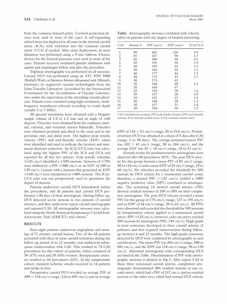
Table. Arteriographic stenoses correlated with velocityvalues in patients with any degree of luminal narrowing
CAS Stenosis % PSV (cm/s) EDV (cm/s) ICA/CCA
1 90 401 120 4.92 85 327 98 4.73 85 300 96 3.54 50 195 38 1.95 40 185 52 1.96 30 184 34 1.17 40 177 44 1.58 30 175 43 2.79 30 174 62
10 30 166 40 1.511 20 164 5712 20 158 30 1.413 30 157 20 2.914 20 147 22 1.815 30 140 30 1.316 20 137 31 1.817 20 150 50 2.2
CAS, Carotid artery stenting; PSV, peak systolic velocity; EDV, end-diastolicvelocity; ICA, internal carotid artery; CCA, common carotid artery.
stenosis in the other two, which had normal DUS velocity
JOURNAL OF VASCULAR SURGERYVolume 45, Number 3 Chahwan et al 525
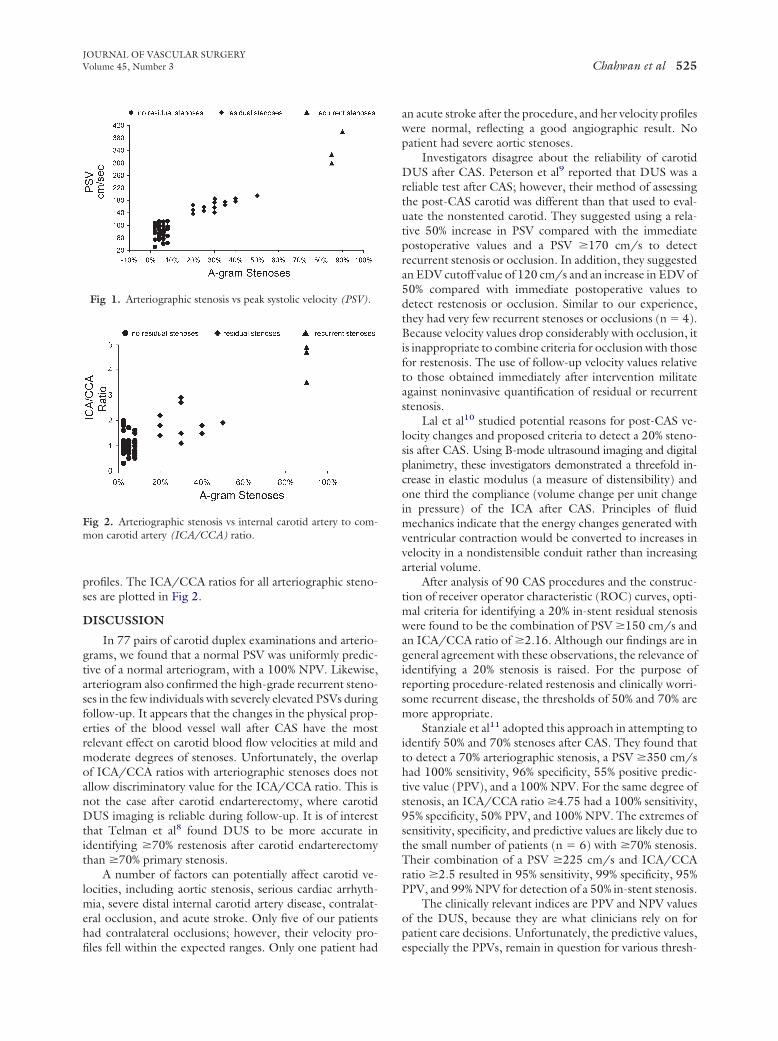
profiles. The ICA/CCA ratios for all arteriographic steno-ses are plotted in Fig 2.
DISCUSSION
In 77 pairs of carotid duplex examinations and arterio-grams, we found that a normal PSV was uniformly predic-tive of a normal arteriogram, with a 100% NPV. Likewise,arteriogram also confirmed the high-grade recurrent steno-ses in the few individuals with severely elevated PSVs duringfollow-up. It appears that the changes in the physical prop-erties of the blood vessel wall after CAS have the mostrelevant effect on carotid blood flow velocities at mild andmoderate degrees of stenoses. Unfortunately, the overlapof ICA/CCA ratios with arteriographic stenoses does notallow discriminatory value for the ICA/CCA ratio. This isnot the case after carotid endarterectomy, where carotidDUS imaging is reliable during follow-up. It is of interestthat Telman et al8 found DUS to be more accurate inidentifying �70% restenosis after carotid endarterectomythan �70% primary stenosis.
A number of factors can potentially affect carotid ve-locities, including aortic stenosis, serious cardiac arrhyth-mia, severe distal internal carotid artery disease, contralat-eral occlusion, and acute stroke. Only five of our patientshad contralateral occlusions; however, their velocity pro-
Fig 1. Arteriographic stenosis vs peak systolic velocity (PSV).
Fig 2. Arteriographic stenosis vs internal carotid artery to com-mon carotid artery (ICA/CCA) ratio.
files fell within the expected ranges. Only one patient had
an acute stroke after the procedure, and her velocity profileswere normal, reflecting a good angiographic result. Nopatient had severe aortic stenoses.
Investigators disagree about the reliability of carotidDUS after CAS. Peterson et al9 reported that DUS was areliable test after CAS; however, their method of assessingthe post-CAS carotid was different than that used to eval-uate the nonstented carotid. They suggested using a rela-tive 50% increase in PSV compared with the immediatepostoperative values and a PSV �170 cm/s to detectrecurrent stenosis or occlusion. In addition, they suggestedan EDV cutoff value of 120 cm/s and an increase in EDV of50% compared with immediate postoperative values todetect restenosis or occlusion. Similar to our experience,they had very few recurrent stenoses or occlusions (n � 4).Because velocity values drop considerably with occlusion, itis inappropriate to combine criteria for occlusion with thosefor restenosis. The use of follow-up velocity values relativeto those obtained immediately after intervention militateagainst noninvasive quantification of residual or recurrentstenosis.
Lal et al10 studied potential reasons for post-CAS ve-locity changes and proposed criteria to detect a 20% steno-sis after CAS. Using B-mode ultrasound imaging and digitalplanimetry, these investigators demonstrated a threefold in-crease in elastic modulus (a measure of distensibility) andone third the compliance (volume change per unit changein pressure) of the ICA after CAS. Principles of fluidmechanics indicate that the energy changes generated withventricular contraction would be converted to increases invelocity in a nondistensible conduit rather than increasingarterial volume.
After analysis of 90 CAS procedures and the construc-tion of receiver operator characteristic (ROC) curves, opti-mal criteria for identifying a 20% in-stent residual stenosiswere found to be the combination of PSV �150 cm/s andan ICA/CCA ratio of �2.16. Although our findings are ingeneral agreement with these observations, the relevance ofidentifying a 20% stenosis is raised. For the purpose ofreporting procedure-related restenosis and clinically worri-some recurrent disease, the thresholds of 50% and 70% aremore appropriate.
Stanziale et al11 adopted this approach in attempting toidentify 50% and 70% stenoses after CAS. They found thatto detect a 70% arteriographic stenosis, a PSV �350 cm/shad 100% sensitivity, 96% specificity, 55% positive predic-tive value (PPV), and a 100% NPV. For the same degree ofstenosis, an ICA/CCA ratio �4.75 had a 100% sensitivity,95% specificity, 50% PPV, and 100% NPV. The extremes ofsensitivity, specificity, and predictive values are likely due tothe small number of patients (n � 6) with �70% stenosis.Their combination of a PSV �225 cm/s and ICA/CCAratio �2.5 resulted in 95% sensitivity, 99% specificity, 95%PPV, and 99% NPV for detection of a 50% in-stent stenosis.
The clinically relevant indices are PPV and NPV valuesof the DUS, because they are what clinicians rely on forpatient care decisions. Unfortunately, the predictive values,
especially the PPVs, remain in question for various thresh-
JOURNAL OF VASCULAR SURGERYMarch 2007526 Chahwan et al
olds of post-CAS stenoses, predominantly because of therelatively small numbers of patients with residual and recur-rent disease confirmed arteriographically.
A weakness of our review is the small number of pa-tients with clinically relevant degrees of stenoses after CAS.Only four patients had �50% stenosis, and only one patienthad a stenosis in the 50% to 69% range. Suggestions aboutvelocity thresholds relative to stenoses must therefore betempered by the realization that as experience is gained,criteria will change.
Because most patients with mild-to-moderate degreesof in-stent stenosis are asymptomatic, the implications foraccurate noninvasive evaluation have more to do withappropriate assessment of CAS as a procedure and ourunderstanding of the natural history of the stented carotidartery than direct impact on patient care. Patients withmoderate degrees of stenosis (5% to 80%) are monitoredwith carotid DUS at 6-month intervals and remain on dailydoses of 81 grams of aspirin and 75 grams of clopidogrel.
CONCLUSION
It appears from our data, and the data reported byothers, that a normal DUS reliably identifies a normal arteryafter CAS and is reliable for the detection of a high-gradestenosis after CAS, a condition having potentially impor-tant implications for patient care. Additional evaluation isrequired from much larger databases to reliably quantitatemoderate degrees of stenoses after CAS and to accuratelyidentify the clinically meaningful thresholds of stenosespostintervention.
AUTHOR CONTRIBUTIONS
Conception and design: SC, MMAnalysis and interpretation: SC, LJData collection: JPP, RCWWriting the article: SC, AJCCritical revision of the article: SC, AJCFinal approval of the article: SC, AJC
Statistical analysis: SCObtained funding: Not applicableOverall responsibility: SC
REFERENCES
1. Ballotta E, Da GG, Abbruzzese E, Saladini M, Renon L, Scannapieco G,et al. Carotid endarterectomy without angiography: can clinical evalu-ation and duplex ultrasonographic scanning alone replace traditionalarteriography for carotid surgery workup? A prospective study. Surgery1999;126:20-7.
2. Moneta GL, Edwards JM, Chitwood RW, Taylor LM Jr, Lee RW,Cummings CA, et al. Correlation of North American SymptomaticCarotid Endarterectomy Trial (NASCET) angiographic definition of70% to 99% internal carotid artery stenosis with duplex scanning. J VascSurg 1993;17:152-7.
3. Yadav JS, Wholey MH, Kuntz RE, Fayad P, Katzen BT, Mishkel GJ,et al. Protected carotid-artery stenting versus endarterectomy in high-risk patients. N Engl J Med 2004;351:1493-501.
4. Gray WA, Hopkins LN, Yadav S, Davis T, Wholey M, Atkinson R, et al.Protected carotid stenting in high-surgical-risk patients: the ARCHeRresults. J Vasc Surg 2006;44:258-68.
5. Vernhet H, Jean B, Lust S, Laroche JP, Bonafe A, Senac JP, et al. Wallmechanics of the stented extracranial carotid artery. Stroke 2003;34:e222-4.
6. Ringer AJ, German JW, Guterman LR, Hopkins LN. Follow-up ofstented carotid arteries by Doppler ultrasound. Neurosurgery 2002;51:639-43.
7. Barnett HJ, Taylor DW, Eliasziw M, Fox AJ, Ferguson GG, Haynes RB,et al. Benefit of carotid endarterectomy in patients with symptomaticmoderate or severe stenosis. North American Symptomatic Carotid End-arterectomy Trial Collaborators. N Engl J Med 1998;339:1415-25.
8. Telman G, Kouperberg E, Sprecher E, Gruberg L, Beyar R, Hoffman A,et al. Duplex ultrasound verified by angiography in patients with severeprimary and restenosis of internal carotid artery. Ann Vasc Surg 2006;20:478-81.
9. Peterson BG, Longo GM, Kibbe MR, Matsumura JS, Blackburn D,Astleford P, et al. Duplex ultrasound remains a reliable test even aftercarotid stenting. Ann Vasc Surg 2005;19:793-7.
10. Lal BK, Hobson RW, Goldstein J, Chakhtoura EY, Duran WN. Carotidartery stenting: is there a need to revise ultrasound velocity criteria?J Vasc Surg 2004;39:58-66.
11. Stanziale SF, Wholey MH, Boules TN, Selzer F, Makaroun MS. Deter-mining in-stent stenosis of carotid arteries by duplex ultrasound criteria.J Endovasc Ther 2005;12:346-53.
Submitted Sep 12, 2006; accepted Nov 16. 2006.