Embed Size (px)
Citation preview
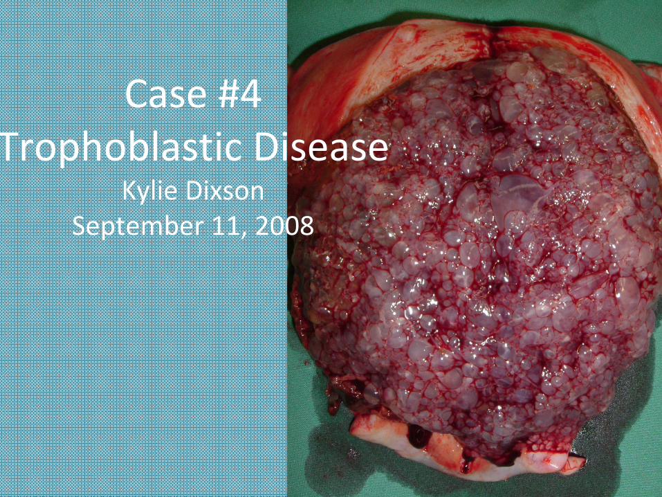
Case #4 Trophoblastic Disease
Kylie Dixson September 11, 2008
Outline
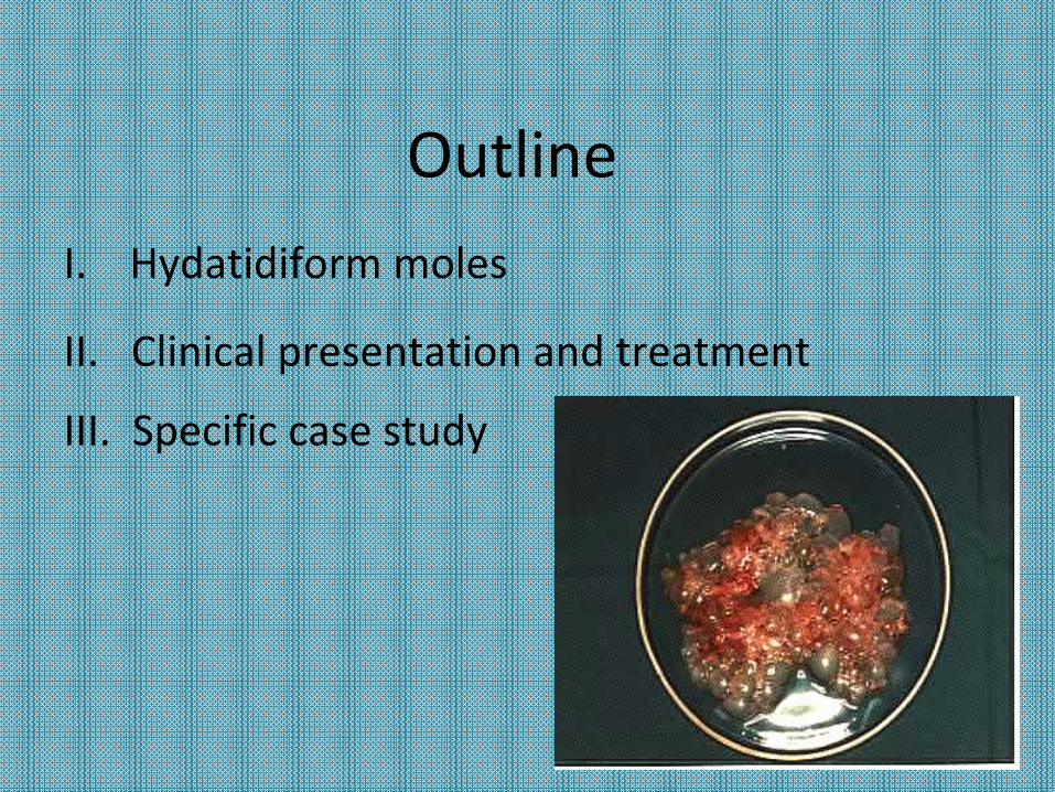
I. Hydatidiform moles
II. Clinical presentation and treatment
III. Specific case study
Complete Hydatidiform Mole1. Occurs when:
(90%) Egg that has lost its nucleus is fertilized by a single sperm : 46, XX(10%) Or by two sperm: 46, XX or 46, XY (all fraternally
derived)
3.
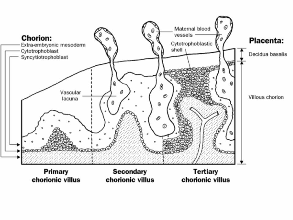
Morphology of the Mole:a. Most chorionic villi have excessive accumulation of fluid
& absent or inadequate development of blood supplyresults in swollen cysts that resemble grapes
b. Trophoblast layer has shown abnormal proliferationc. No embryonic development no fetal parts
2. Develops in the uterus between 3rd
and 5th
weeks of pregnancy‐‐
Occurs in about 0.1 ‐
0.5 percent of pregnancies
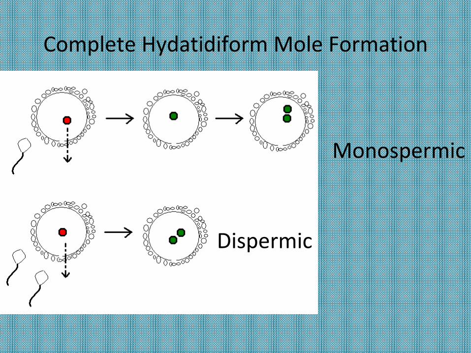
Complete Hydatidiform Mole Formation
Dispermic
Monospermic
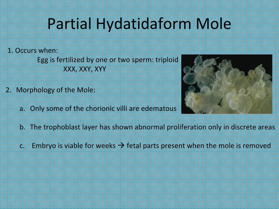
Partial Hydatidaform Mole1. Occurs when:
Egg is fertilized by one or two sperm: triploidXXX, XXY, XYY
2.
Morphology of the Mole:
a.
Only some of the chorionic villi are edematous
b.
The trophoblast layer has shown abnormal proliferation only in discrete areas
c. Embryo is viable for weeks fetal parts present when the mole is removed
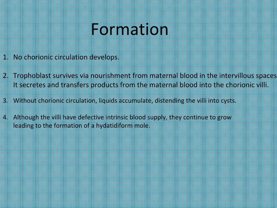
Formation1.
No chorionic circulation develops.
2.
Trophoblast survives via nourishment from maternal blood in the intervillous spacesIt secretes and transfers products from the maternal blood into the chorionic villi.
3.
Without chorionic circulation, liquids accumulate, distending the villi into cysts.
4.
Although the villi have defective intrinsic blood supply, they continue to grow leading to the formation of a hydatidiform mole.
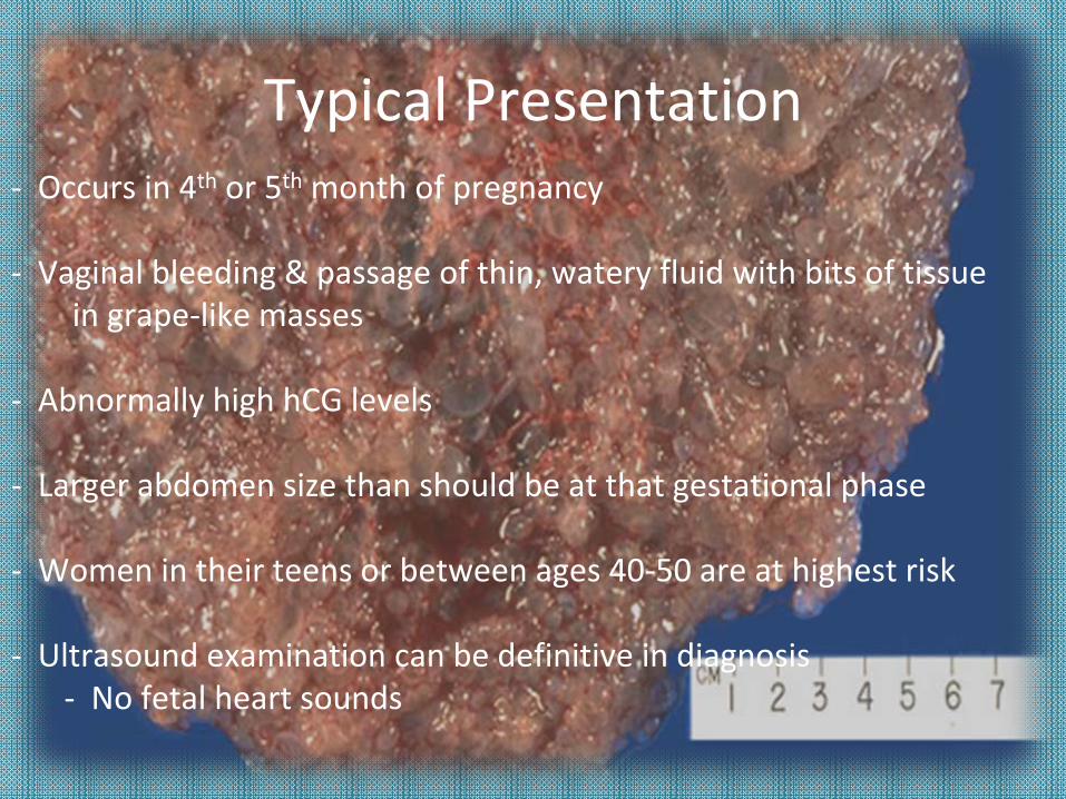
Typical Presentation‐ Occurs in 4th
or 5th
month of pregnancy
‐ Vaginal bleeding & passage of thin, watery fluid with bits of tissuein grape‐like masses
‐ Abnormally high hCG
levels
‐ Larger abdomen size than should be at that gestational phase
‐ Women in their teens or between ages 40‐50 are at highest risk
‐ Ultrasound examination can be definitive in diagnosis‐ No fetal heart sounds
Treatment
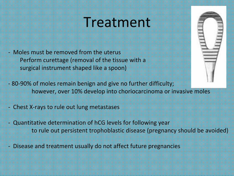
‐ Moles must be removed from the uterus Perform curettage (removal of the tissue with a surgical instrument shaped like a spoon)
‐ 80‐90% of moles remain benign and give no further difficulty; however, over 10% develop into choriocarcinoma
or invasive moles
‐
Chest X‐rays to rule out lung metastases
‐ Quantitative determination of hCG
levels for following yearto rule out persistent trophoblastic
disease (pregnancy should be avoided)
‐
Disease and treatment usually do not affect future pregnancies
Persistent Trophoblastic
Disease‐ Occurs when there is residual trophoblastic
tissue left in the uterus
‐ Can be benign or malignant (choriocarcinoma)
‐ Secrete high levels of hCG, elevated above even hydatidiform
levels
Choriocarcinoma‐ Rapidly invasive and widely metastasizing malignancy
‐ Common sites of metastasis: lungs and vagina
‐ Tumor is soft, fleshy yellow‐white
‐ Tendency to form large areas of ischemic necrosis
‐
Gravida
III: pregnancies regardless of outcome (present included)
‐
Para II: number of pregnancies carried to birth
‐
38 year old female
‐
Chief Complaint: excessive vaginal bleeding lasting less than one day
‐ After regular menstruation, her last menstrual period was 5 months ago (January to May)
Clinical Case Study
‐
Exam in April showed uterus corresponding to normal 3 month pregnancy
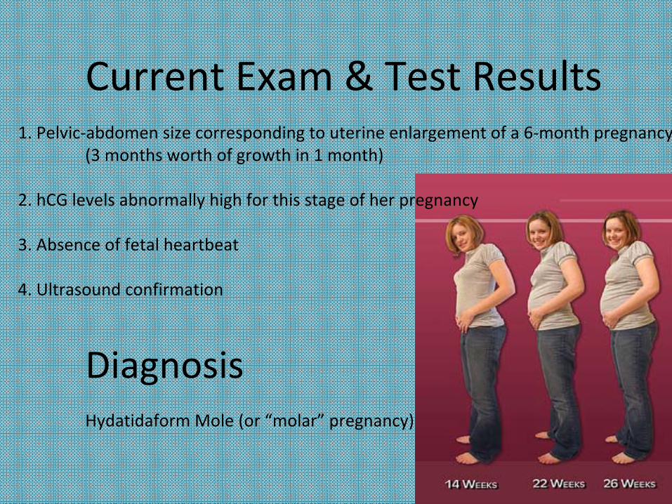
Current Exam & Test Results1. Pelvic‐abdomen size corresponding to uterine enlargement of a 6‐month pregnancy
(3 months worth of growth in 1 month)
2. hCG
levels abnormally high for this stage of her pregnancy
3. Absence of fetal heartbeat
4. Ultrasound confirmation
DiagnosisHydatidaform
Mole (or “molar”
pregnancy)
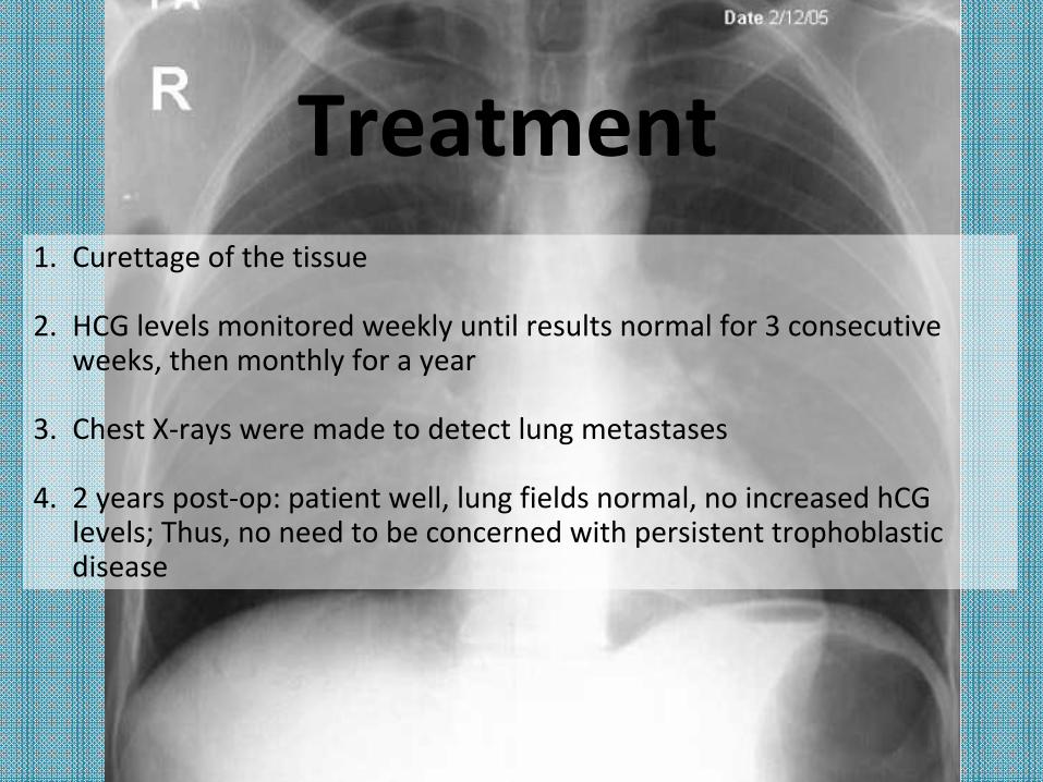
Treatment1.
Curettage of the tissue
2.
HCG levels monitored weekly until results normal for 3 consecutive
weeks, then monthly for a year
3.
Chest X‐rays were made to detect lung metastases
4.
2 years post‐op: patient well, lung fields normal, no increased hCG
levels; Thus, no need to be concerned with persistent trophoblastic
disease

Questions?