Embed Size (px)
Citation preview
Association Between Prior Peripherally Inserted CentralCatheters and Lack of Functioning Arteriovenous Fistulas: ACase-Control Study in Hemodialysis Patients
Mireille El Ters, MD1, Gregory J. Schears, MD2, Sandra J. Taler, MD1, Amy W. Williams,MD1, Robert C. Albright, DO1, Bernice M. Jenson, RN1, Amy L. Mahon, RN3, Andrew H.Stockland, MD4, Sanjay Misra, MD4, Scott L. Nyberg, MD, PhD5, Andrew D. Rule, MD1,6, andMarie C. Hogan, MD, PhD1
1Nephrology and Hypertension Division, Department of Internal Medicine, Mayo Clinic,Rochester, MN2Department of Anesthesiology, Mayo Clinic, Rochester, MN3Department of Nursing, Mayo Clinic, Rochester, MN4Division of Interventional & Vascular Radiology, Department of Radiology, Mayo Clinic,Rochester, MN5Division of Transplantation Surgery, Department of Surgery, Mayo Clinic, Rochester, MN6Division of Epidemiology, Department of Health Sciences, Mayo Clinic, Rochester, MN
AbstractBackground—Although an arteriovenous fistula (AVF) is the hemodialysis access of choice, itsprevalence continues to be lower than recommended in the United States. We assessed theassociation between past peripherally inserted central catheters (PICCs) and lack of functioningAVFs.
Study Design—Case-control study.
Participants & Setting—Prevalent hemodialysis population in 7 Mayo Clinic outpatienthemodialysis units. Cases were without functioning AVFs and controls were with functioningAVFs on January 31, 2011.
Predictors—History of PICCs.
Outcomes—Lack of functioning AVFs.
Results—On January 31, 2011, a total of 425 patients were receiving maintenance hemodialysis,of whom 282 were included in this study. Of these, 120 (42.5%; cases) were dialyzing through atunneled dialysis catheter or synthetic arteriovenous graft and 162 (57.5%; controls) had afunctioning AVF. PICC use was evaluated in both groups and identified in 30% of hemodialysis
© 2012 by the National Kidney Foundation, Inc.
Address correspondence to Marie C. Hogan, MD, PhD, Mayo Clinic, 200 First St SW, Rochester, MN [email protected].
The preliminary results of this study were presented as an abstract for the 2011 meeting of the American Society of Nephrology,November 8–13, 2011, in Philadelphia, PA.
Dr Williams chairs the American Society of Nephrology’s Quality and Patient Safety Task Force, which in April 2012 recommendedthat physicians not place PICCs in patients with stages 3–5 CKD without consulting a nephrologist.
Financial Disclosure: The authors declare that they have no other relevant financial interests.
NIH Public AccessAuthor ManuscriptAm J Kidney Dis. Author manuscript; available in PMC 2013 October 09.
Published in final edited form as:Am J Kidney Dis. 2012 October ; 60(4): 601–608. doi:10.1053/j.ajkd.2012.05.007.
NIH
-PA Author Manuscript
NIH
-PA Author Manuscript
NIH
-PA Author Manuscript
patients, with 54% of these placed after dialysis therapy initiation. Cases were more likely to bewomen (52.5% vs 33.3% in the control group; P = 0.001), with smaller mean vein (4.9 vs 5.8 mm;P < 0.001) and artery diameters (4.6 vs 4.9 mm; P = 0.01) than controls. A PICC was identified in53 (44.2%) cases, but only 32 (19.7%) controls (P < 0.001). We found a strong and independentassociation between PICC use and lack of a functioning AVF (OR, 3.2; 95% CI, 1.9–5.5; P <0.001). This association persisted after adjustment for confounders, including upper-extremityvein and artery diameters, sex, and history of central venous catheter (OR, 2.8; 95% CI, 1.5–5.5; P= 0.002).
Limitations—Retrospective study, participants mostly white.
Conclusion—PICCs are commonly placed in patients with end-stage renal disease and are astrong independent risk factor for lack of functioning AVFs.
INDEX WORDSChronic kidney disease; end-stage renal disease; arteriovenous fistula; hemodialysis; dialysisaccess
Through the Fistula First Breakthrough Initiative and the National Kidney Foundation’sKidney Disease Outcomes Quality Initiative (NKF-KDOQI) guidelines, efforts by theCenters for Medicare & Medicaid Services (CMS) and the NKF have led to an increase inthe overall prevalence of functioning arteriovenous fistulas (AVFs) in hemodialysis patients.When initial patency is established, the superior patency rates associated with autologousAVFs, as well as decreased mortality, morbidity, and cost (compared with a synthetic graftor venous catheter), make it the hemodialysis access of choice.1–9 However, theseprevalence rates are lower than the US target of 66%, as stipulated by the CMS.10–12 Fistulafailure rates also are high and are a major obstacle to achieving this goal.13–16 Therefore,there is a continuing need to examine barriers to improving rates of functioning AVFs inpatients with end-stage renal disease (ESRD).
One potential barrier may be the contribution of prior vascular injury (vascular sclerosis,thrombosis, and stenosis) from previously placed peripherally inserted central catheters(PICCs). Studies that examined complications related to PICC use have reported venousthrombosis rates as high as 58%, with a propensity for thrombosis in the cephalic and basilicveins (both used for AVF creation).17–19 Central vein stenosis also may occur, although lessfrequently.20
These studies have formed the basis for recommendations by renal societies to avoid PICCplacement in patients with advanced chronic kidney disease (CKD).10,21 However, evidencethat PICCs lead to AVF failure in long-term hemodialysis patients is lacking. PICCscontinue to be used in this medically complicated population due to their perceived cost-effectiveness and ease of use.22 We hypothesized that a previous PICC would associate withlack of a functioning AVF independent of characteristics associated with poor vein quality.To our knowledge, this is the first study to systematically examine the association of ahistory of prior PICC placement and the presence of a functioning AVF in a hemodialysispopulation.
METHODSStudy Population
In this case-control study, we included patients with ESRD receiving intermittentmaintenance hemodialysis in the Mayo Clinic Dialysis Services network (within RenalNetwork 11) as of January 31, 2011. Our study was limited to 7 hemodialysis centers in
Ters et al. Page 2
Am J Kidney Dis. Author manuscript; available in PMC 2013 October 09.
NIH
-PA Author Manuscript
NIH
-PA Author Manuscript
NIH
-PA Author Manuscript
Rochester, MN, or nearby cities in which ongoing medical care is provided by Mayo Clinicsites. Only individuals who provided research authorization were included.23 Transienthemodialysis patients (visiting our region) were excluded because they receive long-termcare from other dialysis providers. Finally, we excluded patients with acute kidney injuryrequiring hemodialysis who recovered kidney function and subsequently discontinueddialysis therapy prior to January 31, 2011. The Mayo Clinic Institutional Review Boardapproved this study.
Definition of Case and ControlMedical records were reviewed for the date of initiation of dialysis therapy, cause of ESRD,type and location of hemodialysis access, and date and anatomical placement of thearteriovenous access, up to January 31, 2011. Cases were defined as patients who lacked afunctioning AVF (ie, were dialyzed through a tunneled hemodialysis catheter or syntheticarteriovenous graft). This group included patients with prior failed AVFs or patients deemednot suitable or who declined this type of access. Controls were patients actively undergoinghemodialysis through an AVF.
Exposure VariableTwo separate electronic databases were queried for the date, location, and indication foreach PICC placed by nurses (2002–2011) and interventional radiology (1997–2011) at theMayo Clinic in Rochester, MN. We identified any PICC placed prior to AVF surgery, anyPICC placed prior to long-term hemodialysis therapy initiation, and any PICC placed as ofJanuary 31, 2011. Medical records were reviewed to confirm specific indications for PICCplacement.
Other CharacteristicsMedical records were reviewed for demographics, hospitalizations, and comorbidconditions. Comorbid conditions included diabetes mellitus, coronary artery disease (CAD),peripheral vascular disease, and congestive heart failure (CHF). Comorbid conditions werevalidated based on medications and physician notes (for diabetes mellitus),echocardiography results for systolic or diastolic function (for CHF, defined as ejectionfraction <50% or evidence of diastolic dysfunction with clinical history of CHF), cardiaccatheterization or cardiovascular surgical report (evidence of CAD with or withoutrevascularization procedures), and vascular radiology reports (evidence of peripheralvascular disease with or without interventions).
Upper-extremity vein and artery size were obtained from records of venous mapping studiesperformed routinely in our center on each patient preparing for hemodialysis accessplacement. In cases in which multiple venous mapping studies were available for oneindividual, we used the first report. At our center, venous mapping always includes brachial,radial, and ulnar arteries and cephalic and basilic vein diameters (from distal to proximal, ie,from wrist level to upper humerus level) and is performed initially on the nondominant armand, if the diameters of an artery or vein are thought to be inadequate (<2 mm for artery and<2.5 mm for vein), on the contralateral side. For this analysis, we used maximal vein andartery size on either side. Finally, we collected dates of all prior central venous access(es),including central venous catheters, temporary hemodialysis catheters, tunneled hemodialysiscatheters, and pacemakers, by reviewing all procedure notes.
Statistical AnalysisCharacteristics of cases and controls were summarized as absolute number with percentageor mean ± standard deviation. χ2 test was used to assess statistically significant differences
Ters et al. Page 3
Am J Kidney Dis. Author manuscript; available in PMC 2013 October 09.
NIH
-PA Author Manuscript
NIH
-PA Author Manuscript
NIH
-PA Author Manuscript
between groups for discrete variables; t test was used for continuous variables. We used alogistic regression model to determine the association between prior PICC placement andlack of a functioning AVF in prevalent hemodialysis patients. Statistical models wereadjusted for each characteristic (demographics, venous mapping, prior catheters, andcomorbid conditions). Multivariate analyses (reported as odds ratio [OR] with 95%confidence interval [CI]) were performed using a model adjusted for characteristics thatshowed statistically significant differences between cases and controls. Three separateanalyses were performed, defined as: (1) PICC anytime, (2) PICC before hemodialysistherapy initiation, or (3) PICC before hemodialysis therapy initiation or AVF surgery ineither group. PICC locations were assessed in controls with left-sided AVFs, and separately,in controls with right-arm AVFs. (4) We also repeated the analysis grouping patients withESRD with arteriovenous grafts as controls instead of cases.
To confirm the robustness of our results, we performed a sensitivity analysis by excludingpatients who had a PICC placed for difficult access (n = 15) from the univariate andmultivariate models. To further address the possibility of a PICC being placed for poorvenous quality, we reviewed vein diameters of patients with a prior PICC placed after thevenous mapping (in which case PICC had no effect on results of the venous mappingbecause it occurred after that) compared with those with no prior PICC placement. We alsoevaluated vein diameters of patients with a PICC placed prior to the venous mapping (inwhich case it may have affected results of that vein mapping) versus those with a PICCplaced after the venous mapping.
Statistical analyses were performed using JMP, version 9.0 (SAS Institute, www.sas.com).
RESULTSPatient Characteristics
A total of 425 patients undergoing dialysis in the Mayo Clinic Dialysis Services networkwere identified. After exclusion criteria were applied, 282 patients were included in thisstudy (Fig 1). Cases were 108 patients undergoing hemodialysis through a tunneled dialysiscatheter, and 12, through a synthetic arteriovenous graft. For cases, 54 (45%) had a priorattempt of AVF creation and maturation that failed, 16 (13.3%) had an immature AVF, and50 (41.7%) never had a prior attempt at AVF creation. Of the 50 with no attempted AVF, 14(28%) had refused placement of an arteriovenous access, 10 (20%) had an arteriovenousgraft, 16 (32%) were not medically or surgically acceptable candidates, and 10 (20%) had noreason documented. Patients undergoing hemodialysis through a successfully functioningAVF were considered controls. For controls (n = 162), the most common AVF location wasbrachial-cephalic (n = 115; 71%), followed by brachial-basilic (n = 25; 15.4%) and radial-cephalic (n = 22; 13.6%).
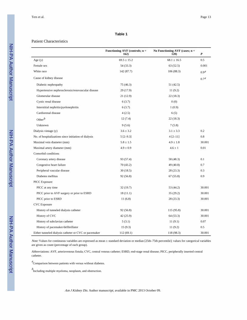
Cases were more likely to be women (63 [52.5%] cases vs 54 [33.3%] controls; P = 0.001)and have smaller mean vein (4.9 mm in cases vs 5.8 mm in controls; P < 0.001) and arterydiameters (4.6 mm in cases vs 4.9 mm in controls; P = 0.01; Table 1). There was nostatistically significant difference between the case and control groups in the proportion ofpatients with CAD, CHF, peripheral vascular disease, diabetes mellitus, or hospitalizationsafter starting dialysis therapy (Table 1).
Figure 1 shows the exposure of interest (PICCs) in cases and controls. Prior to January 11,2011, PICC insertion occurred in 85 of 282 patients (30%) in the hemodialysis cohort: 53patients in the case group (44.2%) versus 32 (19.7%) in the control group (P < 0.001). Wethen examined PICCs placed prior to the AVF surgery date (functional AVF in the controlgroup and failed AVF in cases) or prior to ESRD and found that a history of PICC
Ters et al. Page 4
Am J Kidney Dis. Author manuscript; available in PMC 2013 October 09.
NIH
-PA Author Manuscript
NIH
-PA Author Manuscript
NIH
-PA Author Manuscript
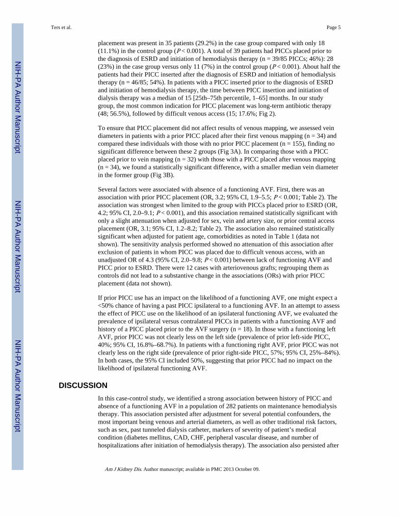
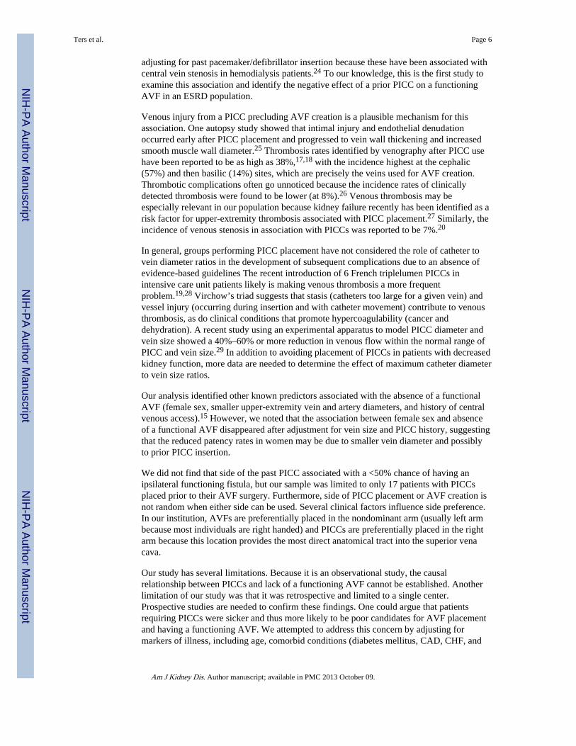
placement was present in 35 patients (29.2%) in the case group compared with only 18(11.1%) in the control group (P < 0.001). A total of 39 patients had PICCs placed prior tothe diagnosis of ESRD and initiation of hemodialysis therapy (n = 39/85 PICCs; 46%): 28(23%) in the case group versus only 11 (7%) in the control group (P < 0.001). About half thepatients had their PICC inserted after the diagnosis of ESRD and initiation of hemodialysistherapy (n = 46/85; 54%). In patients with a PICC inserted prior to the diagnosis of ESRDand initiation of hemodialysis therapy, the time between PICC insertion and initiation ofdialysis therapy was a median of 15 [25th–75th percentile, 1–65] months. In our studygroup, the most common indication for PICC placement was long-term antibiotic therapy(48; 56.5%), followed by difficult venous access (15; 17.6%; Fig 2).
To ensure that PICC placement did not affect results of venous mapping, we assessed veindiameters in patients with a prior PICC placed after their first venous mapping (n = 34) andcompared these individuals with those with no prior PICC placement (n = 155), finding nosignificant difference between these 2 groups (Fig 3A). In comparing those with a PICCplaced prior to vein mapping (n = 32) with those with a PICC placed after venous mapping(n = 34), we found a statistically significant difference, with a smaller median vein diameterin the former group (Fig 3B).
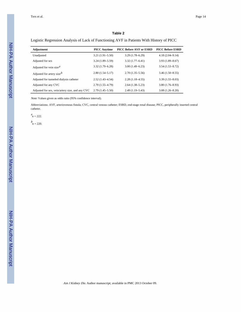
Several factors were associated with absence of a functioning AVF. First, there was anassociation with prior PICC placement (OR, 3.2; 95% CI, 1.9–5.5; P < 0.001; Table 2). Theassociation was strongest when limited to the group with PICCs placed prior to ESRD (OR,4.2; 95% CI, 2.0–9.1; P < 0.001), and this association remained statistically significant withonly a slight attenuation when adjusted for sex, vein and artery size, or prior central accessplacement (OR, 3.1; 95% CI, 1.2–8.2; Table 2). The association also remained statisticallysignificant when adjusted for patient age, comorbidities as noted in Table 1 (data notshown). The sensitivity analysis performed showed no attenuation of this association afterexclusion of patients in whom PICC was placed due to difficult venous access, with anunadjusted OR of 4.3 (95% CI, 2.0–9.8; P < 0.001) between lack of functioning AVF andPICC prior to ESRD. There were 12 cases with arteriovenous grafts; regrouping them ascontrols did not lead to a substantive change in the associations (ORs) with prior PICCplacement (data not shown).
If prior PICC use has an impact on the likelihood of a functioning AVF, one might expect a<50% chance of having a past PICC ipsilateral to a functioning AVF. In an attempt to assessthe effect of PICC use on the likelihood of an ipsilateral functioning AVF, we evaluated theprevalence of ipsilateral versus contralateral PICCs in patients with a functioning AVF andhistory of a PICC placed prior to the AVF surgery (n = 18). In those with a functioning leftAVF, prior PICC was not clearly less on the left side (prevalence of prior left-side PICC,40%; 95% CI, 16.8%–68.7%). In patients with a functioning right AVF, prior PICC was notclearly less on the right side (prevalence of prior right-side PICC, 57%; 95% CI, 25%–84%).In both cases, the 95% CI included 50%, suggesting that prior PICC had no impact on thelikelihood of ipsilateral functioning AVF.
DISCUSSIONIn this case-control study, we identified a strong association between history of PICC andabsence of a functioning AVF in a population of 282 patients on maintenance hemodialysistherapy. This association persisted after adjustment for several potential confounders, themost important being venous and arterial diameters, as well as other traditional risk factors,such as sex, past tunneled dialysis catheter, markers of severity of patient’s medicalcondition (diabetes mellitus, CAD, CHF, peripheral vascular disease, and number ofhospitalizations after initiation of hemodialysis therapy). The association also persisted after
Ters et al. Page 5
Am J Kidney Dis. Author manuscript; available in PMC 2013 October 09.
NIH
-PA Author Manuscript
NIH
-PA Author Manuscript
NIH
-PA Author Manuscript
adjusting for past pacemaker/defibrillator insertion because these have been associated withcentral vein stenosis in hemodialysis patients.24 To our knowledge, this is the first study toexamine this association and identify the negative effect of a prior PICC on a functioningAVF in an ESRD population.
Venous injury from a PICC precluding AVF creation is a plausible mechanism for thisassociation. One autopsy study showed that intimal injury and endothelial denudationoccurred early after PICC placement and progressed to vein wall thickening and increasedsmooth muscle wall diameter.25 Thrombosis rates identified by venography after PICC usehave been reported to be as high as 38%,17,18 with the incidence highest at the cephalic(57%) and then basilic (14%) sites, which are precisely the veins used for AVF creation.Thrombotic complications often go unnoticed because the incidence rates of clinicallydetected thrombosis were found to be lower (at 8%).26 Venous thrombosis may beespecially relevant in our population because kidney failure recently has been identified as arisk factor for upper-extremity thrombosis associated with PICC placement.27 Similarly, theincidence of venous stenosis in association with PICCs was reported to be 7%.20
In general, groups performing PICC placement have not considered the role of catheter tovein diameter ratios in the development of subsequent complications due to an absence ofevidence-based guidelines The recent introduction of 6 French triplelumen PICCs inintensive care unit patients likely is making venous thrombosis a more frequentproblem.19,28 Virchow’s triad suggests that stasis (catheters too large for a given vein) andvessel injury (occurring during insertion and with catheter movement) contribute to venousthrombosis, as do clinical conditions that promote hypercoagulability (cancer anddehydration). A recent study using an experimental apparatus to model PICC diameter andvein size showed a 40%–60% or more reduction in venous flow within the normal range ofPICC and vein size.29 In addition to avoiding placement of PICCs in patients with decreasedkidney function, more data are needed to determine the effect of maximum catheter diameterto vein size ratios.
Our analysis identified other known predictors associated with the absence of a functionalAVF (female sex, smaller upper-extremity vein and artery diameters, and history of centralvenous access).15 However, we noted that the association between female sex and absenceof a functional AVF disappeared after adjustment for vein size and PICC history, suggestingthat the reduced patency rates in women may be due to smaller vein diameter and possiblyto prior PICC insertion.
We did not find that side of the past PICC associated with a <50% chance of having anipsilateral functioning fistula, but our sample was limited to only 17 patients with PICCsplaced prior to their AVF surgery. Furthermore, side of PICC placement or AVF creation isnot random when either side can be used. Several clinical factors influence side preference.In our institution, AVFs are preferentially placed in the nondominant arm (usually left armbecause most individuals are right handed) and PICCs are preferentially placed in the rightarm because this location provides the most direct anatomical tract into the superior venacava.
Our study has several limitations. Because it is an observational study, the causalrelationship between PICCs and lack of a functioning AVF cannot be established. Anotherlimitation of our study was that it was retrospective and limited to a single center.Prospective studies are needed to confirm these findings. One could argue that patientsrequiring PICCs were sicker and thus more likely to be poor candidates for AVF placementand having a functioning AVF. We attempted to address this concern by adjusting formarkers of illness, including age, comorbid conditions (diabetes mellitus, CAD, CHF, and
Ters et al. Page 6
Am J Kidney Dis. Author manuscript; available in PMC 2013 October 09.
NIH
-PA Author Manuscript
NIH
-PA Author Manuscript
NIH
-PA Author Manuscript
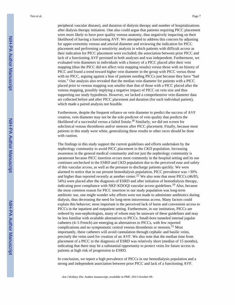
peripheral vascular disease), and duration of dialysis therapy and number of hospitalizationsafter dialysis therapy initiation. One also could argue that patients requiring PICC placementwere more likely to have poor quality venous anatomy, thus negatively impacting on theirlikelihood of having a functioning AVF. We attempted to address this concern by adjustingfor upper-extremity venous and arterial diameter and reviewing the indication for PICCplacement and performing a sensitivity analysis in which patients with difficult access astheir indication for PICC placement were excluded; the association between prior PICC andlack of a functioning AVF persisted in both analyses and was independent. Furthermore, weevaluated vein diameters in individuals with a history of a PICC placed after their veinmapping (thus the PICC did not affect vein mapping results) versus those with no history ofPICC and found a trend toward higher vein diameter in the group with PICC versus thosewith no PICC, arguing against a bias of patients needing PICCs just because they have “badveins.” Our analysis also revealed that the median vein diameter for patients with a PICCplaced prior to venous mapping was smaller than that of those with a PICC placed after thevenous mapping, possibly implying a negative impact of PICC on vein size and thussupporting our study hypothesis. However, we lacked a comprehensive vein diameter dataset collected before and after PICC placement and duration (for each individual patient),which made a paired analysis not feasible.
Furthermore, despite the frequent reliance on vein diameter to predict the success of AVFcreation, vein diameter may not be the sole predictor of vein quality that predicts thelikelihood of a successful versus a failed fistula.30 Similarly, we did not screen forsubclinical venous thrombosis and/or stenosis after PICC placement. Finally, because mostpatients in this study were white, generalizing these results to other races should be donewith caution.
The findings in this study support the current guidelines and efforts undertaken by thenephrology community to avoid PICC placement in the CKD population. Increasingawareness in the general medical community and not just the nephrology community isparamount because PICC insertion occurs most commonly in the hospital setting and its usecontinues unchecked in the ESRD and CKD population due to the perceived ease and safetyof this vascular access, as well as the pressure to discharge patients quickly. We werealarmed to notice that in our present hemodialysis population, PICC prevalence was >30%and higher than reported recently at another center.22 We also note that most PICCs (46/85;54%) were placed after the diagnosis of ESRD and after initiation of hemodialysis therapy,indicating poor compliance with NKF-KDOQI vascular access guidelines.10 Also, becausethe most common reason for PICC insertion in our study population was long-termantibiotic use, one might wonder why efforts were not made to administer antibiotics duringdialysis, thus decreasing the need for long-term intravenous access. Many factors couldexplain this behavior; most important is the perceived lack of harm and convenient access toPICCs in the inpatient and outpatient setting. Furthermore, in our institution, PICCs areordered by non-nephrologists, many of whom may be unaware of these guidelines and maybe less familiar with available alternatives to PICCs. Small-bore tunneled internal jugularcatheters (4–5 French) are emerging as alternatives to PICCs, with few reportedcomplications and no symptomatic central venous thrombosis or stenosis.31 Mostimportantly, these catheters will avoid cannulation through cephalic and basilic veins,precisely the veins used for creation of an AVF. We also note that the median time fromplacement of a PICC to the diagnosis of ESRD was relatively short (median of 15 months),indicating that there may be a substantial opportunity to protect veins for future access inpatients at high risk of progression to ESRD.
In conclusion, we report a high prevalence of PICCs in our hemodialysis population and astrong and independent association between prior PICC and lack of a functioning AVF.
Ters et al. Page 7
Am J Kidney Dis. Author manuscript; available in PMC 2013 October 09.
NIH
-PA Author Manuscript
NIH
-PA Author Manuscript
NIH
-PA Author Manuscript
PICCs are known to cause peripheral and central venous injury, which may preclude futureAVF access. Effective processes, including education of hospitalists, critical care specialists,internists, patients, and families, are needed to promote alternative venous access in ourCKD population to preserve future venous access.
AcknowledgmentsWe thank Cynthia A. Handberg and Dawn P. Bergen for secretarial assistance.
Support: This publication was supported by National Institutes of Health (NIH)/National Center for ResearchResources CTSA grant UL1 RR024150. Its contents are solely the responsibility of the authors and do notnecessarily represent the official views of the NIH.
References1. Huber TS, Carter JW, Carter RL, Seeger JM. Patency of autogenous and polytetrafluoroethylene
upper extremity arteriovenous hemodialysis accesses: a systematic review. J Vasc Surg. 2003;38(5):1005–1011. [PubMed: 14603208]
2. Oliver MJ, Rothwell DM, Fung K, Hux JE, Lok CE. Late creation of vascular access forhemodialysis and increased risk of sepsis. J Am Soc Nephrol. 2004; 15(7):1936–1942. [PubMed:15213284]
3. Ortega T, Ortega F, Diaz-Corte C, Rebollo P, Ma Baltar J, Alvarez-Grande J. The timelyconstruction of arteriovenous fistulae: a key to reducing morbidity and mortality and to improvingcost management. Nephrol Dial Transplant. 2005; 20(3):598–603. [PubMed: 15647308]
4. Perera GB, Mueller MP, Kubaska SM, Wilson SE, Lawrence PF, Fujitani RM. Superiority ofautogenous arteriovenous hemodialysis access: maintenance of function with fewer secondaryinterventions. Ann Vasc Surg. 2004; 18(1):66–73. [PubMed: 14727162]
5. Schon D, Blume SW, Niebauer K, Hollenbeak CS, de Lissovoy G. Increasing the use ofarteriovenous fistula in hemodialysis: economic benefits and economic barriers. Clin J Am SocNephrol. 2007; 2(2):268–276. [PubMed: 17699424]
6. Woods JD, Turenne MN, Strawderman RL, et al. Vascular access survival among incidenthemodialysis patients in the United States. Am J Kidney Dis. 1997; 30(1):50–57. [PubMed:9214401]
7. Tordoir JH. Dialysis: vascular access type defines survival in patients on dialysis. Nat Rev Nephrol.2011; 7(9):489–490. [PubMed: 21769104]
8. Allon M, Daugirdas J, Depner TA, Greene T, Ornt D, Schwab SJ. Effect of change in vascularaccess on patient mortality in hemodialysis patients. Am J Kidney Dis. 2006; 47(3):469–477.[PubMed: 16490626]
9. Dhingra RK, Young EW, Hulbert-Shearon TE, Leavey SF, Port FK. Type of vascular access andmortality in U.S. hemodialysis patients. Kidney Int. 2001; 60(4):1443–1451. [PubMed: 11576358]
10. National Kidney Foundation. K/DOQI Clinical Practice Guidelines and Clinical PracticeRecommendations 2006 updates: Hemodialysis Adequacy Peritoneal Dialysis Adequacy VascularAccess. Am J Kidney Dis. 2006; (48 suppl 1):S227–S350.
11. AVF: the first choice for hemodialysis. Fistula First Breakthrough Initiative; 2011. ArteriovenousFistula First. No author listedhttp://www.fistulafirst.org/ [Accessed December 31, 2011]
12. Rayner HC, Besarab A, Brown WW, Disney A, Saito A, Pisoni RL. Vascular access results fromthe Dialysis Outcomes and Practice Patterns Study (DOPPS): performance against Kidney DiseaseOutcomes Quality Initiative (K/DOQI) Clinical Practice Guidelines. Am J Kidney Dis. 2004; 44(5suppl 2):S22–S26.
13. Schinstock CA, Albright RC, Williams AW, et al. Outcomes of arteriovenous fistula creation afterthe fistula first initiative. Clin J Am Soc Nephrol. 2011; 6(8):1996–2002. [PubMed: 21737851]
14. Miller PE, Tolwani A, Luscy CP, et al. Predictors of adequacy of arteriovenous fistulas inhemodialysis patients. Kidney Int. 1999; 56(1):275–280. [PubMed: 10411703]
15. Miller CD, Robbin ML, Allon M. Gender differences in outcomes of arteriovenous fistulas inhemodialysis patients. Kidney Int. 2003; 63(1):346–352. [PubMed: 12472802]
Ters et al. Page 8
Am J Kidney Dis. Author manuscript; available in PMC 2013 October 09.
NIH
-PA Author Manuscript
NIH
-PA Author Manuscript
NIH
-PA Author Manuscript
16. Obialo CI, Tagoe AT, Martin PC, Asche-Crowe PE. Adequacy and survival of autogenousarteriovenous fistula in African American hemodialysis patients. ASAIO J. 2003; 49(4):435–439.[PubMed: 12918587]
17. Abdullah BJ, Mohammad N, Sangkar JV, et al. Incidence of upper limb venous thrombosisassociated with peripherally inserted central catheters (PICC). Br J Radiol. 2005; 78(931):596–600. [PubMed: 15961840]
18. Allen AW, Megargell JL, Brown DB, et al. Venous thrombosis associated with the placement ofperipherally inserted central catheters. J Vasc Interv Radiol. 2000; 11(10):1309–1314. [PubMed:11099241]
19. Trerotola SO, Stavropoulos SW, Mondschein JI, et al. Triple-lumen peripherally inserted centralcatheter in patients in the critical care unit: prospective evaluation. Radiology. 2010; 256(1):312–320. [PubMed: 20574104]
20. Gonsalves CF, Eschelman DJ, Sullivan KL, DuBois N, Bonn J. Incidence of central vein stenosisand occlusion following upper extremity PICC and port placement. Cardiovasc Intervent Radiol.2003; 26(2):123–127. [PubMed: 12616419]
21. Hoggard J, Saad T, Schon D, Vesely TM, Royer T. Guidelines for venous access in patients withchronic kidney disease. A position statement from the American Society of Diagnostic andInterventional Nephrology, Clinical Practice Committee and the Association for Vascular Access.Semin Dial. 2008; 21(2):186–191. [PubMed: 18364015]
22. Butler PJ, Sood S, Mojibian H, Tal MG. Previous PICC placement may be associated withcatheter-related infections in hemodialysis patients. Cardiovasc Intervent Radiol. 2011; 34(1):120–123. [PubMed: 20857109]
23. Minnesota Health Records Act. Disclosure of Health Records for External Research. §144.295(MN 2011).
24. Drew DA, Meyer KB, Weiner DE. Transvenous cardiac device wires and vascular access inhemodialysis patients. Am J Kidney Dis. 2011; 58(3):494–496. [PubMed: 21664734]
25. Forauer AR, Theoharis C. Histologic changes in the human vein wall adjacent to indwelling centralvenous catheters. J Vasc Interv Radiol. 2003; 14(9 pt 1):1163–1168. [PubMed: 14514808]
26. Fletcher JJ, Stetler W, Wilson TJ. The clinical significance of peripherally inserted central venouscatheter-related deep vein thrombosis. Neurocrit Care. 2011; 15(3):454–460. [PubMed: 21541826]
27. Marnejon T, Angelo D, Abu Abdou A, Gemmel D. Risk factors for upper extremity venousthrombosis associated with peripherally inserted central venous catheters. J Vasc Access. 2012;13(2):231–238. [PubMed: 22266584]
28. Evans RS, Sharp JH, Linford LH, et al. Risk of symptomatic DVT associated with peripherallyinserted central catheters. Chest. 2010; 138(4):803–810. [PubMed: 20923799]
29. Nifong TP, McDevitt TJ. The effect of catheter to vein ratio on blood flow rates in a simulatedmodel of peripherally inserted central venous catheters. Chest. 2011; 140(1):48–53. [PubMed:21349931]
30. Maya ID, O’Neal JC, Young CJ, Barker-Finkel J, Allon M. Outcomes of brachiocephalic fistulas,transposed brachiobasilic fistulas, and upper arm grafts. Clin J Am Soc Nephrol. 2009; 4(1):86–92. [PubMed: 18945990]
31. Sasadeusz KJ, Trerotola SO, Shah H, et al. Tunneled jugular small-bore central catheters as analternative to peripherally inserted central catheters for intermediate-term venous access in patientswith hemodialysis and chronic renal insufficiency. Radiology. 1999; 213(1):303–306. [PubMed:10540677]
Ters et al. Page 9
Am J Kidney Dis. Author manuscript; available in PMC 2013 October 09.
NIH
-PA Author Manuscript
NIH
-PA Author Manuscript
NIH
-PA Author Manuscript
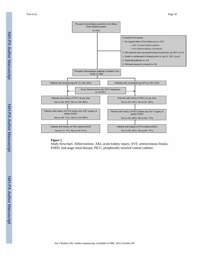
Figure 1.Study flowchart. Abbreviations: AKI, acute kidney injury; AVF, arteriovenous fistula;ESRD, end-stage renal disease; PICC, peripherally inserted central catheter.
Ters et al. Page 10
Am J Kidney Dis. Author manuscript; available in PMC 2013 October 09.
NIH
-PA Author Manuscript
NIH
-PA Author Manuscript
NIH
-PA Author Manuscript
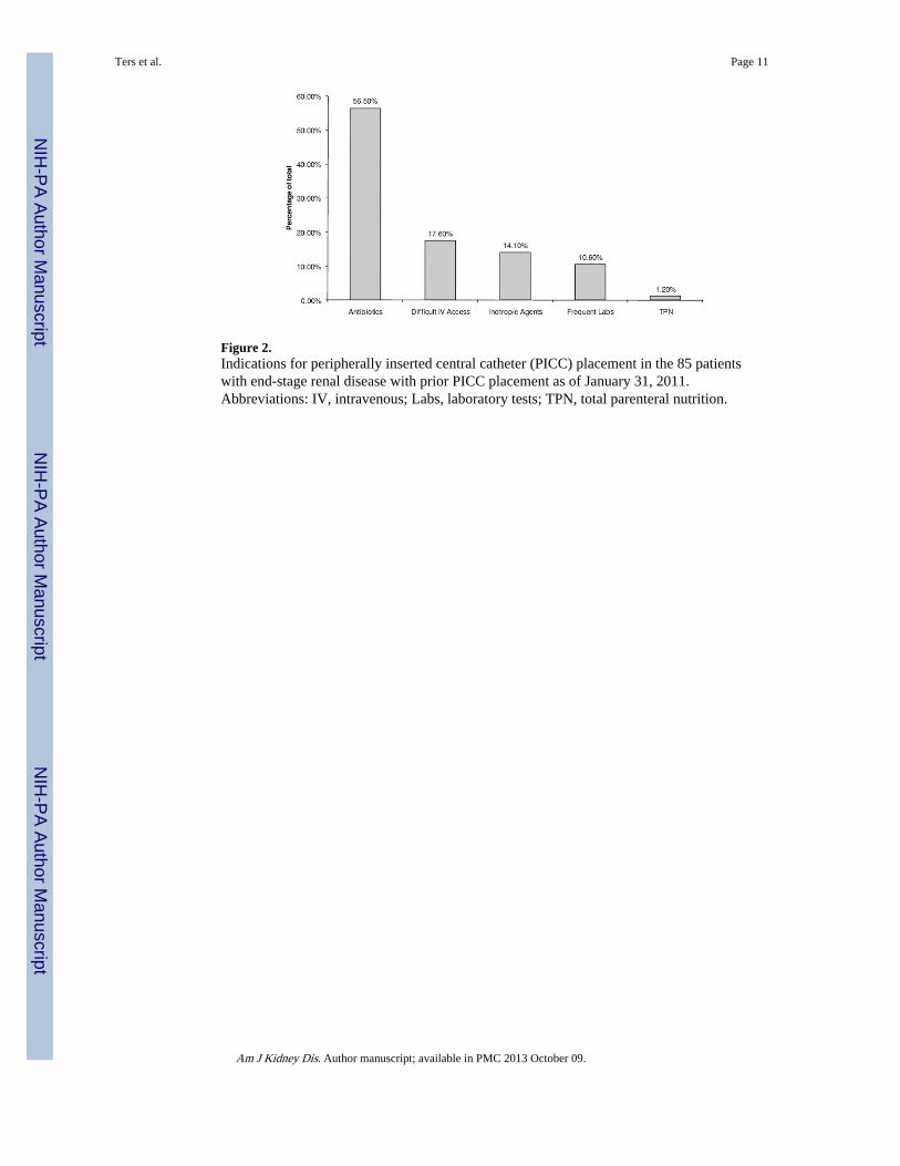
Figure 2.Indications for peripherally inserted central catheter (PICC) placement in the 85 patientswith end-stage renal disease with prior PICC placement as of January 31, 2011.Abbreviations: IV, intravenous; Labs, laboratory tests; TPN, total parenteral nutrition.
Ters et al. Page 11
Am J Kidney Dis. Author manuscript; available in PMC 2013 October 09.
NIH
-PA Author Manuscript
NIH
-PA Author Manuscript
NIH
-PA Author Manuscript
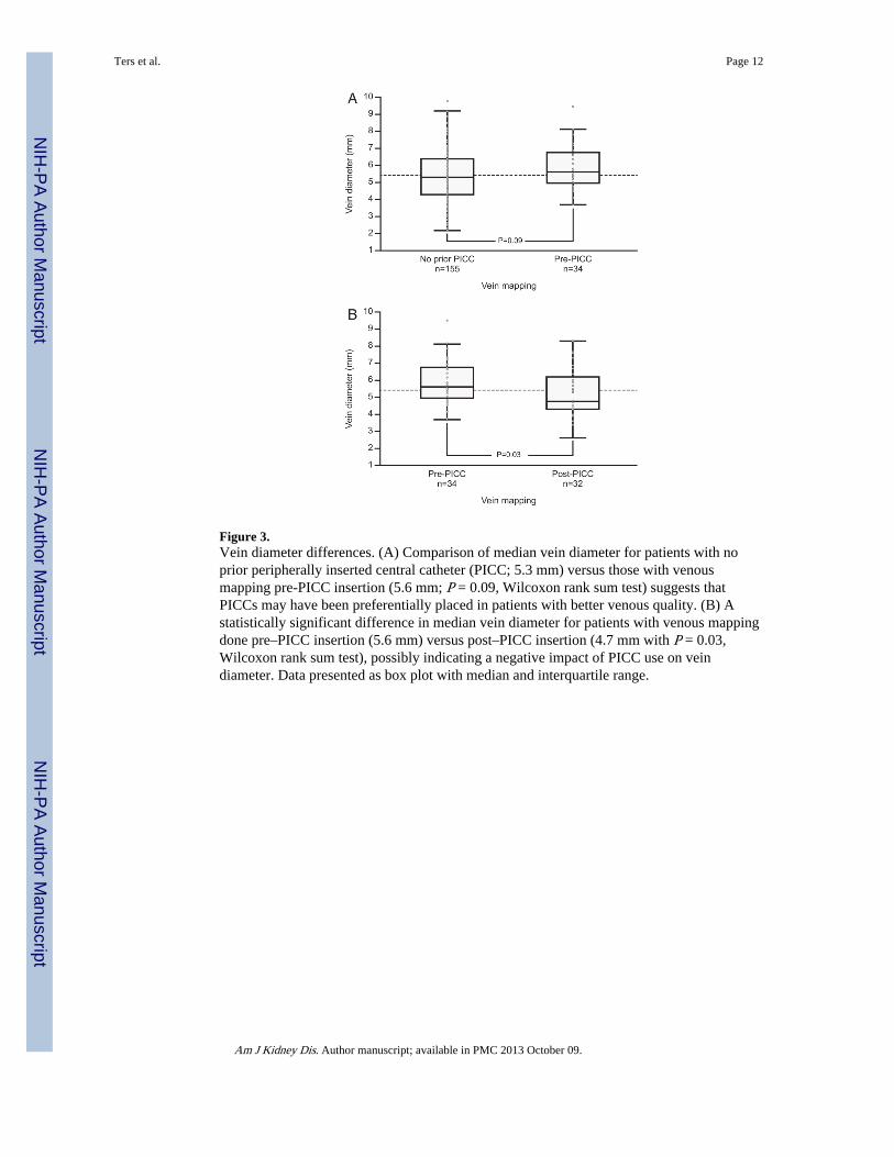
Figure 3.Vein diameter differences. (A) Comparison of median vein diameter for patients with noprior peripherally inserted central catheter (PICC; 5.3 mm) versus those with venousmapping pre-PICC insertion (5.6 mm; P = 0.09, Wilcoxon rank sum test) suggests thatPICCs may have been preferentially placed in patients with better venous quality. (B) Astatistically significant difference in median vein diameter for patients with venous mappingdone pre–PICC insertion (5.6 mm) versus post–PICC insertion (4.7 mm with P = 0.03,Wilcoxon rank sum test), possibly indicating a negative impact of PICC use on veindiameter. Data presented as box plot with median and interquartile range.
Ters et al. Page 12
Am J Kidney Dis. Author manuscript; available in PMC 2013 October 09.
NIH
-PA Author Manuscript
NIH
-PA Author Manuscript
NIH
-PA Author Manuscript
NIH
-PA Author Manuscript
NIH
-PA Author Manuscript
NIH
-PA Author Manuscript
Ters et al. Page 13
Table 1
Patient Characteristics
Functioning AVF (controls; n =162)
No Functioning AVF (cases; n =120) P
Age (y) 69.5 ± 15.2 68.1 ± 16.5 0.5
Female sex 54 (33.3) 63 (52.5) 0.001
White race 142 (87.7) 106 (88.3) 0.9a
Cause of kidney disease 0.7a
Diabetic nephropathy 75 (46.3) 51 (42.5)
Hypertensive nephrosclerosis/renovascular disease 29 (17.9) 11 (9.2)
Glomerular disease 21 (12.9) 22 (18.3)
Cystic renal disease 6 (3.7) 0 (0)
Interstitial nephritis/pyelonephritis 6 (3.7) 1 (0.9)
Cardiorenal disease 4 (2.5) 6 (5)
Otherb 12 (7.4) 22 (18.3)
Unknown 9 (5.6) 7 (5.8)
Dialysis vintage (y) 3.6 ± 3.2 3.1 ± 3.3 0.2
No. of hospitalizations since initiation of dialysis 5 [2–9.3] 4 [2–11] 0.8
Maximal vein diameter (mm) 5.8 ± 1.5 4.9 ± 1.8 30.001
Maximal artery diameter (mm) 4.9 ± 0.9 4.6 ± 1 0.01
Comorbid conditions
Coronary artery disease 93 (57.4) 58 (48.3) 0.1
Congestive heart failure 70 (43.2) 49 (40.8) 0.7
Peripheral vascular disease 30 (18.5) 28 (23.3) 0.3
Diabetes mellitus 92 (56.8) 67 (55.8) 0.9
PICC Exposure
PICC at any time 32 (19.7) 53 (44.2) 30.001
PICC prior to AVF surgery or prior to ESRD 18 (11.1) 35 (29.2) 30.001
PICC prior to ESRD 11 (6.8) 28 (23.3) 30.001
CVC Exposure
History of tunneled dialysis catheter 92 (56.8) 115 (95.8) 30.001
History of CVC 42 (25.9) 64 (53.3) 30.001
History of subclavian catheter 5 (3.1) 11 (9.1) 0.07
History of pacemaker/defibrillator 15 (9.3) 11 (9.2) 0.5
Either tunneled dialysis catheter or CVC or pacemaker 112 (69.1) 118 (98.3) 30.001
Note: Values for continuous variables are expressed as mean ± standard deviation or median [25th–75th percentile]; values for categorical variablesare given as count (percentage of each group).
Abbreviations: AVF, arteriovenous fistula; CVC, central venous catheter; ESRD, end-stage renal disease; PICC, peripherally inserted centralcatheter.
aComparison between patients with versus without diabetes.
bIncluding multiple myeloma, neoplasm, and obstruction.
Am J Kidney Dis. Author manuscript; available in PMC 2013 October 09.
NIH
-PA Author Manuscript
NIH
-PA Author Manuscript
NIH
-PA Author Manuscript
Ters et al. Page 14
Table 2
Logistic Regression Analysis of Lack of Functioning AVF in Patients With History of PICC
Adjustment PICC Anytime PICC Before AVF or ESRD PICC Before ESRD
Unadjusted 3.21 (1.91–5.50) 3.29 (1.78–6.29) 4.18 (2.04–9.14)
Adjusted for sex 3.24 (1.89–5.59) 3.32 (1.77–6.41) 3.93 (1.89–8.67)
Adjusted for vein sizea 3.32 (1.79–6.28) 3.00 (1.49–6.23) 3.54 (1.53–8.72)
Adjusted for artery sizeb 2.80 (1.54–5.17) 2.70 (1.35–5.56) 3.46 (1.50–8.55)
Adjusted for tunneled dialysis catheter 2.52 (1.43–4.54) 2.28 (1.18–4.55) 3.39 (1.55–8.03)
Adjusted for any CVC 2.70 (1.55–4.79) 2.64 (1.38–5.23) 3.80 (1.76–8.93)
Adjusted for sex, vein/artery size, and any CVC 2.79 (1.45–5.50) 2.49 (1.19–5.43) 3.08 (1.26–8.20)
Note: Values given as odds ratio (95% confidence interval).
Abbreviations: AVF, arteriovenous fistula; CVC, central venous catheter; ESRD, end-stage renal disease; PICC, peripherally inserted centralcatheter.
an = 222.
bn = 220.
Am J Kidney Dis. Author manuscript; available in PMC 2013 October 09.


![Effects of supervised exercise on depressive symptoms in hemodialysis … · 2017. 12. 15. · hemodialysis worldwide [1]. Depressive symptoms are common among hemodialysis patients,](https://img.pdfslide.net/doc/110x75/612e737f1ecc51586942d268/effects-of-supervised-exercise-on-depressive-symptoms-in-hemodialysis-2017-12.jpg)