Embed Size (px)
Citation preview
Int J Clin Exp Med 2017;10(1):1371-1375www.ijcem.com /ISSN:1940-5901/IJCEM0038373
Case Report A case of predominant lymphoepithelioma-like carcinoma of the renal pelvis and literature review
Yuemei Xu, Yao Fu, Zhiwen Li, Yihua Wang, Jiong Shi
Department of Pathology, Nanjing Drum Tower Hospital, The Affiliated Hospital of Nanjing University Medical School, Nanjing, China
Received August 21, 2016; Accepted October 15, 2016; Epub January 15, 2017; Published January 30, 2017
Abstract: Lymphoepithelioma, as is well known, is a form of undifferentiated epithelial tumor, which is primarily de-scribed in the nasopharynx and characterized by syncytial sheets of malignant epithelial cells with a prominent re-active lymphoid infiltrate in background. A carcinoma that shows similar histological features but arises outside the nasopharynx is called lymphoepithelioma-like carcinoma (LELC). Primary LELC of the renal pelvis is very rare and there is only limited information in the published reports. We managed a case of a 59-year-old man, who presented with asymptomatic gross hematuria for more than one month. MRI showed that right kidney accounted for a soft tis-sue signal mass with a diameter of 2.3 cm in renal pelvis. He was treated with the Robot assisted Radical resection of the right tumor and was diagnosed with a pT3 N0 cM0, the high grade of renal invasive urothelial carcinoma, with predominant Lymphoepithelioma-like carcinoma differentiation. Unlike nasopharyngeal lymphoepithelioma, in situ hybridization analysis of this urinary LELC was negative for the Epstein-Barr virus. Here we report on one more case of primary LELC of the renal pelvis, and review of the published reports, particularly those describing the expression of Epstein-Barr virus.
Keywords: Lymphoepithelioma-like carcinoma, renal pelvis, Epstein-Barr virus
Introduction
Lymphoepithelioma is a form of undifferentiat-ed epithelial tumor, characterized by syncytial nests of malignant epithelial cells with a promi-nent reactive lymphoid infiltrate in background, and primarily described in the nasopharynx [1]. Tumors that show the similar histological features but arise outside the nasopharynx are called lymphoepithelioma-like carcinoma (LELC). According to some reports, lymphoepi-thelioma-like carcinoma may be pure, predomi-nant, or focally admixed with typical urothelial carcinoma or other variants [2, 3]. Other organs include the tonsil, salivary glands, thymus, lungs, stomach, skin, uterine cervix, breast, prostate, and the urinary tract [4]. In most cases, lymphoepithelioma-like carcinoma is related to Epstein-Barr virus (EBV), especially in the thymus gland (thymoma with lymphoid hyperplasia), but no association has been reported between lymphoepithelioma-like car-cinoma of the bladder and EBV [5, 6]. To the best of our knowledge, this is the tenth case of
lymphoepithelioma-like carcinoma of the renal pelvis in the English literature [1, 4, 7-12].
Case report
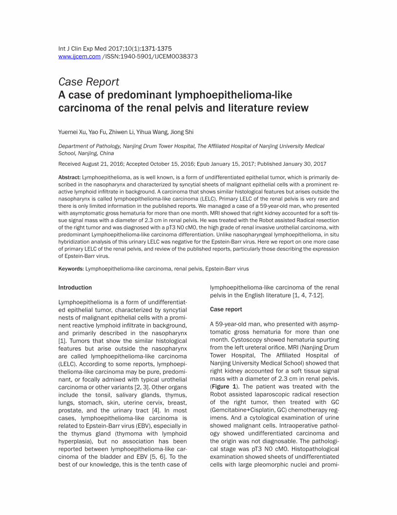
A 59-year-old man, who presented with asymp-tomatic gross hematuria for more than one month. Cystoscopy showed hematuria spurting from the left ureteral orifice. MRI (Nanjing Drum Tower Hospital, The Affiliated Hospital of Nanjing University Medical School) showed that right kidney accounted for a soft tissue signal mass with a diameter of 2.3 cm in renal pelvis. (Figure 1). The patient was treated with the Robot assisted laparoscopic radical resection of the right tumor, then treated with GC (Gemcitabine+Cisplatin, GC) chemotherapy reg-imens. And a cytological examination of urine showed malignant cells. Intraoperative pathol-ogy showed undifferentiated carcinoma and the origin was not diagnosable. The pathologi-cal stage was pT3 N0 cM0. Histopathological examination showed sheets of undifferentiated cells with large pleomorphic nuclei and promi-
Predominant lymphoepithelioma-like carcinoma of the renal pelvis
1372 Int J Clin Exp Med 2017;10(1):1371-1375
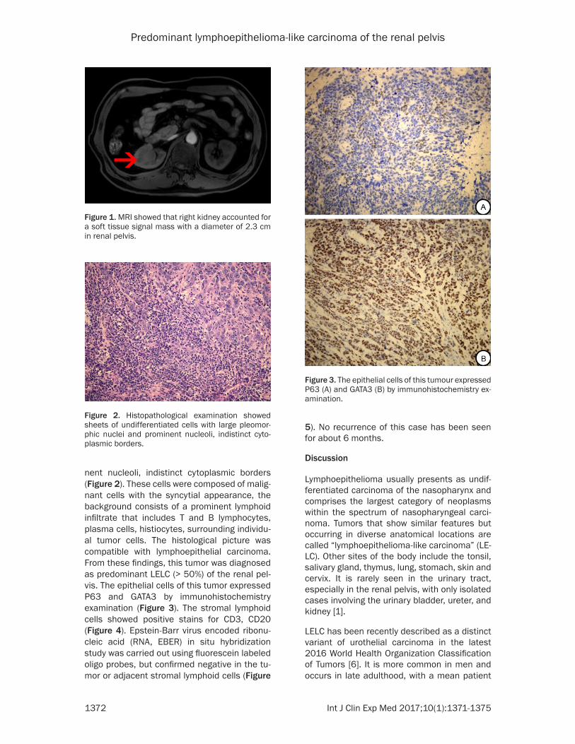
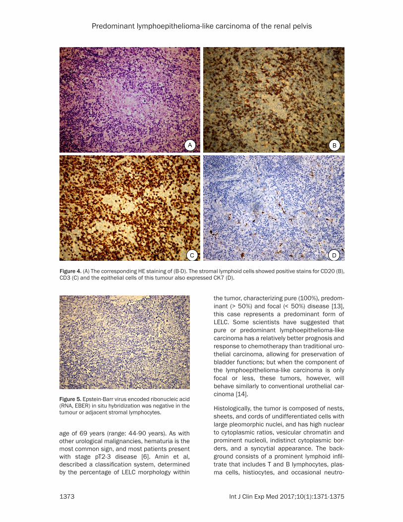
nent nucleoli, indistinct cytoplasmic borders (Figure 2). These cells were composed of malig-nant cells with the syncytial appearance, the background consists of a prominent lymphoid infiltrate that includes T and B lymphocytes, plasma cells, histiocytes, surrounding individu-al tumor cells. The histological picture was compatible with lymphoepithelial carcinoma. From these findings, this tumor was diagnosed as predominant LELC (> 50%) of the renal pel-vis. The epithelial cells of this tumor expressed P63 and GATA3 by immunohistochemistry examination (Figure 3). The stromal lymphoid cells showed positive stains for CD3, CD20 (Figure 4). Epstein-Barr virus encoded ribonu-cleic acid (RNA, EBER) in situ hybridization study was carried out using fluorescein labeled oligo probes, but confirmed negative in the tu- mor or adjacent stromal lymphoid cells (Figure
5). No recurrence of this case has been seen for about 6 months.
Discussion
Lymphoepithelioma usually presents as undif-ferentiated carcinoma of the nasopharynx and comprises the largest category of neoplasms within the spectrum of nasopharyngeal carci-noma. Tumors that show similar features but occurring in diverse anatomical locations are called “lymphoepithelioma-like carcinoma” (LE- LC). Other sites of the body include the tonsil, salivary gland, thymus, lung, stomach, skin and cervix. It is rarely seen in the urinary tract, especially in the renal pelvis, with only isolated cases involving the urinary bladder, ureter, and kidney [1].
LELC has been recently described as a distinct variant of urothelial carcinoma in the latest 2016 World Health Organization Classification of Tumors [6]. It is more common in men and occurs in late adulthood, with a mean patient
Figure 1. MRI showed that right kidney accounted for a soft tissue signal mass with a diameter of 2.3 cm in renal pelvis.
Figure 2. Histopathological examination showed sheets of undifferentiated cells with large pleomor-phic nuclei and prominent nucleoli, indistinct cyto-plasmic borders.
Figure 3. The epithelial cells of this tumour expressed P63 (A) and GATA3 (B) by immunohistochemistry ex-amination.
Predominant lymphoepithelioma-like carcinoma of the renal pelvis
1373 Int J Clin Exp Med 2017;10(1):1371-1375
age of 69 years (range: 44-90 years). As with other urological malignancies, hematuria is the most common sign, and most patients present with stage pT2-3 disease [6]. Amin et al, described a classification system, determined by the percentage of LELC morphology within
the tumor, characterizing pure (100%), predom-inant (> 50%) and focal (< 50%) disease [13], this case represents a predominant form of LELC. Some scientists have suggested that pure or predominant lymphoepithelioma-like carcinoma has a relatively better prognosis and response to chemotherapy than traditional uro-thelial carcinoma, allowing for preservation of bladder functions; but when the component of the lymphoepithelioma-like carcinoma is only focal or less, these tumors, however, will behave similarly to conventional urothelial car-cinoma [14].
Histologically, the tumor is composed of nests, sheets, and cords of undifferentiated cells with large pleomorphic nuclei, and has high nuclear to cytoplasmic ratios, vesicular chromatin and prominent nucleoli, indistinct cytoplasmic bor-ders, and a syncytial appearance. The back-ground consists of a prominent lymphoid infil-trate that includes T and B lymphocytes, plas-ma cells, histiocytes, and occasional neutro-
Figure 4. (A) The corresponding HE staining of (B-D). The stromal lymphoid cells showed positive stains for CD20 (B), CD3 (C) and the epithelial cells of this tumour also expressed CK7 (D).
Figure 5. Epstein-Barr virus encoded ribonucleic acid (RNA, EBER) in situ hybridization was negative in the tumour or adjacent stromal lymphocytes.
Predominant lymphoepithelioma-like carcinoma of the renal pelvis
1374 Int J Clin Exp Med 2017;10(1):1371-1375
phils or eosinophils, with eosinophils being prominent in rare cases. The epithelial cells of this tumor are immunoreactive with several cytokeratin markers and often express p63 and GATA3.
Unlike in the head and neck location, no asso-ciation has been reported between the urinary tract LELC and Epstein-Barr virus [5]. In our case, EBV encoded ribonucleic acid (RNA, EBER) in situ hybridization study was also nega-tive. In most of the published cases, EBV was reported to be positive in the organs which have direct external exposure such as naso-pharynx, and negative in the organs which are completely internal, such as kidney. This may contribute further evidence to no association between EBV and the urinary tract LELC [15].
LELC of the urinary tract is usually diagnosed in less-advanced clinical stages [16]. The progno-sis is favorable well for patients presenting with the pure and predominant forms with a diploid DNA pattern and very poor for patients present-ing with focal LELC [5].
Clinico-pathological characteristics of patients with LELC of the renal pelvis are shown in Table 1. Including this case, there have been ten reported cases of LELC of the renal pelvis in the English literature. Of these ten patients, six were female and the mean age was 72. Most patients presented with painless hematuria and had an advanced stage (pT3) of disease. Almost all tumors were negative for EBV exami-nation. Seven patients were managed with radi-cal surgery, three were treated with nephroure-terectomy and two patients also had auxiliary
radiotherapy. Due to the rarity of LELC of the renal pelvis, there are no better guidelines regarding the management of this disease, it is interesting to note that from the case series in Table 1, two of the three patients who under-went radical nephrectomy alone died with the disease. However, all those who underwent radical nephroureterectomy and auxiliary che-motherapy or radiotherapy had no evidence of disease on follow-up.
This may support the theory that it is related to urothelial carcinoma, LELC of the urinary tract requires radical resection of the entire upper urinary tract. This conclusion has been sup-ported by Haga and colleagues [1].
Disclosure of conflict of interest
None.
Address correspondence to: Jiong Shi, Department of Pathology, Nanjing Drum Tower Hospital, The Affiliated Hospital of Nanjing University Medical School, 321 Zhongshan Road, Nanjing 210008, Jiangsu Province, China. Tel: +86-25-83304616-10169; Fax: +86-25-83304616-10169; E-mail: [email protected]
References
[1] Haga K, Aoyagi T, Kashiwagi A, Yamashiro K and Nagamori S. Lymphoepithelioma-like car-cinoma of the renal pelvis. Int J Urol 2007; 14: 851-853.
[2] Mayer EK, Beckley I and Winkler MH. Lympho-epithelioma-like carcinoma of the urinary blad-der--diagnostic and clinical implications. Nat Clin Pract Urol 2007; 4: 167-171.
Table 1. The clinicopathological characteristics of reported LELC of the renal pelvisCase No. Sex Age Symptom Loca-
tionSize (cm) T S EBV
status HC Follow-up Out-come Year Reference
1 M 70 Hematuria UN UN RN+RT UN N Pre 6 years NED 1998 Fukunaga et al [7].2 F 79 Hematuria R 4 NU T3N0M0 N Pre 6 months NED 1999 Cohen et al [8].3 F 72 UN UN UN RN T3 N Pre 3 months DWD 2006 Perez-Motiel et al [9].4 M 68 UN UN UN RN T3 N Focal 12 months DWD 2006 Perez-Motiel et al [9].5 F 75 Hematuria L 3.5 NU T1N1M0 N Pure 36 months NED 2007 Haga et al [1].6 UN UN UN UN UN UN UN UN UN UN UN 2007 Tamas et al [10].7 F 75 Hematuria L 3.7 RN T3N0M0 N UN 6 months NED 2007 Yamada et al [11].8 F 75 Hematuria R 4.5 NU T3N1M0 UN Pre 6 months NED 2013 Modi et al [4].9 F 65 Hematuria R 5.3 RN T3N0M0 N Pre 7 months NED 2014 Ahn et al [12].10 M 59 Hematuria R 2.3 RN T3N1M0 N Pre 6 months NED 2016 Present caseT: treatment; S: stage; EBV: Epstein-Barr virus; HC: histological classification; M: male; F: female; R: right; L: left; UN: unknown; RN: radical ne-phrectomy; RT: radiation therapy; N: negative; NU: nephroureterectomy; Pre: predominant; NED: no evidence of disease; DWD: died with disease.
Predominant lymphoepithelioma-like carcinoma of the renal pelvis
1375 Int J Clin Exp Med 2017;10(1):1371-1375
[3] Cai G and Parwani AV. Cytomorphology of lym-phoepithelioma-like carcinoma of the urinary bladder: report of two cases. Diagn Cytopathol 2008; 36: 600-603.
[4] Modi H, Beckley I, Bhattarai S, Spencer J and Cartledge J. Lymphoepithelioma-like carcino-ma of the renal pelvis: pathological and thera-peutic implications. Can Urol Assoc J 2013; 7: E590-593.
[5] Terai A, Terada N, Ichioka K, Matsui Y, Yo-shimura K and Wani Y. Lymphoepithelioma-like carcinoma of the ureter. Urology 2005; 66: 1109.
[6] Humphrey PA, Moch H, Cubilla AL, Ulbright TM and Reuter VE. The 2016 WHO classification of tumours of the urinary system and male geni-tal organs-gart B: prostate and bladder tu-mours. Eur Urol 2016; 70: 106-19.
[7] Fukunaga M and Ushigome S. Lymphoepitheli-oma-like carcinoma of the renal pelvis: a case report with immunohistochemical analysis and in situ hybridization for the Epstein-Barr viral genome. Mod Pathol 1998; 11: 1252-1256.
[8] Cohen RJ, Stanley JC and Dawkins HJ. Lympho-epithelioma-like carcinoma of the renal pelvis. Pathology 1999; 31: 434-435.
[9] Perez-Montiel D, Wakely PE, Hes O, Michal M and Suster S. High-grade urothelial carcinoma of the renal pelvis: clinicopathologic study of 108 cases with emphasis on unusual morpho-logic variants. Mod Pathol 2006; 19: 494-503.
[10] Tamas EF, Nielsen ME, Schoenberg MP and Epstein JI. Lymphoepithelioma-like carcinoma of the urinary tract: a clinicopathological study of 30 pure and mixed cases. Mod Pathol 2007; 20: 828-834.
[11] Yamada Y, Fujimura T, Yamaguchi T, Nishimat-su H, Hirano Y, Kawamura T, Teshima S, Takeu-chi T and Kitamura T. Lymphoepithelioma-like carcinoma of the renal pelvis. Int J Urol 2007; 14: 1093-1094.
[12] Ahn H, Sim J, Kim H, Yi K, Han H, Chung Y, Rehman A and Paik SS. Lymphoepithelioma-like carcinoma of the renal pelvis: a case re-port and review of the literature. Korean J Pathol 2014; 48: 458-461.
[13] Pantelides NM, Ivaz SL, Falconer A, Hazell S, Winkler M, Hrouda D and Mayer EK. Lympho-epithelioma-like carcinoma of the urinary blad-der: a case report and review of systemic treat-ment options. Urol Ann 2012; 4: 45-47.
[14] Williamson SR, Zhang S, Lopez-Beltran A, Shah RB, Montironi R, Tan PH, Wang M, Baldridge LA, MacLennan GT and Cheng L. Lymphoepi-thelioma-like carcinoma of the urinary bladder: clinicopathologic, immunohistochemical, and molecular features. Am J Surg Pathol 2011; 35: 474-483.
[15] Gulley ML, Amin MB, Nicholls JM, Banks PM, Ayala AG, Srigley JR, Eagan PA and Ro JY. Ep-stein-Barr virus is detected in undifferentiated nasopharyngeal carcinoma but not in lympho-epithelioma-like carcinoma of the urinary blad-der. Hum Pathol 1995; 26: 1207-1214.
[16] Holmang S, Borghede G and Johansson SL. Bladder carcinoma with lymphoepithelioma-like differentiation: a report of 9 cases. J Urol 1998; 159: 779-782.


![Lymphoepithelioma-like gastric carcinoma: A case report ... · like gastric carcinoma (LELGC), first described by Watanabe et al[2] in 1976 as gastric carcinoma with a lymphoid stroma,](https://img.pdfslide.net/doc/110x75/5fc7c574c9fbf527a569fd63/lymphoepithelioma-like-gastric-carcinoma-a-case-report-like-gastric-carcinoma.jpg)