Embed Size (px)
Citation preview
Hindawi Publishing CorporationCase Reports in PediatricsVolume 2013, Article ID 218124, 4 pageshttp://dx.doi.org/10.1155/2013/218124
Case ReportAnatomical Asplenia in Cat Eye Syndrome:An Expansion of the Disease Spectrum
DeepakBabu Chellapandian1 and Adele Schneider2
1 Department of Pediatrics and Adolescent Medicine, Einstein Medical Center, Philadelphia, PA 19141, USA2Clinical Genetics, Einstein Medical Center, Philadelphia, PA 19141, USA
Correspondence should be addressed to DeepakBabu Chellapandian; [email protected]
Received 21 February 2013; Accepted 14 March 2013
Academic Editors: P. Pavone and M. Raes
Copyright © 2013 D. Chellapandian and A. Schneider. This is an open access article distributed under the Creative CommonsAttribution License, which permits unrestricted use, distribution, and reproduction in any medium, provided the original work isproperly cited.
We report a patient withCat eye syndrome (CES) associatedwith anatomical asplenia. To the best of our knowledge, there have beenno prior reports of this association. Screening for asplenia in CES is potentially important, as asplenia places patients at increasedrisk for life-threatening bacterial infections. Hence patients with CES without a spleen may require the same routine precautionsas any other asplenic patients, with penicillin prophylaxis and immunizations to protect against encapsulated organisms such asStreptococcus pneumoniae, Haemophilus influenzae type b, and Neisseria meningitidis.
1. Introduction
Cat eye syndrome (OMIM 115470) is a rare chromosomaldisorder presenting as a clinically recognizable pattern ofcongenital abnormalities, first described in 1965 by Schachen-mann et al. The classical features are ocular coloboma,anorectal, heart, and renal malformations, ears with pre-auricular tags and/or pits and variable intellectual disabilities[1, 2]. The name “cat eye” was introduced because the iriscoloboma resembles the pupil shape of cats. CES is oftenassociated with significant phenotypic variability, rangingfrom patients with almost normal phenotype to those withsevere abnormalities [1]. None of the features are consistentlypresent. Only 41% of CES patients have the combination ofiris coloboma, anal anomalies, and preauricular pits/tags [1].Thus, many patients cannot be identified as having CES byphenotype alone.Here, we describe amale infant with clinicalfeatures of cat eye syndrome, confirmed by cytogeneticand fluorescence in situ hybridization (FISH) analysis, whopresented with anatomical asplenia.
2. Case Report
The proband is a full term male, born to a gravida 2 para1 mother by Caesarian section. At the patient’s birth, the
mother and father’s ages were 22 and 20, respectively. Thepregnancy was a result of natural conception and uncompli-cated by any teratogens.Theprenatal ultrasoundwas reportedas normal. The parents were healthy and unrelated. Themother has a healthy 3-year-old daughter from a previousrelationship. Review of the family history did not reveal anyother individuals with developmental delay or congenitalmalformations.
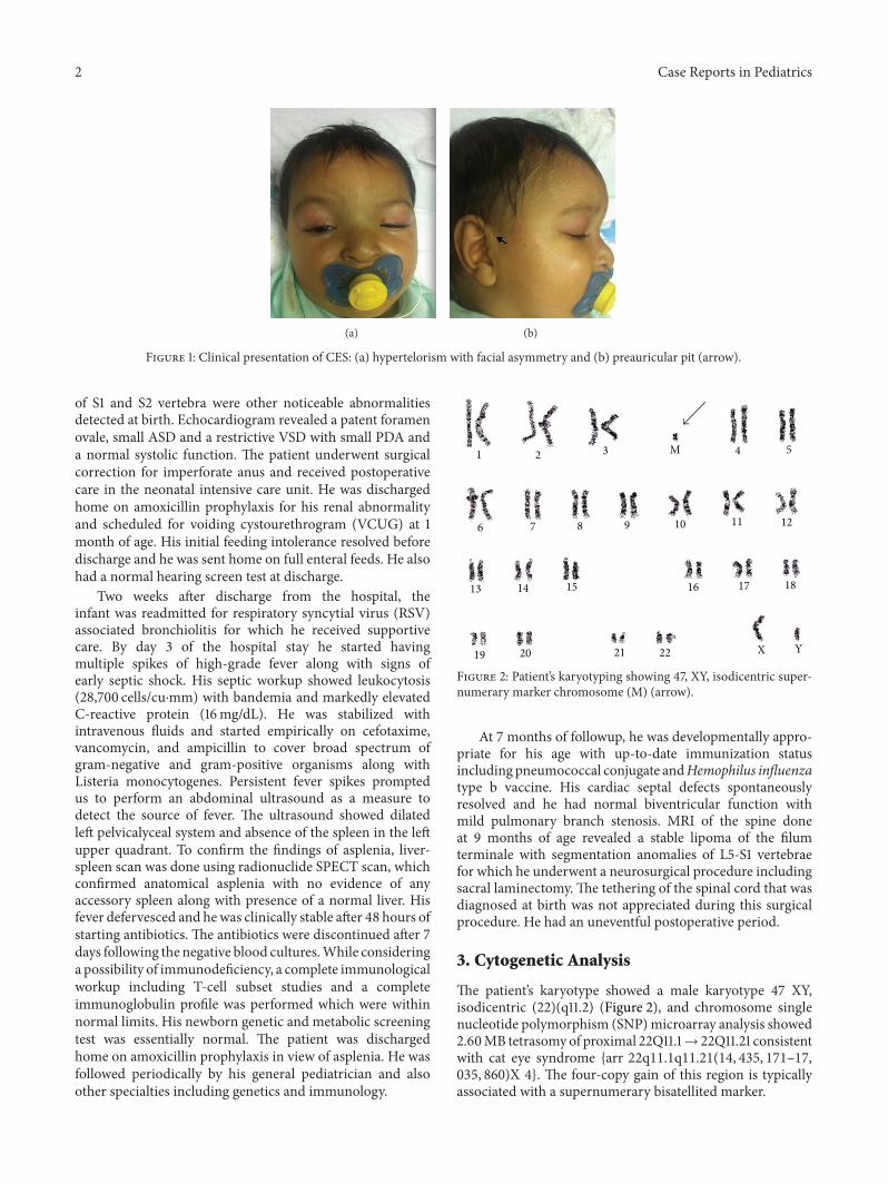
The patient was referred to our center from an out-side hospital soon after birth for surgical management ofimperforate anus. His birth weight was 3345 grams (75thcentile), length was 52.5 cm (>90th centile), and head cir-cumference was 35.5 cm (90th centile). The initial physicalexamination on admission showed the following dysmor-phic features: downslanting palpebral fissures, hypertelorism(Figure 1(a)) with interpupillary distance of 5 cm and innercanthal measurement of 3 cm (both are above 97th centilefor age), mild facial asymmetry (right side > left side),slight difference between the size of the ears (<0.5 cm),anteverted and low set ears with bilateral preauricular pits(Figure 1(b)), micrognathia, and bilateral 5th finger clin-odactyly. His initial detailed ophthalmological examinationshowed iris coloboma with normal retinal vascularization. Agrade II/VI systolic murmur, imperforate anus with perinealfistula, left renal pelviectasis, tethered cord, and partial fusion
2 Case Reports in Pediatrics
(a) (b)
Figure 1: Clinical presentation of CES: (a) hypertelorism with facial asymmetry and (b) preauricular pit (arrow).
of S1 and S2 vertebra were other noticeable abnormalitiesdetected at birth. Echocardiogram revealed a patent foramenovale, small ASD and a restrictive VSD with small PDA anda normal systolic function. The patient underwent surgicalcorrection for imperforate anus and received postoperativecare in the neonatal intensive care unit. He was dischargedhome on amoxicillin prophylaxis for his renal abnormalityand scheduled for voiding cystourethrogram (VCUG) at 1month of age. His initial feeding intolerance resolved beforedischarge and he was sent home on full enteral feeds. He alsohad a normal hearing screen test at discharge.
Two weeks after discharge from the hospital, theinfant was readmitted for respiratory syncytial virus (RSV)associated bronchiolitis for which he received supportivecare. By day 3 of the hospital stay he started havingmultiple spikes of high-grade fever along with signs ofearly septic shock. His septic workup showed leukocytosis(28,700 cells/cu⋅mm) with bandemia and markedly elevatedC-reactive protein (16mg/dL). He was stabilized withintravenous fluids and started empirically on cefotaxime,vancomycin, and ampicillin to cover broad spectrum ofgram-negative and gram-positive organisms along withListeria monocytogenes. Persistent fever spikes promptedus to perform an abdominal ultrasound as a measure todetect the source of fever. The ultrasound showed dilatedleft pelvicalyceal system and absence of the spleen in the leftupper quadrant. To confirm the findings of asplenia, liver-spleen scan was done using radionuclide SPECT scan, whichconfirmed anatomical asplenia with no evidence of anyaccessory spleen along with presence of a normal liver. Hisfever defervesced and hewas clinically stable after 48 hours ofstarting antibiotics. The antibiotics were discontinued after 7days following the negative blood cultures.While consideringa possibility of immunodeficiency, a complete immunologicalworkup including T-cell subset studies and a completeimmunoglobulin profile was performed which were withinnormal limits. His newborn genetic and metabolic screeningtest was essentially normal. The patient was dischargedhome on amoxicillin prophylaxis in view of asplenia. He wasfollowed periodically by his general pediatrician and alsoother specialties including genetics and immunology.
1 2 3 4 5
6 7 8 9 10 11 12
13 14 15 16 17 18
19 20 21 22
M
YX
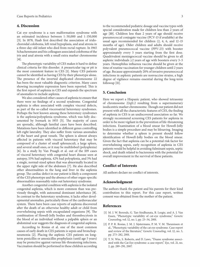
Figure 2: Patient’s karyotyping showing 47, XY, isodicentric super-numerary marker chromosome (M) (arrow).
At 7 months of followup, he was developmentally appro-priate for his age with up-to-date immunization statusincluding pneumococcal conjugate andHemophilus influenzatype b vaccine. His cardiac septal defects spontaneouslyresolved and he had normal biventricular function withmild pulmonary branch stenosis. MRI of the spine doneat 9 months of age revealed a stable lipoma of the filumterminale with segmentation anomalies of L5-S1 vertebraefor which he underwent a neurosurgical procedure includingsacral laminectomy.The tethering of the spinal cord that wasdiagnosed at birth was not appreciated during this surgicalprocedure. He had an uneventful postoperative period.
3. Cytogenetic Analysis
The patient’s karyotype showed a male karyotype 47 XY,isodicentric (22)(q11.2) (Figure 2), and chromosome singlenucleotide polymorphism (SNP)microarray analysis showed2.60MB tetrasomyof proximal 22Q11.1→ 22Q11.21 consistentwith cat eye syndrome {arr 22q11.1q11.21(14, 435, 171–17,035, 860)X 4}. The four-copy gain of this region is typicallyassociated with a supernumerary bisatellited marker.
Case Reports in Pediatrics 3
4. Discussion
Cat eye syndrome is a rare malformation syndrome withan estimated incidence between 1 : 50,000 and 1 : 150,000[3]. In 1878, Haab first described the association of irido-choroidal coloboma, left renal hypoplasia, and anal atresia ina three-day-old infant who died from rectal rupture. In 1965Schachenmann and his colleagues associated coloboma of theiris and anal atresia with a small extra marker chromosome[4].
The phenotypic variability of CES makes it hard to defineclinical criteria for this disorder. A preauricular tag or pit isthe most consistent feature in CES [1]. Many CES patientscannot be identified as having CES by their phenotype alone.The presence of the inverted duplicated chromosome 22has been the most valuable diagnostic criterion. Many casesshowing incomplete expression have been reported. This isthe first report of asplenia in CES and expands the spectrumof anomalies to include asplenia.
We also considered other causes of asplenia to ensure thatthere were no findings of a second syndrome. Congenitalasplenia is often associated with complex visceral defects,as part of the so-called visceroatrial heterotaxy syndromes.Perhaps the best known among these heterotaxy syndromesis the asplenia/polysplenia syndrome, which was fully doc-umented by Ivemark in 1955 [5]. The majority of casesare sporadic, although familial cases have been described.Mutations have been identified in different genes controllingleft-right laterality. They also suffer from various anomaliesof the heart and great vessels. The spleen is almost alwaysaffected in patients with visceral heterotaxy that may becomposed of a cluster of small splenunculi, a large spleen,and several small ones, or it may be multilobed (polysplenia)[6]. In a study by Van Praagh et al. out of the 109 casesof visceral heterotaxy with congenital heart disease seen atautopsy, 53% had asplenia, 42% had polysplenia, and 5% hada single, normal-sized spleen that was abnormally located inthe upper right side of the abdomen [7]. He also describedother abnormalities in the lung and liver in the aspleniagroup.The cardiac defect in our patient is likely a componentof the CES phenotype and the absence of other organ-specificabnormalities reasonably rules out heterotaxy syndrome.
Another congenital conditionwith asplenia is the isolatedcongenital asplenia, which is more common than was pre-viously thought, with autosomal dominant inheritance [8].In contrast to the heterotaxy syndrome, it lacks other devel-opmental anomalies, particularly those of the cardiovascularsystem. There have been case reports of asplenia discoveredafter the death of an otherwise healthy adult or child fromoverwhelming sepsis with encapsulated organisms [9]. Thecombination of Howell-Jolly bodies and thrombocytosis inthe blood of an individual without a palpable spleen or anabdominal scar suggests the diagnosis of congenital asplenia.
According to Rosias et al. one of the most commoncauses of early death in CES patients is sepsis and bronchop-neumonia [2]. Placing the asplenic CES patients on long-term penicillin or amoxicillin prophylaxis (until age 5 years)may be protective against various life-threatening infections.Vaccination should be performed in these children according
to the recommended pediatric dosage and vaccine types withspecial consideration made for children less than 2 years ofage [10]. Children less than 2 years of age should receivepneumococcal conjugate vaccine (PCV-13 if available) at theusual ages recommended for children (2, 4, 6, and 12–15months of age). Older children and adults should receivepolyvalent pneumococcal vaccine (PPV-23) with boosterapproximately every 5 years starting from the first dose.Quadrivalent meningococcal vaccine should be given to allasplenic individuals ≥2 years of age with boosters every 3–5years. Hemophilus influenza vaccine should be given at thetime of routine vaccination for younger children until 2 yearsof age. Because approximately 20% of invasive pneumococcalinfections in asplenic patients are nonvaccine strains, a highdegree of vigilance remains essential during the long-termfollowup [11].
5. Conclusion
Here we report a Hispanic patient, who showed tetrasomyof chromosome 22q11.2 resulting from a supernumeraryisodicentricmarker chromosome.Thoughour patient did notpresent with all the characteristic features of CES, the findingof asplenia in CES is an undiscovered association so far. Westrongly recommend screening CES patients for asplenia inorder to bemore vigilant in the prevention of life-threateninginfections. Examination of a blood smear for Howell-Jollybodies is a simple procedure and may be lifesaving. Imagingto determine whether a spleen is present should followidentification of Howell-Jolly bodies on the blood smear.Given the fact that asplenia can be a potential risk factor foroverwhelming sepsis, early recognition of asplenia in CESpatients would be helpful in avoiding fulminant sepsis, septicshock, and death related to bacteremia with the potential foroverall improvement in the survival of these patients.
Conflict of Interests
All authors declare no conflict of interests.
Acknowledgment
The authors thank the patient and his parents for their kindcontribution to this report. For this case report, writtenconsent was obtained from the mother of the patient.
References
[1] M. J. W. Berends, G. Tan-Sindhunata, B. Leegte, and A. J. VanEssen, “Phenotypic variability of cat-eye syndrome,” GeneticCounseling, vol. 12, no. 1, pp. 23–34, 2001.
[2] P. P. R. Rosias, J. M. J. Sijstermans, P. M. V. M. Theunissen etal., “Phenotypic variability of the cat eye syndrome. Case reportand review of the literature,” Genetic Counseling, vol. 12, no. 3,pp. 273–282, 2001.
[3] T. N. Win, S. Roberts, and D. Laws, “Duane syndrome associ-ated with the Cat Eye syndrome: a case report,” Eye, vol. 21, no.2, pp. 289–291, 2007.
4 Case Reports in Pediatrics
[4] G. Schachenmann, W. Schmid, M. Fraccaro et al., “Chromo-somes in Coloboma and Anal Atresia,”The Lancet, vol. 286, no.7406, p. 290, 1965.
[5] B. I. Ivemark, “Implications of agenesis of the spleen on thepathogenesis of conotruncus anomalies in childhood; an analy-sis of the heart malformations in the splenic agenesis syndrome,with fourteen new cases,” Acta paediatrica. Supplementum, vol.44, supplement 104, pp. 7–110, 1955.
[6] J. E. Keane, V Lock, andD. C. Fyler,Nadas’ Pediatric Cardiology,Elsevier, 2nd edition, 2006.
[7] S. van Praagh, J. Kreutzer, L. Alday, and R. van Praagh, “Sys-temic andpulmonary venous connections in visceral heterotaxywith emphasis on the diagnosis of the atrial situs: a study of 109postmortemcases,” inDevelopmental CardiologyMorphogenesisand Function Futura, E. B. Clark andA. Takas, Eds., pp. 671–727,New York, NY, USA, 1990.
[8] N. Mahlaoui, V. Minard-Colin, C. Picard et al., “Isolatedcongenital asplenia: a French nationwide retrospective surveyof 20 cases,” Journal of Pediatrics, vol. 158, no. 1, pp. 142–148,2011.
[9] B.Gilbert, C.Menetrey,V. Belin, P. Brosset, L.DeLumley, andA.Fisher, “Familial isolated congenital asplenia: a rare, frequentlyhereditary dominant condition, often detected too late as acause of overwhelming pneumococcal sepsis. Report of a newcase and review of 31 others,” European Journal of Pediatrics, vol.161, no. 7, pp. 368–372, 2002.
[10] The Centers for Disease Control and Prevention (CDC), 2010,http://www.cdc.gov/vaccines/.
[11] G. E. Schutze, E. O. Mason, W. J. Barson et al., “Invasivepneumococcal infections in children with asplenia,” PediatricInfectious Disease Journal, vol. 21, no. 4, pp. 278–282, 2002.

Submit your manuscripts athttp://www.hindawi.com
Stem CellsInternational
Hindawi Publishing Corporationhttp://www.hindawi.com Volume 2014
Hindawi Publishing Corporationhttp://www.hindawi.com Volume 2014
MEDIATORSINFLAMMATION
of
Hindawi Publishing Corporationhttp://www.hindawi.com Volume 2014
Behavioural Neurology
EndocrinologyInternational Journal of
Hindawi Publishing Corporationhttp://www.hindawi.com Volume 2014
Hindawi Publishing Corporationhttp://www.hindawi.com Volume 2014
Disease Markers
Hindawi Publishing Corporationhttp://www.hindawi.com Volume 2014
BioMed Research International
OncologyJournal of
Hindawi Publishing Corporationhttp://www.hindawi.com Volume 2014
Hindawi Publishing Corporationhttp://www.hindawi.com Volume 2014
Oxidative Medicine and Cellular Longevity
Hindawi Publishing Corporationhttp://www.hindawi.com Volume 2014
PPAR Research
The Scientific World JournalHindawi Publishing Corporation http://www.hindawi.com Volume 2014
Immunology ResearchHindawi Publishing Corporationhttp://www.hindawi.com Volume 2014
Journal of
ObesityJournal of
Hindawi Publishing Corporationhttp://www.hindawi.com Volume 2014
Hindawi Publishing Corporationhttp://www.hindawi.com Volume 2014
Computational and Mathematical Methods in Medicine
OphthalmologyJournal of
Hindawi Publishing Corporationhttp://www.hindawi.com Volume 2014
Diabetes ResearchJournal of
Hindawi Publishing Corporationhttp://www.hindawi.com Volume 2014
Hindawi Publishing Corporationhttp://www.hindawi.com Volume 2014
Research and TreatmentAIDS
Hindawi Publishing Corporationhttp://www.hindawi.com Volume 2014
Gastroenterology Research and Practice
Hindawi Publishing Corporationhttp://www.hindawi.com Volume 2014
Parkinson’s Disease
Evidence-Based Complementary and Alternative Medicine
Volume 2014Hindawi Publishing Corporationhttp://www.hindawi.com