Embed Size (px)
Citation preview
Int J Clin Exp Pathol 2013;6(6):1128-1131www.ijcep.com /ISSN:1936-2625/IJCEP1209031
Case Report Autopsy findings of fatal cryptogenic organizing pneumonia
Tadashi Terada
Department of Pathology, Shizuoka City Shimizu Hospital, Shimizu, Shizuoka, Japan
Received September 29, 2012; Accepted March 25, 2013; Epub May 15, 2013; Published June 1, 2013
Abstract: Autopsy cases of cryptogenic organizing pneumonia (COP) have been rarely reported. A 73-year-old Japanese man consulted to a hospital because of flu-like sickness. He was diagnosed as pneumonia, and treated by antibiotics. He was referred to our hospital for further treatment. Chest X-P showed pneumonia involving the whole lungs. Blood laboratory test showed leukocytosis, increased CRP, and decreased PaO2. Despite of steroid therapy, he showed a downhill course and died one month after the first manifestation. The clinical diagnosis was acute pneumonia or ARDS. At autopsy, the both lungs were voluminous. The weight of lungs was 1050 g in the left lung and 1300 g in the right lung. The both lungs were entirely affected. The lungs were hard and little air was recognized. Microscopically, almost all alveolar spaces contained Masson’s bodies. Bronchiolitis obliterans was not recognized. The alveolar walls were not affected. The Masson’s bodies showed collagenization with lymphocytic infiltration. Hyalinization of Masson’s bodies with little inflammatory infiltration was frequently seen. Cartilagenous metaplasia and ossification of Masson’s bodies were seen in some places. The pulmonary arteries were affected by fibrosis, and occasionally showed thrombosis. The pathological diagnosis was COP. The heart weighted 500 g, and showed right ventricular hypertrophy (cor pulmonale). Other pathologic changes were pleural effusion (left, 800 ml: right, 1200 ml), acute liver congestion, prostatic hypertrophy, colon adenoma, and hypercellular bone marrow. The cause of death was respiratory failure due to COP and pleural effusion. In conclusion, the author reported an autopsy case of fatal COP.
Keywords: Lung, cryptogenic organizing pneumonia, histopathology
Introduction
Focal organizing pneumonia is frequently seen in patients with bronchopneumonia. However, diffuse organizing pneumonia without features of acute bronchopneumonia was rare. In the past, such condition was called bronchiolitis obliterans organizing pneumonia (BOOP) [1, 2]. However, constrictive bronchiolitis obliterans was similar to BOOP, and BOOP without bron-chiolitis obliterans was present. Therefore, dif-fuse idiopathic organizing pneumonia without bronchiolitis obliterans was called cryptogenic organizing pneumonia (COP) [1, 2]. COP is very rare. Patients with COP show relatively well prognosis [1-4]. Steroid is very effective in COP [1-4]. Fatal COP is very rare, and there have been few reports of autopsy cases of fatal COP. The author herein reports a case of fatal COP with an emphasis of pathologic findings.
Case report
A 73-year-old Japanese man consulted to a hospital because of flu-like sickness. He was diagnosed as pneumonia, and treated by antibi-otics. However, no improvement was obtained, and respiratory failure emerged. Therefore, he was referred to our hospital for further treat-ment. On admission, physical examination showed fever, cyanosis, and rale in both lungs. Chest X-P showed pneumonia involving the whole lungs. Blood laboratory test showed leu-kocytosis (28,500 /μl), increased CRP (29.7 mg/dl), and decreased PaO2 (27.5 mmHg). No organisms were obtained from the sputum and blood. No eosinophilia was noted. He showed no collagen diseases, and autoantibodies were all negative. No causes of the pneumonia were identified. He was in the state of respiratory fail-ure, and oxygen administration was performed.
Cryptogenic organizing pneumonia
1129 Int J Clin Exp Pathol 2013;6(6):1128-1131
Then, he was treated by respirator, and by anti-biotics and steroids. Despite the steroid thera-py, he showed a downhill course, and died one month after the first manifestation. The clinical diagnosis was acute pneumonia or ARDS.
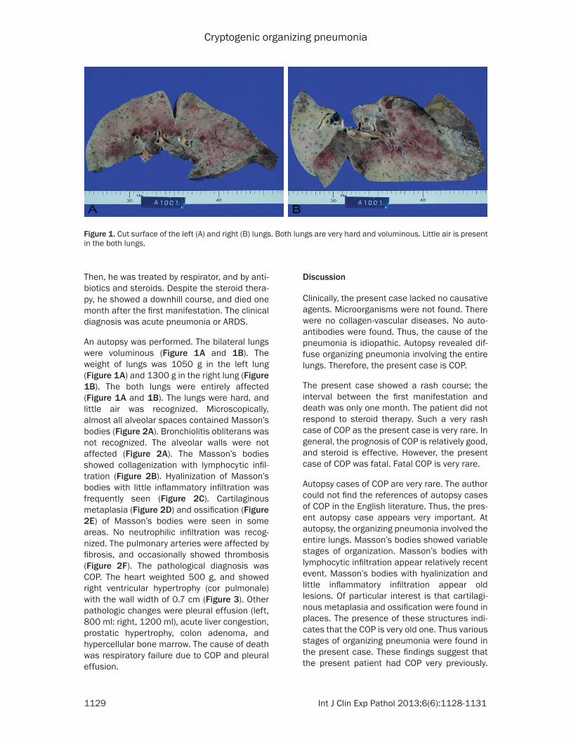
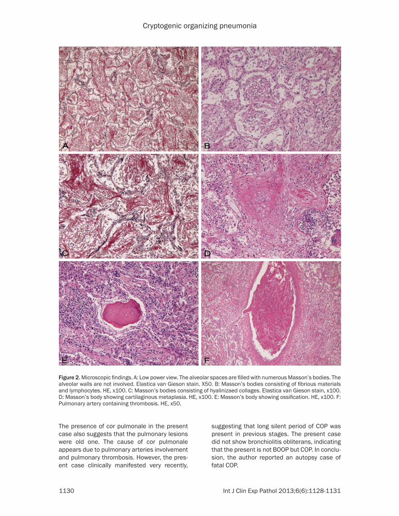
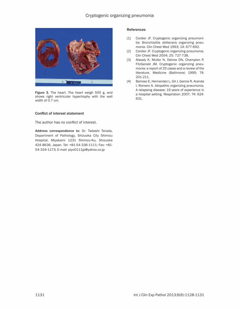
An autopsy was performed. The bilateral lungs were voluminous (Figure 1A and 1B). The weight of lungs was 1050 g in the left lung (Figure 1A) and 1300 g in the right lung (Figure 1B). The both lungs were entirely affected (Figure 1A and 1B). The lungs were hard, and little air was recognized. Microscopically, almost all alveolar spaces contained Masson’s bodies (Figure 2A). Bronchiolitis obliterans was not recognized. The alveolar walls were not affected (Figure 2A). The Masson’s bodies showed collagenization with lymphocytic infil-tration (Figure 2B). Hyalinization of Masson’s bodies with little inflammatory infiltration was frequently seen (Figure 2C). Cartilaginous metaplasia (Figure 2D) and ossification (Figure 2E) of Masson’s bodies were seen in some areas. No neutrophilic infiltration was recog-nized. The pulmonary arteries were affected by fibrosis, and occasionally showed thrombosis (Figure 2F). The pathological diagnosis was COP. The heart weighted 500 g, and showed right ventricular hypertrophy (cor pulmonale) with the wall width of 0.7 cm (Figure 3). Other pathologic changes were pleural effusion (left, 800 ml: right, 1200 ml), acute liver congestion, prostatic hypertrophy, colon adenoma, and hypercellular bone marrow. The cause of death was respiratory failure due to COP and pleural effusion.
Discussion
Clinically, the present case lacked no causative agents. Microorganisms were not found. There were no collagen-vascular diseases. No auto-antibodies were found. Thus, the cause of the pneumonia is idiopathic. Autopsy revealed dif-fuse organizing pneumonia involving the entire lungs. Therefore, the present case is COP.
The present case showed a rash course; the interval between the first manifestation and death was only one month. The patient did not respond to steroid therapy. Such a very rash case of COP as the present case is very rare. In general, the prognosis of COP is relatively good, and steroid is effective. However, the present case of COP was fatal. Fatal COP is very rare.
Autopsy cases of COP are very rare. The author could not find the references of autopsy cases of COP in the English literature. Thus, the pres-ent autopsy case appears very important. At autopsy, the organizing pneumonia involved the entire lungs. Masson’s bodies showed variable stages of organization. Masson’s bodies with lymphocytic infiltration appear relatively recent event. Masson’s bodies with hyalinization and little inflammatory infiltration appear old lesions. Of particular interest is that cartilagi-nous metaplasia and ossification were found in places. The presence of these structures indi-cates that the COP is very old one. Thus various stages of organizing pneumonia were found in the present case. These findings suggest that the present patient had COP very previously.
Figure 1. Cut surface of the left (A) and right (B) lungs. Both lungs are very hard and voluminous. Little air is present in the both lungs.
Cryptogenic organizing pneumonia
1130 Int J Clin Exp Pathol 2013;6(6):1128-1131
The presence of cor pulmonale in the present case also suggests that the pulmonary lesions were old one. The cause of cor pulmonale appears due to pulmonary arteries involvement and pulmonary thrombosis. However, the pres-ent case clinically manifested very recently,
suggesting that long silent period of COP was present in previous stages. The present case did not show bronchiolitis obliterans, indicating that the present is not BOOP but COP. In conclu-sion, the author reported an autopsy case of fatal COP.
Figure 2. Microscopic findings. A: Low power view. The alveolar spaces are filled with numerous Masson’s bodies. The alveolar walls are not involved. Elastica van Gieson stain. X50. B: Masson’s bodies consisting of fibrious materials and lymphocytes. HE, x100. C: Masson’s bodies consisting of hyalinizaed collages. Elastica van Gieson stain, x100. D: Masson’s body showing cartilaginous metaplasia. HE, x100. E: Masson’s body showing ossification. HE, x100. F: Pulmonary artery containing thrombosis. HE, x50.
Cryptogenic organizing pneumonia
1131 Int J Clin Exp Pathol 2013;6(6):1128-1131
References
[1] Cordier JF. Cryptogenic organizing pneumoni-tis: Bronchiolitis obliterans organizing pneu-monia. Clin Chest Med 1993; 14: 677-692.
[2] Cordier JF. Cryptogenic organizing pneumonia. Clin Chest Med 2004; 25: 727-738.
[3] Alasaly K, Muller N, Ostrow DN, Champion P, FitzGerald JM. Cryptogenic organizing pneu-monia: a report of 25 cases and a review of the literature. Medicine (Baltimore) 1995; 74: 201-211.
[4] Barroso E, Hernandez L, Gil J, Garcia R, Aranda I, Romero S. Idiopathic organizing pneumonia. A relapsing disease: 19 years of experience in a hospital setting. Respiration 2007; 74: 624-631.
Conflict of interest statement
The author has no conflict of interest.
Address correspondence to: Dr. Tadashi Terada, Department of Pathology, Shizuoka City Shimizu Hospital, Miyakami 1231 Shimizu-Ku, Shizuoka 424-8636, Japan. Tel: +81-54-336-1111; Fax: +81-54-334-1173; E-mail: [email protected]
Figure 3. The heart. The heart weigh 500 g, and shows right ventricular hypertrophy with the wall width of 0.7 cm.