Embed Size (px)
Citation preview
WORLD JOURNAL OF SURGICAL ONCOLOGY
Wang et al. World Journal of Surgical Oncology 2014, 12:133http://www.wjso.com/content/12/1/133
CASE REPORT Open Access
Gastritis cystica profunda recurrence after surgicalresection: 2-year follow-upLei Wang1, Hua Yan1, Da-Chun Cao1, Li Huo1, Hai-Zhong Huo2, Bing Wang2, Ying Chen3 and Hai-Lin Liu1*
Abstract
Background: Gastritis cystica profunda (GCP) is an uncommon disease characterized by multiple cystic gastricglands within the submucosa of the stomach.
Case description: Here, we present a case of a 63-year-old man with intermittent epigastric discomfort in whomgastroscopy revealed multiple irregular elevated nodular lesions with smooth surfaces at the anterior of the antrum.Surgical resection of the nodular lesions was performed, and the diagnosis of gastritis cystica profunda (GCP) wasconfirmed by histological examination. Another elevated nodular lesion approximately 10 mm in diameter with anulcer was found on the gastric side of the remnant stomach near the resection side from 6 to 24 months after thesurgical resection. Endoscopic ultrasonography (EUS) and repeated biopsies of the new elevated lesion wereperformed. Homogeneous, anechoic masses originating from the submucosa without gastric adenocarcinoma inhistological examination showed GCP recurrence may occur.
Conclusions: We report a case of GCP recurrence within 6 months after surgical resection. GCP should beconsidered in the differential diagnosis of elevated lesions in the stomach.
Keywords: Gastritis cystica profunda, Gastric cancer, Endoscopic ultrasonography
BackgroundGastritis cystica profunda (GCP) is an uncommon diseasecharacterized by multiple cystic gastric glands within thesubmucosa of the stomach. GCP seemed to be limited topreviously operated stomachs in prior reports, but it hasbeen found in unoperated stomachs in several recent arti-cles [1-3]. Here, we report a case of GCP that occurred ina patient who had not previously undergone gastric sur-gery, and the GCP recurred in just 6 months after surgicalresection.
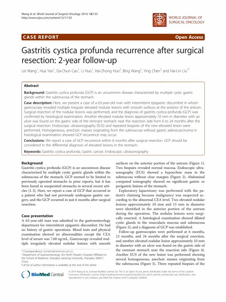
Case presentationA 63-year-old man was admitted to the gastroenterologydepartment for intermittent epigastric discomfort. He hadno history of gastric operations. Blood tests and physicalexamination showed no abnormalities except the CEAlevel of serum was 7.68 ng/mL. Gastroscopy revealed mul-tiple irregularly elevated nodular lesions with smooth
* Correspondence: [email protected] of Gastroenterology, the Ninth People’s Hospital Affiliated tothe School of Medicine, Shanghai Jiaotong University, Shanghai 200011,ChinaFull list of author information is available at the end of the article
© 2014 Wang et al.; licensee BioMed Central LCommons Attribution License (http://creativecreproduction in any medium, provided the or
surfaces on the anterior portion of the antrum (Figure 1).Two biopsies revealed normal mucosa. Endoscopic ultra-sonography (EUS) showed a hypoechoic mass in thesubmucosa without clear margins (Figure 2). Abdominalcomputed tomography showed no significant gastric orperigastric lesions of the stomach.Exploratory laparotomy was performed with the pa-
tient’s claiming because malignancy was suspected ac-cording to the abnormal CEA level. Two elevated nodularlesions approximately 10 mm and 15 mm in diameterwere identified in the anterior portion of the antrumduring the operation. The nodular lesions were surgi-cally resected. A histological examination showed dilatedcystic glands in the muscularis mucosa and submucosa(Figure 3), and a diagnosis of GCP was established.Follow-up gastroscopies were performed at 6 months,
12 months, and 24 months after the surgical resection,and another elevated nodular lesion approximately 10 mmin diameter with an ulcer was found on the gastric side ofthe remnant stomach near the resection side (Figure 4).Another EUS of the new lesion was performed showingseveral homogeneous, anechoic masses originating fromthe submucosa (Figure 5). Three repeated biopsies of the
td. This is an Open Access article distributed under the terms of the Creativeommons.org/licenses/by/2.0), which permits unrestricted use, distribution, andiginal work is properly credited.
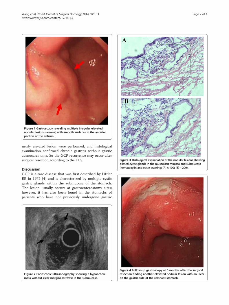
Figure 1 Gastroscopy revealing multiple irregular elevatednodular lesions (arrows) with smooth surfaces in the anteriorportion of the antrum.
Figure 3 Histological examination of the nodular lesions showingdilated cystic glands in the muscularis mucosa and submucosa(hematoxylin and eosin staining; (A) × 100; (B) × 200).
Wang et al. World Journal of Surgical Oncology 2014, 12:133 Page 2 of 4http://www.wjso.com/content/12/1/133
newly elevated lesion were performed, and histologicalexamination confirmed chronic gastritis without gastricadenocarcinoma. So the GCP recurrence may occur aftersurgical resection according to the EUS.
DiscussionGCP is a rare disease that was first described by LittlerER in 1972 [4] and is characterized by multiple cysticgastric glands within the submucosa of the stomach.The lesion usually occurs at gastroenterostomy sites;however, it has also been found in the stomachs ofpatients who have not previously undergone gastric
Figure 2 Endoscopic ultrasonography showing a hypoechoicmass without clear margins (arrows) in the submucosa.
Figure 4 Follow-up gastroscopy at 6 months after the surgicalresection finding another elevated nodular lesion with an ulceron the gastric side of the remnant stomach.
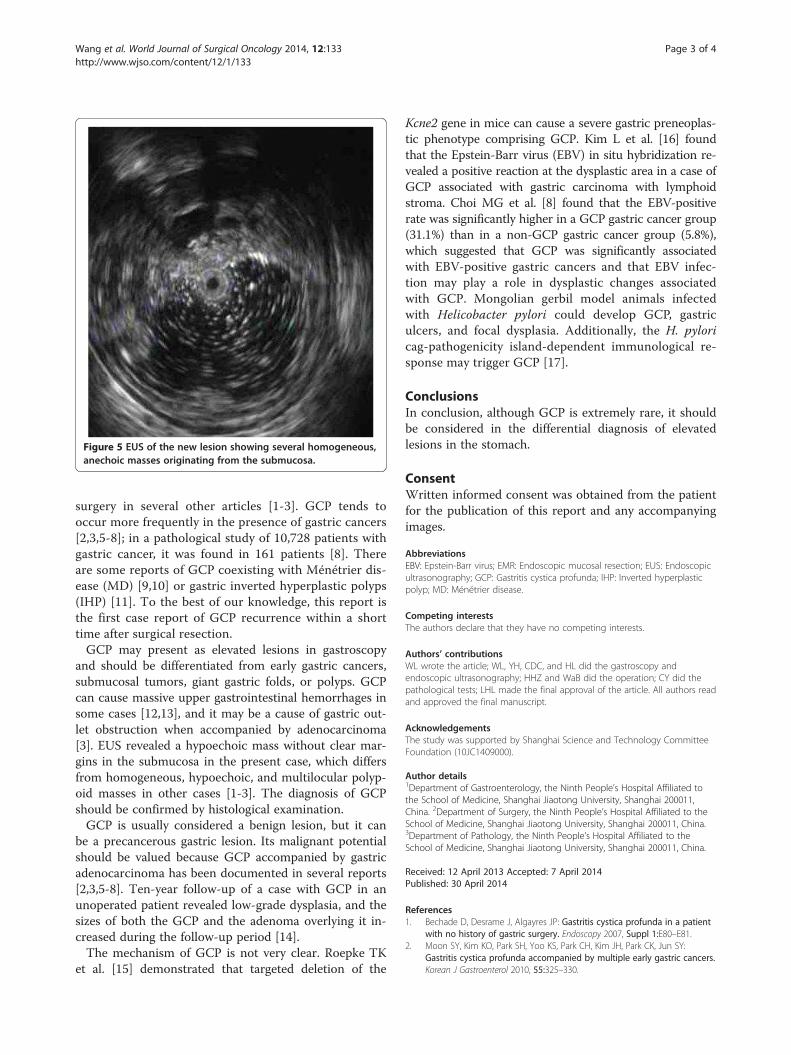
Figure 5 EUS of the new lesion showing several homogeneous,anechoic masses originating from the submucosa.
Wang et al. World Journal of Surgical Oncology 2014, 12:133 Page 3 of 4http://www.wjso.com/content/12/1/133
surgery in several other articles [1-3]. GCP tends tooccur more frequently in the presence of gastric cancers[2,3,5-8]; in a pathological study of 10,728 patients withgastric cancer, it was found in 161 patients [8]. Thereare some reports of GCP coexisting with Ménétrier dis-ease (MD) [9,10] or gastric inverted hyperplastic polyps(IHP) [11]. To the best of our knowledge, this report isthe first case report of GCP recurrence within a shorttime after surgical resection.GCP may present as elevated lesions in gastroscopy
and should be differentiated from early gastric cancers,submucosal tumors, giant gastric folds, or polyps. GCPcan cause massive upper gastrointestinal hemorrhages insome cases [12,13], and it may be a cause of gastric out-let obstruction when accompanied by adenocarcinoma[3]. EUS revealed a hypoechoic mass without clear mar-gins in the submucosa in the present case, which differsfrom homogeneous, hypoechoic, and multilocular polyp-oid masses in other cases [1-3]. The diagnosis of GCPshould be confirmed by histological examination.GCP is usually considered a benign lesion, but it can
be a precancerous gastric lesion. Its malignant potentialshould be valued because GCP accompanied by gastricadenocarcinoma has been documented in several reports[2,3,5-8]. Ten-year follow-up of a case with GCP in anunoperated patient revealed low-grade dysplasia, and thesizes of both the GCP and the adenoma overlying it in-creased during the follow-up period [14].The mechanism of GCP is not very clear. Roepke TK
et al. [15] demonstrated that targeted deletion of the
Kcne2 gene in mice can cause a severe gastric preneoplas-tic phenotype comprising GCP. Kim L et al. [16] foundthat the Epstein-Barr virus (EBV) in situ hybridization re-vealed a positive reaction at the dysplastic area in a case ofGCP associated with gastric carcinoma with lymphoidstroma. Choi MG et al. [8] found that the EBV-positiverate was significantly higher in a GCP gastric cancer group(31.1%) than in a non-GCP gastric cancer group (5.8%),which suggested that GCP was significantly associatedwith EBV-positive gastric cancers and that EBV infec-tion may play a role in dysplastic changes associatedwith GCP. Mongolian gerbil model animals infectedwith Helicobacter pylori could develop GCP, gastriculcers, and focal dysplasia. Additionally, the H. pyloricag-pathogenicity island-dependent immunological re-sponse may trigger GCP [17].
ConclusionsIn conclusion, although GCP is extremely rare, it shouldbe considered in the differential diagnosis of elevatedlesions in the stomach.
ConsentWritten informed consent was obtained from the patientfor the publication of this report and any accompanyingimages.
AbbreviationsEBV: Epstein-Barr virus; EMR: Endoscopic mucosal resection; EUS: Endoscopicultrasonography; GCP: Gastritis cystica profunda; IHP: Inverted hyperplasticpolyp; MD: Ménétrier disease.
Competing interestsThe authors declare that they have no competing interests.
Authors’ contributionsWL wrote the article; WL, YH, CDC, and HL did the gastroscopy andendoscopic ultrasonography; HHZ and WaB did the operation; CY did thepathological tests; LHL made the final approval of the article. All authors readand approved the final manuscript.
AcknowledgementsThe study was supported by Shanghai Science and Technology CommitteeFoundation (10JC1409000).
Author details1Department of Gastroenterology, the Ninth People’s Hospital Affiliated tothe School of Medicine, Shanghai Jiaotong University, Shanghai 200011,China. 2Department of Surgery, the Ninth People’s Hospital Affiliated to theSchool of Medicine, Shanghai Jiaotong University, Shanghai 200011, China.3Department of Pathology, the Ninth People’s Hospital Affiliated to theSchool of Medicine, Shanghai Jiaotong University, Shanghai 200011, China.
Received: 12 April 2013 Accepted: 7 April 2014Published: 30 April 2014
References1. Bechade D, Desrame J, Algayres JP: Gastritis cystica profunda in a patient
with no history of gastric surgery. Endoscopy 2007, Suppl 1:E80–E81.2. Moon SY, Kim KO, Park SH, Yoo KS, Park CH, Kim JH, Park CK, Jun SY:
Gastritis cystica profunda accompanied by multiple early gastric cancers.Korean J Gastroenterol 2010, 55:325–330.
Wang et al. World Journal of Surgical Oncology 2014, 12:133 Page 4 of 4http://www.wjso.com/content/12/1/133
3. Matsumoto T, Wada M, Imai Y, Inokuma T: A rare cause of gastric outletobstruction: gastritis cystica profunda accompanied by adenocarcinoma.Endoscopy 2012, Suppl 2:E138–E139.
4. Littler ER, Gleibermann E: Gastritis cystica polyposa (gastric mucosalprolapse at gastroenterostomy site, with cystic and infiltrative epithelialhyperplasia). Cancer 1972, 29:205–209.
5. Deery S, Yates R, Hata J, Shi C, Parikh AA: Gastric adenocarcinomaassociated with gastritis cystica profunda in an unoperated stomach.Am Surg 2012, 78:E379–E380.
6. Odze RD, Greenson J, Lauwers G, Goldblum J: Gastritis cystica profundaversus invasive adenocarcinoma. Am J Surg Pathol 2012, 36:316.
7. Tsuji T, Iwahashi M, Nakamori M, Ueda K, Ishida K, Naka T, Ojima T,Akamatsu H, Yamaue H: Multiple early gastric cancer with gastritis cysticaprofunda showing various histological types. Hepatogastroenterology 2008,55:1150–1152.
8. Choi MG, Jeong JY, Kim KM, Bae JM, Noh JH, Sohn TS, Kim S: Clinicalsignificance of gastritis cystica profunda and its association withEpstein-barr virus in gastric cancer. Cancer 2012, 118:5227–5233.
9. Soares JB, Bastos P, Goncalves R: Menetrier disease with antrum polyposisand gastritis cystica profunda. Endoscopy 2012, Suppl 2:E56–E57.
10. Lim JK, Jang YJ, Jung MK, Ryeom HK, Kim GC, Bae J: Menetrier diseasemanifested by polyposis in the gastric antrum and coexisting withgastritis cystica profunda. Gastrointest Endosc 2010, 72:1098–1100.
11. Yamashita M, Hirokawa M, Nakasono M, Kiyoku H, Sano N, Fujii M, KoyamaT, Yoshida S, Sano T: Gastric inverted hyperplastic polyp report of fourcases and relation to gastritis cystica profunda. APMIS 2002, 110:717–723.
12. Kurland J, DuBois S, Behling C, Savides T: Severe upper-GI bleed caused bygastritis cystica profunda. Gastrointest Endosc 2006, 63:716–717.
13. Itte V, Mallick IH, Moore PJ: Massive gastrointestinal haemorrhage due togastritis cystica profunda. Cases J 2008, 1:85.
14. Kim JH, Jang SY, Hwang JA, Ha SH, Choi WG, Park JS, Han EM: A ten-yearfollow-up of a case with gastric adenoma accompanied with gastritiscystica profunda treated by endoscopic submucosal dissection.Korean J Gastroenterol 2012, 59:366–371.
15. Roepke TK, Purtell K, King EC, La Perle KM, Lerner DJ, Abbott GW: Targeteddeletion of Kcne2 causes gastritis cystica profunda and gastric neoplasia.PLoS One 2010, 5:e11451.
16. Kim L, Kim JM, Hur YS, Shin YW, Park IS, Choi SJ, Han JY, Chu YC, Kim KH:Extended gastritis cystica profunda associated with epstein-barrvirus-positive dysplasia and carcinoma with lymphoid stroma.Pathol Int 2012, 62:351–355.
17. Wiedemann T, Loell E, Mueller S, Stoeckelhuber M, Stolte M, Haas R,Rieder G: Helicobacter pylori cag-pathogenicity island-dependent earlyimmunological response triggers later precancerous gastric changes inmongolian gerbils. PLoS One 2009, 4:e4754.
doi:10.1186/1477-7819-12-133Cite this article as: Wang et al.: Gastritis cystica profunda recurrenceafter surgical resection: 2-year follow-up. World Journal of Surgical Oncology2014 12:133.
Submit your next manuscript to BioMed Centraland take full advantage of:
• Convenient online submission
• Thorough peer review
• No space constraints or color figure charges
• Immediate publication on acceptance
• Inclusion in PubMed, CAS, Scopus and Google Scholar
• Research which is freely available for redistribution
Submit your manuscript at www.biomedcentral.com/submit