Embed Size (px)
Citation preview
Zhu et al. Journal of Cardiothoracic Surgery 2013, 8:146http://www.cardiothoracicsurgery.org/content/8/1/146
CASE REPORT Open Access
Glomus tumor of uncertain malignant potentialarising in the bronchusYa-Zhen Zhu1,3, Wei-Ping Li1, Zhi-Yuan Wang1, Hai-Feng Yang1, Qing-Lian He1, Hong-Guang Zhu2
and Guang-Juan Zheng1,2,3*
Abstract
Glomus tumor is usually a small, benign tumor and typically occurs in the dermis or subcutis or soft tissue of theextremities and rarely in the visceral locations. Its occurrence in the main bronchus is extremely rare. The currentcase reported a 30-year-old woman with dyspnea on exertion and hemoptysis, she had a glomus tumor which haslarge size, deep location and exhibits an infiltrative margin as well as increased atypical mitotic figures. Thesecharacteristics suggest malignant behavior. However, there is little data regarding glomus tumors arising in thebronchus, the need for caution in diagnosing this case as a malignant glomus tumor must be highlighted.Therefore, the diagnosis of bronchial glomus tumor of uncertain malignant potential was favored. To the best ofour knowledge, both the type and the location of this glomus tumor are extremely rare. Accumulation of morecases are needed to clarify their diagnosis and significance since there is little data regarding glomus tumors arisingin the bronchus.
Keywords: Glomus tumor, Uncertain malignant potential, Bronchus
BackgroundGlomus tumor is usually a small, benign tumor and typ-ically occurs in the dermis or subcutis or soft tissue ofthe extremities and rarely in the visceral locations. Glo-mus tumors in the trachea, bronchi, and lung overall aresufficiently uncommon that they are not tabulated in theWorld Health Organization classification of lung tumors[1]. To date, only a few more than 20 cases of tracheo-bronchial glomus tumors have been reported [2,3]among the total, and most of them are benign. We de-scribe an additional case of this rare entity and this casediffers from the published cases in being a glomus tumorof uncertain malignant potential.
Case presentationA 30-year-old Chinese waitress presented to our hospitalcomplaining of polypnea on exertion for over one year,worsening for 20 days, and hemoptysis for 5 days. Her
* Correspondence: [email protected] of Pathology, Guangdong provincial hospital of TCM,Guangzhou University of Chinese Medicine, 111 Dade Road, Guangzhou510120, China2Department of Pathology, Shanghai Medical College, Fudan University,Shanghai, ChinaFull list of author information is available at the end of the article
© 2013 Zhu et al.; licensee BioMed Central LtdCommons Attribution License (http://creativecreproduction in any medium, provided the or
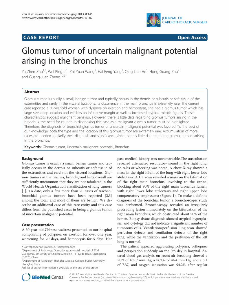
past medical history was unremarkable.The auscultationrevealed attenuated respiratory sound in the right lung,no rales or wheezing was noted. A chest X-ray showed amass in the right hilum of the lung with right lower lobeatelectasis. A CT scan revealed a mass on the bifurcationof the right main bronchus, involving to the carina,blocking about 90% of the right main bronchus lumen,with right lower lobe atelectasis and right upper lobecompensatory emphysema (Figure 1). To make a definitediagnosis of the bronchial tumor, a bronchoscopic studywas performed. Bronchoscopy revealed an irregularlyprotruding lesion immediately on the bifurcation of theright main bronchus, which obstructed about 90% of thelumen. Biopsy tissue diagnosis showed atypical hyperpla-sia, and cytology did not indicate a significant number oftumorous cells. Ventilation/perfusion lung scan showedperfusion defects and ventilation defects of the rightlung, while the ventilation and the perfusion of the leftlung is normal.The patient appeared aggravating polypnea, orthopnea
and perspiration suddenly on the 5th day in hospital. Ar-terial blood gas analysis on room air breathing showed aPO2 of 105.7 mm Hg, a PCO2 of 44.4 mm Hg, and a pHof 7.37, and oxygen saturation was 94.2%. after regular
. This is an Open Access article distributed under the terms of the Creativeommons.org/licenses/by/2.0), which permits unrestricted use, distribution, andiginal work is properly cited.
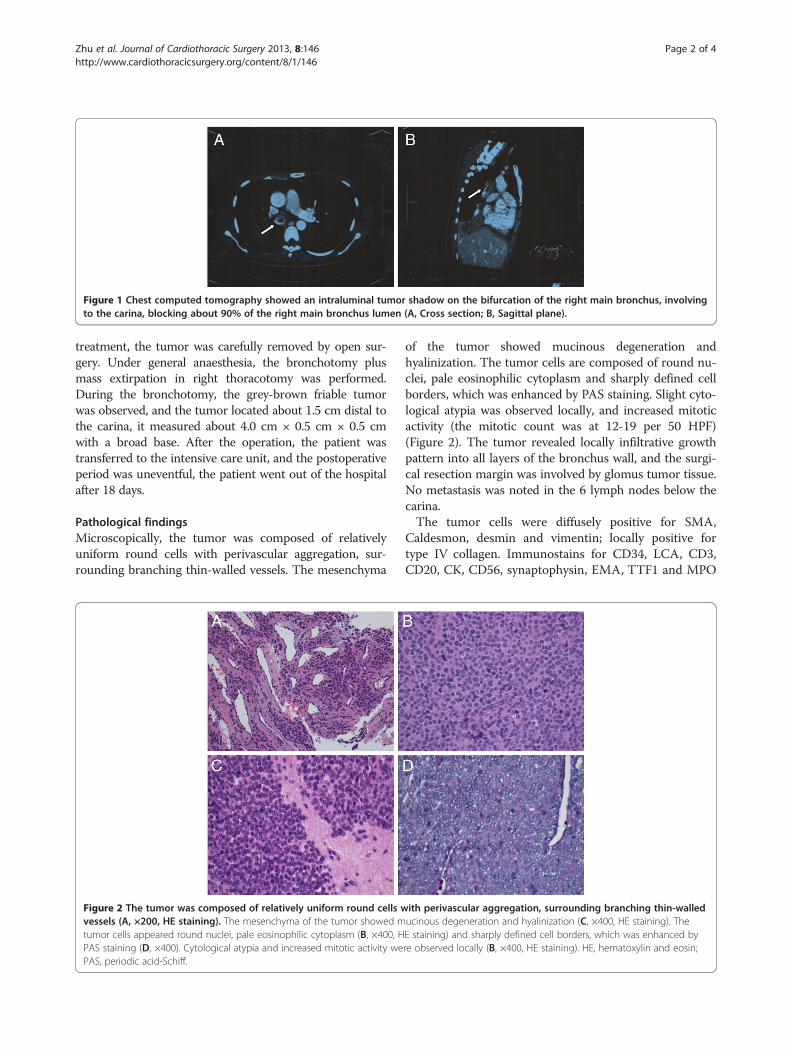
Figure 1 Chest computed tomography showed an intraluminal tumor shadow on the bifurcation of the right main bronchus, involvingto the carina, blocking about 90% of the right main bronchus lumen (A, Cross section; B, Sagittal plane).
Zhu et al. Journal of Cardiothoracic Surgery 2013, 8:146 Page 2 of 4http://www.cardiothoracicsurgery.org/content/8/1/146
treatment, the tumor was carefully removed by open sur-gery. Under general anaesthesia, the bronchotomy plusmass extirpation in right thoracotomy was performed.During the bronchotomy, the grey-brown friable tumorwas observed, and the tumor located about 1.5 cm distal tothe carina, it measured about 4.0 cm × 0.5 cm × 0.5 cmwith a broad base. After the operation, the patient wastransferred to the intensive care unit, and the postoperativeperiod was uneventful, the patient went out of the hospitalafter 18 days.
Pathological findingsMicroscopically, the tumor was composed of relativelyuniform round cells with perivascular aggregation, sur-rounding branching thin-walled vessels. The mesenchyma
Figure 2 The tumor was composed of relatively uniform round cells wvessels (A, ×200, HE staining). The mesenchyma of the tumor showed mtumor cells appeared round nuclei, pale eosinophilic cytoplasm (B, ×400, HPAS staining (D, ×400). Cytological atypia and increased mitotic activity wePAS, periodic acid-Schiff.
of the tumor showed mucinous degeneration andhyalinization. The tumor cells are composed of round nu-clei, pale eosinophilic cytoplasm and sharply defined cellborders, which was enhanced by PAS staining. Slight cyto-logical atypia was observed locally, and increased mitoticactivity (the mitotic count was at 12-19 per 50 HPF)(Figure 2). The tumor revealed locally infiltrative growthpattern into all layers of the bronchus wall, and the surgi-cal resection margin was involved by glomus tumor tissue.No metastasis was noted in the 6 lymph nodes below thecarina.The tumor cells were diffusely positive for SMA,
Caldesmon, desmin and vimentin; locally positive fortype IV collagen. Immunostains for CD34, LCA, CD3,CD20, CK, CD56, synaptophysin, EMA, TTF1 and MPO
ith perivascular aggregation, surrounding branching thin-walleducinous degeneration and hyalinization (C, ×400, HE staining). TheE staining) and sharply defined cell borders, which was enhanced byre observed locally (B, ×400, HE staining). HE, hematoxylin and eosin;
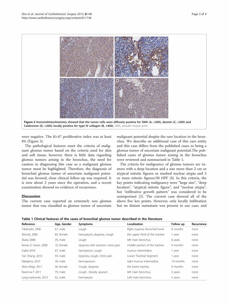
Figure 3 Immunohistochemistry showed that the tumor cells were diffusely positive for SMA (A, ×200), desmin (C, ×200) andCaldesmon (D, ×200); locally positive for type IV collagen (B, ×400). SMA, smooth muscle actin.
Zhu et al. Journal of Cardiothoracic Surgery 2013, 8:146 Page 3 of 4http://www.cardiothoracicsurgery.org/content/8/1/146
were negative. The Ki-67 proliferative index was at least8% (Figure 3).The pathological features meet the criteria of malig-
nant glomus tumor based on the criteria used for skinand soft tissue, however, there is little data regardingglomus tumors arising in the bronchus, the need forcaution in diagnosing this case as a malignant glomustumor must be highlighted. Therefore, the diagnosis ofbronchial glomus tumor of uncertain malignant poten-tial was favored, close clinical follow-up was required. Itis now about 2 years since the operation, and a recentexamination showed no evidence of recurrence.
DiscussionThe current case reported an extremely rare glomustumor that was classified as glomus tumor of uncertain
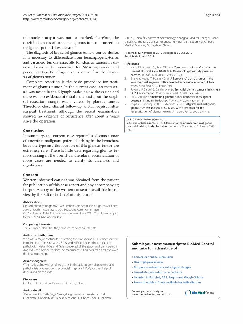
Table 1 Clinical features of the cases of bronchial glomus tum
Reference Age, Gender Symptoms
Takahashi, 2006 67, male cough
Altinok, 2006 83, female hemoptysis, dyspnea, cough
Akata 2008 39, male cough
Kenan E. Haver, 2008 10, female dyspnea with exertion, chest p
Inaba 2010 67, male hemoptysis, cough
Yan Shang, 2010 59, male dyspnea, cough, chest pain
Nakajima, 2010 30, male hemosputum
Akira Mogi, 2011 56, female Cough, dyspnea
Ravenna F 2011 79, male cough , bloody sputum
Lang-Lazdunski, 2012 62, male hemoptysis
malignant potential despite the rare location in the bron-chus. We describe an additional case of this rare entityand this case differs from the published cases in being aglomus tumor of uncertain malignant potential.The pub-lished cases of glomus tumor arising in the bronchuswere reviewed and summarized in Table 1.The criteria for malignancy of glomus tumors are tu-
mors with a deep location and a size more than 2 cm oratypical mitotic figures or marked nuclear atypia and 5or more mitotic figures/50 HPF [4]. In this criteria, thekey points indicating malignancy were “large size”, “deeplocation”, “atypical mitotic figure”, and “nuclear atypia”,but “infiltrative growth pattern” was considered to beunimportant [5]. The current case showed all of theabove five key points. However, only locally Infiltrationbut no distant metastasis was present in our case, and
or described in the literature
Localization Follow up Recurrence
Right superior bronchial trunk 8 months none
the upper third of the trachea 1 year none
left main bronchus 6 years none
ain middle portion of the trachea 9 months none
truncus intermedius 1 year none
Lower Tracheal Segment 1 year none
right truncus intermedius 10 months none
the lower trachea 9 months none
left main bronchus 5 years none
Left main bronchus 5 years none
Zhu et al. Journal of Cardiothoracic Surgery 2013, 8:146 Page 4 of 4http://www.cardiothoracicsurgery.org/content/8/1/146
the nuclear atypia was not so marked, therefore, thecareful diagnosis of bronchial glomus tumor of uncertainmalignant potential was favored.The diagnosis of bronchial glomus tumors can be elusive.
It is necessary to differentiate from hemangiopericytomasand carcinoid tumors especially for glomus tumors in un-usual locations. Immunostains for SMA expression andpericellular type IV collagen expression confirm the diagno-sis of glomus tumor.Complete resection is the basic procedure for treat-
ment of glomus tumor. In the current case, no metasta-sis was noted in the 6 lymph nodes below the carina andthere was no evidence of distal metastasis, but the surgi-cal resection margin was involved by glomus tumor.Therefore, close clinical follow-up is still required aftersurgical treatment, although the recent examinationshowed no evidence of recurrence after about 2 yearssince the operation.
ConclusionIn summary, the current case reported a glomus tumorof uncertain malignant potential arising in the bronchus,both the type and the location of this glomus tumor areextremely rare. There is little data regarding glomus tu-mors arising in the bronchus, therefore, accumulation ofmore cases are needed to clarify its diagnosis andsignificance.
ConsentWritten informed consent was obtained from the patientfor publication of this case report and any accompanyingimages. A copy of the written consent is available for re-view by the Editor-in-Chief of this journal.
AbbreviationsCT: Computed tomography; PAS: Periodic acid-Schiff; HPF: High-power fields;SMA: Smooth muscle actin; LCA: Leukocyte common antigen;CK: Cytokeratin; EMA: Epithelial membrane antigen; TTF1: Thyroid transcriptorfactor 1; MPO: Myeloperoxidase.
Competing interestsThe authors declare that they have no competing interests.
Authors’ contributionsY-ZZ was a major contributor in writing the manuscript. Q-LH carried out theimmunohistochemistry. W-PL, Z-YW and H-FY collected the clinical andpathological data, H-GZ and G-JZ conceived of the study, and participated indiagnosis and helped to draft the manuscript. All authors read and approvedthe final manuscript.
AcknowledgmentWe greatly acknowledge all surgeons in thoracic surgery department andpathologists of Guangdong provincial hospital of TCM, for their helpfuldiscussions on this case.
DisclosureConflicts of Interest and Source of Funding: None.
Author details1Department of Pathology, Guangdong provincial hospital of TCM,Guangzhou University of Chinese Medicine, 111 Dade Road, Guangzhou
510120, China. 2Department of Pathology, Shanghai Medical College, FudanUniversity, Shanghai, China. 3Guangdong Provincial Academy of ChineseMedical Sciences, Guangzhou, China.
Received: 12 November 2012 Accepted: 6 June 2013Published: 7 June 2013
References1. Haver KE, Hartnick CJ, Ryan DP, et al: Case records of the Massachusetts
General Hospital. Case 10-2008. A 10-year-old girl with dyspnea onexertion. N Engl J Med 2008, 358:1382–1390.
2. Shang Y, Huang Y, Huang HD, et al: Removal of glomus tumor in thelower tracheal segment with a flexible bronchoscope: report of twocases. Intern Med 2010, 49:865–869.
3. Ravenna F, Saturni S, Casalini A, et al: Bronchial glomus tumor mimicking aCOPD exacerbation. Monaldi Arch Chest Dis 2011, 75:194–198.
4. Gill J, Van Vliet C: Infiltrating glomus tumor of uncertain malignantpotential arising in the kidney. Hum Pathol 2010, 41:145–149.
5. Folpe AL, Fanburg-Smith JC, Miettinen M, et al: Atypical and malignantglomus tumors: analysis of 52 cases, with a proposal for thereclassification of glomus tumors. Am J Surg Pathol 2001, 25:1–12.
doi:10.1186/1749-8090-8-146Cite this article as: Zhu et al.: Glomus tumor of uncertain malignantpotential arising in the bronchus. Journal of Cardiothoracic Surgery 20138:146.
Submit your next manuscript to BioMed Centraland take full advantage of:
• Convenient online submission
• Thorough peer review
• No space constraints or color figure charges
• Immediate publication on acceptance
• Inclusion in PubMed, CAS, Scopus and Google Scholar
• Research which is freely available for redistribution
Submit your manuscript at www.biomedcentral.com/submit