Embed Size (px)
Citation preview
CASE REPORT Open Access
Herlyn-werner-wunderlich syndrome: MRIfindings, radiological guide (two cases andliterature review), and differential diagnosisRiccardo Del Vescovo*, Sofia Battisti, Valerio Di Paola, Claudia L Piccolo, Roberto L Cazzato, Ilaria Sansoni,Rosario F Grasso and Bruno Beomonte Zobel
Abstract
Background: Herlyn-Werner-Wunderlich (HWW) syndrome is a very rare congenital anomaly of the urogenital tractinvolving Müllerian ducts and Wolffian structures, and it is characterized by the triad of didelphys uterus,obstructed hemivagina and ipsilateral renal agenesis. It generally occurs at puberty and exhibits non-specific andvariable symptoms with acute or pelvic pain shortly following menarche, causing a delay in the diagnosis.Moreover, the diagnosis is complicated by the infrequency of this syndrome, because Müllerian duct anomalies(MDA) are infrequently encountered in a routine clinical setting.
Cases presentation: two cases of HWW syndrome in adolescents and a differential diagnosis for one case of adifferent MDA, and the impact of magnetic resonance (MR) imaging technology to achieve the correct diagnosis.
Conclusions: MR imaging is a very suitable diagnostic tool in order to perform the correct diagnosis of HWWsyndrome.
BackgroundThe association of renal agenesis with ipsilateral blindhemivagina and didelphys uterus is reported as Herlyn-Werner-Wunderlich (HWW) syndrome; this triadwas initially disclosed in an English report published in2006 [1].The incidence of didelphys uterus, related to HWW, is
approximately 1/2,000 to 1/28,000, and it is accompa-nied by unilateral renal agenesis in 43% of cases. Theincidence of unilateral renal agenesis is 1/1,100, and 25-50% of affected women exhibits associated genitalabnormalities [2].HWW syndrome represents a type of Müllerian duct
anomalies (MDA) associated with mesonephric ductanomalies. MDA are congenital entities resulting fromnon-development (agenesia or hypoplasia), defective ver-tical or lateral fusion, or resorption failure of the Mül-lerian (paramesonephric) ducts [3].MDA are estimated to have an overall prevalence of
2% to 3% among all women, with an incidence of 1/
200-600 among fertile women. Hypoplasia, as well asagenesis of the uterus and proximal vagina, accounts for5%-10% of Müllerian duct anomalies, whereas didelphysuterus accounts for approximately 11% of Müllerianduct anomalies. Renal tract anomalies are associatedwith MDA in as many as 30% of cases [4].A complete or partial vaginal septum is present in
75% of women with didelphys uterus [5].The exact cause, pathogenesis and embryologic origin
of HWW syndrome are unclear and remain a subject ofdiscussion [6].HWW syndrome is usually discovered at puberty with
non-specific symptoms, like increasing pelvic pain, dys-menorrhea and palpable mass due to the associated hae-matocolpos or hematometra, which result from retained,longstanding menstrual flow in the obstrucucted vagina.It rarely occurs with primary infertility in early adult-
hood when the vaginal septum is incomplete [7].
It is really difficult to achieve to an accurate diagno-sis because menstruation is often regular and whenpatient complains symptoms of cyclic dysmenorrhea,they are usually given anti-inflammatory drugs and
* Correspondence: [email protected] of Radiology, Campus Bio-Medico Univeristy of Rome, Italy
Del Vescovo et al. BMC Medical Imaging 2012, 12:4http://www.biomedcentral.com/1471-2342/12/4
© 2012 Del Vescovo et al; licensee BioMed Central Ltd. This is an Open Access article distributed under the terms of the CreativeCommons Attribution License (http://creativecommons.org/licenses/by/2.0), which permits unrestricted use, distribution, andreproduction in any medium, provided the original work is properly cited.
oral-contraceptives, thus causing a delay in the diag-nosis because they reduce or eliminate menses; ulti-mately, HWW is an uncommon syndrome, not oftenthought of as a diagnostic possibility [8].
The potential complications of this syndrome are dis-tinct in acute complications, such as pyohematocolpos,pyosalpinx, or pelviperitonitis, and long-term complica-tions, such as endometriosis, pelvic adhesions andincreased risk of abortion or infertility [9,10].The American Society for Reproductive Medicine
(ASRM) established a classification of Müllerian ductanomalies in 1988 in order to group them in entirety(Table 1) [11].According to this classification, HWW syndrome
appears to include the addition of III uterine anomaly toIa vaginal anomaly and renal agenesis (Table 2).
Case presentationPatient 1A 16-years-old female presented with a history of severeabdomino-pelvic pain, which increased lasting 2 to 7days with each of her menstrual cycles, hindering herdaily activities.Gynecologic history indicated menarche at 13 years of
age, followed by 3 years of irregular menses.The patient denied any recent abdominal trauma,
abnormal vaginal bleeding, nausea, vomiting or diarrhea.A bimanual physical examination indicated a right-
sided cystic and tender pelvic mass, movable, mildly ten-der to palpation. The medical staff recommended
further evaluation such as US, which showed theabsence of right kidney and the possibility of uterineanomaly; so Magnetic Resonance Imaging (MRI) wasperformed in order to evaluate the possible genito-urin-ary anomaly.The MRI examination was performed on a 1.5 Tesla
MR clinical scanner (Siemens, Avanto Erlangen Ger-many). The patient’s mother gave her informed consentfor MRI exam. Images were acquired on multipleplanes with T1-weighted (TR 714 ms; TE 12 ms) andT2-weighted Turbo Spin Echo (TR 5530 ms; TE 150ms) sequences; fat-suppressed T2-weighted TSE images(TR 6810 ms; TE 150 ms), Flash3D T1-weighted (TR5.2; TE 2.5) and HASTE sequences (TR 700; TE 89). Acontrast agent was not used as it was not considerednecessary.MRI imaging showed a uterine-vaginal anomaly con-
sisting of didelphys uterus and double vagina, one ofwhich was obstructed (Figure 1); consequently, therewas accumulation of fluid exhibiting a signal intensitysimilar to methaemoglobin in the right uterus (slightlydilated, 14 mm diameter of the lumen) and in the rightobstructed vagina (which appeared to be considerablydilated, 43 × 34 × 35 mm).In the right ovary an expanding mass of 31 mm was
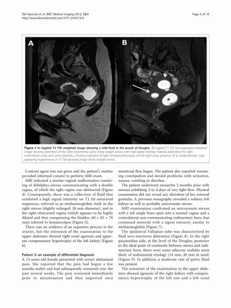
hyperintense on T1-weighted images and mildly hypoin-tense or hyperintense on T2-weighted images (Figure2A). The mass was characterized by a typical “shading”sign (Figure 2B) and exhibited features compatible withan endometriosic cyst; there was mild fluid in the pouchof Douglas.
Table 1 Classification of müllerian duct anomalies
MULLERIAN AGENESIS OR HYPOLPLASIA a) vaginab) cervixc) fundusd) fallopian tubee) combined agenesis or hypoplasia (two or more findings fromclasses I-A through I-D)
UNICORNUATE UTERUS (Agenesis or hypoplasia of one of the twoMullerian ducts)
• rudimentary horn with an endometrial cavity that communicateswith the (single-horned) uterus;• rudimentary horn with an endometrial cavity that does notcommunicate with the uterus;• rudimentary horn with no endometrial cavity;no rudimentary horn
UTERUS DYDELPHYS(Failure of lateral fusion between vagina and two Mullerian ducts)
BICORNUATE UTERUS(Incomplete fusion of uterine horns at the level of the fundus)
a) complete (septum extends to the internal or external os);b) partial (septum is confined to the fundal region)
SEPTATE UTERUS(Incomplete or absent resorption of uterovaginal septum)
a) complete (septum extends to the internal os);b) partial (septum does not reach the internal os)
ARCHUATE UTERUS(Light indent of the fundus of the uterus due to almost completeresorption of the uterovaginal septum)
UTERUS EXPOSED TO DIETHYILSTILBESTROL-RELATED UTERINEANOMALIES
a) T-shaped uterus;b) T-shaped uterus with dilated horns;c) Uterine hypoplasia
Del Vescovo et al. BMC Medical Imaging 2012, 12:4http://www.biomedcentral.com/1471-2342/12/4
Page 2 of 10
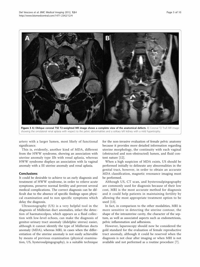
There was no evidence of an expansive process inthe left ovary. The extension of examination to theupper abdomen revealed right renal agenesis andapparent compensatory hypertrophy of the left kidney(Figure 3).
Patient 2A 15-years-old woman presented with severe abdominalpain that occurred since the patient achieved menarcheat 14 years of age; the patient also had irregular
menstruation with severe dysmenorrhea and shereported that the pain worsened immediately prior tomenstruation and improved with menstrual flow.Upon physical examination, the findings were non-
specific, with the exception of lower abdominal tender-ness, arising from the pelvis, mildly tender to palpation.A gynecologic examination did not reveal any anomaliesof her external genitalia or hymen.So, MRI was then performed, using the same sequences
protocol employed for Patient 1.
Table 2 Hww syndrome: mr findings
HWWSYNDROME
MR FINDINGS
Uterus didelphys It is characterized by two symmetric, widely divergent uterine horns and two cervixes, with an enlarged cavity filled by bloody/proteinaceous fluid due to haematocolpos.It appears hyperintense signal on T1 fat saturated sequences.
Hemivagina It is an obstructed vagina due to a longitudinal septum which occludes one cervix and isolates it with consequent hematometra.
Renal aplasya It is tipically omolateral to the vaginal anomaly (right side prevalence), with possible controlateral renal hypertrophy due tocompensation but in site.
Ovaries Possible presence of endometriosic cysts with hyperintense signal on T1 fat sat sequences and with “shading sign” on T2-weighted images and a gradual variation of signal intensity due to chronic bleeding with accumulation of high concentration ofiron and protein in the endometrioma(endometriotic lesions appear hyperintense on T1-weighted images and mildly hypointense or hyperintense on T2-weightedimages).Possible presence of functional cysts with no clinical significance, which appear hyperintense on T2 sequences and hypo/isointense on T1 sequences.
Figure 1 Herlyn-Werner-Wunderlich syndrome (C) Drawing illustrates the triad (renal agenesis, didelphys uterus, and obstructedhemivagina and in this case the presence of an endometriosic cyst. A 16-years-old girl presenting the triad of didelphys uterus (class IIIMDA), an obstructed right hemivagina (class I MDA), and ipsilateral renal agenesis. (A) Axial turbo spin-echo T2 weighted (B) Axial turbo spin-echo T1 fat-saturated weighted MR image showing centrally a hematocolpos (asterisk), a finding corresponding to the obstructed righthemivagina. Mild dilation of the right endometrial cavity (curved arrow) with fluid exhibiting a signal intensity similar to methaemoglobin due tohaematometra.
Del Vescovo et al. BMC Medical Imaging 2012, 12:4http://www.biomedcentral.com/1471-2342/12/4
Page 3 of 10
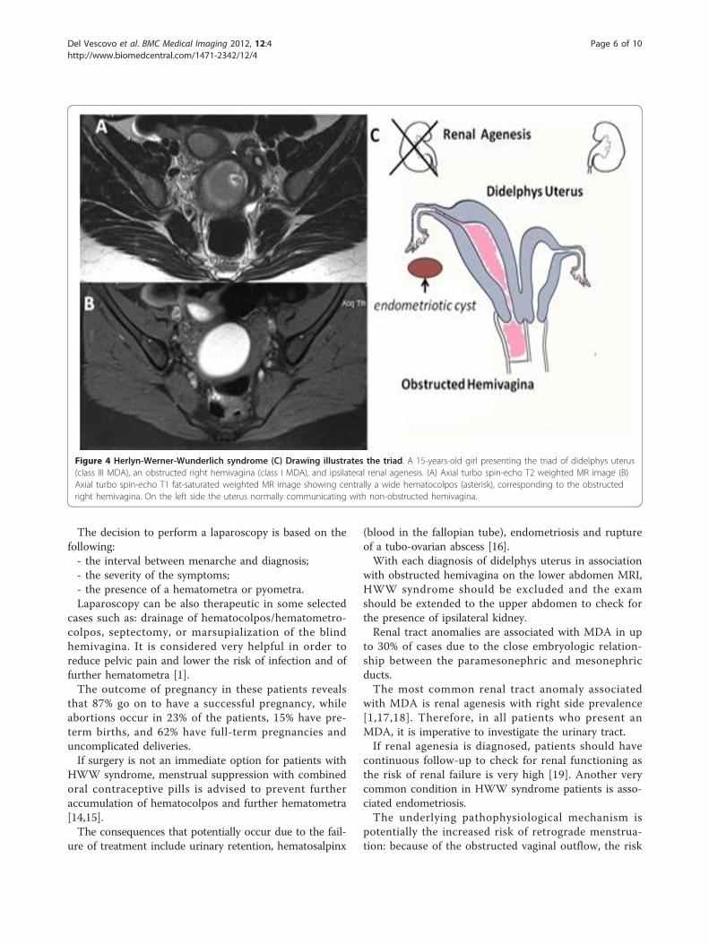
Contrast agent was not given and the patient’s motherprovided informed consent to perform MRI exam.MRI indicated a uterine-vaginal malformation consist-
ing of didelphys uterus communicating with a doublevagina, of which the right vagina was obstructed (Figure4). Consequently, there was a collection of fluid thatexhibited a high signal intensity on T1 fat-saturatedsequences, referred to as methaemoglobin, both in theright uterus (slightly enlarged: 18 mm diameter), and inthe right obstructed vagina (which appears to be highlydilated and thus compressing the bladder, 60 × 65 × 76mm) referred to hematocolpos (Figure 5).There was no evidence of an expansive process in the
ovaries, but the extension of the examination to theupper abdomen showed right renal agenesis and appar-ent compensatory hypertrophy of the left kidney (Figure6).
Patient 3: an example of differential diagnosisA 13-years-old female presented with severe abdominalpain. She reported that the pain had begun a fewmonths earlier and had subsequently worsened over thepast several weeks. The pain worsened immediatelyprior to menstruation and then improved once
menstrual flow began. The patient also reported worsen-ing constipation and denied problems with urination,nausea, vomiting or diarrhea.The patient underwent menarche 2 months prior with
menses exhibiting 3 to 4 days of very light flow. Physicalexamination did not reveal any alteration of her externalgenitalia. A previous sonography revealed a solitary leftkidney as well as probable unicornuate uterus.MRI examination confirmed an unicornuate uterus
with a left single horn open into a normal vagina and acontrolateral non-communicating rudimentary horn thatcontained material with a signal intensity similar tomethaemoglobin (Figure 7).The ipsilateral Fallopian tube was characterized by
fluid sero-mucinous distension (Figure 8). In the rightparamedian side, at the level of the Douglas, posteriorto the ideal point of continuity between uterus and rudi-mentary horn, there were some adjacent nodules mostlikely of endometrial etiology (14 mm, 38 mm in total)(Figure 9). In addition, a moderate rate of pelvic fluidwas present.The extension of the examination to the upper abdo-
men showed agenesis of the right kidney with compen-satory hypertrophy of the left one and a left renal
Figure 2 A) Sagittal T2 TSE weighted image showing a mild fluid in the pouch of Douglas. (B) Sagittal T1 TSE fat-suppressed weightedimage showing distention of the right endometrial cavity (long straight arrow) with high-signal-intensity material distending the rightendometrial cavity and cervix (asterisk), a finding indicative of right hematometrocolpos; on the right ovary presence of an endometriosic cystsappearing hyperintense on T1 fat-saturated image (short straight arrow).
Del Vescovo et al. BMC Medical Imaging 2012, 12:4http://www.biomedcentral.com/1471-2342/12/4
Page 4 of 10
artery with a larger lumen, most likely of functionalsignificance.This is, evidently, another kind of MDA, different
from the HWW syndrome, showing an association withuterine anomaly type IIb with renal aplasia, whereasHWW syndrome displays an association with Ia vaginalanomaly with a III uterine anomaly and renal aplasia.
ConclusionsIt could be desirable to achieve to an early diagnosis andtreatment of HWW syndrome, in order to relieve acutesymptoms, preserve normal fertility and prevent severalmedical complications. The correct diagnosis can be dif-ficult due to the absence of specific findings upon physi-cal examination and to non specific symptoms whichdelay the diagnosis.Ultrasonography (US) is a very helpful tool in the
diagnosis of Müllerian duct anomalies, infact the detec-tion of haematocolpos, which appears as a fluid collec-tion with low-level echoes, can make the diagnosis ofgenitor-urinary tract anomaly (didelphic uterus) easier,although it cannot identify the type of Müllerian ductsanomaly (MDA), whereas MRI, in cases when the differ-entiation of the uterine anomaly is not easily achievableby means of previous examination (physical examina-tion, US, hysterosalpingography), is a suitable technique
for the non-invasive evaluation of female pelvic anatomybecause it provides more detailed information regardinguterine morphology, the continuity with each vaginal(obstructed and non-obstructed) lumen, and fluid con-tent nature [12].When a high suspicion of MDA exists, US should be
performed initially to delineate any abnormalities in thegenital tract, however, in order to obtain an accurateMDA classification, magnetic resonance imaging mustbe performed.Although US, CT scan, and hysterosalpingography
are commonly used for diagnosis because of their lowcost, MRI is the most accurate method for diagnosisand it could help patients in maintaining fertility byallowing the most appropriate treatment option to beused [13].In fact, in comparison to the other modalities, MRI is
more sensitive in detecting the uterine contour, theshape of the intrauterine cavity, the character of the sep-tum, as well as associated aspects such as endometriosis,pelvic inflammation and adhesions.However, laparoscopy should now be considered the
gold standard for the evaluation of female reproductivetract anomaly, although it could be reserved when thediagnosis is not clear after imaging or when MRI is notavailable and not performed as a routine procedure [7].
Figure 3 A) Oblique coronal TSE T2-weighted MR image shows a complete view of the anatomical defects. B) Coronal T2 Trufi MR imageshowing the omolateral renal aplasia with respect to the pelvic abnormalities and a solitary left kidney with a mild hypertrophy.
Del Vescovo et al. BMC Medical Imaging 2012, 12:4http://www.biomedcentral.com/1471-2342/12/4
Page 5 of 10
The decision to perform a laparoscopy is based on thefollowing:- the interval between menarche and diagnosis;- the severity of the symptoms;- the presence of a hematometra or pyometra.Laparoscopy can be also therapeutic in some selected
cases such as: drainage of hematocolpos/hematometro-colpos, septectomy, or marsupialization of the blindhemivagina. It is considered very helpful in order toreduce pelvic pain and lower the risk of infection and offurther hematometra [1].The outcome of pregnancy in these patients reveals
that 87% go on to have a successful pregnancy, whileabortions occur in 23% of the patients, 15% have pre-term births, and 62% have full-term pregnancies anduncomplicated deliveries.If surgery is not an immediate option for patients with
HWW syndrome, menstrual suppression with combinedoral contraceptive pills is advised to prevent furtheraccumulation of hematocolpos and further hematometra[14,15].The consequences that potentially occur due to the fail-
ure of treatment include urinary retention, hematosalpinx
(blood in the fallopian tube), endometriosis and ruptureof a tubo-ovarian abscess [16].With each diagnosis of didelphys uterus in association
with obstructed hemivagina on the lower abdomen MRI,HWW syndrome should be excluded and the examshould be extended to the upper abdomen to check forthe presence of ipsilateral kidney.Renal tract anomalies are associated with MDA in up
to 30% of cases due to the close embryologic relation-ship between the paramesonephric and mesonephricducts.The most common renal tract anomaly associated
with MDA is renal agenesis with right side prevalence[1,17,18]. Therefore, in all patients who present anMDA, it is imperative to investigate the urinary tract.If renal agenesia is diagnosed, patients should have
continuous follow-up to check for renal functioning asthe risk of renal failure is very high [19]. Another verycommon condition in HWW syndrome patients is asso-ciated endometriosis.The underlying pathophysiological mechanism is
potentially the increased risk of retrograde menstrua-tion: because of the obstructed vaginal outflow, the risk
Figure 4 Herlyn-Werner-Wunderlich syndrome (C) Drawing illustrates the triad. A 15-years-old girl presenting the triad of didelphys uterus(class III MDA), an obstructed right hemivagina (class I MDA), and ipsilateral renal agenesis. (A) Axial turbo spin-echo T2 weighted MR image (B)Axial turbo spin-echo T1 fat-saturated weighted MR image showing centrally a wide hematocolpos (asterisk), corresponding to the obstructedright hemivagina. On the left side the uterus normally communicating with non-obstructed hemivagina.
Del Vescovo et al. BMC Medical Imaging 2012, 12:4http://www.biomedcentral.com/1471-2342/12/4
Page 6 of 10
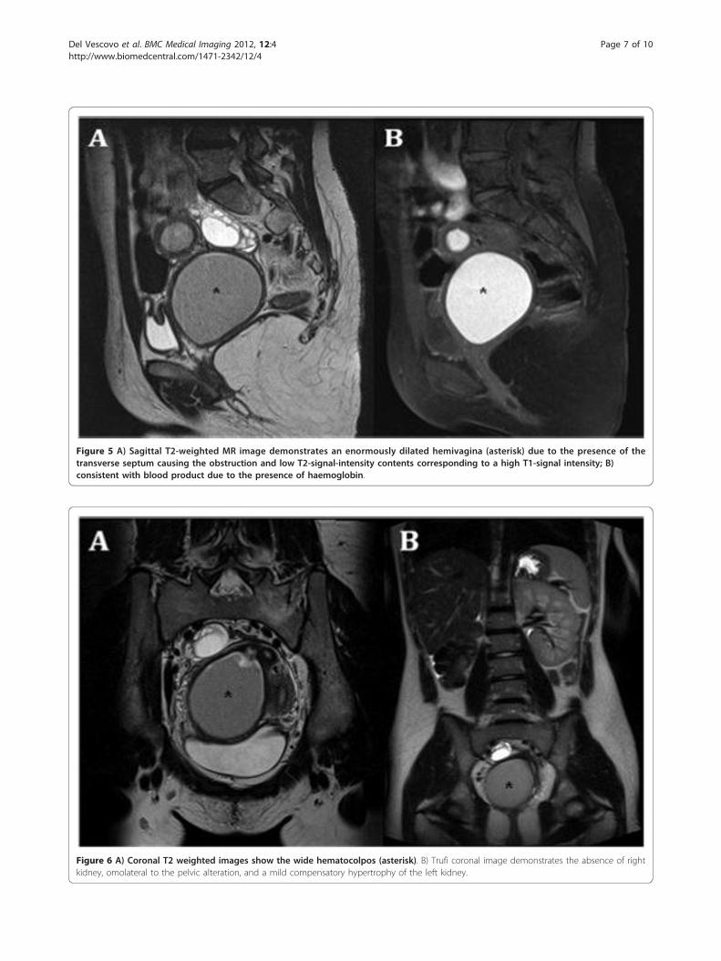
Figure 5 A) Sagittal T2-weighted MR image demonstrates an enormously dilated hemivagina (asterisk) due to the presence of thetransverse septum causing the obstruction and low T2-signal-intensity contents corresponding to a high T1-signal intensity; B)consistent with blood product due to the presence of haemoglobin.
Figure 6 A) Coronal T2 weighted images show the wide hematocolpos (asterisk). B) Trufi coronal image demonstrates the absence of rightkidney, omolateral to the pelvic alteration, and a mild compensatory hypertrophy of the left kidney.
Del Vescovo et al. BMC Medical Imaging 2012, 12:4http://www.biomedcentral.com/1471-2342/12/4
Page 7 of 10
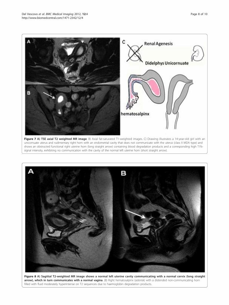
Figure 7 A) TSE axial T2 weighted MR image. B) Axial fat-saturated T1-weighted images. C) Drawing illustrates a 14-year-old girl with anunicornuate uterus and rudimentary right horn with an endometrial cavity that does not communicate with the uterus (class II MDA type) andshows an obstructed functional right uterine horn (long straight arrow) containing blood degradation products and a corresponding high T1fs-signal intensity, exhibiting no communication with the cavity of the normal left uterine horn (short straight arrow).
Figure 8 A) Sagittal T2-weighted MR image shows a normal left uterine cavity communicating with a normal cervix (long straightarrow), which in turn communicates with a normal vagina. (B) Right hematosalpinx (asterisk) with a distended non-communicating hornfilled with fluid moderately hyperintense on T2 sequences due to haemoglobin degradation products.
Del Vescovo et al. BMC Medical Imaging 2012, 12:4http://www.biomedcentral.com/1471-2342/12/4
Page 8 of 10
of endometrial tissue reflux through the fallopian tube,which can result in retrograde endometriosis, is higher,confirming the theory postulated by Sampson [20].Several reports have confirmed that an association
with endometriosis is present in obstructive Mülleriananomalies such as HWW syndrome, but it has not beenconfirmed to date in non-obstructive malformations.Therefore, it is imperative that MRI technology shouldbe used to evaluate the presence of endometriosis.Additionally, the presence of associated hematocolpos
and hematometra, as well as the presence of an irregu-larly shaped or sized adnexal structure, or potentiallyexpanding masses inside, need to be detected.Another kind of obstructive Müllerian anomalies, dif-
ferent from the HWW syndrome, is OHVIRA syn-drome, characterized by a variety of associateddisorders. Infact, while the classic presentation includesobstructed hemivagina and ipsilateral renal anomaly(OHVIRA), some authors reported other renal anoma-lies, such as duplicated kidneys, dysplastic kidneys orretrovescical bands [21]. There have been also fewreports of varied uterine anatomy; infact, a series of 42cases of OHVIRA syndrome described, in 22% of cases,a septate uteri [15], but the majorority of cases areduplicated or didelphys uterus, although it has beendescribed a case with two cervices, two uterine cavitiesand a single uterus [22].
ConsentWritten informed consent was obtained from the patientfor publication of this case report and accompanyingimages. A copy of the written consent is available forreview by the Editor-in-Chief of this journal.
AbbreviationsHWW syndrome: Herlyn-Werner-Wunderlich syndrome; MDA: Müllerian DuctAnomalies; US: ultrasonography: MR: Magnetic Resonance; MRI: MagneticResonance Imaging; ASRM: American Society for Reproductive Medicine; CT:Computed Tomography.
Authors’ contributionsSB, VDP, RLC and CP analysed and interpreted the patient data. RDV, IS, RFGand BBZ performed the abdominal magnetic resonance. RDV, SB and VDPwere the major contributors in writing the manuscript. All authors read andapproved the final manuscript.
Competing interestsThe authors declare that they have no competing interests.
Received: 9 April 2011 Accepted: 9 March 2012 Published: 9 March 2012
References1. Gholoum S, Puligandla PS, Hui T, Su W, Quiros E, Laberge JM: Management
and outcome of patients with combined vaginal septum, bifid uterusand ipsilateral renal agenesis (Herlin-Werner-Wunderlich syndrome. JPediatr Surg 2006, 41(5):987-92.
2. Alan JW, Louis RK: Campbell-Walsh Urology. Philadelphia: Saunders;,93270-6.
3. Orazi C, Lucchetti MC, Schingo PM, Marchetti P, Ferro F: Herlyn-Werner-Wunderlich syndrome: uterus didelphys, blind hemivagina and
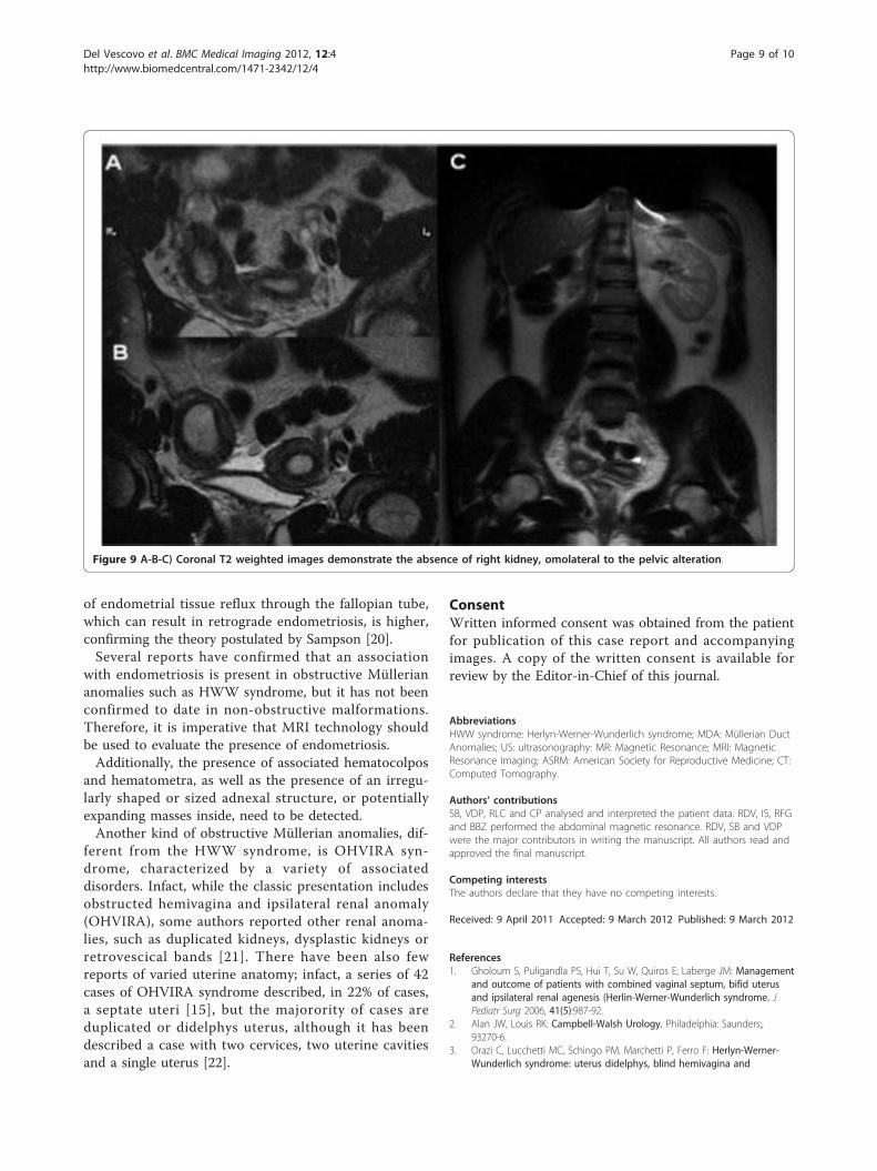
Figure 9 A-B-C) Coronal T2 weighted images demonstrate the absence of right kidney, omolateral to the pelvic alteration.
Del Vescovo et al. BMC Medical Imaging 2012, 12:4http://www.biomedcentral.com/1471-2342/12/4
Page 9 of 10
ipsilateral renal agenesis. Sonographic and MR findings in 11 cases.Pediatr Radiol 2007, 37:657-65.
4. Lee BH, Kim JW, Oh SI, et al: 3 cases of uterus didelphys with obstructedhemivagina and ipsilateral renal agenesis. Korean J Obstet Gynecol 1997,40:1489-95.
5. Heinonen PK: Clinical implications of the didelphic uterus: long-termfollow-up of 49 cases. Eur J Obstet Gynecol Reprod Biol 2000, 91:183-90.
6. Candiani GB, Fedele L, Candiani M: Double uterus, blind hemivagina andipsilateral renal agenesis: 36 cases and long-term follow-up. ObstetGynecol 1997, 90:26-32.
7. Park Noh Hyuck, Park Hee Jin, Park Chan Sup, Park Sung Il: Herlyn-Werner-Wunderlich Syndrome with Unilateral Hemivaginal Obstruction,Ipsilateral Renal Agenesis and Contralateral Renal Thin GBM Disease: ACase Report with Radiological Follow Up. J Korean Soc Radiol 2010,62:383-388.
8. Zurawin RK, Dietrich JE, Heard MJ, Edwards CL: Didelphic uterus andobstructed hemivagina with renal agenesis: case report and review ofthe literature. J Pediatr Adolesc Gynecol 2004, 17:137-41.
9. Kim TE, et al: Hysteroscopic resection of the vaginal septum in uterusdidelphys with obstructed hemivagina: a case report. J Korean Med Sci2007, 22:766-769.
10. Grimbizis GF, et al: Clinical implications of uterine malformations andhysteroscopic treatment results. Hum Reprod Update 2001, 7:161-174.
11. American Fertility Society: The American Fertility Society classifications ofadnexal adhesions, distal tubal occlusion, tubal occlusion secondary totubal ligation, tubal pregnancies. Müllerian duct anomalies andintrauterine adhesions. Fertil Steril 1988, 49:944-55.
12. Rana R, Pasrija S, Puri M: Herlyn-Werner-Wunderlich syndrome withpregnancy: a rare presentation. Congenit Anom 2008, 48:142-3.
13. Tanaka YO, Kurosaki Y, Kobayashi T, et al: Uterus didelphys associated withobstructed hemivagina and ipsilateral renal agenesis: MR findings inseven cases. Abdom Imaging 1998, 23(4):437-441.
14. Foglia RP, Kim SH, Cleveland RH, Donahoe PK: Complications of vaginalatresia in association with a duplicated Müllerian duct. J Pediatr Surg1987, 22(7):653-656.
15. Haddad B, Barranger E, Paniel BJ: Blind hemivagina: long-term follow-upand reproductive performance in 42 cases. Hum Reprod 1999,14(8):1962-1964.
16. Kiechl-Kohlendorfer U, Geley TE, Unsinn KM, Ganer I: Diagnosing neonatalfemale genital anomalies using saline-enhanced sonography. AJR Am JRoentgenol 2001, 177:1041-1044.
17. Burgis J: Obstructive Müllerian anomalies: Case report, diagnosis, andmanagement. Am J Obstet Gynecol 2001, 185:338-44.
18. Gruenwald P: Relation of the growing Müllerian duct to the Wolffianduct and its importance for the genesis of malformation. Anat Rec 1941,81:1-20.
19. Park Noh Hyuck, Park Hee Jin, Park Chan Sup, Park Sung Il: Herlyn-Werner-Wunderlich Syndrome with Unilateral Hemivaginal Obstruction,Ipsilateral Renal Agenesis and Contralateral Renal Thin GBM Disease: ACase Report with Radiological Follow Up. J Korean Soc Radiol 2010,62:383-388.
20. Brosens IA, Brosens JJ: Endometriosis. Eur J Obstet Gynecol Reprod Biol 2000,90(2):159-64.
21. Shavell VI, Montgomery SE, Johnson SC, Diamond MP, Berman JM:Complete septate uterus, obstructed hemivagina and ipsilateral renalanomaly: pregnancy course complicated by a rare urogenital anomaly.Arch Gynecol Obstet 2009, 280:449-52.
22. Shah KDivya, Laufer RMarc: Obstructed hemivagina and ipsilateral renalanomaly (OHVIRA) syndrome with a single uterus. Fertility and Sterility2011, 96:e39-e41.
Pre-publication historyThe pre-publication history for this paper can be accessed here:http://www.biomedcentral.com/1471-2342/12/4/prepub
doi:10.1186/1471-2342-12-4Cite this article as: Del Vescovo et al.: Herlyn-werner-wunderlichsyndrome: MRI findings, radiological guide (two cases and literaturereview), and differential diagnosis. BMC Medical Imaging 2012 12:4.
Submit your next manuscript to BioMed Centraland take full advantage of:
• Convenient online submission
• Thorough peer review
• No space constraints or color figure charges
• Immediate publication on acceptance
• Inclusion in PubMed, CAS, Scopus and Google Scholar
• Research which is freely available for redistribution
Submit your manuscript at www.biomedcentral.com/submit
Del Vescovo et al. BMC Medical Imaging 2012, 12:4http://www.biomedcentral.com/1471-2342/12/4
Page 10 of 10


![wunderlich R1200R_ITA[1]](https://img.pdfslide.net/doc/110x75/5571f8ba49795991698df7dd/wunderlich-r1200rita1.jpg)