Embed Size (px)
Citation preview
Case ReportPigmented Lesion of Buccal Mucosa
Manas Bajpai,1 Malay Kumar,2 Manish Kumar,3 and Deshant Agarwal1
1 Department of Oral and Maxillofacial Pathology, NIMS Dental College, Jaipur, Rajasthan, India2Department of Oral and Maxillofacial Pathology, Ahmedabad Dental College, Ahmadabad, Gujarat, India3 Department of Prosthodontics, NIMS Dental College, Jaipur, Rajasthan, India
Correspondence should be addressed to Malay Kumar; [email protected]
Received 1 May 2014; Revised 11 June 2014; Accepted 11 June 2014; Published 6 August 2014
Academic Editor: David W. Eisele
Copyright © 2014 Manas Bajpai et al. This is an open access article distributed under the Creative Commons Attribution License,which permits unrestricted use, distribution, and reproduction in any medium, provided the original work is properly cited.
Pigmented lesions are commonly found in the mouth. Such lesions represent a variety of clinical entities, ranging from physiologicchanges to manifestation of systemic illness and malignant neoplasm. Diagnosis of such lesions requires a proper case history,extraoral and intraoral examination, and, in some cases, biopsy, aspiration cytology, and laboratory investigations. Here we presenta case of purple lesion on the buccal mucosa of a 34-year-old male patient which was provisionally diagnosed as mucocele but onthe basis of histopathological picture it was finally diagnosed as angiofibroma, and we also discuss the clinical and histopathologicaldifferential diagnosis.
1. Introduction
Pigmented lesions are commonly found in the mouth. Suchlesions represent a variety of entities, ranging from racialpigmentation to manifestation of systemic illness (Addison’sdisease) and benign (hemangioma, angiofibroma) andmalig-nant neoplasms (Kaposi’s sarcoma) [1].
Angiofibromas are uncommon, highly vascular benignbut locally aggressive tumors that characteristically arisewithin the nasopharynx and are predominantly seen tooccur in young adolescent males [2]. Nucci et al. describedangiofibroma as an uncommon benign mesenchymal tumorin 1997 [3]. The term extranasopharyngeal angiofibroma hasbeen applied to vascular, fibrous nodules occurring outsidethe nasopharynx. As on 2009, 56 extranasopharyngeal fibro-mas have been reported, with the most common site ofpresentation being the maxillary sinus [4]. Juvenile angiofi-broma is the most common benign tumor of nasopharynx.It is believed that juvenile angiofibroma is the testosteronedependent tumor [5]. Pathogenesis of angiofibroma is notvery clearly understood. Various predisposing factors havebeen proposed in literature such as infection, trauma, arte-riovenous malformation and hormones [6].
2. Case Report
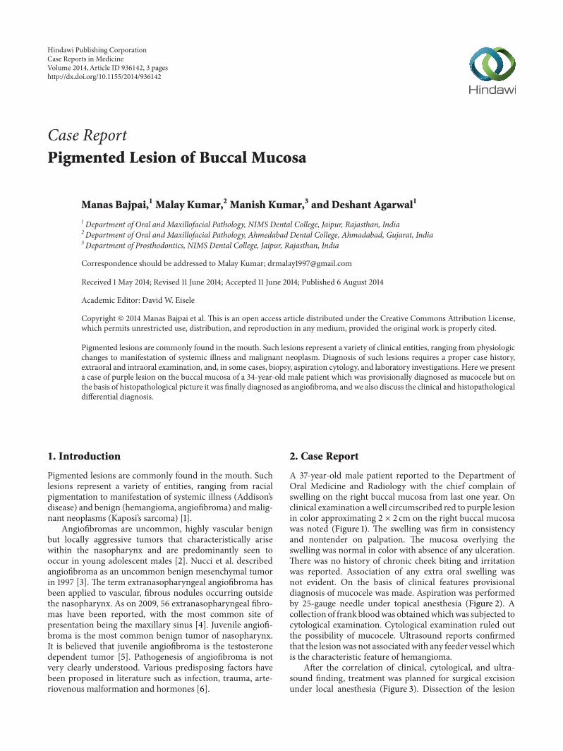
A 37-year-old male patient reported to the Department ofOral Medicine and Radiology with the chief complain ofswelling on the right buccal mucosa from last one year. Onclinical examination awell circumscribed red to purple lesionin color approximating 2 × 2 cm on the right buccal mucosawas noted (Figure 1). The swelling was firm in consistencyand nontender on palpation. The mucosa overlying theswelling was normal in color with absence of any ulceration.There was no history of chronic cheek biting and irritationwas reported. Association of any extra oral swelling wasnot evident. On the basis of clinical features provisionaldiagnosis of mucocele was made. Aspiration was performedby 25-gauge needle under topical anesthesia (Figure 2). Acollection of frank bloodwas obtainedwhichwas subjected tocytological examination. Cytological examination ruled outthe possibility of mucocele. Ultrasound reports confirmedthat the lesionwas not associatedwith any feeder vessel whichis the characteristic feature of hemangioma.
After the correlation of clinical, cytological, and ultra-sound finding, treatment was planned for surgical excisionunder local anesthesia (Figure 3). Dissection of the lesion
Hindawi Publishing CorporationCase Reports in MedicineVolume 2014, Article ID 936142, 3 pageshttp://dx.doi.org/10.1155/2014/936142
2 Case Reports in Medicine
Figure 1: Purple lesion of buccal mucosa.
Figure 2: Aspiration consists of blood.
Figure 3: Postoperative picture.
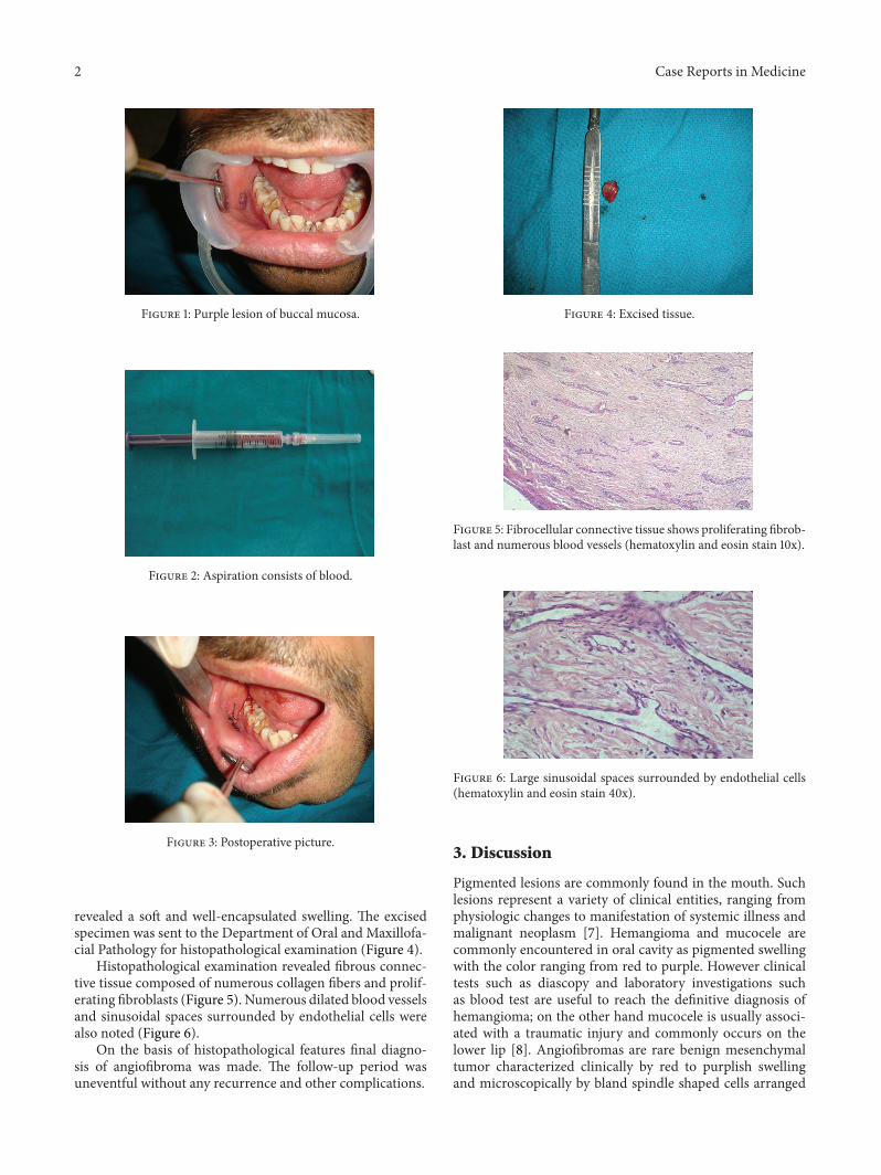
revealed a soft and well-encapsulated swelling. The excisedspecimen was sent to the Department of Oral and Maxillofa-cial Pathology for histopathological examination (Figure 4).
Histopathological examination revealed fibrous connec-tive tissue composed of numerous collagen fibers and prolif-erating fibroblasts (Figure 5). Numerous dilated blood vesselsand sinusoidal spaces surrounded by endothelial cells werealso noted (Figure 6).
On the basis of histopathological features final diagno-sis of angiofibroma was made. The follow-up period wasuneventful without any recurrence and other complications.
Figure 4: Excised tissue.
Figure 5: Fibrocellular connective tissue shows proliferating fibrob-last and numerous blood vessels (hematoxylin and eosin stain 10x).
Figure 6: Large sinusoidal spaces surrounded by endothelial cells(hematoxylin and eosin stain 40x).
3. Discussion
Pigmented lesions are commonly found in the mouth. Suchlesions represent a variety of clinical entities, ranging fromphysiologic changes to manifestation of systemic illness andmalignant neoplasm [7]. Hemangioma and mucocele arecommonly encountered in oral cavity as pigmented swellingwith the color ranging from red to purple. However clinicaltests such as diascopy and laboratory investigations suchas blood test are useful to reach the definitive diagnosis ofhemangioma; on the other hand mucocele is usually associ-ated with a traumatic injury and commonly occurs on thelower lip [8]. Angiofibromas are rare benign mesenchymaltumor characterized clinically by red to purplish swellingand microscopically by bland spindle shaped cells arranged
Case Reports in Medicine 3
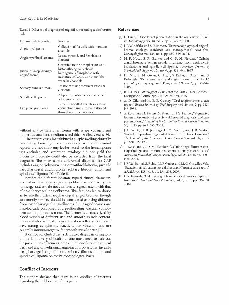
Table 1: Differential diagnosis of angiofibroma and specific features[11].
Differential diagnosis Features
Angiomyolipoma Collection of fat cells with musculararteriole
Angiomyofibroblastoma Loose, myxoid, and fibroblasticelement
Juvenile nasopharyngealangiofibroma
Cornified to the nasopharynx andhistopathologically showshomogenous fibroplasias withimmature collagen, and sinus-likevascular channels
Solitary fibrous tumors Do not exhibit prominent vascularelements
Spindle cell lipoma Adipocytes intimately interspersedwith spindle cells
Pyogenic granulomaLarge thin-walled vessels in a looseconnective tissue stroma infiltratedthroughout by leukocytes
without any pattern in a stroma with wispy collagen andnumerous small and medium-sized thick-walled vessels [9].
The present case also exhibited a purple swelling clinicallyresembling hemangioma or mucocele as the ultrasoundreports did not show any feeder vessel so the hemangiomawas excluded and aspiration cytology did not yield themucin so mucocele could also be excluded from the finaldiagnosis. The microscopic differential diagnosis for CAFincludes angiomyolipoma, angiomyofibroblastoma, juvenilenasopharyngeal angiofibroma, solitary fibrous tumor, andspindle cell lipoma [10] (Table 1).
Besides the different location, typical clinical character-istics of extranasopharyngeal angiofibromas, such as, symp-toms, age, and sex, do not conform to a great extent with thatof nasopharyngeal angiofibroma. This fact has led to doubtas to whether extranasopharyngeal angiofibromas, thoughstructurally similar, should be considered as being differentfrom nasopharyngeal angiofibroma [5]. Angiofibromas arehistologically composed of a proliferating vascular compo-nent set in a fibrous stroma. The former is characterized byblood vessels of different size and smooth muscle content.Immunohistochemical analysis has shown that stromal cellshave strong cytoplasmic reactivity for vimentin and aregenerally immunonegative for smooth muscle actin [8].
It can be concluded that a definitive diagnosis of angiofi-broma is not very difficult but one must need to rule outthe possibilities of hemangioma and mucocele on the clinicalbasis and angiomyolipoma, angiomyofibroblastoma, juvenilenasopharyngeal angiofibroma, solitary fibrous tumor, andspindle cell lipoma on the histopathological basis.
Conflict of Interests
The authors declare that there is no conflict of interestsregarding the publication of this paper.
References
[1] D. Eisen, “Disorders of pigmentation in the oral cavity,” Clinicsin Dermatology, vol. 18, no. 5, pp. 579–587, 2000.
[2] J. P. Windfuhr and S. Remmert, “Extranasopharyngeal angiofi-broma: etiology, incidence and management,” Acta Oto-Laryngologica, vol. 124, no. 8, pp. 880–889, 2004.
[3] M. R. Nucci, S. R. Granter, and C. D. M. Fletcher, “Cellularangiofibroma: a benign neoplasm distinct from angiomyofi-broblastoma and spindle cell lipoma,” American Journal ofSurgical Pathology, vol. 21, no. 6, pp. 636–644, 1997.
[4] H. Dere, K. M. Ozcan, G. Ergul, S. Bahar, I. Ozcan, and S.Kulacoglu, “Extranasopharyngeal angiofibroma of the cheek,”Journal of Laryngology and Otology, vol. 120, no. 2, pp. 141–144,2006.
[5] R. B. Lucas, Pathology of Tumours of the Oral Tissues, ChurchillLivingstone, Edinburgh, UK, 3rd edition, 1976.
[6] A. D. Giles and M. B. E. Gosney, “Oral angiomyoma: a casereport,” British Journal of Oral Surgery, vol. 20, no. 2, pp. 142–146, 1982.
[7] A. Kauzman,M. Pavone, N. Blanas, and G. Bradley, “Pigmentedlesions of the oral cavity: review, differential diagnosis, and casepresentations,” Journal of the Canadian Dental Association, vol.70, no. 10, pp. 682–683, 2004.
[8] J. C. Whitt, D. R. Jennings, D. M. Arendt, and J. R. Vinton,“Rapidly expanding pigmented lesion of the buccal mucosa,”The Journal of the American Dental Association, vol. 117, no. 5,pp. 620–622, 1988.
[9] Y. Iwasa and C. D. M. Fletcher, “Cellular angiofibroma: clin-icopathologic and immunohistochemical analysis of 51 cases,”American Journal of Surgical Pathology, vol. 28, no. 11, pp. 1426–1435, 2004.
[10] J. F. Val-Bernal, S. Rubio, M. F. Garijo, andM. C. Gonzalez-Vela,“Extragenital subcutaneous cellular angiofibroma: case report,”APMIS, vol. 115, no. 3, pp. 254–258, 2007.
[11] L. R. Eversole, “Cellular angiofibroma of oral mucosa: report oftwo cases,” Head and Neck Pathology, vol. 3, no. 2, pp. 136–139,2009.

Submit your manuscripts athttp://www.hindawi.com
Stem CellsInternational
Hindawi Publishing Corporationhttp://www.hindawi.com Volume 2014
Hindawi Publishing Corporationhttp://www.hindawi.com Volume 2014
MEDIATORSINFLAMMATION
of
Hindawi Publishing Corporationhttp://www.hindawi.com Volume 2014
Behavioural Neurology
EndocrinologyInternational Journal of
Hindawi Publishing Corporationhttp://www.hindawi.com Volume 2014
Hindawi Publishing Corporationhttp://www.hindawi.com Volume 2014
Disease Markers
Hindawi Publishing Corporationhttp://www.hindawi.com Volume 2014
BioMed Research International
OncologyJournal of
Hindawi Publishing Corporationhttp://www.hindawi.com Volume 2014
Hindawi Publishing Corporationhttp://www.hindawi.com Volume 2014
Oxidative Medicine and Cellular Longevity
Hindawi Publishing Corporationhttp://www.hindawi.com Volume 2014
PPAR Research
The Scientific World JournalHindawi Publishing Corporation http://www.hindawi.com Volume 2014
Immunology ResearchHindawi Publishing Corporationhttp://www.hindawi.com Volume 2014
Journal of
ObesityJournal of
Hindawi Publishing Corporationhttp://www.hindawi.com Volume 2014
Hindawi Publishing Corporationhttp://www.hindawi.com Volume 2014
Computational and Mathematical Methods in Medicine
OphthalmologyJournal of
Hindawi Publishing Corporationhttp://www.hindawi.com Volume 2014
Diabetes ResearchJournal of
Hindawi Publishing Corporationhttp://www.hindawi.com Volume 2014
Hindawi Publishing Corporationhttp://www.hindawi.com Volume 2014
Research and TreatmentAIDS
Hindawi Publishing Corporationhttp://www.hindawi.com Volume 2014
Gastroenterology Research and Practice
Hindawi Publishing Corporationhttp://www.hindawi.com Volume 2014
Parkinson’s Disease
Evidence-Based Complementary and Alternative Medicine
Volume 2014Hindawi Publishing Corporationhttp://www.hindawi.com