Embed Size (px)
Citation preview
Case ReportPneumomediastinum Associated with Pneumopericardium andEpidural Pneumatosis
Ozlem Bilir, Ozcan Yavasi, Gokhan Ersunan, Kamil Kayayurt, and Baris Giakoup
Department of Emergency Medicine, Recep Tayyip Erdogan University, Rize Research and Training Hospital, 53020 Rize, Turkey
Correspondence should be addressed to Ozcan Yavasi; [email protected]
Received 19 March 2014; Accepted 2 May 2014; Published 13 May 2014
Academic Editor: Chih Cheng Lai
Copyright © 2014 Ozlem Bilir et al. This is an open access article distributed under the Creative Commons Attribution License,which permits unrestricted use, distribution, and reproduction in any medium, provided the original work is properly cited.
Spontaneous pneumomediastinum is a relatively rare benign condition. It may rarely be associated with one or combination ofpneumothorax, epidural pneumatosis, pneumopericardium, or subcutaneous emphysema. We present a unique case with four ofthe radiological findings in a 9-year-old male child who presented to our emergency department with his parents with complaintsof unproductive cough, dyspnea, and swelling on chest wall. Bilateral subcutaneous emphysema was palpated on anterior chestwall from sternum to midaxillary regions. His anteroposterior and lateral chest radiogram revealed subcutaneous emphysema andpneumomediastinum. His thorax computed tomography to rule out life-threatening conditions revealed bilateral subcutaneous,mediastinal, pericardial, and epidural emphysema without pneumothorax. He was transferred to pediatric intensive care unitfor close monitorization and conservative treatment. He was followed-up by chest radiographs. He was relieved from symptomsand signs around the fifth day and he was discharged at the seventh day. Diagnosis of pneumomediastinum is often made basedon physical findings and plain radiographs. It may not be as catastrophic as it is seen. Close cardiopulmonary monitorization ismandatory for complications and accompanying conditions. Most patients with uncomplicated spontaneous pneumomediastinumrespond well to oxygen and conservative management without any specific treatment.
1. Introduction
Pneumomediastinum, pneumopericardium, epidural pneu-matosis, and subcutaneous emphysema are disorders charac-terized by the presence of free air or gas in the related spaces.They are usually self-limited conditions unless tension pneu-momediastinum, tension pneumothorax, air tamponade andcardiac herniation, and esophageal rupture accompany thesebenign disorders [1, 2]. The combination of pneumome-diastinum with epidural pneumatosis, pneumopericardium,and subcutaneous emphysema is a very rare condition. Wepresent a unique case with four of the radiological findings ina 9-year-old male child.
2. Case Report
A 9-year-old male child with complaints of unproductivecough, dyspnea, and swelling on chest wall presented to ouremergency department with his parents. He did not state anobvious chest pain. His complaints had started 12 hours ago.He does not have any trauma history. His history and family
history were unremarkable and he had never been diagnosedwith bronchial asthma before. Hewas conscious and in sittingposition because of dyspnea and had expiratory wheezing.His weight was 33 kg (between 75th and 90th percentiles)and height was 138 cm (between 75th and 90th percentiles).His vital signs were blood pressure, 100/60mmHg; pulse rate,140 beats/minute; respiratory rate, 45/minute; temperature,36,9∘C; and oxygen saturation, 80% at room air. Bilateralsubcutaneous emphysema was palpated on anterior chestwall from sternum to midaxillary regions. On auscultationhe had rhonchus on both hemithoraces, prolonged expir-ium, and Hamman’s sign. An intravenous line was insertedand 8 L/minutes oxygen and nebulized salbutamol werestarted. Arterial blood gas analysis revealed pH, 7.43; PaCO
2,
45mmHg; and PaO2, 75mmHg. Other laboratory findings
were as follows: white blood cell count, 5.40K/uL; neu-trophil count, 1.52 K/uL; hemoglobin, 13.5mg/dL; platelet,266K/uL; C-reactive protein, 0.55mg/dL; and immunoglob-ulin E, 419 IU/mL (normal range; 0–165 IU/mL). His satu-ration was elevated to 90%. His electrocardiogram showedsinus tachycardia. His anteroposterior and lateral chest
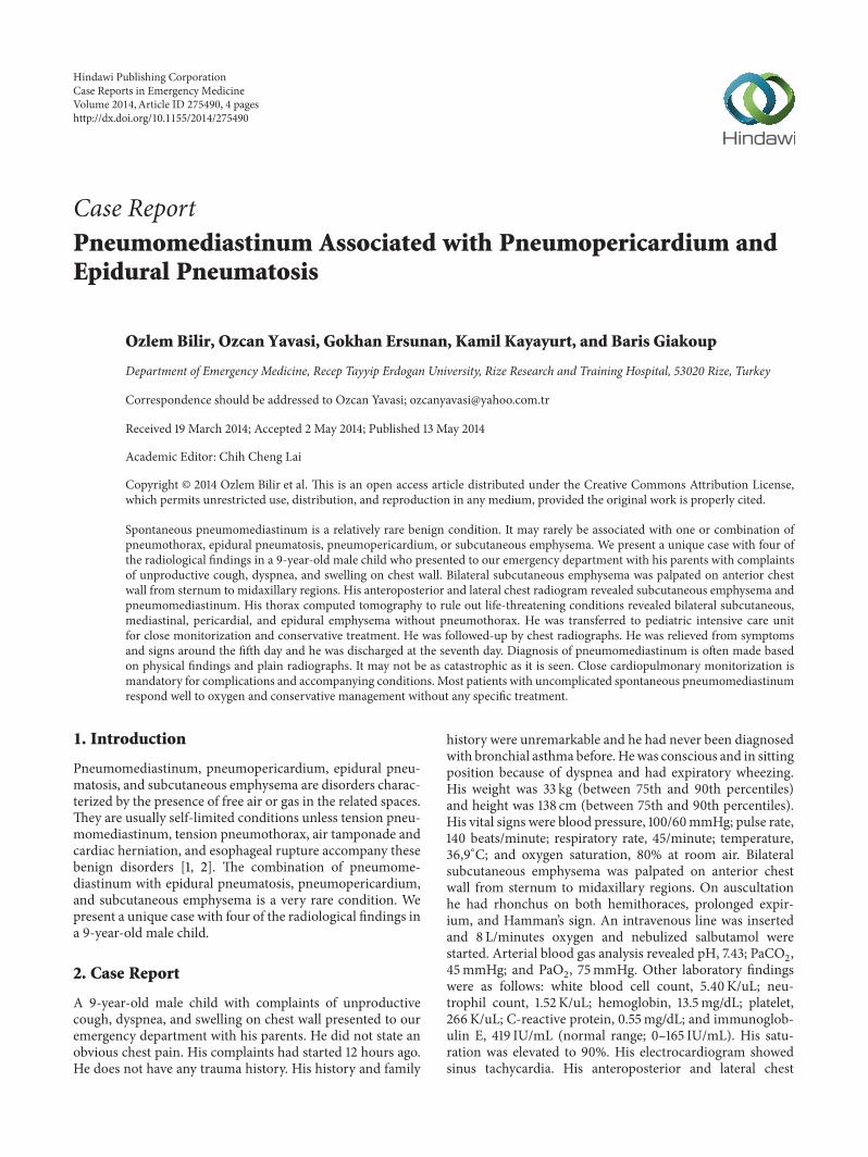
Hindawi Publishing CorporationCase Reports in Emergency MedicineVolume 2014, Article ID 275490, 4 pageshttp://dx.doi.org/10.1155/2014/275490
2 Case Reports in Emergency Medicine
(a) (b)
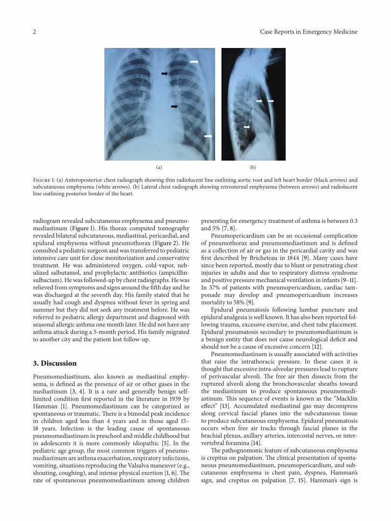
Figure 1: (a) Anteroposterior chest radiograph showing thin radiolucent line outlining aortic root and left heart border (black arrows) andsubcutaneous emphysema (white arrows). (b) Lateral chest radiograph showing retrosternal emphysema (between arrows) and radiolucentline outlining posterior border of the heart.
radiogram revealed subcutaneous emphysema and pneumo-mediastinum (Figure 1). His thorax computed tomographyrevealed bilateral subcutaneous, mediastinal, pericardial, andepidural emphysema without pneumothorax (Figure 2). Heconsulted a pediatric surgeon andwas transferred to pediatricintensive care unit for close monitorization and conservativetreatment. He was administered oxygen, cold-vapor, neb-ulized salbutamol, and prophylactic antibiotics (ampicillin-sulbactam).Hewas followed-up by chest radiographs.Hewasrelieved from symptoms and signs around the fifth day and hewas discharged at the seventh day. His family stated that heusually had cough and dyspnea without fever in spring andsummer but they did not seek any treatment before. He wasreferred to pediatric allergy department and diagnosed withseasonal allergic asthma onemonth later. He did not have anyasthma attack during a 5-month period. His family migratedto another city and the patient lost follow-up.
3. Discussion
Pneumomediastinum, also known as mediastinal emphy-sema, is defined as the presence of air or other gases in themediastinum [3, 4]. It is a rare and generally benign self-limited condition first reported in the literature in 1939 byHamman [1]. Pneumomediastinum can be categorized asspontaneous or traumatic. There is a bimodal peak incidencein children aged less than 4 years and in those aged 15–18 years. Infection is the leading cause of spontaneouspneumomediastinum in preschool andmiddle childhood butin adolescents it is more commonly idiopathic [5]. In thepediatric age group, the most common triggers of pneumo-mediastinumare asthma exacerbation, respiratory infections,vomiting, situations reproducing theValsalvamaneuver (e.g.,shouting, coughing), and intense physical exertion [1, 6].Therate of spontaneous pneumomediastinum among children
presenting for emergency treatment of asthma is between 0.3and 5% [7, 8].
Pneumopericardium can be an occasional complicationof pneumothorax and pneumomediastinum and is definedas a collection of air or gas in the pericardial cavity and wasfirst described by Bricheteau in 1844 [9]. Many cases havesince been reported, mostly due to blunt or penetrating chestinjuries in adults and due to respiratory distress syndromeand positive pressuremechanical ventilation in infants [9–11].In 37% of patients with pneumopericardium, cardiac tam-ponade may develop and pneumopericardium increasesmortality to 58% [9].
Epidural pneumatosis following lumbar puncture andepidural analgesia is well known. It has also been reported fol-lowing trauma, excessive exercise, and chest tube placement.Epidural pneumatosis secondary to pneumomediastinum isa benign entity that does not cause neurological deficit andshould not be a cause of excessive concern [12].
Pneumomediastinum is usually associated with activitiesthat raise the intrathoracic pressure. In these cases it isthought that excessive intra-alveolar pressures lead to ruptureof perivascular alveoli. The free air then dissects from theruptured alveoli along the bronchovascular sheaths towardthe mediastinum to produce spontaneous pneumomedi-astinum. This sequence of events is known as the “Macklineffect” [13]. Accumulated mediastinal gas may decompressalong cervical fascial planes into the subcutaneous tissueto produce subcutaneous emphysema. Epidural pneumatosisoccurs when free air tracks through fascial planes in thebrachial plexus, axillary arteries, intercostal nerves, or inter-vertebral foramina [14].
The pathognomonic feature of subcutaneous emphysemais crepitus on palpation. The clinical presentation of sponta-neous pneumomediastinum, pneumopericardium, and sub-cutaneous emphysema is chest pain, dyspnea, Hamman’ssign, and crepitus on palpation [7, 15]. Hamman’s sign is
Case Reports in Emergency Medicine 3
(a) (b)
(c) (d)
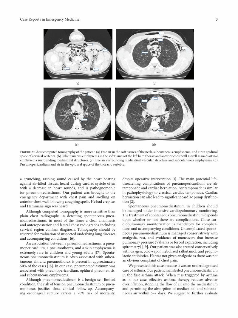
Figure 2: Chest computed tomography of the patient. (a) Free air in the soft tissues of the neck, subcutaneous emphysema, and air in epiduralspace of cervical vertebra. (b) Subcutaneous emphysema in the soft tissues of the left hemithorax and anterior chest wall as well as mediastinalemphysema surrounding mediastinal structures. (c) Free air surrounding mediastinal vascular structure and subcutaneous emphysema. (d)Pneumopericardium and air in the epidural space of the thoracic vertebra.
a crunching, rasping sound caused by the heart beatingagainst air-filled tissues, heard during cardiac systole oftenwith a decrease in heart sounds, and is pathognomonicfor pneumomediastinum. Our patient was brought to theemergency department with chest pain and swelling onanterior chest wall following coughing spells. He had crepitusand Hamman’s sign was heard.
Although computed tomography is more sensitive thanplain chest radiographs in detecting spontaneous pneu-momediastinum, in most of the times a clear anamnesisand anteroposterior and lateral chest radiographs includingcervical region confirm diagnosis. Tomography should bereserved for evaluation of suspected underlying lung diseasesand accompanying conditions [16].
An association between a pneumomediastinum, a pneu-mopericardium, a pneumothorax, and a skin emphysema isextremely rare in children and young adults [17]. Sponta-neous pneumomediastinum is often associated with subcu-taneous air, and pneumothorax is present in approximately50% of the cases [18]. In our case, pneumomediastinum wasassociated with pneumopericardium, epidural pneumatosis,and subcutaneous emphysema.
Although pneumomediastinum is a benign self-limitedcondition, the risk of tension pneumomediastinum or pneu-mothorax justifies close clinical follow-up. Accompany-ing esophageal rupture carries a 70% risk of mortality,
despite operative intervention [1]. The main potential life-threatening complications of pneumopericardium are airtamponade and cardiac herniation. Air tamponade is similarin pathophysiology to classical cardiac tamponade. Cardiacherniation can also lead to significant cardiac pump dysfunc-tion [2].
Spontaneous pneumomediastinum in children shouldbe managed under intensive cardiopulmonary monitoring.The treatment of spontaneous pneumomediastinum dependsupon whether or not there are complications. Close car-diopulmonary monitorization is mandatory for complica-tions and accompanying conditions. Uncomplicated sponta-neous pneumomediastinum is managed conservatively withanalgesia, rest, and avoidance of maneuvers that increasepulmonary pressure (Valsalva or forced expiration, includingspirometry) [19]. Our patient was also treated conservativelywith oxygen, cold-vapor, nebulized salbutamol, and prophy-lactic antibiotics. He was not given analgesic as there was notan obvious complaint of chest pain.
We presented this case because it was an underdiagnosedcase of asthma. Our patientmanifested pneumomediastinumin the first asthma attack. When it is triggered by asthmaas in our case, effective asthma therapy reduces alveolaroverinflation, stopping the flow of air into the mediastinumand permitting the absorption of mediastinal and subcuta-neous air within 5–7 days. We suggest to further evaluate
4 Case Reports in Emergency Medicine
the children for asthma and its complications such as pneu-momediastinum who present with cough and dyspnea to theemergency departments. Diagnosis of pneumomediastinumin young asthmatic children is not always easy as they are notoften able to complain about chest pain, which is a significantclinical sign in adults.
Conflict of Interests
The authors declare that there is no conflict of interestsregarding the publication of this paper.
References
[1] I. Abolnik, I. S. Lossos, and R. Breuer, “Spontaneous pneumo-mediastinum; A report of 25 cases,” Chest, vol. 100, no. 1, pp.93–95, 1991.
[2] G. Fulda, C. E. M. Brathwaite, A. Rodriguez, S. Z. Turney, C.M. Dunham, and R. A. Cowley, “Blunt traumatic rupture ofthe heart and pericardium: a ten-year experience (1979–1989),”Journal of Trauma, vol. 31, no. 2, pp. 167–173, 1991.
[3] D. R. Park and E. Vallienes, Pneumomediastinum and Medias-tinitis, Elsevier Saunders, Philadelphia, Pa, USA, 5th edition,2010.
[4] F. G. A. Versteegh and I. A. J. M. Broeders, “Spontaneous pneu-momediastinum in children,” European Journal of Pediatrics,vol. 150, no. 5, pp. 304–307, 1991.
[5] C.-Y. Lee, C.-C. Wu, and C.-Y. Lin, “Etiologies of spontaneouspneumomediastinum in children of different ages,” Pediatricsand Neonatology, vol. 50, no. 5, pp. 190–195, 2009.
[6] M. Chalumeau, L. Le Clainche, N. Sayeg et al., “Spontaneouspneumomediastinum in children,” Pediatric Pulmonology, vol.31, no. 1, pp. 67–75, 2001.
[7] A. M. Stack and G. L. Caputo, “Pneumomediastinum inchildhood asthma,” Pediatr Emergency Care, vol. 12, no. 2, pp.98–101, 1996.
[8] P. A. Eggleston, B. H. Ward, W. E. Pierson, and C. W. Bierman,“Radiographic abnormalities in acute asthma in children,”Pediatrics, vol. 54, no. 4, pp. 442–449, 1974.
[9] R. G. Cummings, R. L. R. Wesly, D. H. Adams, and J. E. Lowe,“Pneumopericardium resulting in cardiac tamponade,” Annalsof Thoracic Surgery, vol. 37, no. 6, pp. 511–518, 1984.
[10] P. J. Capizzi, M. Martin, and M. P. Bannon, “Tension pneu-mopericardium following blunt injury,” Journal of Trauma, vol.39, no. 4, pp. 775–780, 1995.
[11] D. Katzir, E. Klinovsky, V. Kent, A. Shucri, and Y. Gilboa,“Spontaneous pneumopericardium: case report and review ofthe literature,” Cardiology, vol. 76, no. 4, pp. 305–308, 1989.
[12] P. D. Koelliker and L. A. Brannam, “Epidural pneumatosisassociated with spontaneous pneumomediastinum: case reportand review of the literature,” Journal of EmergencyMedicine, vol.17, no. 2, pp. 247–250, 1999.
[13] M. T. Macklin and C. C. Macklin, “Malignant interstitialemphysema of the lungs and mediastinum as an importantoccult complication in many respiratory diseases and otherconditions,”Medicine, vol. 23, pp. 281–288, 1944.
[14] W. B. Hall and R. M. Aris, “Epidural pneumatosis and diffusesoft tissue free air as a complication of diabetic ketoacidosis,”American Journal of Respiratory and Critical Care Medicine, vol.185, no. 4, p. e5, 2012.
[15] M. Caceres, S. Z. Ali, R. Braud, D.Weiman, andH. E. Garrett Jr.,“Spontaneous pneumomediastinum: a comparative study andreview of the literature,” Annals of Thoracic Surgery, vol. 86, no.3, pp. 962–966, 2008.
[16] T. Kaneki, K. Kubo, A. Kawashima, T. Koizumi, M. Sekiguchi,and S. Sone, “Spontaneous pneumomediastinum in 33 patients:yield of chest computed tomography for the diagnosis of themild type,” Respiration, vol. 67, no. 4, pp. 408–411, 2000.
[17] C. Eberle, K. Junger, K.-M. Debatin, and M. Wabitsch, “Spon-taneously occurring pneumomediastinum related to a pneu-mopericardium, a pneumothorax and a skin emphysema in a12-year old boy,” Klinische Padiatrie, vol. 222, no. 1, pp. 40–44,2010.
[18] P. Monksfield, O. Whiteside, S. Jaffe, N. Steventon, and C.Milford, “Pneumomediastinum, an unusual complication offacial trauma,” Ear, Nose and Throat Journal, vol. 84, no. 5, pp.298–301, 2005.
[19] I. Macia, J. Moya, R. Ramos et al., “Spontaneous pneumomedi-astinum: 41 cases,” European Journal of Cardio-Thoracic Surgery,vol. 31, no. 6, pp. 1110–1114, 2007.

Submit your manuscripts athttp://www.hindawi.com
Stem CellsInternational
Hindawi Publishing Corporationhttp://www.hindawi.com Volume 2014
Hindawi Publishing Corporationhttp://www.hindawi.com Volume 2014
MEDIATORSINFLAMMATION
of
Hindawi Publishing Corporationhttp://www.hindawi.com Volume 2014
Behavioural Neurology
EndocrinologyInternational Journal of
Hindawi Publishing Corporationhttp://www.hindawi.com Volume 2014
Hindawi Publishing Corporationhttp://www.hindawi.com Volume 2014
Disease Markers
Hindawi Publishing Corporationhttp://www.hindawi.com Volume 2014
BioMed Research International
OncologyJournal of
Hindawi Publishing Corporationhttp://www.hindawi.com Volume 2014
Hindawi Publishing Corporationhttp://www.hindawi.com Volume 2014
Oxidative Medicine and Cellular Longevity
Hindawi Publishing Corporationhttp://www.hindawi.com Volume 2014
PPAR Research
The Scientific World JournalHindawi Publishing Corporation http://www.hindawi.com Volume 2014
Immunology ResearchHindawi Publishing Corporationhttp://www.hindawi.com Volume 2014
Journal of
ObesityJournal of
Hindawi Publishing Corporationhttp://www.hindawi.com Volume 2014
Hindawi Publishing Corporationhttp://www.hindawi.com Volume 2014
Computational and Mathematical Methods in Medicine
OphthalmologyJournal of
Hindawi Publishing Corporationhttp://www.hindawi.com Volume 2014
Diabetes ResearchJournal of
Hindawi Publishing Corporationhttp://www.hindawi.com Volume 2014
Hindawi Publishing Corporationhttp://www.hindawi.com Volume 2014
Research and TreatmentAIDS
Hindawi Publishing Corporationhttp://www.hindawi.com Volume 2014
Gastroenterology Research and Practice
Hindawi Publishing Corporationhttp://www.hindawi.com Volume 2014
Parkinson’s Disease
Evidence-Based Complementary and Alternative Medicine
Volume 2014Hindawi Publishing Corporationhttp://www.hindawi.com

![Case Report Subcutaneous Emphysema, …downloads.hindawi.com/journals/criem/2015/134816.pdfpneumothorax, pneumomediastinum, pneumopericardium, or subcutaneous emphysema [ ]. Diagnosis](https://img.pdfslide.net/doc/110x75/5f4072ff5627821a5534fd08/case-report-subcutaneous-emphysema-pneumothorax-pneumomediastinum-pneumopericardium.jpg)