Embed Size (px)
Citation preview
S44 Annals of Medical and Health Sciences Research | November 2013 | Vol 3 | Supplement 1 |
Address for correspondence: Dr. Owolabi Lukman Femi, Department of Medicine, Neurology Unit, Aminu Kano Teaching Hospital, PMB 3452, Kano, Nigeria. E‑mail: [email protected]
Introduction
Progressive supranuclear palsy (PSP) is a degenerative disorder that causes postural instability, supranuclear vertical ophthalmoplegia, Parkinsonism unresponsive to Levodopa, pseudobulbar palsy and cognitive impairment.[1,2] It was first described as a distinct syndrome by John Steele, J Clifford Richardson and Jerzy Olszewski in 1963, but, in the last two decades, there have been further refinements at the clinical, neuroimaging, pharmacologic and molecular levels. In spite of this development, however, there have been no laboratory markers for the diagnosis of PSP; thus, neuropathological examination remains the “gold standard” for its diagnosis. Besides, PSP may be associated with more than one neuropathological diagnosis, such as Alzheimer’s or Parkinson’s disease[3] and because of its increasingly recognized clinical diversity, it may be difficult, particularly in resource‑poor settings, to diagnose PSP. Consequently, the underdiagnosis or misdiagnosis of PSP neurodegenerative disorder is not uncommon.[4]
Recent studies have used imaging techniques in an effort to refine the clinical diagnosis.[5,6] Neuroimaging techniques
are increasingly becoming common in Nigeria, particularly in the tertiary health institutions; therefore, acquaintance of health practitioners with the usefulness of the brain magnetic resonance imaging (MRI) technique in diagnosing PSP will certainly ease the diagnostic difficulty associated with the condition.
The goal of this clinico‑radiological case report of PSP in a Nigerian man is to, from a practical point of view, highlight the usefulness of brain MRI in arriving at a diagnosis of PSP and also review the relevant literature.
Case Report
The patient is an 82‑year‑old clergyman, who developed progressively worsening generalized body stiffness, slowing of movement, postural instability resulting in occasional falls, visual disturbance, drooling of saliva, and slurred speech of 7‑years duration. There were associated “messy” eating, owing to the inability to look down at the plate of food, and visual hallucination. There were no associated tremor, dysphagia, and motor weakness. The symptoms became worse 2 years before presentation to our facility. He was a known hypertensive diagnosed 10 years previously; however, his blood pressure was controlled with amlodipine and lisinopril. He had also been treated for benign prostatic hyperplasia. Examination in our hospital revealed a fully conscious elderly man with bradykinesia, decreased fine motor skills, and hypophonia. His gait/posture was characterized by flexion at the waist, not swinging while walking and retrocollis. His face was blank and
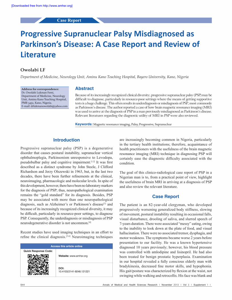
Progressive Supranuclear Palsy Misdiagnosed as Parkinson’s Disease: A Case Report and Review of Literature
Owolabi LFDepartment of Medicine, Neurology Unit, Aminu Kano Teaching Hospital, Bayero University, Kano, Nigeria
AbstractBecause of its increasingly recognized clinical diversity, progressive supranuclear palsy (PSP) may be difficult to diagnose, particularly in resource‑poor settings where the means of getting supportive tests is a huge challenge. This often results in underdiagnosis or misdiagnosis of PSP, most commonly as Parkinson’s disease. The author reported a case of how brain magnetic resonance imaging (MRI) was used to arrive at the diagnosis of PSP in a man previously misdiagnosed as Parkinson’s disease. Relevant literatures regarding the diagnostic utility of MRI in PSP were also reviewed.
Keywords: Magnetic resonance imaging, Palsy, Progressive, Supranuclear
Access this article online
Quick Response Code:
Website: www.amhsr.org
DOI: 10.4103/2141-9248.121221
Case Report
[Downloaded free from http://www.amhsr.org]
Owolabi: Misdiagnosed progressive supranuclear palsy; utility of MRI
Annals of Medical and Health Sciences Research | November 2013 | Vol 3 | Supplement 1 | S45
mask‑like, with a soft, slurred and monotonous speech. There was defective pronunciation of consonants and lack of variation in his speech. Glabella and palmomental reflex, as well as “aplause sign,” were all present. However, no abnormality was found on the cranial nerves, motor system (apart from rigidity and tremor) and sensory system examinations. Cerebellar signs were also absent. His blood pressure was 120/70 mmHg. Findings on other systemic examinations were within normal limits. Initially, the diagnosis was that of Parkinson’s disease (PD) to keep in view PSP. He was placed on Levodopa/Carbidopa without improvement in clinical condition and, subsequently, pramipexole (dopamine agonist) and selegiline (monoamine oxidase inhibitor) were added. However, there was no appreciable improvement in his clinical condition. Additional features detected on reassessment of the patient following failure of anti‑Parkinsonian drugs were significant rigidity with axial preponderance, vertical gaze palsy with preservation of lateral eye movements and mild cognitive impairment (mainly memory and calculation). Glabella and palmomental reflex as well as “aplause sign” were present.
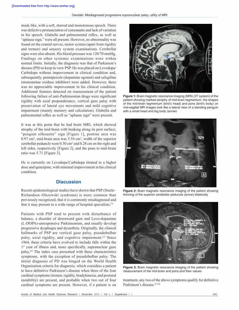
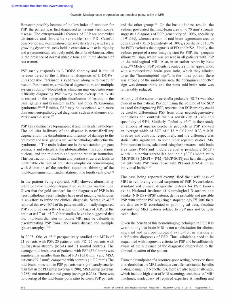
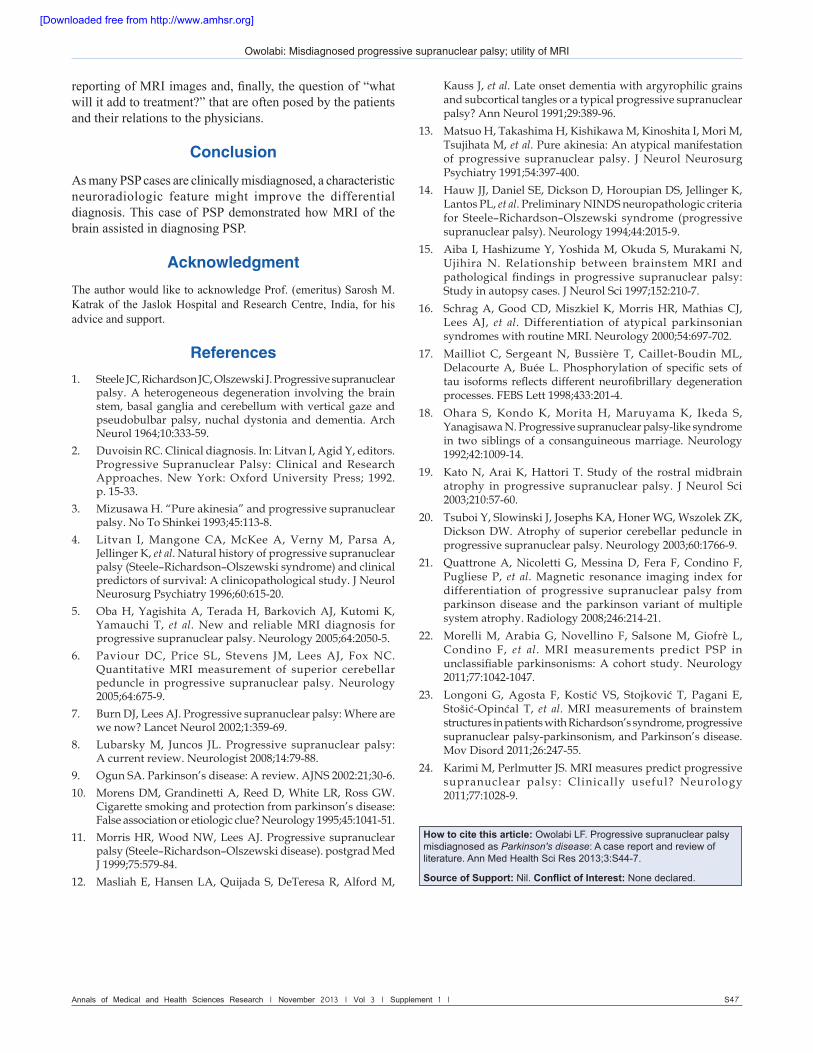
It was at this point that he had brain MRI, which showed atrophy of the mid‑brain with beaking along its post surface; “penguin silhouette” sign [Figure 1], pontine area was 0.97 cm2, mid‑brain area was 5.54 cm2, width of the superior cerebellar peduncle were 0.30 cm2 and 0.28 cm on the right and left sides, respectively [Figure 2], and the pons to mid‑brain ratio was 5.71 [Figure 3].
He is currently on Levodopa/Carbidopa titrated to a higher dose and quiterpine, with minimal improvement in his clinical condition.
Discussion
Recent epidemiological studies have shown that PSP (Steele–Richardson–Olszewski syndrome) is more common than previously recognized, that it is commonly misdiagnosed and that it may present to a wide range of hospital specialists.[7]
Patients with PSP tend to present with disturbance of balance, a disorder of downward gaze and Levo‑dopamine (L‑DOPA)‑unresponsive Parkinsonism, and usually develop progressive dysphagia and dysarthria. Originally, the clinical hallmarks of PSP are vertical gaze palsy, pseudobulbar palsy, axial rigidity, and cognitive impairment.[2] Since 1964, these criteria have evolved to include falls within the 1st year of illness and, more specifically, supranuclear gaze palsy.[8] The index case presented with these characteristics symptoms, with the exception of pseudobulbar palsy. The initial diagnosis of PD was hinged on the World Health Organization criteria for diagnosis, which considers a patient to have definitive Parkinson’s disease when three of the four cardinal symptoms (tremor, rigidity, bradykinesia, and postural instability) are present, and probable when two out of four cardinal symptoms are present. However, if a patient is on
treatment, any two of the above symptoms qualify for definitive Parkinson’s disease.[9,10]
Figure 1: Brain magnetic resonance imaging (MRI) (3T system) of the patient showing marked atrophy of mid-brain tegmentum, the shapes of the mid-brain tegmentum (bird’s head) and pons (bird’s body) on mid-sagittal MR images look like a lateral view of a standing penguin with a small head and big body (arrow)
Figure 2: Brain magnetic resonance imaging of the patient showing thinning of the superior cerebellar peduncle (arrow) bilaterally
Figure 3: Brain magnetic resonance imaging of the patient showing measurement of the mid-brain and pons and their values
[Downloaded free from http://www.amhsr.org]
Owolabi: Misdiagnosed progressive supranuclear palsy; utility of MRI
S46 Annals of Medical and Health Sciences Research | November 2013 | Vol 3 | Supplement 1 |
However, possibly because of the low index of suspicion for PSP, the patient was first diagnosed as having Parkinsons’s disease. The extrapyramidal features of PSP are somewhat distinctive and should be separable from PD. Careful observation and examination often reveals a taut spastic face, a growling dysarthria, neck held in extension with axial rigidity and a symmetrical, relatively mild, distal bradykinesia, often in the presence of normal muscle tone and in the absence of rest tremor.
PSP rarely responds to L‑DOPA therapy and it should be considered in the differential diagnosis of L‑DOPA‑ unresponsive Parkinson’s syndrome along with vascular pseudo‑Parkinsonism, corticobasal degeneration, and multiple system atrophy.[11] Nonetheless, clinicians may encounter some difficulty diagnosing PSP owing to the overlap that exists in respect of the topographic distribution of lesions in the basal ganglia and brainstem in PSP and other Parkinsonian syndromes.[12,13] Besides, PSP may be associated with more than one neuropathological diagnosis, such as Alzheimer’s or Parkinson’s disease.[3]
PSP has a distinctive topographical and molecular pathology. The cellular hallmark of the disease is neurofibrillary degeneration; the distribution and intensity of damage to the brainstem and basal ganglia are used to pathologically define the syndrome.[14] The main lesions are in the substantianigra pars compacta and reticulata, the globuspallidus, the subthalamic nucleus, and the mid‑brain and pontine reticular formation. This destruction of mid‑brain and pontine structures leads to identifiable changes of brainstem atrophy on neuroimaging with dilatation of the cerebral aqueduct, thinning of the mid‑brain tegmentum, and dilatation of the fourth ventricle.[15]
In the patient being reported, MRI showed abnormality referable to the mid‑brain tegmentum, ventricles, and the pons. Given that the gold standard for the diagnosis of PSP is its neuropathology, recent studies have used imaging techniques in an effort to refine the clinical diagnosis. Schrag et al.[16]
reported that over 70% of the patients with clinically diagnosed PSP could be correctly classified on the basis of MRI of the brain at 0·5 T or 1·5 T. Other studies have also suggested that low mid‑brain diameter on routine MRI may be valuable in discriminating PSP from Parkinson’s disease and multiple system atrophy.[17,18]
In 2005, Oba et al.[5] prospectively studied the MRIs of 21 patients with PSP, 23 patients with PD, 25 patients with multisystem atrophy (MSA) and 31 normal controls. The average mid‑brain area of patients with PSP (56.0 mm2) was significantly smaller than that of PD (103.0 mm2) and MSA patients (97.2 mm2) compared with controls (117.7 mm2).The mid‑brain–pons ratio in PSP patients was significantly smaller than that in the PD group (average 0.208), MSA group (average 0.266) and normal control group (average 0.236). There was no overlap of the mid‑brain–pons ratio between PSP patients
and the other groups.[5] On the basis of these results, the authors postulated that mid‑brain area of < 70 mm2 strongly suggests a diagnosis of PSP (sensitivity of 100%, specificity of 91.3%), whereas a ratio of mid‑brain tegmentum area to pons area of < 0.15 (sensitivity of 100%, specificity of 100% for PSP) excludes the diagnosis of PD and MSA. Finally, the authors proposed a new imaging sign for PSP, the “penguin silhouette” sign, which was present in all patients with PSP on the mid‑sagittal MRI. Also, in an earlier report by Kato et al.,[19] MRIs of PSP patients revealed a similar appearance, with a reduced mid‑brain–pons ratio, which they alluded to as the “hummingbird sign”. In the index patient, there was atrophy of the mid‑brain area, the “penguin silhouette” sign was demonstrable and the pons–mid‑brain ratio was remarkably reduced.
Atrophy of the superior cerebella peduncle (SCP) was also evident in this patient. Paviour, using the volume of the SCP as a tool for diagnosing PSP, reported that SCP atrophy could be used to differentiate PSP from other neurodegenerative conditions and controls with a sensitivity of 74% and specificity of 94%. Similarly, Tsuboi et al.[20] in their study on atrophy of superior cerebellar peduncle in PSP, showed an average width of SCP of 0.16 ± 0.01 and 0.33 ± 0.01 in cases and controls, respectively, and the difference was statistically significant. In some other separate studies, MR Parkinsonism index, calculated using the pons area – mid‑brain area ratio (P/M) and middle cerebellar penduncle (MCP) width – superior cerebellar peduncle (SCP) width ratio (MCP/SCP) [MRPI = (P/M)·(MCP/SCP)] can help distinguish patients with PSP from those with PD and MSA‑P on an individual basis.[21‑23]
The case being reported exemplified the usefulness of MRI in reinforcing clinical suspicion of PSP. Nevertheless, standardized clinical diagnostic criteria for PSP, known as the National Institute of Neurological Disorders and Stroke (NINDS)–SPSP criteria, define probable and possible PSP, with definite PSP requiring histopathology.[24] Until there are data on MRI correlated to pathological data, absolute certainty on MRI features related to PSP may not be fully established.
Given the benefit of this neuroimaging technique in PSP, it is worth noting that brain MRI is not a substitution for clinical appraisal and neuropathological evaluation in arriving at a definitive diagnosis of PSP. Thus, clinicians need to be acquainted with diagnostic criteria for PSP and be sufficiently aware of the relevance of the diagnostic observation to the clinical situation of the patient.
From the standpoint of a resource‑poor setting, however, there is no doubt that the MRI technique can offer substantial benefits in diagnosing PSP. Nonetheless, there are also huge challenges, which include high cost of MRI scanning, scantiness of MRI machines, inadequacy of required expertise at handling and
[Downloaded free from http://www.amhsr.org]
Owolabi: Misdiagnosed progressive supranuclear palsy; utility of MRI
Annals of Medical and Health Sciences Research | November 2013 | Vol 3 | Supplement 1 | S47
reporting of MRI images and, finally, the question of “what will it add to treatment?” that are often posed by the patients and their relations to the physicians.
Conclusion
As many PSP cases are clinically misdiagnosed, a characteristic neuroradiologic feature might improve the differential diagnosis. This case of PSP demonstrated how MRI of the brain assisted in diagnosing PSP.
AcknowledgmentThe author would like to acknowledge Prof. (emeritus) Sarosh M. Katrak of the Jaslok Hospital and Research Centre, India, for his advice and support.
References1. Steele JC, Richardson JC, Olszewski J. Progressive supranuclear
palsy. A heterogeneous degeneration involving the brainstem, basal ganglia and cerebellum with vertical gaze andpseudobulbar palsy, nuchal dystonia and dementia. ArchNeurol 1964;10:333‑59.
2. Duvoisin RC. Clinical diagnosis. In: Litvan I, Agid Y, editors. Progressive Supranuclear Palsy: Clinical and ResearchApproaches. New York: Oxford University Press; 1992.p. 15‑33.
3. Mizusawa H. “Pure akinesia” and progressive supranuclear palsy. No To Shinkei 1993;45:113‑8.
4. Litvan I, Mangone CA, McKee A, Verny M, Parsa A,Jellinger K, et al. Natural history of progressive supranuclear palsy (Steele–Richardson–Olszewski syndrome) and clinical predictors of survival: A clinicopathological study. J NeurolNeurosurg Psychiatry 1996;60:615‑20.
5. Oba H, Yagishita A, Terada H, Barkovich AJ, Kutomi K,Yamauchi T, et al. New and reliable MRI diagnosis forprogressive supranuclear palsy. Neurology 2005;64:2050‑5.
6. Paviour DC, Price SL, Stevens JM, Lees AJ, Fox NC.Quantitative MRI measurement of superior cerebellarpeduncle in progressive supranuclear palsy. Neurology2005;64:675‑9.
7. Burn DJ, Lees AJ. Progressive supranuclear palsy: Where are we now? Lancet Neurol 2002;1:359‑69.
8. Lubarsky M, Juncos JL. Progressive supranuclear palsy:A current review. Neurologist 2008;14:79‑88.
9. Ogun SA. Parkinson’s disease: A review. AJNS 2002:21;30‑6.10. Morens DM, Grandinetti A, Reed D, White LR, Ross GW.
Cigarette smoking and protection from parkinson’s disease:False association or etiologic clue? Neurology 1995;45:1041‑51.
11. Morris HR, Wood NW, Lees AJ. Progressive supranuclearpalsy (Steele–Richardson–Olszewski disease). postgrad Med J 1999;75:579‑84.
12. Masliah E, Hansen LA, Quijada S, DeTeresa R, Alford M,
Kauss J, et al. Late onset dementia with argyrophilic grains and subcortical tangles or a typical progressive supranuclear palsy? Ann Neurol 1991;29:389‑96.
13. Matsuo H, Takashima H, Kishikawa M, Kinoshita I, Mori M, Tsujihata M, et al. Pure akinesia: An atypical manifestationof progressive supranuclear palsy. J Neurol NeurosurgPsychiatry 1991;54:397‑400.
14. Hauw JJ, Daniel SE, Dickson D, Horoupian DS, Jellinger K,Lantos PL, et al. Preliminary NINDS neuropathologic criteria for Steele–Richardson–Olszewski syndrome (progressivesupranuclear palsy). Neurology 1994;44:2015‑9.
15. Aiba I, Hashizume Y, Yoshida M, Okuda S, Murakami N,Ujihira N. Relationship between brainstem MRI andpathological findings in progressive supranuclear palsy:Study in autopsy cases. J Neurol Sci 1997;152:210‑7.
16. Schrag A, Good CD, Miszkiel K, Morris HR, Mathias CJ,Lees AJ, et al. Differentiation of atypical parkinsoniansyndromes with routine MRI. Neurology 2000;54:697‑702.
17. Mailliot C, Sergeant N, Bussière T, Caillet‑Boudin ML,Delacourte A, Buée L. Phosphorylation of specific sets oftau isoforms reflects different neurofibrillary degenerationprocesses. FEBS Lett 1998;433:201‑4.
18. Ohara S, Kondo K, Morita H, Maruyama K, Ikeda S,Yanagisawa N. Progressive supranuclear palsy‑like syndrome in two siblings of a consanguineous marriage. Neurology1992;42:1009‑14.
19. Kato N, Arai K, Hattori T. Study of the rostral midbrainatrophy in progressive supranuclear palsy. J Neurol Sci2003;210:57‑60.
20. Tsuboi Y, Slowinski J, Josephs KA, Honer WG, Wszolek ZK,Dickson DW. Atrophy of superior cerebellar peduncle inprogressive supranuclear palsy. Neurology 2003;60:1766‑9.
21. Quattrone A, Nicoletti G, Messina D, Fera F, Condino F,Pugliese P, et al. Magnetic resonance imaging index fordifferentiation of progressive supranuclear palsy fromparkinson disease and the parkinson variant of multiplesystem atrophy. Radiology 2008;246:214‑21.
22. Morelli M, Arabia G, Novellino F, Salsone M, Giofrè L,Condino F, et al. MRI measurements predict PSP inunclassifiable parkinsonisms: A cohort study. Neurology2011;77:1042‑1047.
23. Longoni G, Agosta F, Kostić VS, Stojković T, Pagani E,Stošić‑Opinćal T, et al. MRI measurements of brainstemstructures in patients with Richardson’s syndrome, progressive supranuclear palsy‑parkinsonism, and Parkinson’s disease.Mov Disord 2011;26:247‑55.
24. Karimi M, Perlmutter JS. MRI measures predict progressivesupranuclear palsy: Clinically useful? Neurology2011;77:1028‑9.
How to cite this article: Owolabi LF. Progressive supranuclear palsy misdiagnosed as Parkinson's disease: A case report and review of literature. Ann Med Health Sci Res 2013;3:S44-7.
Source of Support: Nil. Conflict of Interest: None declared.
[Downloaded free from http://www.amhsr.org]