Embed Size (px)
Citation preview
219
Cauda equina-conus medullaris syndrome as an isolated presenting symptom of intravascular large B-cell lymphoma: Case report and review of the literatureMeng-Chen Wu MD, Chin-Hsien Lin MD PhD
Department of Neurology, National Taiwan University Hospital, Taipei, Taiwan Abstract
Intravascular large B-cell lymphoma (IVLBCL) is a rare non-Hodgkin lymphoma with variable clinical manifestations. Although neurological symptoms are common in patients with IVLBCL, isolated cauda equina-conus medullaris syndrome is rarely reported. We herein report a case of IVLBCL whose initial presentation was cauda equina-conus medullaris syndrome with neither dermatological nor hematological manifestations. A 54-year-old man without known immune-compromised state presented with progressive ascending numbness and weakness of bilateral legs and urine incontinence for 2 months. Lumbar-sacral magnetic resonance images showed gadolinium-enhanced conus medullaris and cauda equina nerve roots. Cerebrospinal fluid analysis revealed lymphocyte predominant pleocytosis and elevated protein level without malignant cells. Focal seizure and mental status changes followed several weeks later. Brain biopsy led to the diagnosis of IVLBCL. Conclusions: IVLBCL should be included in the differential diagnosis of patients with isolated cauda equina-conus medullaris syndrome. A survey of previously published cases in the literature also showed that early initiation of chemotherapy has better outcome.
Neurology Asia 2014; 19(2) : 219 – 225
Address correspondence to: Dr. Chin-Hsien Lin, Department of Neurology, National Taiwan University Hospital, Taipei 100, Taiwan. Tel: +886-2-23123456 ext 65335, Fax: +886-2-23418395, E-mail: [email protected]
INTRODUCTION
Intravascular large B-cell lymphoma (IVLBCL) is a rare form of extranodal non-Hodgkin lymphoma. Disseminated disease with a variety of organ involvement complicates the clinical presentations and leads to diagnosis difficulty.1 The nervous system and skin are the most frequently involved organs in western patients but are uncommon in Asians.2 Asian patients with IVLBCL usually presents as bone marrow involvement with hematological manifestations.3,4 The reasons for the ethnic difference in clinical presentation are unclear.2-4 Isolated cauda equina or conus medullaris syndrome is rarely reported as the presenting symptom of IVLBCL.5 We report an IVLBCL case whose initial presentation was cauda equina-conus medullaris syndrome without other system involvement. We also reviewed the previously reported cases reported in the literature.
CASE REPORT
A 54-year-old man had history of well-controlled hypertension, hyperlipidemia and polycystic kidney disease. He presented with a two-month history of progressive numbness, pain and weakness of
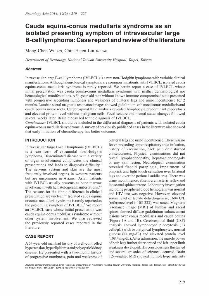
bilateral legs and urine incontinence. There was no fever, preceding upper respiratory tract infection, history of vaccination, back pain or disturbed consciousness. Physical examinations did not reveal lymphadenopathy, hepatosplenomegaly or any skin lesion. Neurological examination revealed flaccid paraplegia, impairment of pinprick and light touch sensation over bilateral legs and over the perianal saddle area. There was urine incontinence, absent cremasteric reflex and loose anal sphincter tone. Laboratory investigation including peripheral blood hemogram was normal and HIV test was negative. However, elevated serum level of lactate dehydrogenase, 1604 U/L (reference level is 105-333), was noted. Magnetic resonance image (MRI) of lumbar and sacral spines showed diffuse gadolinium enhancement lesions over conus medullaris and cauda equina (Figure 1A and 1B). Cerebrospinal fluid (CSF) analysis showed lymphocyte pleocytosis (13 cells/μL) with two atypical lymphocytes, normal glucose (44 mg/dL) and elevated protein level (108.4 mg/dL). After admission, the muscle power of both legs further deteriorated and left upper limb weakness developed. His consciousness fluctuated and several episodes of seizures occurred. Brain T2-weighted MRI showed multiple hyperintensity
Neurology Asia June 2014
220
Figure 1. Lumbar-sacral spine MRI without (A) and with (B) gadolinium contrast with arrows to indicate gadolinium contrast enhanced lesions of the conus medullaris and cauda equina nerve roots.
Figure 2. FLAIR Brain MRI of the patient on admission and two months after chemotherapy. (A) On admission, FLAIR showed multiple abnormal T2 hyperintense lesions over bilateral subcortical white matter, with some of the lesions showing mild diffusion restriction and contrast enhancement. (B) Two months after chemotherapy, the follow-up brain image showed left medial frontal post-operative change from the open-brain biopsy, and mild regression of bilateral subcortical white matter lesions as compared to (A).
lesions at left frontal, bilateral subcortical white matter areas, cerebellum, and pons. Some of these showed contrast enhancement (Figure 2A). Open brain biopsy at the left frontal pole was performed. Pathology showed neoplastic lymphoid cells lodged within the lumen of small
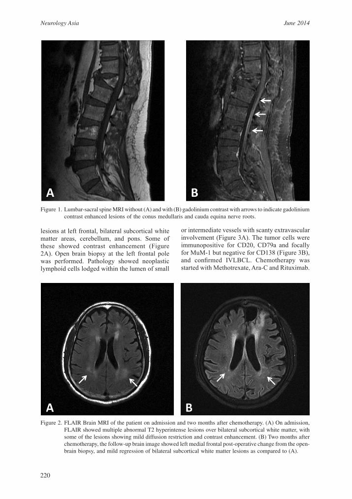
or intermediate vessels with scanty extravascular involvement (Figure 3A). The tumor cells were immunopositive for CD20, CD79a and focally for MuM-1 but negative for CD138 (Figure 3B), and confirmed IVLBCL. Chemotherapy was started with Methotrexate, Ara-C and Rituximab.
221
Consciousness improved and follow-up brain MRI 2 months later demonstrated diminished cerebral white matter lesions (Figure 2B). However, pancytopenia developed after a high dose of methotrexate and he died from sepsis 8 months after the onset of symptoms.
DISCUSSION
We report a patient with IVLBCL presenting as cauda equina-conus medullaris syndrome without evidence of hepatosplenomegaly or bone marrow involvement. Isolated neurological symptoms are estimated to be less than 30% in CNS IVLBCL.1-3,6 Cauda equina/conus medullaris syndrome accounts for only 5%.7 Deficits are caused by invasion of the lymphoid tumor cell into the small vessels of either the central nervous system or the peripheral nerves, and lead to microcirculatory dysfunction and subsequent neuronal damage. Given that isolated cauda equina or conus medullaris syndrome is a rare presenting symptom in patients with IVLBCL, we searched MEDLINE for previously published cases. The terms we used for keyword search included intravascular lymphoma, cauda equina, conus medullaris, spinal cord, and paraplegia. A total of 23 related cases were identified from 1991 to 2013 (Table 1). When combined with our index patient, the median onset age of neurological symptoms was 64.4 years old (range 41-86 years) and mostly were men. Most cases had paraparesis with sensory and sphincter dysfunction. Notably, 9 patients (38%) had pain as part of their initial symptoms, either radicular pain or low back pain, which is probably related to ischemic change of nerve root caused by
tumor cell thrombosis.1 Among these 24 IVLBCL patients, 10 patients showed isolated cauda equina or conus medullaris involvement (42%), 5 patients had isolated thoracic cord involvement (21%), 6 had long segment involvement from the thoracic region to the conus medullaris/cauda equina level (25%). None had cervical cord involvement. With disease progression, 3 of the 5 patients (60%) with initial thoracic cord lesion reported subsequent involvement of conus medullaris or cauda equina. In contrast, among the 10 patients with initial conus medullaris or cauda equina involvement, only 2 patients (20%) had subsequent thoracic cord involvement. These observations suggest that IVLBCL has a predilection for the lower spinal cord invasion with subsequent caudal-rostral involvement of the adjacent spinal cord. Clinical outcome of IVLBCL was dismal before the use of chemotherapy, especially rituximab. Patients receiving chemotherapy had a longer survival duration and less systemic dissemination of the disease than patients who did not receive chemotherapy.9 Shimada et al. reported a retrospective analysis of 106 patients with IVLBCL and found that the 2-year survival rate was 66% and 46% in patients receiving and not receiving rituximab, respectively.3 These findings suggest that rituximab-containing chemotherapy was crucial for patients with IVLBCL. In the case series reviewed, the survival duration of patients with and without chemotherapy was 17 months and 6 months, respectively. The most common chemotherapy regimens were anthracycline-based chemotherapy, CHOP (cyclophosphamide, doxorubicin, vincristine, and prednisolone) and rituximab (Table 1). Thus, early chemotherapy
Figure 3. Left frontal lobe open-brain biopsy of the patient. (A) Arrows indicate IVLBCL neoplastic lymphoid cells with large nucleoli and mitosis lodged within lumina of vessels (hematoxylin and eosin stain, original magnification × 200). (B) Immunohistochemical staining show numerous CD20+ lymphoma cells filled in the lumen of small vessels (original magnification × 100).
Neurology Asia June 2014
222
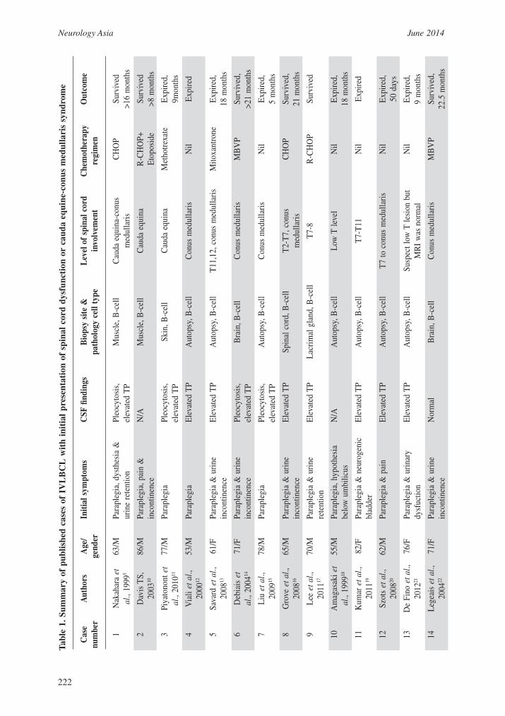
Tab
le 1
. Sum
mar
y of
pub
lishe
d ca
ses
of I
VL
BC
L w
ith
init
ial
pres
enta
tion
of
spin
al c
ord
dysf
unct
ion
or c
auda
equ
ine-
conu
s m
edul
lari
s sy
ndro
me
C
ase
Aut
hors
A
ge/
Initi
al s
ympt
oms
CSF
find
ings
B
iops
y si
te &
L
evel
of
spin
al c
ord
C
hem
othe
rapy
O
utco
me
num
ber
ge
nder
pa
thol
ogy
cell
type
in
volv
emen
t re
gim
en
1
Nak
ahar
a et
63
/M
Para
pleg
ia, d
ysth
esia
&
Pleo
cyto
sis,
M
uscl
e, B
-cel
l C
auda
equ
ina-
conu
s C
HO
P Su
rviv
ed
al.,
1999
5
urin
e re
tent
ion
elev
ated
TP
m
edul
lari
s
>16
mon
ths
2
Dav
is T
S,
86/M
Pa
rapl
egia
, pai
n &
N
/A
Mus
cle,
B-c
ell
Cau
da e
quin
a R
-CH
OP+
Su
rviv
ed
2003
10
in
cont
inen
ce
Et
opos
ide
>8 m
onth
s
3
Piya
tono
nt e
t 77
/M
Para
pleg
ia
Pleo
cyto
sis,
Sk
in, B
-cel
l C
auda
equ
ina
Met
hotr
exat
e Ex
pire
d,
al.,
2010
11
elev
ated
TP
9m
onth
s
4
Via
li et
al.,
53
/M
Para
pleg
ia
Elev
ated
TP
Aut
opsy
, B-c
ell
Con
us m
edul
lari
s N
il Ex
pire
d
20
0012
5
Sava
rd e
t al.,
61
/F
Para
pleg
ia &
uri
ne
Elev
ated
TP
Aut
opsy
, B-c
ell
T11,
12, c
onus
med
ulla
ris
Mito
xant
rone
Ex
pire
d,
2008
13
in
cont
inen
ce
18 m
onth
s
6
Deb
iais
et
71/F
Pa
rapl
egia
& u
rine
Pl
eocy
tosi
s,
Bra
in, B
-cel
l C
onus
med
ulla
ris
MB
VP
Surv
ived
,
al
., 20
0414
inco
ntin
ence
el
evat
ed T
P
>21
mon
ths
7
Liu
et a
l.,
78/M
Pa
rapl
egia
Pl
eocy
tosi
s,
Aut
opsy
, B-c
ell
Con
us m
edul
lari
s N
il Ex
pire
d,
2009
15
elev
ated
TP
5
mon
ths
8
Gro
ve e
t al.,
65
/M
Para
pleg
ia &
uri
ne
Elev
ated
TP
Spin
al c
ord,
B-c
ell
T2-T
7, c
onus
C
HO
P Su
rviv
ed,
2008
16
in
cont
inen
ce
med
ulla
ris
21
mon
ths
9
Lee
et a
l.,
70/M
Pa
rapl
egia
& u
rine
El
evat
ed T
P La
crim
al g
land
, B-c
ell
T7-8
R
-CH
OP
Surv
ived
2011
17
re
tent
ion
10
A
mag
asak
i et
55
/M
Para
pleg
ia, h
ypot
hesi
a N
/A
Aut
opsy
, B-c
ell
Low
T le
vel
Nil
Expi
red,
al
., 19
9918
belo
w u
mbi
licus
18
mon
ths
11
K
umar
et a
l.,
82/F
Pa
rapl
egia
& n
euro
geni
c El
evat
ed T
P A
utop
sy, B
-cel
l T7
-T11
N
il Ex
pire
d
20
1119
blad
der
12
Sz
ots
et a
l.,
62/M
Pa
rapl
egia
& p
ain
Elev
ated
TP
Aut
opsy
, B-c
ell
T7 to
con
us m
edul
lari
s N
il Ex
pire
d,
2008
20
50 d
ays
13
D
e Fi
no e
t al.,
76
/F
Para
pleg
ia &
uri
nary
El
evat
ed T
P A
utop
sy, B
-cel
l Su
spec
t low
T le
sion
but
N
il Ex
pire
d,
2012
21
dy
sfuc
tion
MR
I w
as n
orm
al
9
mon
ths
14
Le
geai
s et
al.,
71
/F
Para
pleg
ia &
uri
ne
Nor
mal
B
rain
, B-c
ell
Con
us m
edul
lari
s M
BV
P Su
rviv
ed,
2004
22
in
cont
inen
ce
22.5
mon
ths
223
Tab
le 1
. Sum
mar
y of
pub
lishe
d ca
ses
of I
VL
BC
L w
ith
init
ial
pres
enta
tion
of
spin
al c
ord
dysf
unct
ion
or c
auda
equ
ine-
conu
s m
edul
lari
s sy
ndro
me
C
ase
Aut
hors
A
ge/
Initi
al s
ympt
oms
CSF
find
ings
B
iops
y si
te &
L
evel
of
spin
al c
ord
C
hem
othe
rapy
O
utco
me
num
ber
ge
nder
pa
thol
ogy
cell
type
in
volv
emen
t re
gim
en
15
S
cully
et a
l.,
43/M
Pa
rapl
egia
& u
rine
Pl
eocy
tosi
s,
Bra
in, B
-cel
l T
cord
, con
us m
edul
lari
s M
etho
trex
ate
Expi
red
1995
23
in
cont
inen
ce
elev
ated
TP
16
Sc
hwar
z et
al.,
41
/M
Peri
neal
sen
sory
loss
&
Pleo
cyto
sis,
A
utop
sy, B
-cel
l C
onus
med
ulla
ris
Cyc
loph
osph
amid
e Ex
pire
d,
2002
24
ur
ine
inco
ntin
ence
el
evat
ed T
P
13 m
onth
s
17
C
lark
et a
l.,
63/M
Pa
rapl
egia
& v
oidi
ng
N/A
A
utop
sy, B
-cel
l T8
leve
l N
il Ex
pire
d
19
9125
diffi
culty
18
Y
ang
et a
l.,
70/M
Pa
rapl
egia
& s
phin
cter
El
evat
ed T
P A
utop
sy, B
-cel
l Su
spec
t low
T le
sion
but
N
il Ex
pire
d,
2008
26
dy
sfun
ctio
n
M
RI
was
nor
mal
3 m
onth
s
19
A
bbas
i et a
l.,
58/M
Pa
rapl
egia
& u
rine
El
evat
ed T
P R
enal
, B-c
ell
T6-T
10
R-C
HO
P Su
rviv
ed,
2014
27
re
tent
ion
3 ye
ars
20
W
arin
g et
al.,
74
/M
Uri
ne r
eten
tion
Elev
ated
TP
Aut
opsy
, B-c
ell
Low
er T
to u
pper
L
Nil
Expi
red,
19
9928
sp
inal
cor
d
3 m
onth
s
21
T
akiz
awa
et a
l., 52
/M
Para
pleg
ia, p
ain
& u
rine
El
evat
ed T
P M
uscl
e, B
-cel
l Lo
wer
T to
con
us
R-C
HO
P Su
rviv
ed,
2007
29
in
cont
inen
ce
med
ulla
ris
3.
5 ye
ars
22
A
buzi
nada
h et
al.,
70/M
Pe
rine
al n
umbn
ess
with
Pl
eocy
tosi
s,
S2 n
erve
roo
t, C
auda
equ
ina
R-C
HO
P+
Surv
ived
,
20
1230
urin
e re
tent
ion
elev
ated
TP
B-c
ell
M
etho
trex
ate
12 m
onth
s
23
Lo
zsad
i et a
l.,
62/M
Pa
rapl
egia
& s
enso
ry le
vel
Elev
ated
TP
Bra
in, B
-cel
l T5
N
il Ex
pire
d,
2005
7
at T
6
4.
5 m
onth
s
24
C
urre
nt r
epor
t 54
/M
Para
pleg
ia &
uri
ne
Pleo
cyto
sis,
B
rain
, B-c
ell
Cau
da e
quin
e-co
nus
Met
hoth
rexa
te+
Ex
pire
d,
inco
ntin
ence
el
evat
ed T
P
med
ulla
ris
C
ytar
abin
e+R
itux
8 m
onth
s
im
ab
CS
F: c
ereb
ral s
pina
l flui
d; T
P: T
otal
pro
tein
(mg/
dL);
N/A
: not
ava
ilab
le; T
:Tho
raci
c; C
HO
P: c
yclo
phos
pham
ide,
dox
orub
icin
, vin
cris
tine
and
pre
dnis
one;
R-C
HO
P: R
itux
imab
, cy
clop
hosp
ham
ide,
dox
orub
icin
, vi
ncri
stin
e an
d pr
edni
sone
; M
BV
P:
met
hotr
exat
e, c
arm
usti
ne,
Eto
posi
de a
nd m
ethy
lpre
dnis
olon
e
Neurology Asia June 2014
224
treatment in patients with IVLBCL appear to have better outcome. In conclusion, we report a case of IVLBCL whose initial presentation was isolated cauda equina-conus medullaris syndrome. Review of literature showed that patients with isolated spinal cord dysfunction as the leading symptom are mostly men and there is a predilection of the lymphoid tumor to affect the conus medullaris and cauda equina. Early initiation of chemotherapy is associated with better outcome.
ACKNOWLEDGEMENTS
We thank the patient who gave informed consent to the publication of related images.
DISCLOSURE
Conflicts of interests: None
REFERENCES 1. Shimada K, Kinoshita T, Naoe T, Nakamura S.
Presentation and management of intravascular large B-cell lymphoma. Lancet Oncol 2009; 10:895-902.
2. Ferreri AJ, Campo E, Seymour JF, et al. Intravascular lymphoma: clinical presentation, natural history, management and prognostic factors in a series of 38 cases, with special emphasis on the ‘cutaneous variant’. Br J Haematol 2004; 127:173-83.
3. Shimada K, Matsue K, Yamamoto K, et al. Retrospective analysis of intravascular large B-cell lymphoma treated with rituximab-containing chemotherapy as reported by the IVL study group in Japan. J Clin Oncol 2008; 26:3189-95.
4. Shimada K, Murase T, Matsue K, et al. Central nervous system involvement in intravascular large B-cell lymphoma: A retrospective analysis of 109 patients. Cancer Sci 2010; 101:1480-6.
5. Nakahara T, Saito T, Muroi A, et al. Intravascular lymphomatosis presenting as an ascending cauda equina conus medullaris syndrome: Remission after biweekly CHOP therapy. J Neurol Neurosurg Psychiatry 1999; 67:403-6.
6. Glass J, Hochberg FH, Miller DC. Intravascular lymphomatosis: A systemic disease with neurologic manifestations. Cancer 1993; 71:3156-64.
7. Lozsadi DA, Wieshmann U, Enevoldson TP. Neurological presentation of intravascular lymphoma: report of two cases and discussionof diagnostic challenges. Eur J Neurol 2005; 12:710-4.
8. Ponzoni M, Ferreri AJ, Campo E, et al. Definition, Diagnosis, and Management of Intravascular Large B-Cell Lymphoma: Proposals and Perspectives From an International Consensus Meeting. J Clin Oncol 2007; 25:3168-73.
9. DiGiuseppe JA, Nelson WG, Seifter EJ, Boitnott JK, Mann RB. Intravascular lymphomatosis: a clinicopathologic study of 10 cases and assessment of response to chemotherapy. J Clin Oncol 1994; 12:2573-9.
10. Davis TS. Intravascular Lymphoma Presenting with Cauda Equina Syndrome: Treated with CHOP and Rituxan. Leukemia & Lymphoma 2003; 44:887-8.
11. Piyatanont K, Bamrungrak K, Watcharananan S, et al. Intravascular B-cell lymphoma presenting with cauda equina syndrome: the role of skin biopsy. Eur J Dermatol 2010; 20:821-2.
12. Viali S, Hutchinson DO, Hawkins TE, et al. Presentation of intravascular lymphomatosis as lumbosacral polyradiculopathy. Muscle Nerve 2000; 23:1295-300.
13. Savard M, Verreault S, Gould PV, et al. Intravascular lymphoma with conus medullaris syndrome followed by encephalopathy. Can J Neurol Sci 2008; 35:366-71.
14. Debiais S, Bonnaud I, Cottier JP, et al. A spinal cord intravascular lymphomatosis with exceptionally good outcome. Neurology 2004; 63:1329-30.
15. Liu H, Koyanagi I, Chiba H, et al. Spinal cord infarct as the initial clinical presentation of intravascular malignant lymphomatosis. J Clin Neurosci 2009; 16:570-3.
16. Grove CS, Robbins PD, Kermode AG. Intravascular lymphoma presenting as progressive paraparesis. J Clin Neurosci 2008; 15;1056-8.
17. Lee BS, Frankfort BJ, Eberhart CG, Weinberg RS. Diagnosis of intravascular lymphoma by a novel biopsy site. Ophthalmology 2011; 118:586-90.
18. Amagasaki K1, Yamazaki H, Ohmori K, Koizumi H, Hashizume K, Sasaguchi N. Malignant intravascular lymphomatosis associated with venous stenosis. Case report. J Neurosurg 1999; 90:355-8.
19. Kumar N, Keegan BM, Rodriguez FJ, Hammack JE, Kantarci OH. Intravascular lymphoma presenting as a longitudinally-extensive myelitis: Diagnostic challenges and etiologic clues. J Neurol Sci 2011; 303:146-9.
20. Szots M, Szomor A, Kover F, et al. Intravascular lymphomatosis of the nervous system. J Neurol 2008; 255:1590-2.
21. De Fino C, Arena V, Hohaus S, Di Iorio R, Bozzoli V, Mirabella M. Intravascular large B-cell lymphoma presenting as slowly progressive paraparesis with normal MRI features. J Neurol Sci 2012; 314:171-4.
22. Legeais M, Gallas S, Cottier JP, Herbreteau D. Paraplegia and sensory deficit caused by angiotropic large cell lymphoma. Am J Neuroradiol 2004; 25:1831-5.
23. Scully RE, Mark EJ, McNeely WF, et al. Case records of the Massachusetts General Hospital: Case 31-1995. A 43-year-old man with multifocal neurologic problems and confusion. N Engl J Med 1995; 333:992-9.
24. Schwarz S, Zoubaa S, Knauth M, Sommer C, Storch-Hagenlocher B. Intravascular lymphomatosis presenting with a conus medullaris syndrome mimicking disseminated encephalomyelitis. J Neurooncol 2002; 4:187-91.
25. Clark WC, Dohan FCJr, Moss T, Schweitzer JB. Immunocytochemical evidence of lymphocytic derivation of neoplastic cells in malignant angioendotheliomatosis. J Neurosurg 1991; 74:757-62.
26. Yang T, Tian L, Li Q, et al. A case of intravascular
225
B-cell lymphoma presenting as myelopathy and diagnosed post mortem. J Neurol Sci 2008; 272:196-8.
27. Abbasi H, Bell SL, Stewart W, Neelakantan A, Webb S. A mystery solved. Pract Neurol 2014; 14:107-9.
28. Waring WS, Wharton SB, Grant R, McIntyre M. Angiotropic large B-cell lymphoma with clinical features resembling subacute combined degeneration of the cord. Clin Neurol Neurosurg 1999; 101:275-9.
29. Takizawa S, Shirasugi Y, Nakamura N, et al. An Atypical Form of Asian Variant of Intravascular Large B-cell Lymphoma Presenting with Myelopathy Alone for 4 Months Prior to Pancytopenia. Intern Med 2007; 46:1879-80.
30. Abuzinadah A, Almalik Y, Shabani-Rad MT, et al. Cauda equina syndrome secondary to intravascular lymphoma. Neurol Clin Pract 2012; 2:158-61.


![Conus-cauda Syndrom Edit.pptx [Autosaved]](https://img.pdfslide.net/doc/110x75/5695d2f21a28ab9b029c487d/conus-cauda-syndrom-editpptx-autosaved.jpg)