Embed Size (px)
Citation preview
SHORT COMMUNICATION
cDNA cloning and physical mapping of porcine 3b-hydroxysteroiddehydrogenase/D5-D4 isomerase
A. von Teichman*, H. Joerg*, P. Werner², B. Brenig³ and G. Stranzinger**Institute of Animal Science, Swiss Federal Institute of Technology, ETH Zentrum, Zurich, Switzerland. ²St Luke's-Roosevelt Hospital Center,
New York, USA. ³Institute of Veterinary Medicine, University of GoÈ ttingen, GoÈ ttingen, Germany
Summary The 3b-hydroxysteroid dehydrogenase/D5-D4-isomerase (3b-HSD) enzymes are
essential for the biosynthesis of steroid hormones. The 3b-HSD gene family has been
reported to encode for different isoenzymes which function either as dehydrogenase/
isomerase or as reductase. The 3b-HSD enzymes are involved in the formation of the
pheromone androstenone (5a-androst-16-ene-3-one) which contributes to the
unpleasant odour present in the meat of uncastrated boars. An reverse-transcription±
polymerase chain reaction (RT±PCR) probe from porcine testicular tissue of a 3b-HSD
enzyme was used to screen a porcine adipose tissue cDNA library. Both strands of the
positive clones were sequenced and the putative coding sequence of 1122 nucleotides
encodes 374 amino acids. Comparison of the putative open reading frame with the
bovine and the human type I homologues revealed 85.6 and 79.3% identity,
respectively. Fluorescence in situ hybridization (FISH) was performed with a labelled
PAC clone containing the gene of interest. The 3b-HSD gene was mapped to the
porcine chromosome 4q16-4q21 which is in accordance with the comparative gene
map.
Keywords 3b-hydroxysteroid dehydrogenase/isomerase, androstenone, boar taint,
porcine chromosome 4, steroidogenesis.
The 3b-hydroxysteroid dehydrogenase/D5-D4-isomerase
(3b-HSD) isoenzymes play an essential role in the biosyn-
thesis of all classes of hormonal steroids in classical ste-
roidogenic, as well as in peripheral tissues (Bain et al. 1991;
Lachance et al. 1991; Abbaszade et al. 1995). The isoen-
zymes catalyse the oxidative conversion of D5-3b-hydroxy-
steroids to the D4-3-keto con®guration and they also
function as reductase. The 3b-HSD is involved in the for-
mation of the pheromone androstenone (5a-androst-16-
ene-3-one) which contributes to the unpleasant odour
present in the meat from uncastrated boars, also known as
`boar taint'.
The structures of several cDNAs encoding 3b-HSD
isoenzymes have been characterized in both the human and
other vertebrate species. The isoenzyme types I and II are
found in humans (Luu-The et al. 1989; Lachance et al.
1990; Lorence et al. 1990; RheÂaume et al. 1991), isoen-
zyme types I, II, III, IV, V and VI in the mouse (Bain et al.
1991, 1993; Abbaszade et al. 1995, 1997), isoenzyme
types I, II, III and IV in the rat (Simard et al. 1991a, 1993)
and isoenzyme types I, II and III in the hamster (Rogerson
et al. 1995). Macaque (Simard et al. 1991b), bovine (Zhao
et al. 1989), equine (Boerboom & Sirois 1997), chicken
(Nakabayashi et al. 1995) and rainbow trout (Sakai et al.
1994) 3b-HSD genes have also been studied, but to date
only one gene has been cloned from these species.
Two functionally distinct groups have been reported
in the mouse. Types I, III, VI, and most likely type II
function as dehydrogenase (EC 1.1.1.145) and isomerase
(D5 ® D4, EC 5.3.3.1), whereas types IV and V only
function as reductase.
The different types of the 3b-HSD gene usually appear to
be expressed in an organ-speci®c manner. For example, the
human type I 3b-HSD has been detected in the placenta,
Address for correspondence
G. Stranzinger, Department of Animal Science, Swiss Federal Institute
of Technology, ETH Zentrum, 8092 Zurich, Switzerland.
E-mail: [email protected]
Accepted for publication 4 August 2000
Ó 2001 International Society for Animal Genetics, Animal Genetics, 32, 298±302
skin and mammary gland (Lorence et al. 1990; RheÂaume
et al. 1991), whereas type II is predominantly expressed in
the adrenals as well as the gonads (Lachance et al. 1991;
RheÂaume et al. 1991).
In order to characterize the enzyme or enzymes involved
in the production of androstenone, members of the porcine
3b-HSD isoenzymes will be characterized. For this purpose
total RNA was extracted from the testicular tissue of an
adult boar as described by Neuenschwander et al. (1996).
Primers (HSDhumF2 and HSDhumR4, Table 1), chosen
from exon 3 and exon 4, were designed based on the human
3b-HSD type I which shares high identity and similarity to
the human, bovine and equine 3b-HSD gene sequences.
Using the oligo-(dT) primer, reverse transcription (RT) was
performed in a ®nal reaction volume of 25 ll with 2.5 lg
total RNA extracted from porcine testis, 4 lM primer, 1 ´RT buffer, 10U Rnasin (Promega, Madison, WI, USA), 4 mM
Na-Pyrophosphate, 0.25 lM dNTPs and 10U AMV Reverse
Transcriptase (Promega, Madison, WI, USA). Conditions
were at 70 °C for 5 min, cooling to 25 °C in 10 min, 42 °C
for 45 min, 50 °C for 10 min, 55 °C for 10 min, and ®nally
70 °C for 15 min. This was followed by polymerase chain
reaction (PCR) in a ®nal reaction volume of 25 ll of
1 ´ PCR buffer containing 5 ll RT product, 0.4 lM HSD-
humF2, 0.4 lM HSDhumR4, 0.2 mM dNTPs and 2.5U Taq
DNA Polymerase (Amersham Pharmacia Biotech, Buck-
inghamshire, UK). The PCR pro®le was 95 °C for 5 min,
followed by 35 cycles of 95 °C for 30 s, 56 °C for 30 s and
72 °C for 30 s, and a ®nal extension of 72 °C for 7 min. The
PCR product had the expected size of 801 bp and was
sequenced to determine whether the obtained fragment
belonged to the gene of interest. The DNA sequencing was
performed by the dideoxy chain-termination method with
the ABI 377 DNA sequencer (Applied Biosystems, Perkin-
Elmer Corp., Foster City, CA, USA). Alignments were
performed using the National Center for Biotechnology
Information's (NCBI) Basic Local Alignment Search Tool
(BLAST) (Altschul et al. 1997). The obtained sequences
were compared with existing sequences of the 3b-HSD gene
of other species in the GenBank using the GCG sequence
analysis software package version 8.2 (Genetic Computer
Group, Madison, WI, USA). From these sequences it was
possible to create porcine speci®c primers (Table 1).
A porcine adipose tissue cDNA library (Werner et al.
1999) was screened with a probe obtained by reverse-
transcription±polymerase chain reaction (RT±PCR) from
porcine testis using primers HSDhumF2 and HSDpR1
(Table 1), which produces a fragment of 732 bp. The
RT±PCR product was labelled with [a-32P]dATP (Prime-ItÒ
II Random Primer Labeling Kit, Stratagene, La Jolla, CA,
USA) and hybridized to »500 000 phages of the cDNA
library by in situ plaque hybridization. Five clones were
obtained after the second screening. One of these clones
harboured the complete coding sequence of the 3b-HSD
gene (GenBank accession number: AF232699). The other
clones obtained from the screening were truncated lacking
the 5¢-end of the coding sequence. However, as the 3¢-ends
of all ®ve clones were identical, this would suggest that the
cDNA library contains only one member of the 3b-HSD
genes. The putative open reading frame (ORF) with a length
of 1122 nucleotides of this complete clone encodes a 374
amino acids protein with a calculated molecular mass of
41 851 Da. This is in agreement with the human 3b-HSD
type I which was reported to have a molecular mass of
42 216 Da (Luu-The et al. 1989) and the bovine 3b-HSD, a
molecular mass of 42 093 Da (Zhao et al. 1989). All other
types known have been found to contain 373 amino acids.
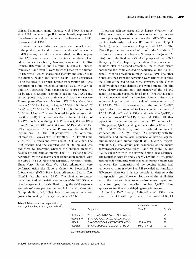
The porcine 3b-HSD coding sequence shares 85.6, 81.7,
79.3, and 79.5% identity and the deduced amino acid
sequence 88.5, 82, 79.3 and 79.2% similarity with the
nucleotide and amino acid sequence of bovine, equine,
human type I and human type II 3b-HSD cDNA, respect-
ively (Fig. 1). The amino acid sequences of the mouse
dehydrogenase/isomerase types I and VI share 76 and
75.9% similarity with the porcine amino acid sequence.
The reductase types IV and V share 71.9 and 71.8% amino
acid sequence similarity with that of the porcine amino acid
sequence. The comparison of the porcine amino acid
sequence to human types I and II revealed no signi®cant
differences, therefore it is not possible to determine the
corresponding type. However, because of the similarities
with the mouse dehydrogenase/isomerase types and
reductase types, the described porcine 3b-HSD clone
appears to function as a dehydrogenase/isomerase.
A porcine PAC library (Al-Bayati et al. 1999) was
screened by PCR with a porcine with the primers HSDpF1
Table 1 Primer sequences (synthesized by
Microsynth GmbH, Balgach, Switzerland).Primer Sequence
Nucleotide position
5' ® 3' Ta (°C)
HSDhumF2 5¢-TGTCAATGTGAAAGGTACCCAGC-3¢ 56
HSDhumR4 5¢-CACAAGGGAACCAACCCACTCC-3 56
HSDpF1 5¢-ACCTCCCCAAAGCTACGATAACC-3¢ 955 ® 974 58
HSDpR1 5¢-CAGATCTCGCTGCGCCTTCTTG-3¢ 1186 ® 1165 58
Ta, Annealing temperature.
Ó 2001 International Society for Animal Genetics, Animal Genetics, 32, 298±302
cDNA cloning and physical mapping 299
and HSDpR1 (Table 1). These primers amplify a fragment of
235 bp, which was sequenced to con®rm that the ampli®-
cation was speci®c. The obtained PAC clone (IVM-
PAC714D9A472) was digested with restriction enzyme
Sau3AI. The digested product was labelled with Biotin-16-
dUTP using the random priming DNA labelling method
with the Prime-ItÒ Fluor Fluorescence Labeling Kit (Strata-
gene, La Jolla, CA, USA). The labelled probe were hybridized
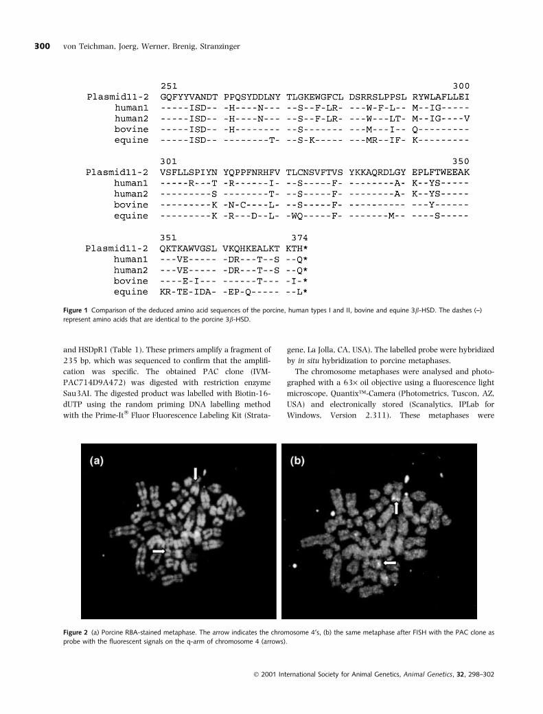
by in situ hybridization to porcine metaphases.
The chromosome metaphases were analysed and photo-
graphed with a 63´ oil objective using a ¯uorescence light
microscope, QuantixÔ-Camera (Photometrics, Tuscon, AZ,
USA) and electronically stored (Scanalytics, IPLab for
Windows, Version 2.311). These metaphases were
Figure 1 Comparison of the deduced amino acid sequences of the porcine, human types I and II, bovine and equine 3b-HSD. The dashes (±)
represent amino acids that are identical to the porcine 3b-HSD.
Figure 2 (a) Porcine RBA-stained metaphase. The arrow indicates the chromosome 4¢s, (b) the same metaphase after FISH with the PAC clone as
probe with the ¯uorescent signals on the q-arm of chromosome 4 (arrows).
Ó 2001 International Society for Animal Genetics, Animal Genetics, 32, 298±302
von Teichman, Joerg, Werner, Brenig, Stranzinger300
compared with the pictures of the same RBA-banded
metaphases. The porcine 3b-HSD gene was mapped to
chromosome 4q16-4q21 (Fig. 2). In the mouse, 3b-HSD
isoforms have been found to be closely linked to chromosome
3 (Bain et al. 1993). In man the two known types have also
been shown to be in close genetic linkage (Russell et al.
1994) and have been allocated to chromosome 1p13
(Morrison et al. 1991). The segment on mouse chromosome
3 containing the 3b-HSD genes shows conservation of gene
order with the corresponding region of human chromosome
1. The mapping of the porcine 3b-HSD gene to chromosome
4q16-4q21 is in agreement with the comparative gene map
(Rettenberger et al. 1995). Human chromosome 1, which
contains the 3b-HSD genes, was shown to correspond to the
evolutionarily conserved fragments located on porcine
chromosomes 4, 6, 9, 10 and 14. Porcine chromosome
4q16-24 contains the NGFB gene which lies at position p13
on human chromosome 1, the same position as the human
3b-HSD genes are located and supporting the presence of
the porcine 3b-HSD found in this position.
The different types of the 3b-HSD gene family appear to
be expressed in different organs, therefore various tissues
will be analysed to identify and characterize further mem-
bers of the 3b-HSD gene family.
Acknowledgements
We would like to thank David Betts for his critical review
and comments to the manuscript. This study was supported
by the ETH Zurich project-number 0-20693-99.
References
Abbaszade I., Clarke T., Park C.H. & Payne A. (1995) The mouse
3b-hydroxysteroid dehydrogenase multigene family includes two
functionally distinct groups of proteins. Molecular Endocrinology
9, 1214±22.
Abbaszade I.G., Arensburg J., Park C.H.J., Kasa-Vubu J.Z., Orly J. &
Payne A.H. (1997) Isolation of a new mouse 3-beta-hydroxys-
teroid dehydrogenase isoform, 3-beta-HSD VI, expressed during
early pregnancy. Endocrinology 138, 1392±9.
Al-Bayati H.K., Duscher S., Kollers S., Rettenberger G., Fries R. &
Brenig B. (1999) Construction and characterization of a porcine
P1-derived arti®cial chromosome (PAC) library covering 3.2
genome equivalents and cytogenetical assignment of six type I
and type II loci. Mammalian Genome 10, 569±72.
Altschul S.F., Madden T.L., Schaffer A.A., Zhang J., Zhang Z., Miller
W. & Lipman D.J. (1997) Gapped BLAST and PSI-BLAST: a new
generation of protein database search programs. Nucleic Acids
Research 25, 3389±402.
Bain P.A., Yoo M., Clarke T., Hammond S.H. & Payne A.H. (1991)
Multiple forms of mouse 3b-hydroxysteroid dehydrogenase/D5-
D4-isomerase and differential expression in gonads adrenal glands
liver and kidneys of both sexes. Proceedings of the National Acad-
emy of Sciences of the USA 88, 8870±4.
Bain P.A., Meisler M.H., Taylor B.A. & Payne A.H. (1993) The genes
encoding gonadal and nongonadal forms of 3b-hydroxysteroid
dehydrogenase/D5-D4-isomerase are closely linked on mouse
chromosome 3. Genomics 16, 219±23.
Boerboom D. & Sirois J. (1997) Molecular cloning and characteri-
sation of equine cytochrome P450 cholesterol side-chain
cleavage and 3b-hydroxysteroid dehydrogenase cDNAs and
regulation of their mRNA in preovulatory follicles. GenBank
accession number: AF031665.
Lachance Y., Luu-The V., Labrie C., Simard J., Dumont M., de
Launoit Y., Guerin S., Leblanc G. & Labrie F. (1990) Character-
ization of human 3 beta-hydroxysteroid dehydrogenase/delta
5-delta 4-isomerase gene and its expression in mammalian cells
[published erratum appears in Journal of Biological Chemistry
267: 3551]. Journal of Biological Chemistry 265, 20469±75.
Lachance Y., Luu-The V., Verreault H., Dumont M., Rheaume E.,
Leblanc G. & Labrie F. (1991) Structure of the human type II, 3
beta-hydroxysteroid dehydrogenase/delta 5-delta 4 isomerase (3
beta-HSD) gene: adrenal and gonadal speci®city. DNA and Cell
Biology 10, 701±11.
Lorence M.C., Corbin C.J., Kamimura N., Mahendroo M.S. & Mason
J.I. (1990) Structural analysis of the gene encoding human
3b-hydroxysteroid dehydrogenase/D5-D4-isomerase. Molecular
Endocrinology 4, 1850±5.
Luu-The V., Lachance Y., Labrie C., Leblanc G., Thomas J.L.,
Strickler R. & Labrie F. (1989) Full length cDNA structure and
deduced amino acid sequence of human 3b-hydroxy-5-ene ster-
oid dehydrogenase. Molecular Endocrinology 3, 1310±2.
Morrison N., Nickson D.A., McBride M.W., Mueller U.W., Boyd E. &
Sutcliffe R.G. (1991) Regional chromosomal assignment
of human 3-beta-hydroxy-5-ene steroid dehydrogenase to 1p13.1
by non-isotopic in situ hybridisation. Human Genetics 87, 223±5.
Nakabayashi O., Nomura O., Nishimori K. & Mizuno S. (1995) The
cDNA cloning and transient expression of a chicken gene enco-
ding a 3 beta-hydroxysteroid dehydrogenase/delta 5->4 isomer-
ase unique to major steroidogenic tissues. Gene 162, 261±5.
Neuenschwander S., Rettenberger G., Meijerink E., Jorg H. &
Stranzinger G. (1996) Partial characterization of porcine obesity
gene (OBS) and its localization to chromosome 18 by somatic cell
hybrids. Animal Genetics 27, 275±8.
Rettenberger G., Klett C., Zechner U., Kunz J., Vogel W. & Hameister
H. (1995) Visualization of the conservation of synteny between
humans and pigs by heterologous chromosomal painting.
Genomics 26, 372±8.
RheÂaume E., Lachance Y., Zhao H.F., Breton N., Dumont M., de
Launoit Y., Trudel C., Luu-The V., Simard J. & Labrie F. (1991)
Structure and expression of a new complementary DNA encoding
the almost exclusive 3b-hydroxysteroid dehydrogenase/D5-D4-
isomerase in human adrenals and gonads. Molecular Endocrinol-
ogy 5, 1147±57.
Rogerson F.M., Lehoux J.G. & Mason J.I. (1995) Expression and
characterization of isoforms of 3 beta-hydroxysteroid dehydro-
genase/delta 5 ® 4-isomerase in the hamster. Journal of Steroid
Biochemistry and Molecular Biology 55, 481±7.
Ó 2001 International Society for Animal Genetics, Animal Genetics, 32, 298±302
cDNA cloning and physical mapping 301
Russell A.J., Wallace A.M., Donaldson M.D.C., Edwards C.R.W. &
Sutcliffe R.G. (1994) Mutation in the human gene for 3b-hy-
droxysteroid dehydrogenase type II leading to male pseudoher-
maphroditism without salt loss. Journal of Molecular Endocrinology
12, 225±37.
Sakai N., Tanaka M., Takahashi M., Fukada S., Mason J.I. &
Nagahama Y. (1994) Ovarian 3b-hydroxysteroid dehydro-
genase/D5-D4 isomerase of rainbow trout: its cDNA cloning and
properties of the enzyme expressed in a mammalian cell. FEBS
Letters 350, 309±13.
Simard J., de Launoit Y. & Labrie F. (1991a) Characterisation of the
structure-activity of the rat type I and type II 3b-hydroxysteroid
dehydrogenase/D5-D4-isomerase by site-directed mutagenesis and
expression in HeLa cells. Journal of Biological Chemistry 166,
14842±5.
Simard J., Melner M.H., Breton N., Low K.G., Zhao H.F., Periman
L.M. & Labrie F. (1991b) Characterization of macaque 3 beta-
hydroxy-5-ene steroid dehydrogenase/delta 5-delta 4 isomerase:
structure and expression in steroidogenic and peripheral
tissues in primate. Molecular and Cellular Endocrinolgy 75,
101±10.
Simard J., CoueÈt J., Durocher F., Labrie Y., Sanchez R., Breton N.,
Turgeon C. & Labrie F. (1993) Structure and tissue-speci®c
expression of a novel member of the rat 3b-hydroxysteroid
dehydrogenase/D5-D4-isomerase (3b-HSD) family: the exclusive
3b-HSD gene expressed in the skin. Journal of Biological Chemistry
268, 19659±68.
Werner P., Neuenschwander S. & Stranzinger G. (1999) Charac-
terisation of the porcine uncoupling proteins 2 and 3 (UCP2 &
UCP3) and their localization to chromosome 9, p. by somatic cell
hybrids. Animal Genetics 30, 221±4.
Zhao H.F., Simard J., Labrie C., Breton N., RheÂaume E., Luu-The V.
& Labrie F. (1989) Molecular cloning, cDNA structure and pre-
dicted amino acid sequence of bovine 3 beta-hydroxy-5-ene
steroid dehydrogenase/delta 5-delta 4 isomerase. FEBS Letters
259, 153±7.
Ó 2001 International Society for Animal Genetics, Animal Genetics, 32, 298±302
von Teichman, Joerg, Werner, Brenig, Stranzinger302


![Evidence for Endogenous Neurosteroid Production in the ... · tetrahydroDOC in the brain by the enzymes 5α reductase and 3α hydroxysteroid dehydrogenase (HSD) [5,12]. Cholesterol](https://img.pdfslide.net/doc/110x75/5fd9b663408dab2eba43865a/evidence-for-endogenous-neurosteroid-production-in-the-tetrahydrodoc-in-the.jpg)